Article

Biosynthesis and Bioactivity of Melanin from the Deep-Sea Hydrothermal Vent Yeast Hortaea werneckii Mo34

1
College of Chemical and Biological Engineering, Shandong University of Science and Technology, Qingdao 266590, China
2
College of Marine Life Sciences, Ocean University of China, Qingdao 266003, China
*
Author to whom correspondence should be addressed.
J. Mar. Sci. Eng. 2025, 13(6), 1004; https://doi.org/10.3390/jmse13061004
Submission received: 23 April 2025 / Revised: 19 May 2025 / Accepted: 20 May 2025 / Published: 22 May 2025
(This article belongs to the Section Marine Biology)

Abstract

:
Importance of this study: Melanin synthesized through the oxidative polymerization of phenolic compounds exhibits a high molecular weight and has many physiological functions and activities. Main results: In this study, the key PKS1-1, PKS1-2, CMR1-1, and CMR1-2 genes for melanin biosynthesis and regulation from the highly genome-duplicated black yeast Hortaea werneckii Mo34, isolated from a deep-sea hydrothermal vent, were heterologously complemented in the ∆pks1 albino mutant K5 and the ∆cmr1 albino mutant CM7-2 of Aureobasidium melanogenum XJ5-1. Melanin formation in all the resulting transformants was restored, confirming that both the PKS1-1 and PKS1-2 genes from H. werneckii Mo34 were likely involved in the DHN melanin biosynthesis of A. melanogenum XJ5-1. Furthermore, the CMR1-1 and CMR1-2 genes from H. werneckii Mo34 could play significant roles in regulating melanin biosynthesis in A. melanogenum XJ5-1. Simultaneously, the expression of the PKS1 and THR1 genes involved in melanin biosynthesis was also enhanced in the transformants complemented with the CMR1-1 and CMR1-2 genes. The purified high-molecular-weight melanin from H. werneckii Mo34 exhibited excellent Fe2⁺-chelating, DPPH radical-scavenging, and superoxide radical-scavenging activities. Additionally, it actively inhibited the growth of Staphylococcus aureus and Pseudomonas putida. Conclusions: The black yeast H. werneckii Mo34 indeed had the DHN melanin biosynthesis pathway and the melanin produced by it had many potential applications.

1. Introduction

H. werneckii is a heavily melanized and polymorphic fungus belonging to the division Ascomycota, the family Teratosphaeriaceae, and the genus Hortaea. It can grow in NaCl solutions ranging from 0% to nearly saturation (5.1 M NaCl) and is among the most salt-adaptable fungal species. Its cells can divide by both fission and budding [1]. The whole genome (51.6 Mb, accession No: AIJO00000000) is duplicated and contains two highly identical gene copies of nearly every protein, attributed to intraspecific hybridization events between ancestors [2]. The genome sizes of two strains of H. werneckii, MC848 and MC873, which also exhibit genome duplication, were isolated from the Mediterranean Sea at depths of 2500 m and 3400 m, and were 50.7 Mb and 51.0 Mb, respectively [3]. However, the genome size of H. werneckii M-3, isolated from marine sediment in the West Pacific, is only 38.2 Mb (accession number: JBAOKB000000000.1) [4]. Unfortunately, because of the thick cell wall, it is very difficult to introduce DNA fragments into its cells, edit its genomes, and elucidate its metabolism and regulation. For example, a vanadium-dependent chloroperoxidase gene of H. werneckii UBOCC-A-208029 isolated from the Rainbow hydrothermal vent field, in the mid-Atlantic ridge, was expressed in E. coli strain BL21(DE3) + pKJE7 + vHPO_Hw [5]. It is widely distributed in thalassohaline and hypersaline waters, magnesium-rich bitterns, seawater, sea sponges, mangrove plants, deep-sea water, shallow-water hydrothermal vents, beach soil, glacial ice, arid inorganic and organic surfaces, and salted foods [6,7,8]. This yeast species is characterized by high adaptability to harsh environments, salt tolerance, elevated melanin production, and wide-spread distribution [2]. The accumulated melanin in the cell walls enhances the yeast’s ability to withstand UV radiation, oxidative stress, extreme desiccation, high temperatures, the toxic effects of heavy metals, and high salt concentrations [9]. This adaptation promotes the competitive abilities and survival of the yeast in extreme environments [9]. Therefore, the produced melanin can be widely applied in the medical, pharmaceutical, cosmetic, and environmental industries.
Melanins synthesized through the oxidative polymerization of phenolic compounds are insoluble pigments with high molecular weights, negatively charged, and generally red, black, or brown in color [9]. The primary pathways for melanin biosynthesis in fungi are the dihydroxynaphthalene (DHN) pathway and the L-3,4-dihydroxyphenylalanine (L-DOPA) pathway. The non-reducing polyketide synthases (NR-PKSs), which consist of one ketosynthase (KS), one acyl transferase (AT), two acyl carrier proteins (ACP), one thioesterase (TE), and one cyclase (CYC), are responsible for melanin biosynthesis in the dihydroxynaphthalene (DHN) pathway, using acetyl-CoA or malonyl-CoA as precursors (Figure 1). The NR-PKS is activated by the post-translational modification of the proteins, catalyzed by phosphopantetheinyl transferases (PPTases). Melanin biosynthesis is regulated by the cell wall integrity (CWI) signaling pathway. The transcriptional activator Swi4 and the melanin-specific transcriptional activator Cmr1, encoded by the CMR1 gene, are the primary transcriptional activators during melanin biosynthesis [9] (Figure 1). Very recently, the NSDD gene encoding a GATA-type transcriptional repressor also has been found to negatively regulate melanin biosynthesis [10]. Consequently, the deletion of the NR-PKS and CMR1 genes can cause the mutants of A. melanogenum XJ5-1, obtained from the Taklimakan Desert in China, to develop albino colonies (characterized by the absence of melanin) [9]. However, little is known about melanin biosynthesis and bioactivity of melanin in H. werneckii isolated from the deep-sea hydrothermal vents [11]. In this study, we found that the NR-PKS and CMR1 genes cloned from H. werneckii Mo34, isolated from deep-sea hydrothermal vents and characterized by tolerance to high temperature, high salt concentration, and acidic environments, could be effectively complemented in A. melanogenum XJ5-1. Furthermore, melanin produced by H. werneckii Mo34 exhibited several bioactivities.

2. Materials and Methods

2.1. Microbial Strains, Media and Plasmids

H. werneckii Mo34, isolated from the deep-sea hydrothermal vents, is a halophilic black yeast with an optimal NaCl concentration of 30.0 g L−1 for its cell growth [11]. It was kindly offered by Dr. Gaëtan Burgaud at Laboratoire Universitaire de Biodiversite’ et Ecologie Microbienne in France. The black yeast, A. melanogenum XJ5-1, was isolated from the Taklimakan Desert in China [9]. The albino mutants K5 and CM7-2, designated ∆pks1 and ∆cmr1, respectively, were generated by deleting the NR-PKS and CMR1 genes from A. melanogenum XJ5-1, respectively [9,10,11]. The overexpression vector pAMEXlox-1 (GenBank accession number MH453956) was constructed in this laboratory [9]. The competent cells of Escherichia coli DH5α, utilized for amplifying plasmids carrying a target gene, were purchased from Tiangen Biotech (Beijing) Co., Ltd., China. The sensitive bacteria included in this study were Staphylococcus aureus, Pseudomonas putida, Bacillus subtilis, Enterobacter cloacae, Pseudomonas aeruginosa, Vibrio harveyi, Vibrio anguillarum, and Acinetobacter baumannii, all of which were cultivated in the liquid 2216E medium or on 2216E agar plates at 37 °C.
E. coli DH5α was cultivated in an LB medium, while E. coli transformants were grown in the LB medium supplemented with 100.0 μg mL−1 of ampicillin (Sinopharm Chemical Reagent Co., Ltd., Shanghai, China) [9]. All the yeast strains were cultured in a YPD medium, and yeast transformants were grown in the YPD medium containing 100.0 μg mL−1 of nourseothricin (Sinopharm Chemical Reagent Co., Ltd., Shanghai, China) [12]. The medium used for melanin production was Potato Dextrose Broth (PDB) or Potato Dextrose Agar (PDA), composed of 200.0 g L−1 of potato extract and 20.0 g L−1 of glucose (Sinopharm Chemical Reagent Co., Ltd., Shanghai, China) and 20.0 g/L−1 agar (Sinopharm Chemical Reagent Co., Ltd., Shanghai, China) (only in PDA).

2.2. Construction of the Recombinant Plasmids Carrying the Native NR-PKS Gene and the Native CMR1 Gene Cloned from the Genomic DNA of H. werneckii Mo34

Genomic DNA was isolated and purified from H. werneckii Mo34 using the methods outlined by Chi et al. [12]. The homologues of the PKS1-1, PKS1-2, CMR1-1, and CMR1-2 genes in the highly genome-duplicated H. werneckii Mo34 were PCR amplified using the primers MO34PKS1se/MO34PKS1an and MO34CMR1se/MO34CMR1an (Table S1), with the genomic DNA serving as the template. The primers MO34PKS1se/MO34PKS1an and MO34CMR1se/MO34CMR1an (Table S1) were designed based on the sequenced genomic DNA (accession No: AIJO00000000) of H. werneckii EXF2000, which was isolated from marine solar salterns along the Adriatic coast [2]. The PCR products were isolated and purified using 10.0 g L−1 agarose gel electrophoresis. The purified PCR products were digested with the enzymes MluI/SalI and SpeI/MluI (Takara Biomedical Technology Co., Ltd., Beijing, China) (Table S1), and the resulting digested PCR products were ligated into the overexpression vector pAMEXlox-1, which had been digested with the same enzymes. The recombinant plasmids were transformed into the competent cells of E. coli DH5α, and the bacterial transformants were cultured on the LB medium supplemented with ampicillin at 37 °C for 12 to 14 h. The amplified recombinant plasmids from the bacterial transformants were identified using PCR techniques with the primers MO34PKS1se/MO34PKS1an and MO34CMR1se/MO34CMR1an (Table S1) and were subsequently sequenced using the primers NATX13F (5′-TCTTGGATCTGCTCATTCTTTT-3′) and NATX13R (5′-GAAACCCTTAGTATGTATTTGTATTTG-3′). The amino acid sequences deduced from the sequenced PCR products were aligned and analyzed using the online BLASTX tool (http://www.ncbi.nlm.nih.gov/BLAST, accessed on 1 January 2020) at NCBI, as well as software listed in Table S2.

2.3. Total RNA Extraction and cDNA Obtainment

H. werneckii Mo34 was grown aerobically in 5.0 mL of the YPD medium at 28 °C and 180 rpm for 48 h; 2.0 mL of the yeast cell culture was transferred into 50.0 mL of the PDB medium, and the new culture was cultivated aerobically at 28 °C and 180 rpm for 48 h. The yeast cells were harvested by centrifugation, after which they were disrupted in liquid nitrogen. Total RNA was extracted from the disrupted yeast cells using the E.Z.N.A. Fungal RNA Kit (Shanghai Yuanmu Bio-Technology Co., Ltd, Shanghai, China) and the extracted RNA was subsequently transcribed into complementary DNA (cDNA) using a FastKing gDNA Dispelling RT SuperMix Kit (Tiangen Biotech (Beijing) Co., Ltd, Beijing, China).

2.4. Construction of the Expression Vectors Carrying Coding Sequences (CDSs) of PKS1-2 Genes and CMR1-2 Genes

Upon analysis of the cloned PCR products, it was determined that the genomic DNA of H. werneckii Mo34 contained two copies of the PKS1-1 gene and PKS1-2 gene, which contained one intron and two exons and two copies of the CMR1-1 gene and CMR1-2 gene, which contained two introns and three exons due to its highly duplicated genomes. The 5′-end DNA fragment CDS1 (5199 bp, with no introns) of the PKS1-1 gene was PCR amplified from the genomic DNA using the primer pairs MO34PKS1se/PKS1se-R (Table S1). Additionally, the DNA fragment CDS2 (1389 bp) of the PKS1-1 gene was PCR amplified from the cDNA of H. werneckii Mo34 using the primer pairs PKS1an-F/MO34PKS1an (Table S1). The two DNA fragments obtained from the PCR amplifications were combined, denatured, annealed, and subjected to a fusion PCR using the primers MO34PKS1se/MO34PKS1an (Table S1), resulting in a full-length coding sequence (CDS) of the PKS1-1 gene (6558 bp, accession number: MN966970). Similarly, the PKS1-2 gene (6546 bp, accession number: MN966971) was obtained using similar methods.
The 5′-end DNA fragment CDS11 (1494 bp) of the CMR1-1 gene was PCR amplified from cDNA using the primer pairs MO34CMR1se/CMR1se-R (Table S1). Additionally, another DNA fragment of the CMR1-1 gene, CDS12 (1494 bp), was PCR amplified from the cDNA of H. werneckii Mo34 using the primer pairs MO34CMR1an/CMR1an-F. The two DNA fragments (CDS11 and CDS12) obtained from the PCR amplifications were combined, denatured, annealed, and subjected to the fusion PCR using the primer pairs MO34CMR1se/MO34CMR1an (Table S1), resulting in a full-length coding sequence (CDS) of the CMR1-1 gene (2948 bp, accession number: MN966972). Similarly, the CMR1-2 gene (2945 bp, accession number: MN966973) was gained using similar techniques.
The cloned PKS1-1 gene, PKS1-2 gene, CMR1-1 gene, and CMR1-2 gene were digested with their corresponding enzymes (Table S1). The obtained DNA fragments were ligated into pAMEX-lox1, which had been digested with the same enzymes, resulting in the production of pAMEXlox-1-Mo34PKS1-1 and pAMEXlox-1-Mo34PKS1-2 (Figure S1A), as well as pAMEXlox-1-Mo34CMR1-1 and pAMEXlox-1-Mo34CMR1-2 (Figure S1B).

2.5. Transformation and Isolation of the Transformants

The plasmids pAMEXlox-1-Mo34PKS1-1, pAMEXlox-1-Mo34PKS1-2, pAMEXlox-1-Mo34CMR1-1, and pAMEXlox-1-Mo34CMR1-2, constructed as described above (Figure S1), were digested with the enzyme SmaI (Takara Biomedical Technology Co., Ltd., Beijing, China). The resulting linear DNA fragments—26S-loxp-Ppgk-NAT-polyA-loxp-MO34PKS1-1-PTEF-18S, 26S-loxp-Ppgk-NAT-polyA-loxp-MO34PKS1-2-PTEF-18S, 26S-loxp-Ppgk-NAT-polyA-loxp-MO34CMR1-1-PTEF-18S, and 26S-loxp-Ppgk-NAT-polyA-loxp-MO34CMR1-2-PTEF-18S—were obtained.
The linear DNA fragments 26S-loxp-Ppgk-NAT-polyA-loxp-MO34PKS1-1-PTEF-18S and 26S-loxp-Ppgk-NAT-polyA-loxp-MO34PKS1-2-PTEF-18S were introduced into the competent cells of the K5 mutant by transformation. In contrast, the linear DNA fragments 26S-loxp-Ppgk-NAT-polyA-loxp-MO34CMR1-1-PTEF-18S and 26S-loxp-Ppgk-NAT-polyA-loxp-MO34CMR1-2-PTEF-18S were transformed into the competent cells of the CM7-2 mutant. All the obtained transformants, including the K5 mutant and the CM7-2 mutant, were cultured on PDA plates at 28 °C for 3 to 5 d. After purification of all the transformants, the transformants Mo34K1-1-3, which carried the PKS1-1 gene, and Mo34K1-2-4, which carried the PKS1-2 gene, from the K5 mutant, as well as the transformants Mo34C1-1-12, which carried the CMR1-1 gene, and Mo34C1-2-5, which carried the CMR1-2 gene, from the CM7-2 mutant, were isolated.

2.6. Confirmation of the Inserted NAT Gene in the Transformants

The albino mutants K5 and CM7-2, along with the transformants Mo34K1-1-3, Mo34K1-2-4, Mo34C1-1-12, and Mo34C1-2-5, were cultured in the liquid YPD medium at 28 °C and 180 rpm for 12 to 16 h. The genomic DNAs of all the strains were extracted as described above, and the inserted NAT gene was PCR amplified using the primer pairs NAT-se/NAT-an (Table S1), with the extracted genomic DNAs serving as the template. The PCR products were identified using 1.0% agarose gel electrophoresis.

2.7. Extraction and Quantitative Determination of Melanin

The wild type strain XJ5-1 and the albino mutants K5 and CM7-2, along with the transformants Mo34K1-1-3, Mo34K1-2-4, Mo34C1-1-12, and Mo34C1-2-5, were cultured in the liquid PDB medium at 28 °C and 180 rpm for 7 d. The yeast-like cells in the cultures were collected and washed with sterile distilled water via centrifugation. An equal amount of each washed cell was suspended in 800 µL of NaOH solution (2.0 M), and the cell suspension was treated at 80 °C for 30 min. The treated cell suspensions were centrifuged at 5000× g for 10 min, and the optical density at 400 nm (OD400nm) of the suitably diluted supernatants was measured [9].

2.8. Determination of the Transcriptional Levels of the Relevant Genes for Melanin Biosynthesis

The wild-type strain XJ5-1, the albino mutants K5 and CM7-2, and the transformants Mo34K1-1-3, Mo34K1-2-4, Mo34C1-1-12, and Mo34C1-2-5 were cultivated in 5.0 mL of the liquid YPD medium at 28 °C and 180 rpm for 2 d; 2.0 mL of the culture was inoculated into 50.0 mL of the fresh liquid PDB medium, and the new culture was cultivated at 28 °C and 180 rpm for 24 h. Total RNA was extracted from the cells of each strain, and the extracted RNA was transcribed into complementary DNA (cDNA) as described above. To assess the transcriptional levels of various genes associated with melanin biosynthesis, quantitative real-time PCR was conducted following the protocols outlined by Liu et al. [13]. A fluorescent real-time reverse transcription (RT)-PCR assay was conducted to analyze the transcriptional levels of the genes involved in melanin biosynthesis across the different strains mentioned above. The previously obtained cDNAs were utilized as samples. The formula RATE = 2−ΔΔCt (where Ct represents the threshold cycle) was employed to calculate the transcriptional levels of various genes. The β-actin gene served as the internal control for the RT-PCR reaction, and the relative transcriptional levels of the genes in the wild-type strain XJ5-1 were considered to be 100%.

2.9. Melanin Production by H. werneckii Mo34, Extraction and Purification of Melanin

H. werneckii Mo34 was grown in the PDB medium at 28 °C and 180 rpm for 7 d. The yeast cells from the culture were harvested and washed with sterile distilled water by centrifugation at 4000× g for 10 min. The washed yeast cells were resuspended in NaOH solution (pH 12.0), and the suspended yeast cells were treated with ultrasonication at 43 °C for 36 min, followed by centrifugation at 4000 × g for 10 min. The pH of the obtained supernatant was adjusted to 2.0 using 3.0 M HCl, followed by centrifugation at 10,000 × g for 20 min. The crude melanin obtained was hydrolyzed in 7.0 M HCl solution at 100 °C to remove impurities. The treated melanin sample was centrifuged at 10,000 × g for 20 min, and the resulting sediments were washed using trichloromethane, ethyl acetate, and ethanol by centrifugation, respectively. The washed sediments were dissolved in 1.0 M NaOH solution, and the pH of the solution was adjusted to 2.0 with 3.0 M HCl solution, followed by centrifugation at 10,000 × g for 20 min. The new sediments were washed three times with sterile distilled water via centrifugation. Finally, the purified melanin was obtained through vacuum freeze-drying at −20 °C.

2.10. Solubility of the Purified Melanin

Twenty milligrams of the purified melanin was mixed with 2.0 mL of distilled water, HCl solution (pH 2.5), NaOH solution (pH 12.0), ethanol, methanol, trichloromethane, acetone, ethyl acetate, butanol, and dimethyl sulfoxide (DMSO). After the mixtures were agitated for 1 h and allowed to stand statically for 0.5 h, the mixture was filtered using a 0.22 μm membrane, and the optical density at 400 nm (OD400nm) of the filtrate was measured. Finally, the solubility of the melanin was calculated.

2.11. Fourier-Transform Infrared Analysis of the Purified Melanin

The purified melanin obtained above was characterized using Fourier-transform infrared (FT-IR) spectroscopy with a Nicolet Nexus FTIR 470 spectrophotometer (Thermo Nicolet Corporation, Waltham, MA, USA). One milligram of the purified melanin sample was mixed with 200 mg of 95% potassium bromide powder. The mixture was desiccated overnight at 50 °C under vacuum. The FT-IR spectra were obtained using the potassium bromide pellets of the purified melanin and standard melanin obtained from (Sigma-Aldrich®, Shanghai, China) over a range of 4000–400 cm−1 at a rate of 16 scans with a resolution of 2 cm−1 [14].

2.12. Determination of the Fe2+-Chelating Ability of the Purified Melanin

The reaction mixture consisted of 2.0 mM FeSO4 (0.1 mL), different concentrations of the purified melanin or EDTA (0, 0.2, 0.4, 0.6, 0.8, and 1.0 mg mL−1), and 3.7 mL of DMSO, incubated at room temperature for 30 s. Then, 0.2 mL of a 5.0 mM ferrozine solution was added to the mixture. The resulting mixture was stirred for 2 min, followed by standing at room temperature for 10 min. The optical density at 562 nm (OD562nm) of the treated mixtures was measured. The chelating activity of the purified melanin against Fe2⁺ was calculated using the following formula: Chelating ability (%) = (AC − AS)/AC × 100, where AC represents the absorbance of the control (distilled water) and AS represents the absorbance of the treated mixture. The IC50 value for Fe2⁺-chelating activity was defined as the concentration of the purified melanin required to chelate 50% of Fe2⁺ under the assay conditions.

2.13. Measurement of Superoxide-Radical Scavenging Activity

The measurement method was based on the ability of any antioxidant to inhibit formazan formation by scavenging superoxide radicals generated in a riboflavin–light–NBT system. One milliliter of various concentrations of the purified melanin solution, DMSO solution, or water was mixed with 3.0 mL of phosphate buffer (50.0 mM, pH 7.8) containing 13.0 mM methionine, 2.0 µM riboflavin, 100.0 µM EDTA, and 75.0 µM butylated hydroxytoluene (BHT). The mixture was then placed in a sealed box equipped with a fluorescent lamp covered with aluminum foil. After 10 min of illumination from a fluorescent lamp (4000 lux), the optical density at 560 nm (OD560nm) values of the mixture was measured, representing the maximal absorbance of blue formazan. Simultaneously, the same mixture kept in the dark was used as a blank. The percentage inhibition of superoxide anion generation was calculated using the following formula:
The superoxide radical scavenging activity (%) = [1 − (A1 − A2)/A0] × 100, where A1 represents the optical density at 560 nm (OD560nm) of the reaction mixture; A2 represents the OD560nm of the reaction mixture kept in the dark; and A0 represents the OD560nm of the reaction mixture without melanin. The IC50 value for superoxide radical scavenging activity is defined as the concentration of melanin required to scavenge 50% of superoxide radicals under the assay conditions.

2.14. Assay of 1,1-Diphenyl-2-picryl-hydrazyl (DPPH) Radical-Scavenging Activity

The free radical-scavenging activity of the purified melanin was measured using the procedure described by Wilson et al. [15]. One milliliter of a methanol solution (95% w/v) containing 0.2 mM DPPH was mixed with 1.0 mL of a methanol solution (95% w/v) containing different concentrations of the purified melanin or vitamin C, and the mixture was agitated vigorously for 2 min. Then, the treated mixture kept in the dark was allowed to stand for 30 min, and the optical density at 517 nm (OD517nm) values of the mixture was measured. The ability to scavenge the DPPH radical was calculated using the following equation:
DPPH radical scavenging activity (%) = [1 − (A1 − A2)/A0] × 100
A0 was the OD517nm value of the mixture containing 1.0 mL of DPPH and 1.0 mL of 95% (w/v) methanol; A1 was the OD517nm value of the mixture containing 1.0 mL of the sample and 1.0 mL of DPPH; A2 was the OD517nm value of the mixture containing 1.0 mL of the sample and 1.0 mL of 95% (w/v) methanol. The IC50 value for DPPH radical scavenging activity was defined as the concentration of melanin required to scavenge 50% of DPPH radicals under the assay conditions.

2.15. Assay of Antibacterial Activity of the Purified Melanin

Five microliters of bacterial suspensions (3 × 10⁸ cells mL−1) grown in the liquid 2216E medium was mixed with 1.0 mL of 2216E medium containing different concentrations of purified melanin. The mixture was aerobically cultivated at 37 °C and 960 rpm for 4 h. Subsequently, the optical density at 600 nm (OD600nm) of the culture was measured. The minimum inhibitory concentration (MIC) was defined as the melanin concentration capable of completely inhibiting bacterial growth.

2.16. Statistical Analysis

The data presented in this research show the average values obtained from triplicate measurements. The statistical tests were conducted using SPSS 22.0 for Windows (SPSS Inc., Chicago, IL, USA).

3. Results and Discussion

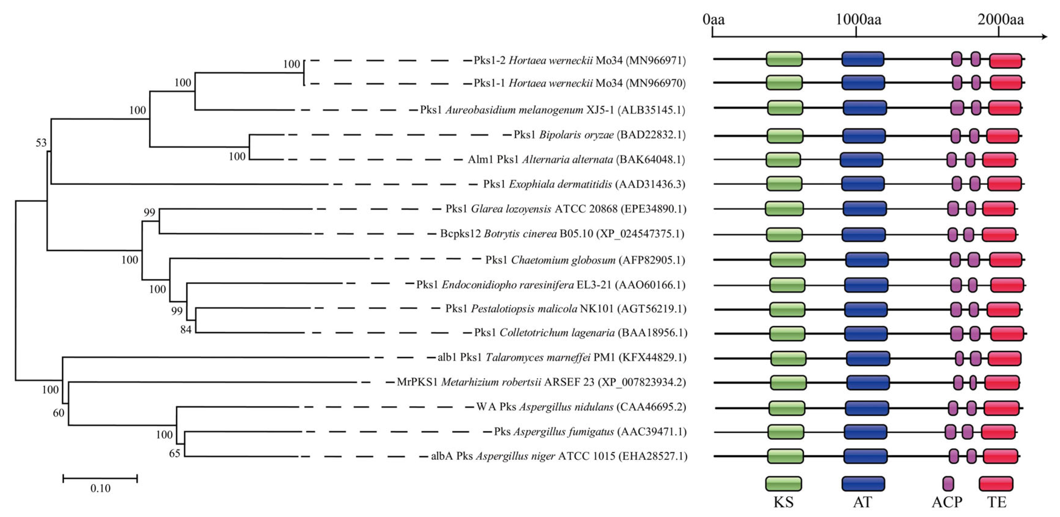
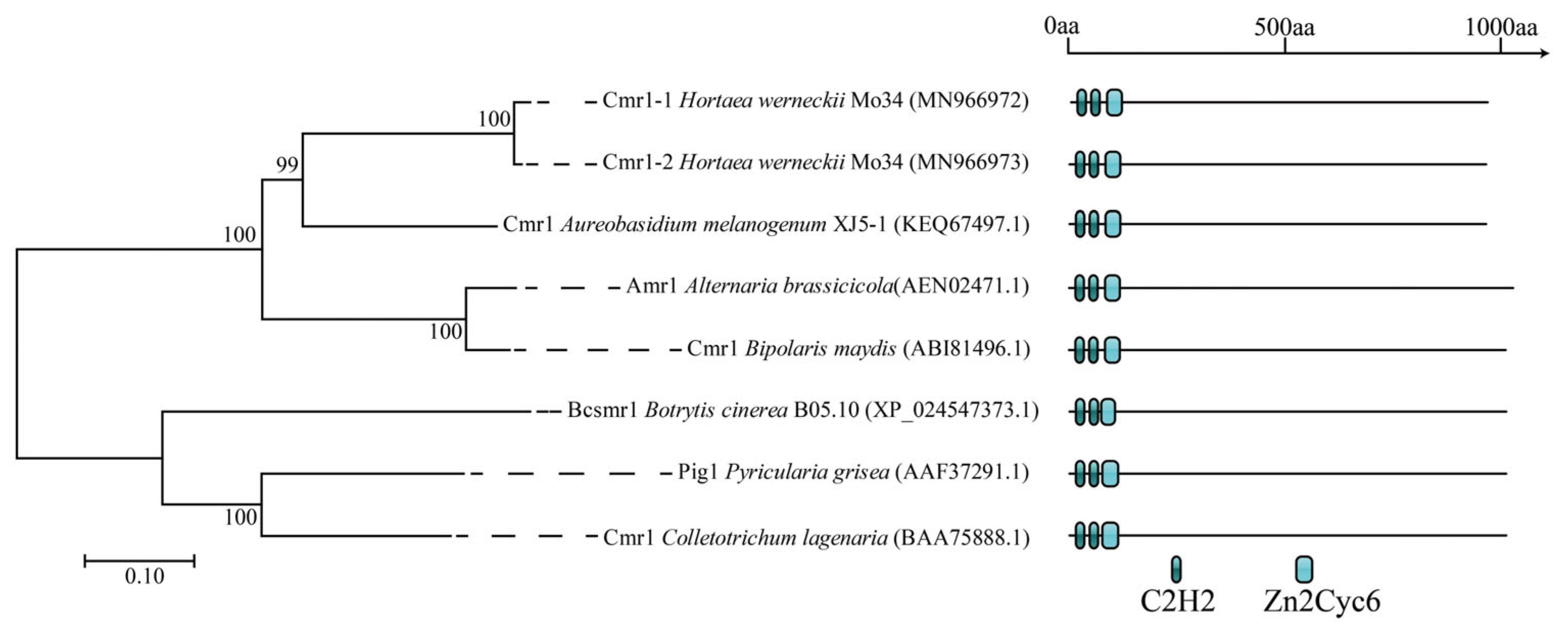
3.1. Characterization of the PKS1-1, PKS1-2, CMR1-1, and CMR1-2 Genes from H. werneckii Mo34

It has been preliminarily confirmed that H. werneckii MZKI B-736, isolated from hypersaline water in a solar saltern on the eastern coast of the Adriatic Sea, possesses the 1,8-dihydroxynaphthalene (DHN) melanin biosynthesis pathway [11,16]. Furthermore, most of the genes in the genomes of H. werneckii isolated from different sources are highly duplicated [2,3]. After the cloning and characterization of the NR-PKS1 genes involved in melanin biosynthesis and the melanin-specific transcriptional activator CMR1 genes from the genomic DNA of H. werneckii Mo34 used in this study, it was found that the genomic DNA of H. werneckii Mo34 contained two copies of the NR-PKS gene (PKS1-1 and PKS1-2 genes, with accession numbers MN966970 and MN966971, respectively) and two copies of the CMR1 gene (CMR1-1 and CMR1-2 genes, with accession numbers MN966972 and MN966973, respectively). The size of the PKS1-1 gene was 6620 bp, with one intron of 62 bp, encoding 2185 amino acids with a molecular weight of 236.0 kDa. In comparison, the size of the PKS1-2 gene was 6608 bp, also with one intron of 62 bp, encoding 2181 amino acids with a molecular weight of 235.5 kDa. Meanwhile, the size of the CMR1-1 gene was 3048 bp, with two introns of 65 bp and 55 bp, encoding 975 amino acids with a molecular weight of 109.0 kDa. In comparison, the size of the CMR1-2 gene was 3045 bp, also with two introns of 65 bp and 55 bp, encoding 974 amino acids with a molecular weight of 108.87 kDa. These results are reasonable, as the species underwent an evolutionarily recent whole-genome duplication, is a hybrid of the same H. werneckii strain, and contains two highly identical gene copies for nearly every protein [2].
After aligning the proteins deduced from the cloned PKS1-1 and PKS1-2 genes with homologous proteins from other melanin-producing fungi and analyzing their conserved domains, the results presented in Figure 2 indicated that the Pks1-1 and Pks1-2 proteins from the Mo34 strain were phylogenetically closely related to the Pks1 protein derived from the PKS1 gene of A. melanogenum XJ5-1, which was isolated from the Taklimakan Desert [10]. In addition, all the deduced proteins contained the following conserved domains: one KS domain, one AT domain, two ACP domains, and one TE domain (Figure 2). This indicated that the DHN-melanin biosynthesis pathways in these two fungi were very similar.
Additionally, the two ACP domains contained the conserved amino acid sequences LGVDSL and MGMDSL, which serve as the binding sites for phosphopantetheine [9]. This indicated that, similar to NR-Pks1 from A. melanogenum XJ5-1, which can produce a large amount of melanin, the activity of Pks1-1/2 from H. werneckii Mo34 was also activated by the protein post-modification under the catalysis of phosphopantetheinyl transferase (PPTase) [9,17]. This suggested that Pks1-1/2 from H. werneckii Mo34 was also likely involved in DHN melanin biosynthesis.
After aligning the proteins deduced from the cloned CMR1 genes with the homologous proteins from other melanin-producing fungi and analyzing their conserved domains, the results presented in Figure 3 indicated that the Cmr1-1 and Cmr1-2 proteins from the Mo34 strain were also phylogenetically closely related to the Cmr1 protein derived from the CMR1 gene of A. melanogenum XJ5-1, which was isolated from the Taklimakan Desert [9]. Furthermore, all the identified proteins contained two Cys2His2 zinc-finger motifs and one Zn2Cys6 binuclear cluster motif for DNA binding (Figure 3). Previous research has established that the two Cys2His2 zinc-finger motifs and the one Zn2Cys6 binuclear cluster motif are essential for DHN melanin biosynthesis in Magnaporthe grisea and A. melanogenum XJ5-1 [9,18]. This indicated that, similar to A. melanogenum XJ5-1 and any other fungi, the DHN melanin biosynthesis in H. werneckii Mo34 was also regulated by the melanin-specific transcriptional activator Cmr1.

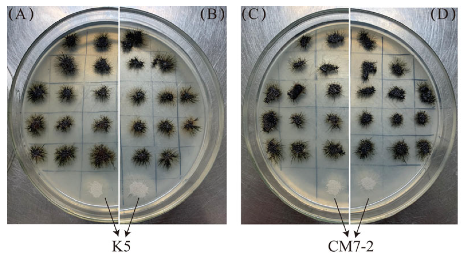
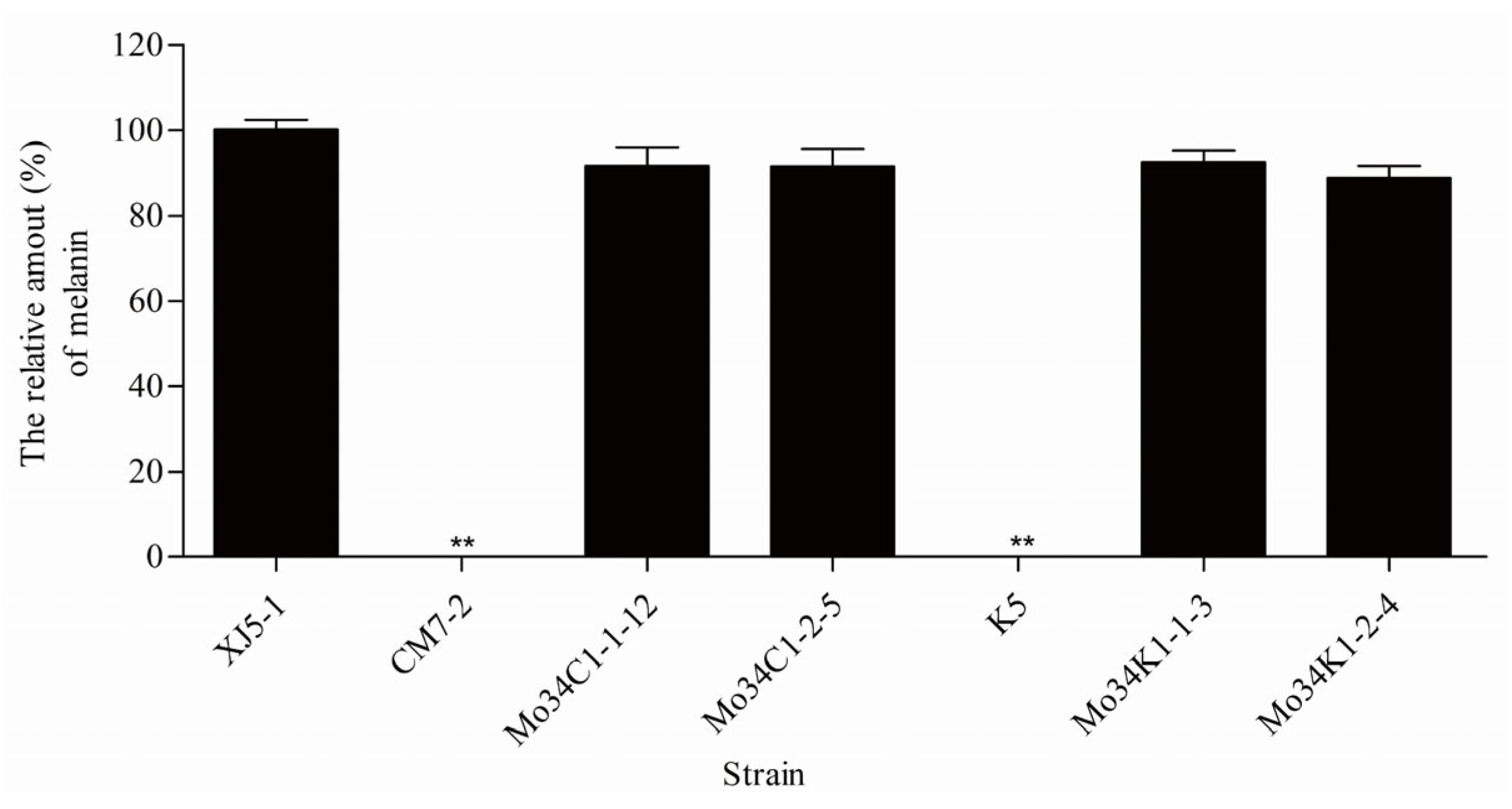
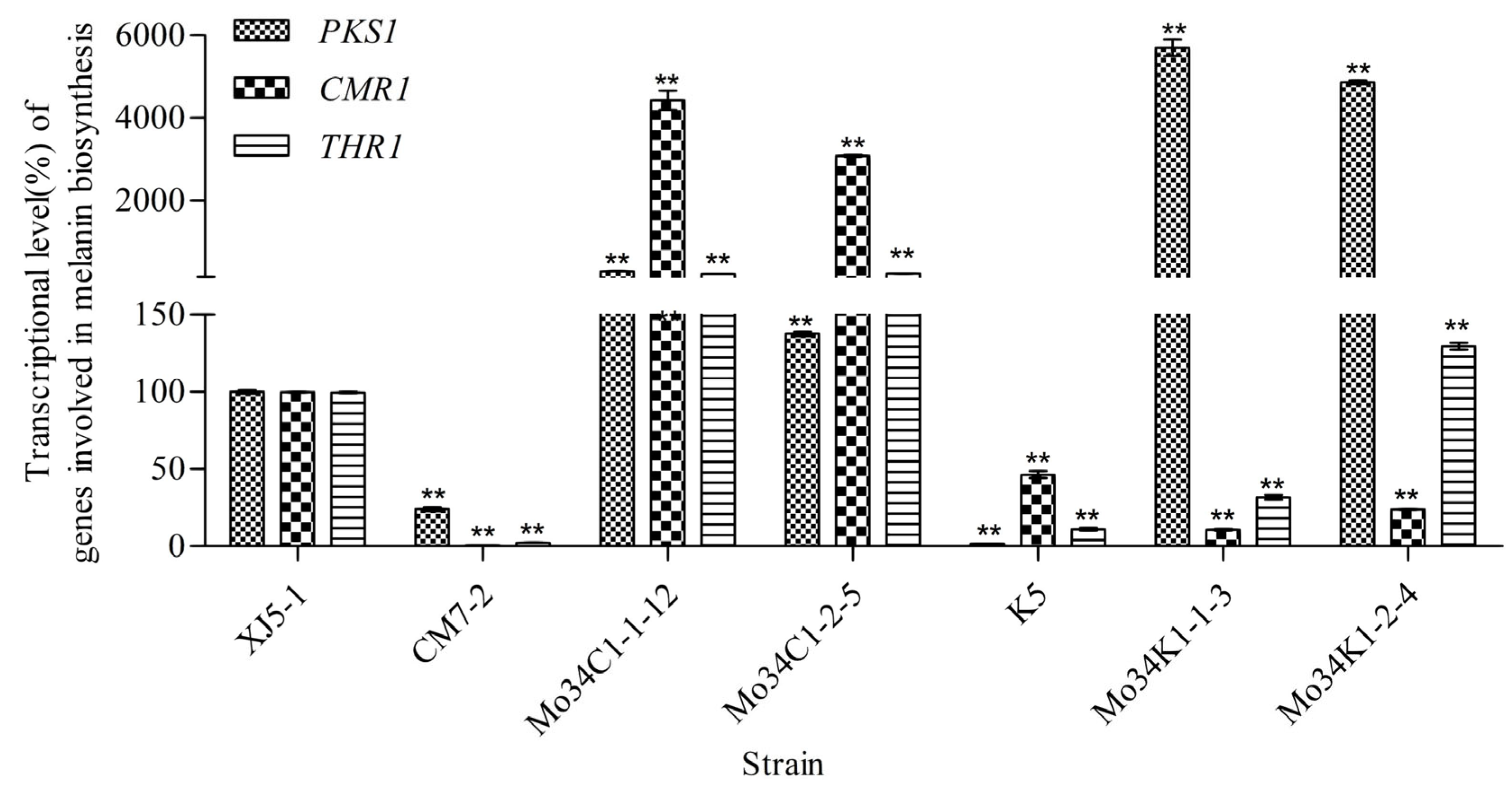
3.2. Expression of the PKS1-1, PKS1-2, CMR1-1, and CMR1-2 Genes from H. werneckii Mo34 in the Albino Mutants of A. melanogenum XJ5-1

In our previous studies [9], it was firmly established that the albino mutant K5, from which the NR-PKS1 gene was completely removed, and the albino mutant CM7-2, from which the CMR1 gene was entirely deleted, are unable to synthesize any melanin. In contrast, their parental strain, A. melanogenum XJ5-1, can produce a considerable amount of melanin. To ascertain the roles of the PKS1-1 and PKS1-2 genes from H. werneckii Mo34 in DHN melanin biosynthesis, as well as the involvement of the CMR1-1 and CMR1-2 genes in the regulation of DHN melanin biosynthesis, we expressed the PKS1-1 and PKS1-2 genes in the albino mutant K5, and the CMR1-1 and CMR1-2 genes in the albino mutant CM7-2, as detailed in the Section 2. The results presented in Figure 4 clearly indicated that all the colonies of the transformants carrying the PKS1-1, PKS1-2, CMR1-1, and CMR1-2 genes exhibited a black coloration, whereas the colonies of the Δpks1 mutant K5 and Δcmr1 mutant CM7-2 still remained white. This indicated that the PKS1-1, PKS1-2, CMR1-1, and CMR1-2 genes in the transformants were actively expressed, resulting in the restoration of melanin biosynthesis. This also indicated that the PKS1-1 and PKS1-2 genes in H. werneckii Mo34 were also implicated in DHN melanin biosynthesis, while the CMR1-1 and CMR1-2 genes in H. werneckii Mo34 were also involved in the regulation of DHN melanin biosynthesis. Thus, this strongly demonstrated that similar to other fungi [9], the black yeast H. werneckii, isolated from the deep-sea hydrothermal vent, possessed the same DHN melanin biosynthesis pathway.
One transformant, Mo34K1-1-3, derived from a colony on the plate A; another transformant, Mo34K1-2-4, from a colony on the plate B; a third transformant, Mo34C1-1-12, from a colony on the plate C; and a fourth transformant, Mo34C1-2-5, from a colony on the plate D (as shown in Figure 4), along with the albino mutants K5 and CM7-2 and the wild-type strain A. melanogenum XJ5-1, were each aerobically cultured in the PDB medium. Melanin was subsequently extracted from the cultured cells and quantitatively measured. The results presented in Figure 5 indicated that the transformants Mo34K1-1-3, Mo34K1-2-4, Mo34C1-1-12, and Mo34C1-2-5 synthesized nearly the same amount of melanin as the wild-type strain A. melanogenum XJ5-1. However, the albino mutants K5 and CM7-2 were unable to synthesize any melanin. This further demonstrated that, like any other fungi [9], the black yeast H. werneckii Mo34, isolated from the deep-sea hydrothermal vents, also possessed the DHN melanin biosynthesis pathway.
To confirm that the PKS1-1, PKS1-2, CMR1-1, and CMR1-2 genes from H. werneckii Mo34 (Figure S1) were indeed integrated into the genomic DNA of the transformants Mo34K1-1-3, Mo34K1-2-4, Mo34C1-1-12, and Mo34C1-2-5, the NAT gene encoding nourseothricin resistance (Figure S1) was PCR amplified using the genomic DNAs from the albino mutants K5 and CM7-2, as well as the transformants Mo34K1-1-3, Mo34K1-2-4, Mo34C1-1-12, and Mo34C1-2-5 as the templates, along with the primer pairs NAT-se/NAT-an (Table S1). The results presented in Figure S2 indicated that no PCR products for the nourseothricin resistance were obtained from the genomic DNA of the albino mutants K5 and CM7-2. In contrast, PCR products (570 bp) of the nourseothricin resistance gene were successfully amplified from the genomic DNAs of the transformants Mo34K1-1-3, Mo34K1-2-4, Mo34C1-1-12, and Mo34C1-2-5. This clearly demonstrated that the linear DNA fragments containing the NAT gene (Figure S1) were successfully integrated into the genomic DNAs of the transformants Mo34K1-1-3, Mo34K1-2-4, Mo34C1-1-12, and Mo34C1-2-5, where they were actively expressed. Thus, the transformants Mo34K1-1-3, Mo34K1-2-4, Mo34C1-1-12, and Mo34C1-2-5 could produce a significant amount of melanin (Figure 4 and Figure 5).
Following the determination of the relative transcriptional levels of the genes associated with melanin biosynthesis, the results presented in Figure 6 indicated that when the relative transcriptional levels of the genes in the wild-type strain XJ5-1 were considered as 100%, the relative transcriptional levels of the CMR1 gene in the albino mutant CM7-1 and the PKS1 gene in the albino mutant K5 were both 0%. This finding indicated that the CMR1 and PKS1 genes in the mutants were indeed effectively removed. In contrast, the relative transcriptional levels of the CMR1, PKS1, and THR1 genes [10,11] in the transformants Mo34C1-1-12 and Mo34C1-2-5 were significantly upregulated, indicating that Cmr1, encoded by the CMR1 gene from H. werneckii Mo34, could indeed promote the expression of the key PKS1 and THR1 genes involved in melanin biosynthesis. However, the relative transcriptional levels of only the PKS1 gene were significantly elevated in the transformants Mo34K1-1-3 and Mo34K1-2-4. These results further underscored the capacity of the transformants Mo34K1-1-3, Mo34K1-2-4, Mo34C1-1-12, and Mo34C1-2-5 to produce substantial amounts of melanin (Figure 4 and Figure 5).

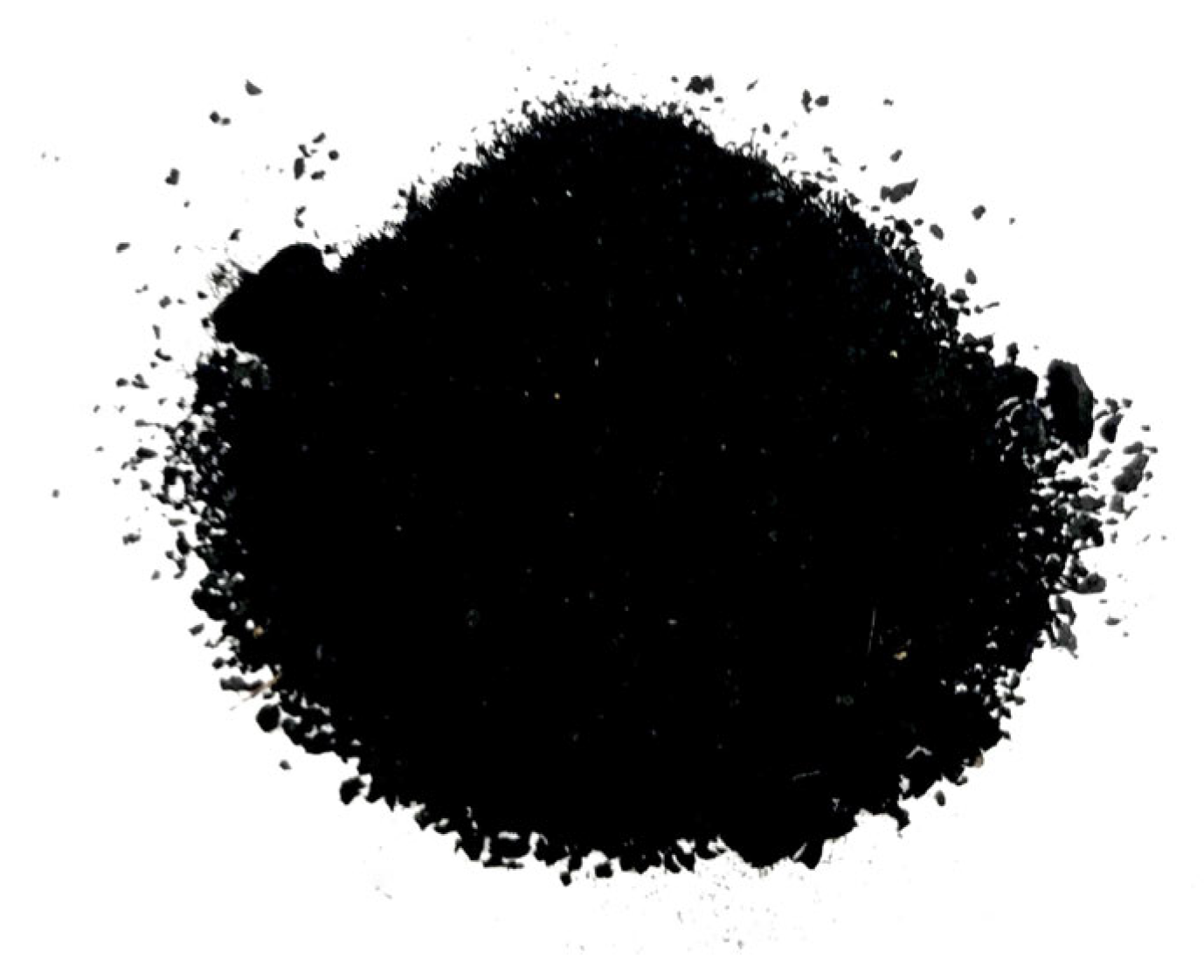
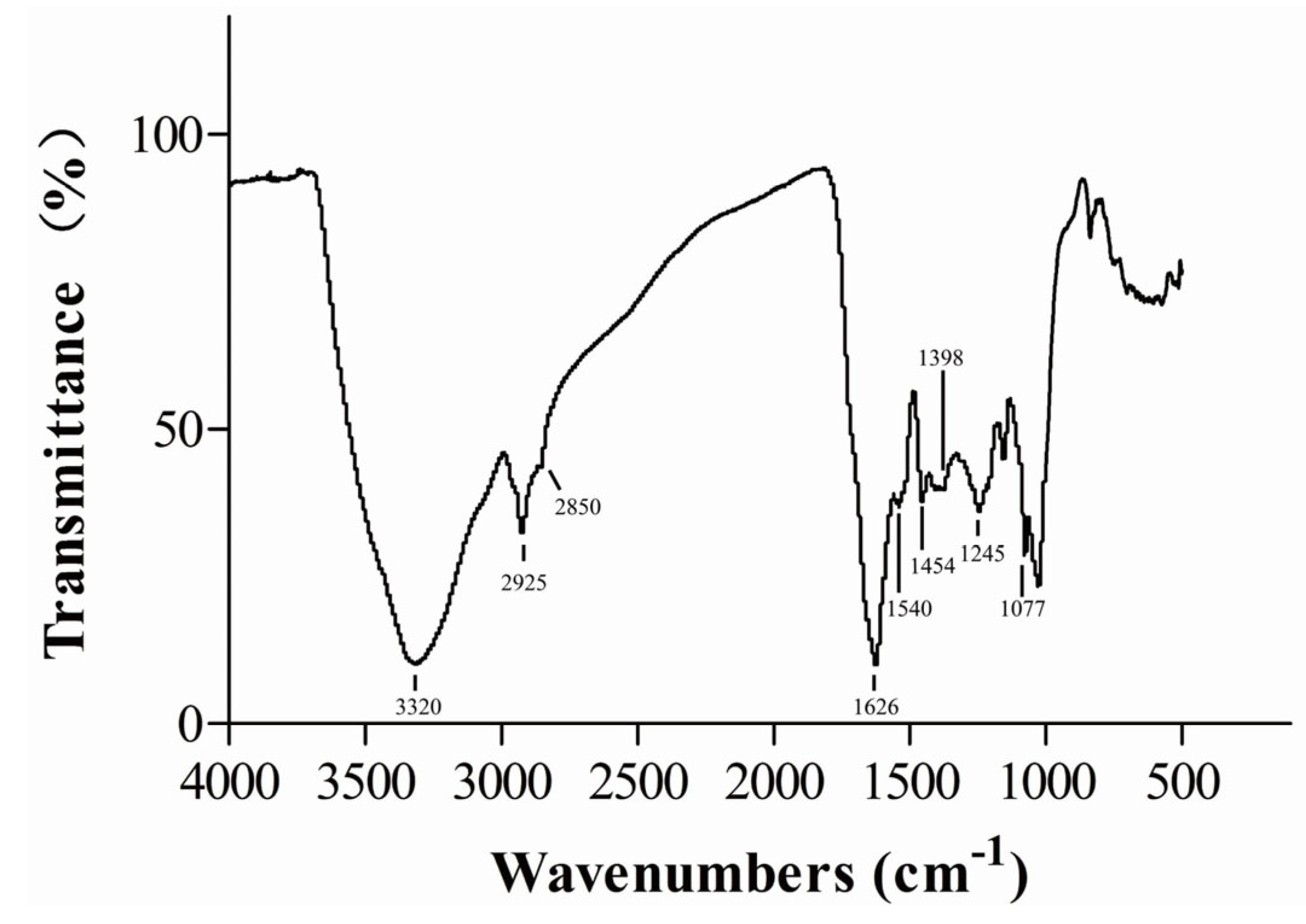
3.3. Chemical and Physical Properties of Melanin Produced by H. werneckii Mo34

Following the extraction and purification of melanin produced by H. werneckii Mo34, the resulting purified melanin sample was presented in Figure 7. As indicated in Figure 1, Figure 2, Figure 3, Figure 4, Figure 5 and Figure 6, H. werneckii Mo34 synthesized melanin via the DHN melanin biosynthesis pathway. Typically, the melanin synthesized via the DHN melanin biosynthesis pathway in Aspergillus fumigatus, Phyllosticta capitalensis, and A. melanogenum XJ5-1 exhibits black or brown coloration, with low sulfur (S) and nitrogen (N) contents [9,19,20]. The result presented in Figure 7 was consistent with the observed color.
The purified melanin could not be dissolved in water, hydrochloric acid (HCl) solution (pH 2.5), or various organic solvents, including ethanol, methanol, trichloromethane, acetone, ethyl acetate, and butanol. However, it could be dissolved in dimethyl sulfoxide (DMSO) and sodium hydroxide (NaOH) solution (pH 12.0). Thus, the solubility of the melanin produced by H. werneckii Mo34 was similar to that of the melanin produced by Auricularia auricula and other fungi [9,21]. Indeed, melanin from H. werneckii R23, isolated from the Arabian Sea and other strains of H. werneckii, exhibit acid precipitation, alkaline solubilization, and insolubility in most organic solvents and water. Additionally, it possessed a moderately high percentage of nitrogen and a detectable proportion of sulfur [15].
After analyzing the purified melanin using Fourier-transform infrared (FT-IR) spectroscopy, the IR spectra presented in Figure 8 revealed a broad band at 3320 cm−1, indicating stretching vibrations of hydroxyl (OH) and amine (NH) groups. Additionally, the absorbance at 2925 cm−1 and 2850 cm−1 likely corresponded to the symmetrical and asymmetrical stretching of CH2 groups, or the aliphatic C–H stretching. The prominent peak at 1626 cm−1 corresponded to the vibrations associated with significant aromatic groups, specifically C=C stretching or C=O stretching. Moreover, the small absorbance values at 1454 cm−1, 1398 cm−1, and 1077 cm−1 were likely attributable to the bending and stretching of amine (NH), ethyl (CH3CH2), or carbonyl (C=O) functional groups. The absorbance values at 1540 cm−1 and 1398 cm−1 strongly suggested the presence of a pyrrole or indole nitrogen–hydrogen (NH) group. Therefore, the purified melanin in this study was also a hybrid polymer with a high molecular weight. Furthermore, the IR spectra of the purified melanin were similar to those of melanins produced by A. auricula, A. fumigatus, P. capitalensis, and the marine black yeast H. werneckii R23 [15,19,21]. The results in Figure 8 indeed showed that the melanin produced by H. werneckii Mo34 also possessed a moderately high percentage of nitrogen.

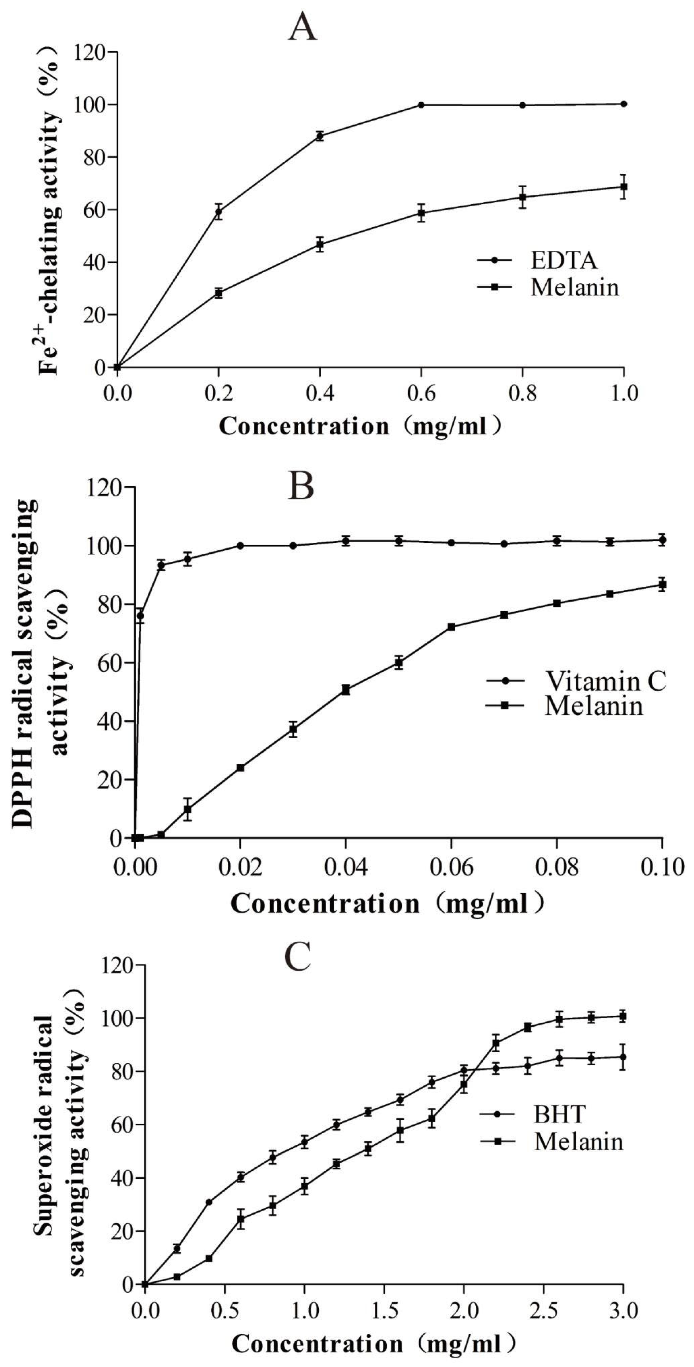
3.4. Bioactivity of the Purified Melanin

Fe2+, a strong oxidizing agent, can initiate pro-oxidant activity during lipid oxidation. Therefore, any compound possessing Fe2+-chelating ability is likely to exhibit strong antioxidant activity. The results shown in Figure 9A indicated that as the concentration of either the purified melanin or EDTA increased, their Fe2+-chelating ability also increased. When the concentration of the purified melanin exceeded 0.8 mg mL−1, its Fe2+-chelating ability reached nearly 60%. The IC50 values for the Fe2+-chelating ability of EDTA and the purified melanin were 0.18 ± 0.02 mg mL−1 and 0.46 ± 0.03 mg mL−1, respectively. These activities were nearly identical to those of melanin isolated from A. auricula [21]. Given that the melanin produced by H. werneckii Mo34 contained numerous -OH groups capable of binding Fe2+ (Figure 8), it was reasonable to conclude that this melanin had high antioxidant activity (Figure 9A).
Because free radicals are highly toxic to food and living organisms, it is crucial for antioxidant agents to possess the ability to eliminate free radicals from both food and biological systems. The DPPH method employed in this study is the simplest and the most reliable technique for evaluating the radical scavenging ability of antioxidants (Section 2.14). After determining the radical scavenging ability of the purified melanin, we found that its IC50 value for radical scavenging was 38.34 ± 7.28 µg mL−1. When the concentrations of the purified melanin and vitamin C were 100 µg mL−1, their radical scavenging abilities were 86.8 ± 4.1% and 101.12 ± 1.20%, respectively (Figure 9B). Therefore, the radical scavenging ability of the purified melanin was higher than that of the melanin produced by A. auricula, palm, the marine black yeast H. werneckii R23, black chicken, and chemically synthesized melanin, and was nearly equal to that of the melanin produced by Streptomyces sp. ZL-24 [22], but lower than that of vitamin C (Figure 9B).
Although the superoxide radical is a relatively weak oxidant, its binding with nitric oxide (NO) and other active substances can create a strong oxidant that induces lipid peroxidation. After measuring the superoxide radical scavenging activities of the purified melanin and the commonly used industrial antioxidant BHT, the results presented in Figure 9C indicated that the IC50 values for the superoxide radical scavenging activity of the purified melanin and BHT were 0.82 ± 0.01 mg mL−1 and 1.23 ± 0.01 mg mL−1, respectively. However, when the concentration of the purified melanin exceeded 2.0 mg mL−1, its superoxide radical scavenging activity surpassed that of BHT (Figure 9C). Furthermore, its superoxide radical scavenging activity even surpassed that of the melanins produced by A. auricula, palm, the marine black yeast H. werneckii R23, black chicken, and chemically synthesized melanin [15].
The bioactivities of the purified melanin, as shown above, demonstrated its high potential for application in the cosmetic industry for protecting the skin against oxidative damage and serving as potent inhibitors of peroxidative damage. In addition, melanin from H. werneckii R23 exhibits strong antioxidant potential as a reactive oxygen species (ROS) scavenger, demonstrated through the in vitro ABTS (2,2-azinobis-3-ethyl-benzothiazoline-6-sulfonic acid) radical scavenging assay [15].
The antimicrobial activity of the purified melanin was also evaluated in this study. The results presented in Table 1 indicated that the purified melanin actively inhibited the growth of the pathogenic bacteria S. aureus and P. putida, with minimum inhibitory concentrations (MICs) of 0.3 mg mL−1 and 1.0 mg mL−1, respectively. It can also be observed that the purified melanin also weakly inhibited the cell growth of B. subtilis, as well as the animal pathogenic bacteria V. harveyi, V. anguillarum, P. aeruginosa, and A. baumannii. It has been reported that melanin can inhibit the formation of biofilm in S. aureus, with a minimum inhibitory concentration (MIC) of 0.30 mg mL−1. Additionally, the melanin from Streptomyces sp. ZL-24 can also inhibit biofilm formation in S. aureus and P. aeruginosa [22,23]. However, the melanin isolated from wine waste at a concentration of 40 mg mL−1 can completely inhibit the growth of B. subtilis G17-89 and Candida gropengiesseri 10228 [24]. Therefore, the purified melanin utilized in this study demonstrated potent antibacterial activity against S. aureus and P. putida. However, it is still completely unknown about the mechanisms of antibacterial activity of the purified melanin.

4. Conclusions

In summary, in this study, the PKS1-1, PKS1-2, CMR1-1, and CMR1-2 genes cloned from the black deep-sea yeast H. werneckii Mo34 could be successfully complemented in the ∆pks1 albino mutant K5 and the ∆cmr1 albino mutant CM7-2 of A. melanogenum XJ5-1, causing restoration of melanin biosynthesis in the complementing strains. Furthermore, Cmr1-1 and Cmr1-2 encoded by the CMR1-1 and CMR1-2 genes from H. werneckii Mo34 could regulate melanin biosynthesis in A. melanogenum XJ5-1. This was the first time for obtaining the genetic evidence to show that H. werneckii Mo34 also had a DHN melanin biosynthesis pathway which was regulated by the specific transcriptional activators Cmr1-1 and Cmr1-2. The purified melanin from H. werneckii Mo34 exhibited excellent Fe2⁺-chelating, DPPH radical-scavenging, superoxide radical-scavenging activities, and potent antibacterial activity against S, aureus and P. putida.

Supplementary Materials

The following supporting information can be downloaded at: https://www.mdpi.com/article/10.3390/jmse13061004/s1, Figure S1: Construction of the plasmids for heterologous expression of the PKS1-1 gene, PKS1-2 gene and CMR1-1 gene and CMR1-2 gene from the strain Mo34; Figure S2: Confirmation of the NAT gene integrated in the genomic DNA of the transformants Mo34K1-1-3 (2), Mo34K1-2-4 (3), Mo34C1-1-12 (5) and Mo34C1-2-5 (6). No such PCR products (lanes 1 and 4) were amplified from the genomic DNAs of the albino mutants K5 and CM7-2. M was D2000 DNA Marker, the DNA sizes from top to bottom were 2000 bp, 1000 bp, 750 bp, 500 bp, 200 bp and 100 bp; Table S1: The primers used in cloning of the PKS1 and CMR1 genes of strain Mo34; Table S2: Websites for bioinformatic analysis; Table S3: The primers used in the Fluorescent real-time PCR.

Author Contributions

Methodology, H.-J.L.; Investigation, H.-J.L.; Writing—review & editing, Z.-M.C.; Supervision, Z.-M.C. All authors have read and agreed to the published version of the manuscript.

Funding

This work was supported by the National Key R&D Program of China, China (2021YFC2103200). National Natural Science Foundation of China, China (Grant No. 31970058), the key research and development of Shandong Province, China (Grant No. 2019GSF107097), and the Fundamental Research Funds for the Central Universities (Grant No. 31500029).

Data Availability Statement

The raw data supporting the conclusions of this article will be made available by the authors on request.

Conflicts of Interest

The authors declare that they have no known competing financial interests or personal relationships that could have appeared to influence the work reported in this paper.

References

  1. Mitchison-Field, L.M.Y.; JVargas-Muniz, J.M.; Stormo, B.M.; Lew, D.J.; Field, C.M.; Gladfelter, A.S. Unconventional cell division cycles from marine derived yeasts. Curr. Biol. 2019, 29, 3439–3456. [Google Scholar] [CrossRef] [PubMed]
  2. Lenassi, M.; Gostincar, C.; Jackman, S.; Turk, M.; Sadowsk, I.; Nislo, C.; Jones, S.; Biro, I.; Gunde-Cimerman, N.; Plemenitas, A. Whole genome duplication and enrichment of metal cation transporters revealed by De Novo genome sequencing of extremely halotolerant black yeast Hortaea werneckii. PLoS ONE 2013, 8, e71328. [Google Scholar] [CrossRef] [PubMed]
  3. Romeo, O.; Marchetta, A.; Giosa, D.; Giuffrè, L.; Urzì, C.; De Leo, F. Whole genome sequencing and comparative genome analysis of the halotolerant deep sea black yeast Hortaea werneckii. Life 2020, 10, 229. [Google Scholar] [CrossRef] [PubMed]
  4. Ling, M.; Zhang, K.; Hu, J.; Huang, X.; Fan, G.; Grossart, H.P.; Luo, Z. Complete genome sequencing of Hortaea werneckii M-3 for identifying polyester polyurethane degrading enzymes. Mar. Genom. 2024, 75, 101111. [Google Scholar] [CrossRef]
  5. Cochereau, B.; Le Strat, Y.; Ji, Q.; Pawtowski, A.; Delage Amélie Weill, L.; Mazéas, L.; Hervé, C.; Burgaud, G.; Gunde-Cimerman, N.; François Pouchus, Y.; et al. Heterologous expression and biochemical characterization of a new chloroperoxidase isolated from the deep-sea hydrothermal vent black yeast Hortaea werneckii UBOCC-A-208029. Mar. Biotechnol. 2023, 25, 519–536. [Google Scholar] [CrossRef]
  6. Gostinčar, C.; Gunde-Cimerman, N. Black yeasts in hypersaline conditions. Appl. Microbiol. Biotechnol. 2024, 108, 252. [Google Scholar] [CrossRef]
  7. Elsayis, A.; Hassan, S.W.M.; Ghanem, K.M.; Khairy, H. Optimization of melanin pigment production from the halotolerant black yeast Hortaea werneckii AS1 isolated from solar salter in Alexandria. BMC Microbiol. 2022, 22, 92. [Google Scholar] [CrossRef]
  8. Salah El-Din Hodhod, M.; Zakaria Gaafar, A.; Alshameri, A.; Qahtan, A.A.; Abdulrahman Noor, A.; Abdel-Wahab, M. Molecular characterization and bioactive potential of newly identified strains of the extremophilic black yeast Hortaea werneckii isolated from red sea mangrove. Biotechnol. Biotechnol. Equip. 2020, 34, 1288–1298. [Google Scholar] [CrossRef]
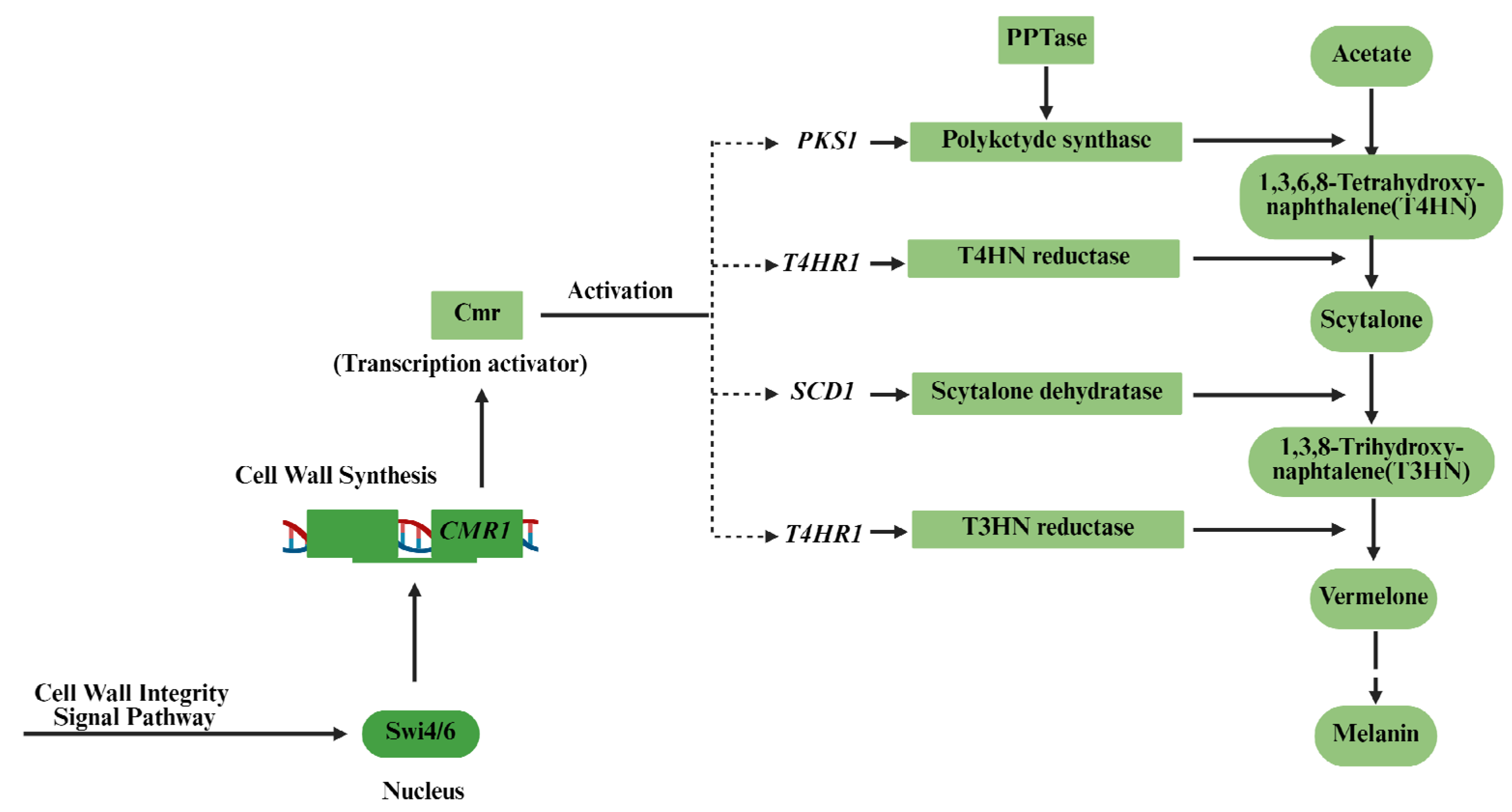
  9. Jiang, H.; Chi, Z.; Liu, G.L.; Hu, Z.; Zhao, S.Z.; Chi, Z.M. Melanin biosynthesis in the desert-derived Aureobasidium melanogenum XJ5-1 is controlled mainly by the CWI signal pathway via a transcriptional activator Cmr1. Curr. Genet. 2020, 66, 173–185. [Google Scholar] [CrossRef]
  10. Chi, Z.; Wei, X.; Ge, N.; Jiang, H.; Liu, G.L.; Chi, Z.M. NsdD, a GATA-type transcription factor is involved in regulation and biosynthesis of macromolecules melanin, pullulan, and polymalate in Aureobasidium melanogenum. Int. J. Biol. Macromol. 2024, 268, 131820. [Google Scholar] [CrossRef]
  11. Burgaud, G.; Arzur, D.; Durand, L.; Cambon-Bonavita, M.A.; Barbie, G. Marine culturable yeasts in deep-sea hydrothermal vents: Species richness and association with fauna. FEMS Microbiol. Ecol. 2010, 73, 121–133. [Google Scholar] [CrossRef] [PubMed]
  12. Chi, Z.; Wang, X.X.; Ma, Z.C.; Buzdar, M.A.; Chi, Z.M. The unique role of siderophore in marine-derived Aureobasidium pullulans HN6.2. Biometals 2012, 25, 219–230. [Google Scholar] [CrossRef]
  13. Liu, G.L.; Wang, D.S.; Wang, L.F.; Zhao, S.F.; Chi, Z.M. Mig1 is involved in mycelial formation and expression of the genes encoding extracellular enzymes in Saccharomycopsis fibuligera A11. Fungal Genet. Biol. 2011, 48, 904–909. [Google Scholar] [CrossRef]
  14. Ma, Z.C.; Liu, N.N.; Chi, Z.; Liu, G.L.; Chi, Z.M. Genetic modification of the marine-isolated yeast Aureobasidium melanogenum P16 for efficient pullulan production from inulin. Mar. Biotechnol. 2015, 17, 511–522. [Google Scholar] [CrossRef]
  15. Wilson, W.; Lowman, D.; Puthumana, J.; Kuriakose, R.; Bright Singh, I.S.; Philip, R. Biocompatible melanin from the marine black yeast Hortaea werneckii R23 with antioxidant and photoprotection property. Braz. J. Microbiol. 2024, 55, 3171–3186. [Google Scholar] [CrossRef]
  16. Kogej, T.; Wheeler, M.H.; Lanisnik Rizner, T.; Gunde-Cimerman, N. Evidence for 1,8-dihydroxynaphthalene melanin in three halophilic black yeasts grown under saline and non-saline conditions. FEMS Microbiol. Lett. 2004, 232, 203–209. [Google Scholar] [CrossRef]
  17. Beld, J.; Sonnenschein, E.C.; Vickery, R.V.; Noel, J.P.; Burkart, M.D. The phosphopantetheinyl transferases: Catalysis of a post-translational modification crucial for life. Nat. Prod. Rep. 2014, 31, 61–108. [Google Scholar] [CrossRef]
  18. Tsuji, G.; Takano, T.K.Y.; Sweigard, J.; Sweigard, J.; Farrall, L.; Furusawa, I.; Horino, O.; Kubo, Y. Novel fungal transcriptional activators, Cmr1p of Colletotrichum lagenarium and Pig1p of Magnaporthe grisea, contain Cys2His2 zinc finger and Zn(II)2Cys6 binuclear cluster DNA-binding motifs and regulate transcription. Mol. Microbiol. 2000, 38, 940–954. [Google Scholar] [CrossRef]
  19. Raman, N.; Ramasamy, S. Genetic validation and spectroscopic detailing of DHN-melanin extracted from an environmental fungus. Biochem. Biophy. Rep. 2017, 12, 98–107. [Google Scholar] [CrossRef]
  20. Suryanarayanan, T.S.; Ravishankar, J.P.; Venkatesan, G.; Murali, T.S. Characterization of the melanin pigment of a cosmopolitan fungal endophyte. Mycol. Res. 2004, 108, 974–978. [Google Scholar] [CrossRef]
  21. Zou, Y.; Hu, W.; Ma, K. Physicochemical properties and antioxidant activities of melanin and fractions from Auricularia auricula fruiting bodies. Food Sci. Biotechnol. 2015, 24, 15–21. [Google Scholar] [CrossRef]
  22. Wang, L.; Li, Y.; Li, Y. Metal ions driven production, characterization and bioactivity of extracellular melanin from Streptomyces sp. ZL-24. Int. J. Biol. Macromol. 2018, 123, 521–530. [Google Scholar]
  23. Xu, C.; Li, J.; Yang, L. Antibacterial activity and a membrane damage mechanism of Lachnum YM30 melanin against Vibrio parahaemolyticus and Staphylococcus aureus. Food Control 2017, 73, 1445–1451. [Google Scholar] [CrossRef]
  24. Minasyan, E.; Aghajanyan, A.; Karapetyan, K.; Khachaturyan, N.; Hovhannisyan, G.; Yeghyan, K.; Tsatury, A. Antimicrobial activity of melanin isolated from wine waste. Indian J. Microbiol. Biotechnol. 2014, 64, 1528–1534. [Google Scholar] [CrossRef]
Figure 1. DHN melanin synthesis pathway in fungi.
Jmse 13 01004 g001
Figure 2. The phylogenetic tree of the polyketide synthases Pks1 from different fungi based on a neighbor-joining analysis and the corresponding domains. Bootstrap values (1000 pseudoreplications) were ≥53%.
Jmse 13 01004 g002
Figure 3. The phylogenetic tree of the melanin-specific transcriptional factors Cmr1 from different fungi based on a neighbor-joining analysis and corresponding domains. Bootstrap values (1000 pseudoreplications) were ≥99%.
Jmse 13 01004 g003
Figure 4. The colonies of the ∆pks1 mutant K5 (A,B) and the ∆cmr1 mutant CM7-2 (C,D) were white while those of all the transformants were black. All the mutants were grown in the PDA plates at 28 °C for 7 days.
Jmse 13 01004 g004
Figure 5. The relative amount of melanin produced by different mutants, transformants in which the corresponding genes had been complemented, and their wild type strain XJ5-1. Data are given as mean ± SD, n = 3. ** Means significantly different. All the yeast-like strains were grown in the liquid PDB medium at 28 °C for 7 d.
Jmse 13 01004 g005
Figure 6. The relative transcriptional levels of different genes in different mutants, transformants, and their wild-type strain XJ5-1. Data are given as mean ± SD, n = 3. ** Meant significantly different. The key THR1 gene (KU844849.1) encoding 1,3,8-trihydroxylaphthalene reductase Thr1 for melanin biosynthesis.
Jmse 13 01004 g006
Figure 7. The purified melanin produced by H. werneckii Mo34.
Jmse 13 01004 g007
Figure 8. Infrared spectra of the purified melanin produced by Mo34.
Jmse 13 01004 g008
Figure 9. Fe2+-chelating activity (A), DPPH radical scavenging activity (B) and superoxide radical scavenging activity (C) of the purified melanin produced by H. werneckii Mo34. The reaction conditions are shown in Section 2.12, Section 2.13, and Section 2.14.
Jmse 13 01004 g009
Table 1. Antibacterial activity of the purified melanin against tested bacteria.
StrainConcentrations (mg/mL)
0.10.150.20.30.51.02.03.0
S. saureus (G+)++++++-----
B. subtilis (G+)+++++++++++
P. putida (G)++++++++---
V. harveyi (G)+++++++++++++++++++++
V. anguillarum (G)+++++++++++++++++++++
P. aeruginosa (G)++++++++++++++++++++++
A. baumannii (G)+++++++++++++++++++++++
E. cloacae (G)+++++++++++++++++++++++
-: no growth; +: considerable arrest; ++: turbid solution; +++: highly turbid solution (strong growth). All the sensitive bacteria were grown on the liquid 2166E medium at 37 °C and 960 rpm for 4 h. G+: Gram positive; G: Gram negative.
Disclaimer/Publisher’s Note: The statements, opinions and data contained in all publications are solely those of the individual author(s) and contributor(s) and not of MDPI and/or the editor(s). MDPI and/or the editor(s) disclaim responsibility for any injury to people or property resulting from any ideas, methods, instructions or products referred to in the content.

