Article

Preliminary Biological Assessments of Some Algae Basis Biomaterials

1
Faculty of Biotechnologies, University of Agronomic Sciences and Veterinary Medicine of Bucharest, 59 Marasti Boulevard, District 1, 011464 Bucharest, Romania
2
Department of Biotechnologies, National Institute of Chemistry and Petrochemistry R&D of Bucharest, 202 Splaiul Independentei Street, District 6, 060021 Bucharest, Romania
3
Department of Quantum Chemistry and Molecular Structure, Institute of Physical Chemistry “Ilie Murgulescu”, 202 Splaiul Independentei St. 060021, 060021 Bucharest, Romania
4
Department of Pharmaceutical Biotechnologies, National Institute of Chemical Pharmaceutical Research and Development, 112 Vitan, 031299 Bucharest, Romania
5
Center of Immunology, Institute of Virology Stefan S, Nicolau, 285 Mihai Bravu Avenue, 030304 Bucharest, Romania
6
Faculty of Pharmacy, Titu Maiorescu University, 16 Gheorghe Sincai Blvd, 040314 Bucharest, Romania
*
Author to whom correspondence should be addressed.
J. Mar. Sci. Eng. 2025, 13(2), 318; https://doi.org/10.3390/jmse13020318
Submission received: 5 January 2025 / Revised: 28 January 2025 / Accepted: 7 February 2025 / Published: 9 February 2025

Abstract

:
Four types of algae—Porphyra umbilicalis, Undaria pinnatifida, Cystoseira barbata, and Chlorella sp.—were used to obtain crude bioproducts enriched in polysaccharides (four bioproducts) and to create formulations enriched with gold cations (four bioproducts). The bioproducts obtained through aqueous extraction from Cystoseira barbata exhibited significant antioxidant activities and a total polyphenol content of (714.17 ± 1.26) mg GAE/L. In the bioproducts derived from the aqueous extract of Porphyra umbilicalis and Undaria pinnatifida, combined with gold ions, gold nanoparticles with sizes of less than 34 nm were formed. In vitro tests performed on the Caco-2 tumour cell line with each of the eight bioproducts, after 24 h of exposure, showed that the crude bioproducts containing polysaccharides derived from Porphyra umbilicalis, Undaria pinnatifida, and Chlorella sp. exhibited cytotoxicity against the Caco-2 cell line. In the case of the HepG2 cell line, after 24 h of exposure, the tests indicated that only the crude polysaccharides derived from Cystoseira barbata exhibited cytotoxic effects. These results indicate the protective effect of the algal polysaccharides against the tumourigenesis processes that may occur in the human digestive system. Regarding the bioproducts containing gold, no cytotoxic effect was observed. However, in the case of the two algal bioproducts containing gold nanoparticles with a size of less than 34 nm, they may represent potential raw materials for electrochemical sensors.

1. Introduction

Currently, algae species such as Porphyra umbilicalis, Undaria pinnatifida, Cystoseira barbata, and Chlorella sp. are widely used as raw materials in the food industry [1,2,3] or as sources for bioactive compounds used in pharmacology and agriculture [4,5].
Porphyra umbilicalis, harvested from North Atlantic, contains fibres with prebiotic, antimicrobial, and antitumour properties, carotenoids, proteins (up to 40%), lipids (8.88 mg/g), polysaccharides such as porphyran, starch, and cellulose (25.37%), and minerals such as Na, K, Ca, and Co [6,7,8].
Undaria pinnatifida, widespread in the Pacific Ocean, Atlantic Ocean, and the Sea of Asia, is rich in minerals (Ca, Fe, Mn), proteins (168 mg/g), lipids (27 mg/g), dietary fibre (307 mg/g), and sterols (136 mg/kg). Undaria pinnatifida contains beta-carotene, B vitamins, fucoidan (a sulfonated polysaccharide), laminarin, and alginates, which are composed of elementary units of fucose, galactose, and glucose, with sulfonated sugars reaching up to 7.8% [9,10,11,12].
Cystoseira barbata, a brown algae species protected at both national and international levels, grows in the clean waters of the Atlantic Ocean, the Mediterranean Sea, and the Black Sea, its presence serving as an indicator of the absence of pollution [13]. Studies performed on the samples collected from the Black Sea indicate that this species contains proteins (12–25 g/kg), alginic acid (18–27%), magnesium (0.19–1.8 mg/g), phosphorus (79–131 mg/kg), and polyphenols (14.3–66.9 mg GAE/g dry biomass) [14]. Other studies have reported a total polyphenol content in Cystoseira barbata of 386 mg GAE/100 g dry biomass, and the presence of polyphenolic compounds such as pyrogallol, gallic acid, chlorogenic acid, and ferulic acid [15]. The fucoidans (sulfonated polysaccharides) isolated through extraction from Tunisian Cystoseira barbata contain sulphates (22%), proteins (5%), monosaccharides such as fucose (44.6%), galactose (33%), and glucose (7.55%) [16]. The extraction of polysaccharides from Cystoseira barbata can be achieved using water (normal or pressurised), organic solvents (ethanol/methanol), or advanced extraction techniques, such as extraction with supercritical fluids, ultrasound-assisted extraction, or extraction with pressurised fluids (50–200 °C, 3.5–20 MPa) [17]. These processes yield fucoidans with a molecular weight between (83.39–183.32) kDa [17]. Purification of the crude polysaccharides is essential to remove sulphates, lipids, proteins, metal cations, and pigments [18]. Studies performed on aqueous extracts obtained from Cystoseira barbata harvested from the Sea of Marmara, the Aegean Sea, and the Black Sea have indicated the presence of bioactive compounds such as terpenoids, fatty acids, steroids, and phlorotannins (polyphenols with 5–7 units of phloroglucinol) [19]. The extracts obtained at 70 °C from 100 g of algae biomass and 900 g of water contain alginate (11.6 g/L), laminarin (4.2 g/L), fucoidan (46 g/L), polyphenols (0.7 g GAE/L), amino acids (2.3 g/L), and mannitol (3 g/L). These extracts exhibit antioxidant properties (ABTS, DPPH, FRAP) and bio-stimulatory effects on Triticum aestivum crops [20]. Fucoidan isolated from Cystoseira barbata exhibits antioxidant effects (DPPH, Fe2+ chelation, Fe3+ reduction) [17], anti-inflammatory, antitumoural, and hepatoprotective activities [4]. Both fucoidan and laminarin show antitumoural activity in vitro against tumour cell lines, such as HCT116, HT-29, and DLD-1. The mechanisms of action are based on the inhibition of proliferation, migration, colony formation, mitochondrial depolarisation, nitric oxide production, and apoptosis induction [17].
Chlorella sp., cultivated in photobioreactors, contains lipids (10–50%) and carotenoids such as lutein (1.8–6 mg/g) and is used in human nutrition. Polysaccharides derived from Chlorella sp. are obtained through extraction with hot water, alcohol [3,21], or by extraction with supercritical fluids at 100–300 °C, which yields bioproducts containing 37% carbohydrates, 52% proteins, and 2.5% oils [22]. The biomass of Chlorella sp. is used in the food industry due to its high content of proteins (<65%), carbohydrates (8.1–65%), lipids (1.6–40%), fibres (1.6–6%), amino acids, fatty acids, vitamins, and pigments, including chlorophylls (1.16–24 mg/g) and carotenoids (0.24–8.21 mg/g) [23]. Carotenoides compounds such as beta-carotene, astaxanthin, and lutein exhibit antioxidant and antitumour properties [23,24,25]. The polysaccharides contained in Chlorella sp. reduce tumour cell proliferation and are effective against breast and skin tumours [23,24,25]. Extraction methods include boiling Chlorella powder with water (mass ratio 1:20), followed by ethanol precipitation and the isolation of polysaccharide compounds, which contain uronic acid, mannose, glucose, xylose, and arabinose as elementary units [24,25,26].
Marine-derived polysaccharides can reduce gold ions (Au3+) to metallic gold. This process can use the extracts obtained from boiling algae in water or direct treatment of algae with Au3+ solutions [27,28,29,30,31,32]. Polysaccharides derived from algae exhibit antioxidant activity comparable to that of extracts from terrestrial plants and serve as precursors for the production of biocompatible biomaterials (sponges, films) in medicine [33,34,35,36].
Algae produce antioxidants (enzymes such as SOD, CAT, APX; vitamins C and E; carotenoids such as lutein and astaxanthin; polyphenols such as gallic acid and quercetin) as part of their defence system [37,38,39,40,41]. However, under the influence of high concentrations of oxygen, carotenoids can act as prooxidants [42,43]. The antioxidant status of vitamins (such as vitamin C and alpha-tocopherol) can be altered in the presence of copper/iron ions and hydrogen peroxide (H2O2). Vitamin C reduces Fe3+ to Fe2+ and Cu2+ to Cu+ through Fenton-like reactions, while alpha-tocopherol, at high concentrations, becomes a radical unless regenerated by vitamin C [44].
Polyphenolic compounds (kaempferol, quercetin, myricetin) are similarly affected by H2O2 in the presence of heavy metals. Analyses performed on Cystoseira barbata harvested from the Black Sea have shown a variable content of this in heavy metals, with copper concentrations reaching 4.19 mg/kg in 2015 [45,46].
Commercial products containing Chlorella sp., Undaria pinnatifida, and Porphyra umbilicalis also contain metal cations such as Cu, Fe, Mn, Mo, and Zn [47,48,49,50,51]. Chlorella sp. contains polysaccharides with antitumour properties, known as immulina and immurela [52], and secretes exopolysaccharides that include D-mannose, glucosamine, D-glucose, and D-galactose [53]. Bioproducts based on polysaccharides obtained from Chlorella pyrenoidosa exhibit antitumour effects on human colon adenocarcinoma cells (HT-29) [52,53,54].
Algae such as Porphyra umbilicalis, Undaria pinnatifida, and Chlorella sp. are currently widely used in food products that involve boiling, particularly in Asian diets [1,2]. They are also incorporated into various processes within the food industry to enhance products and improve their sensory properties [8,12,23].
Statistical research conducted on Asian populations, where algae-based foods requiring boiling form a significant part of the daily diet, has shown that here, the incidence of cancer is significantly lower compared to typical diets of Europeans or populations in Western countries [3]. These findings are supported by in vitro studies using biomaterials based on polysaccharides derived from microalgae such as Chlorella sp., obtained via aqueous boiling, which have demonstrated antiproliferative properties through mechanisms involving DNA damage and apoptosis [25,52].
Given that dried algae such as Porphyra umbilicalis, Undaria pinnatifida, and Chlorella sp. are widely available on the European market, and considering previous research indicating that gold-enriched extracts of algal polysaccharides may exhibit antitumour effects [30,31,52,54], we aimed to develop bioproducts enriched with algal polysaccharides, with or without gold, to assess their preliminary antiproliferative properties. The algal bioproducts were obtained using a simplified procedure that mimics the processes employed in the food industry, where these raw materials are typically boiled.
For this purpose, we used the following algal species as raw materials. Two algal strains from the Romanian market (i.e., Porphyra umbilicalis and Undaria pinnatifida) and one algal strain from the Black Sea (i.e., Cystoseira barbata) were provided by Ovidius University of Constanța. Cystoseira barbata was harvested during flora and fauna inventory studies conducted in the Black Sea. The microalga Chlorella sp. was obtained by multiplication in photobioreactor.
The studies were performed with the following aims:
1.
Obtaining crude polysaccharide extracts (A1–A4) from four algae species—Porphyra umbilicalis (A1), Undaria pinnatifida (A2), Cystoseira barbata (A3), and Chlorella sp. (A4);
2.
Developing gold-enriched bioproducts by adding Au3+ ions in standardised extracts and characterising gold-enriched bioproducts (A1+Au, A2+Au, A3+Au, A4+Au);
3.
Evaluating the antioxidant or prooxidant activities of the algae extracts;
4.
Assessing bioproducts cytotoxicity by tests performed in vitro on three standardised human cell lines: HUVEC, Caco-2, and HepG2.

2. Materials and Methods

2.1. Obtaining Bioproducts with Polysaccharides

The polysaccharide-based bioproducts were obtained from four types of algae; two were acquired from the Romanian market (Plafar, Bucharest, Romania) (i.e., Porphyra umbilicalis; Undaria pinnatifida), one from the Black Sea (Cystoseira barbata), and the last one (Chlorella sp. CCAP 211/102) was obtained by multiplication in a Sartorius PBS 25S photobioreactor (Sartorius Stedim Biotech GmbH, Göttingen, Germany) in Jaworoski culture medium (Table 1). The crude bioproducts enriched in polysaccharides were obtained from each alga, according to the flowchart presented Figure 1a.
In this regard, the dried and crushed algae were subjected to extraction with hot water under reflux for 2 h, at 95 °C. After cooling, the suspension was separated by centrifugation at 8000 rpm (Hettich Universal 320, Nitech, Bucharest, Romania). The supernatant was concentrated using a rotavapor (Heidolph, Schwabach, Germany) at 50 °C.
The crude biomaterial obtained in each case was weighed and then dissolved in a minimal amount of aqueous solution of dimethyl sulfoxide (DMSO) with c = 20% (Merck, Bucharest, Romania). The concentration of each crude extract in the solution obtained in this manner is presented in the Table 1.
The bioproducts containing gold were synthesised from each polysaccharide extract using a 1 mM solution of tetrachloroauric (III) acid trihydrate (Thermo Fisher Scientific, Waltham, MA, USA) prepared with distilled water, as outlined in Figure 1b. To obtain the bioproducts with gold, one part of the algal crude extract was mixed with 4.5 (or 4.55) parts of a 1 mM Au3+ solution. The mixture was left to mature in the dark for 48 h. After this period, each solution was analysed using the DLS technique to determine the particle size. The final gold concentrations in each solution are presented in the Table 1, as mM ± STDEV (standard deviation).

2.2. Antioxidant Methodology

The antioxidant activity (AA) was assessed by chemiluminescence (CL) using luminol (Sigma Aldrich, Bucharest, Romania) and a GLOMAX 20/20 luminometer (model E 5321-PROMEGA, Promega GmbH, Walldorf, Germany), operated at a wavelength of λ = 430 nm. Chemiluminescence studies were conducted on solutions of each sample within a concentration range of 10–30 µL/mL.
Reagents:
  • Luminol (LH2) solution: 5-amino-2,3-dihydro-1,4-phthalazinedione (c = 2.5 × 10⁻⁵ M, prepared in DMSO) (Merck, Bucharest, Romania);
  • TRIS-HCl buffer solution c = 50 mM, pH = 8.5; (Sigma Aldrich, Bucharest, Romania):
  • H2O2, c= 30 mM; (Merck, Bucharest, Romania);
  • Witness (control): a mixture containing 200 μL LH2, 750 μL buffer solution, and 50 μL H2O2.
The testing of each sample was performed by adding the reagents in the following sequence: 200 μL LH2 + 700 μL buffer solution + 50 μL sample + 50 μL H2O2. The antioxidant activity (%) of the extracts, based on the scavenging of oxygen free radicals, was calculated using Equation (1):
A.A. = (Iw − Is/Iw) × 100
Iw and Is represent the intensities of chemiluminescence measured between 5 and 170 s from the reaction start for the witness (Iw) and each sample (Is), respectively. All measurements were performed in triplicate, and the results are expressed as average values with corresponding standard deviation (STDEV).

2.3. Polyphenols Content Evaluation

The polyphenol content was determined using the Folin–Ciocalteu method. A volume of 1.5 mL of water was mixed with 100 μL of the Folin–Ciocalteu reagent (Sigma Aldrich, Bucharest, Romania). Subsequently, 100 μL of the algal extract and 300 μL of a 20% Na2CO3 solution (Merck, Bucharest, Romania) were added to the mixture in the specified order. The solution was then left to stand in the dark for 120 min. After this period, the absorbance was measured spectrophotometrically at 765 nm, using water as the reference. The calibration curve was prepared with gallic acid (Merck, Bucharest, Romania) and bidistilled water. The results were expressed as mgGAE/L ± STDEV.

2.4. Characterisation of Bioproducts Obtained from Au3+ and Aqueous Extracts of Algae

Measurements of particle size in the samples with gold were performed using dynamic light scattering (DLS), also known as Photon Correlation Spectroscopy (PCS). The analyses were conducted on a Nano ZS Red Badge ZEN3600 device (Malvern Instruments Ltd., Worcestershire, UK). Bioproducts containing crude algal extracts, as well as those combined with gold, are not purified. At this stage, since the biological activities observed in this study for the bioproducts with gold are weaker compared to those of the crude extracts, it is not justified to invest additional resources to perform a second or type of measurement.

2.5. Cell Cultures

Three types of standardised adherent human cell lines were used: a human Umbilical Vein Endothelial Cell line (HUVEC cell line, ECACC cat. no. 06090720) as a normal cell line, a human tumoural colon cell line (Caco-2, ATCC HTB-37), and a human hepatocellular carcinoma cell line (HepG2, ATCC HB-8065). The HUVEC cells were maintained at 37 °C and 5% CO2 in endothelial cell growth medium (ECACC cat. no. 06091509), supplemented with 10% fetal bovine serum (FBS, ATCC 30-2020) and 2 mM glutamine. The Caco-2 and HepG2 cell lines were grown and maintained in Eagle’s Minimum Essential Medium (ATCC 30-2003), supplemented with 10% FBS, 2 mM glutamine, 1% penicillin, and 1% streptomycin, at 37 °C in 5% CO2. Sub-confluent cultures (70–80%) were split using trypsin-EDTA (0.25% trypsin, 0.03% EDTA) [35,55,56].

2.6. In Vitro Cytotoxicity Assessment

Normal cells (HUVEC) as well as both tumour cell lines (Caco-2, HepG2) were treated with each bioproduct/bioproduct with gold, using the same volumetric concentrations, which ranged from 0 to 75 µL/mL. After 24 or 48 h, the number of viable cells was quantified using the CellTiter 96 Aqueous One Solution Cell Proliferation Assay, (Promega Co., Madison, WI, USA).
Measurements were recorded as absorbance at λ = 490 nm using an ELISA EZ 400 Biochrom plate reader (Holliston, MA, USA). Results were expressed as the mean values of three determinations ± standard deviation (STDEV). Untreated cells were used as a control.
In parallel, studies were conducted to quantify the influence of inulin (Merck, Bucharest, Romania) as a model polysaccharide, dimethyl sulfoxide, (at maximum and minimum concentrations ranging from 0.1% to 0.003%), as well as the effect of algal crude extracts, with and without cells, on cell growth and on the colorimetric method used for acquiring results.
In all cases, measurements indicated that the influence of these parameters was negligible. We chose to test the same volumetric concentrations of solutions containing polysaccharides to allow for a better comparison of results. Cytotoxicity was considered in this study as the concentration at which cell viability decrease below 70% [55,56,57,58].

2.7. Gold Content Evaluation

The measurements of gold concentration in the bioproducts A1+Au, A2+Au, A3+Au, and A4+Au were performed using an atomic emission spectrometer (ICP-OES, Varian Liberty 110, Perkin Elmer, Waltham, MA, USA), with detection limits for gold ranging from 0.1 to 0.5 µg/mL, operating at a wavelength of 242.795 nm. Before analysis, the samples were treated with 63% HNO3 and 30% H2O2 (Merck, Bucharest, Romania) in a Berghof microwave digester (Romspectra IMPEX SRL, Bucharest, Romania). All measurements were conducted in triplicate. The average value for each concentration was calculated, with the corresponding standard deviation.

2.8. Statistical Analysis

All experiments and measurements were performed in triplicate. The corresponding results are presented as averages with their standard deviations. Experiments conducted on standardised tumour cell lines were analysed statistically using the GraphPad Prism 5 software (GraphPad Prism 5 Software, Inc., Boston, MA, USA) with a confidence interval of 95%. Values were considered statistically significant for p < 0.05. The notations for significance were as follows:
p ≤ 0.05: Notation: *
0.05 < p < 0.001: Notation: **
p ≤ 0.001: Notation: ***
The mathematical models of the antitumour activities exhibited by bioproducts A4 and A3, as a function of three parameters, were performed using 3D Systat 4.0 Software (Inpixon, Palo Alto, CA, USA).

3. Results

The bioproducts obtained through aqueous extractions from the four species of algae (Porphyra umbilicalis, Undaria pinnatifida, Cystoseira barbata, and Chlorella sp.) contain polyphenols in concentrations ranging from (19.84 ± 0.10) mgGAE/L to (714.17 ± 1.26) mg GAE/L (Table 2).
The chemiluminescence tests used to determine the antioxidant effect (Figure 2a–d) showed that the extracts generally exhibit a prooxidant character (Figure 2a–d), except for the extract obtained from Cystoseira barbata (Figure 2c), which exhibited a significant antioxidant effect. The addition of gold ions (Au3+) to the aqueous algae extracts, in the concentration range of 0.74–0.77 mM/L, results in the reduction of gold to its metallic form (Au0).
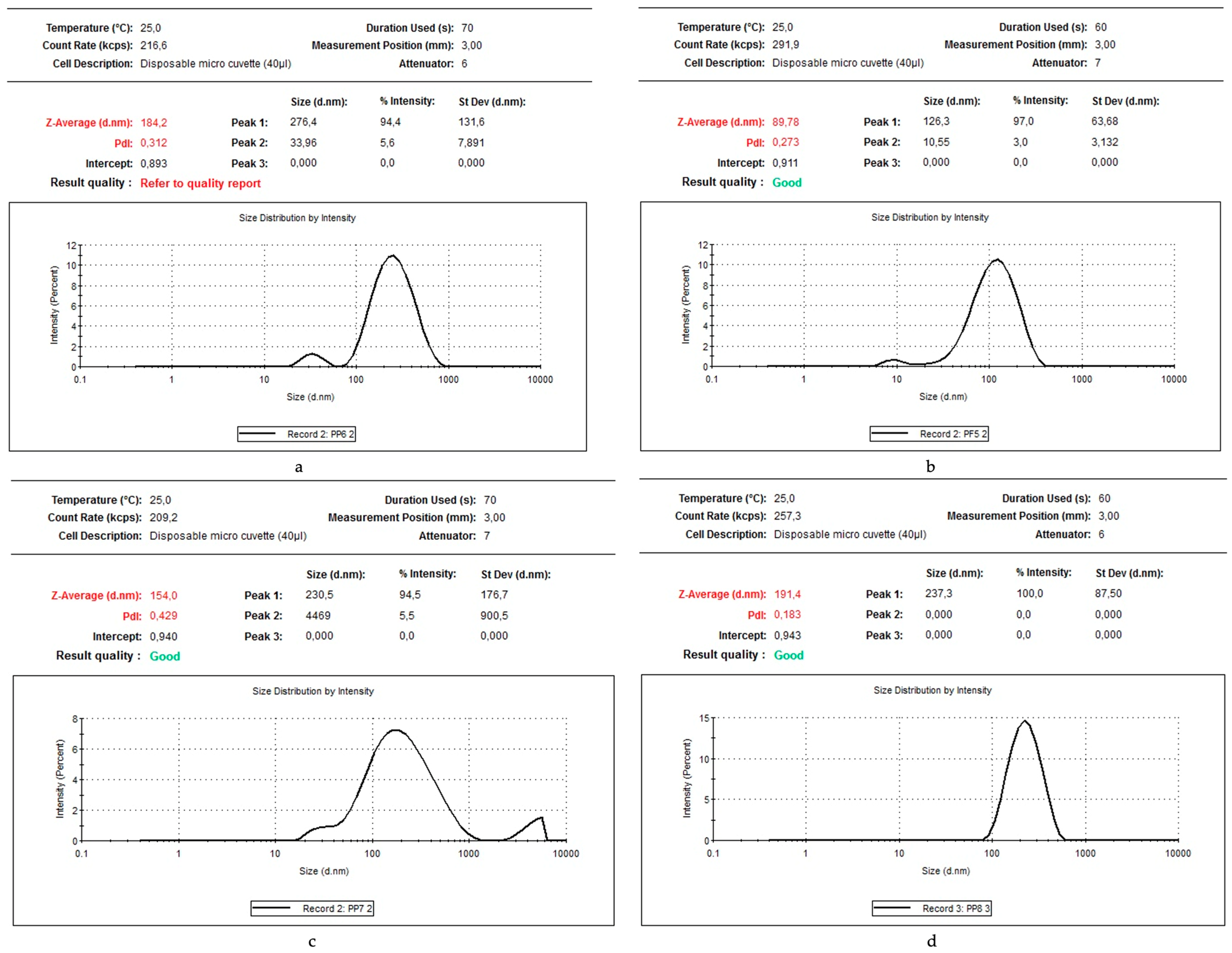
The average particle size obtained in the bioproducts analysed ranged between 89.78 nm and 276.4 nm (Figure 3a–d). In the case of the bioproduct A1+Au, the average size of the metallic gold obtained was 184.2 nm. However, in this system, the intensity of peak 2 from Figure 3a suggests that the primary size of the gold particles generated is dispersed as nanoparticles measuring 33.96 nm (peak 2 from Figure 3a). For the bioproduct derived from Undaria pinnatifida, the average size of the metallic gold particles obtained was 89.78 nm (Figure 3b). However, the intensity of peak 2 from Figure 3b suggests that the major gold particles are dispersed as nanoparticles, with a size of 10.55 nm. In the case of bioproducts A3+Au and A4+Au, the average size of the gold particles generated is greater than 200 nm.
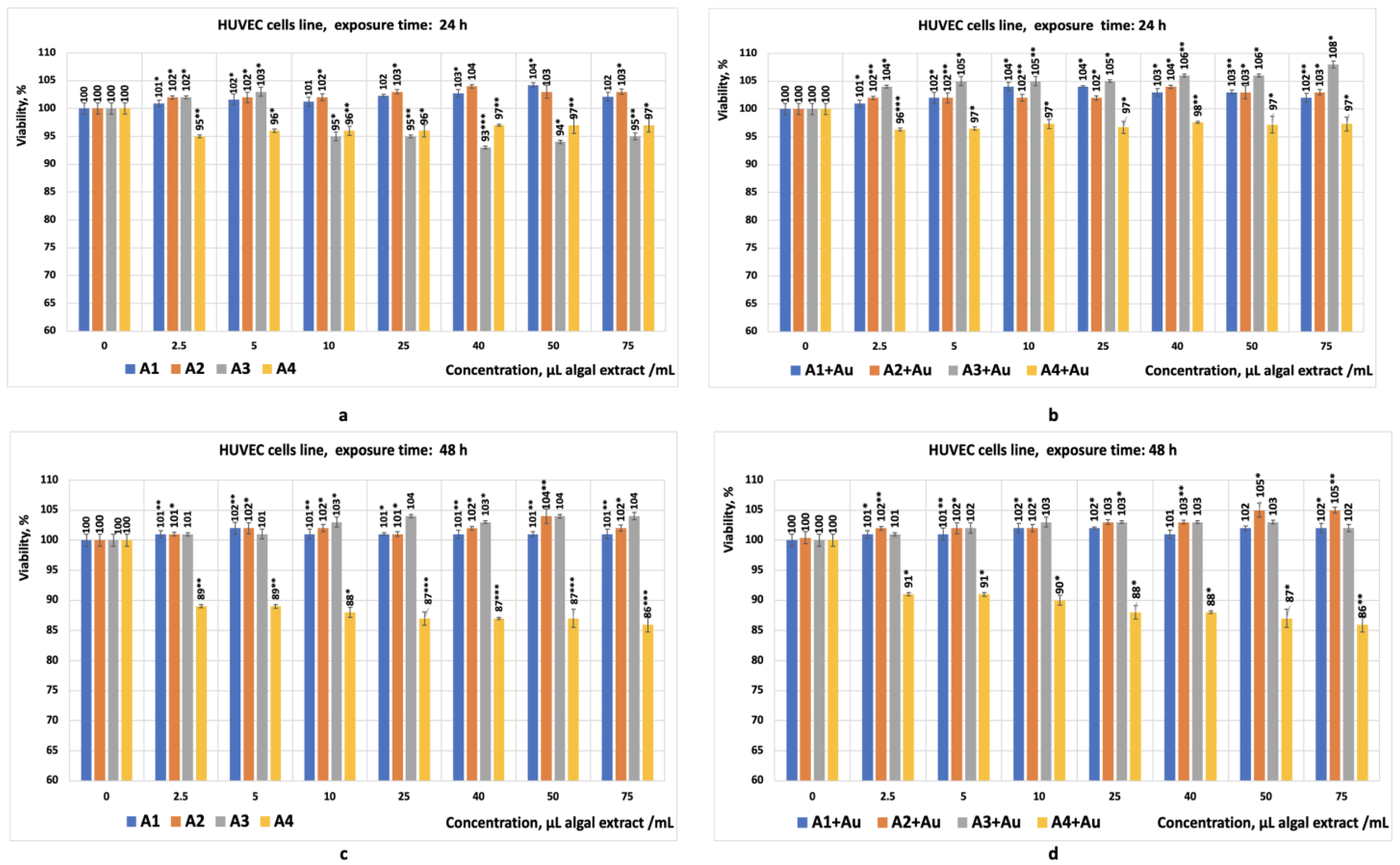
Cytotoxicity tests performed in vitro showed that the eight bioproducts did not exhibit cytotoxic effects on the HUVEC cell line (Figure 4a–d) when exposed for 24–48 h to algal extract at volumetric concentrations ranging from 2.5 to 75 µL/mL.
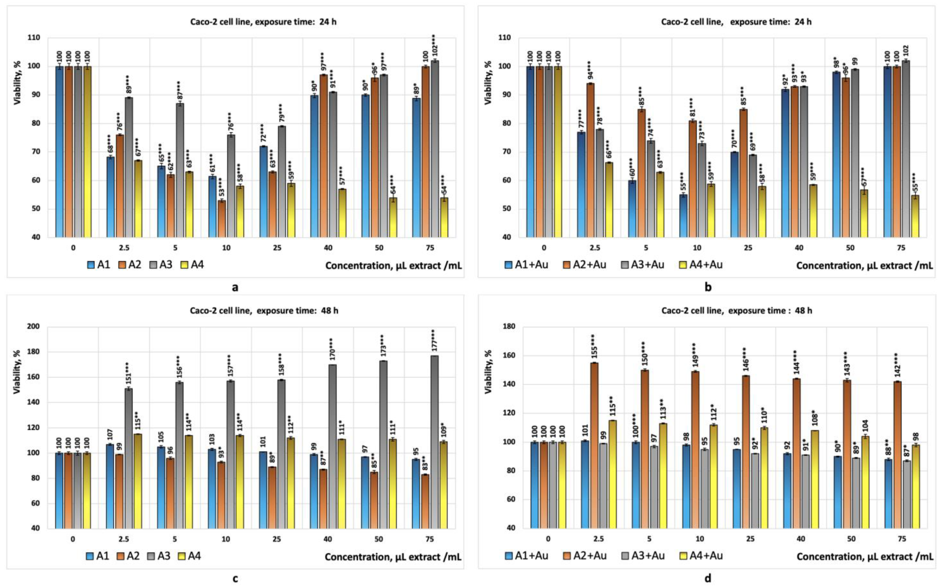
The results obtained from the tests performed on the Caco-2 cell line (Figure 5a–d) suggest significant differences in terms of cytotoxicity and cell viability, depending on the type of bioproduct and the concentration used, as follows:
  • Bioproduct A1 showed cytotoxic effects at concentrations situated between (2.5–10) µL/mL, with the corresponding cell viabilities ranging from 61% to 68% (statistically significant values);
  • Bioproduct A2 exhibited cytotoxicity at concentrations situated between (5–25) µL/mL, with the corresponding cell viabilities ranging from 53% to 65% (statistically significant values);
  • Bioproduct A3 did not exhibit cytotoxic effects within the concentration range situated of (2.5–75) µL/mL. In this case, the cell viabilities range from 76% to 102%;
  • Bioproduct A4 exhibited cytotoxic effects across the entire concentration range studied (2.5–75 µL/mL), with the corresponding cell viabilities ranging from 54% to 67% (statistically significant results);
  • Bioproduct (A1+Au) showed cytotoxic effects at concentrations situated between (5–10) µL/mL, with the corresponding cell viabilities ranging from 55% to 60% (statistically significant values);
  • The bioproduct (A4+Au) exhibited cytotoxicity across the entire concentration range studied, with the cell viabilities ranging from 55% to 66% (statistically significant values).
The preliminary data suggest that bioproduct A4 and bioproduct (A4+Au), derived from Chlorella sp., are the most cytotoxic, while bioproduct A3 does not show significant cytotoxic effects on the Caco-2 cell line under the tested conditions. Bioproducts A1 and A2 exhibit moderate cytotoxic effects. The presence of gold (A1+Au; A2+Au; A3+Au) does not appear to significantly influence cell viability.
After 48 h of exposure of the Caco-2 cell line to the eight bioproducts, the following results were obtained:
The bioproducts A1, A2, A3, and A4 did not exhibit cytotoxic effects (Figure 5c). The bioproduct A3 appears to stimulate Caco-2 cell proliferation (statistically significant values);
The bioproducts A1+Au, A3+Au, and A4+Au did not exhibit cytotoxic effects, but bioproduct (A2+Au) appears to stimulate the proliferation of the Caco-2 cell line (statistically significant values) (Figure 5d);
The tests conducted on the HepG2 cell line, after 24 and 48 h of exposure (Figure 6a–d), generally indicated no cytotoxic effects from the eight bioproducts, except bioproduct A3, which, at a concentration of 75 µL/mL, reduced HepG2 cell viability to 54% (value statistically significant) (Figure 6a,b).

4. Discussion

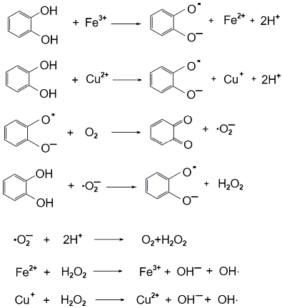
The data obtained from the bioproduct characterisation reveal that the crude extracts, obtained through aqueous extraction, contain polyphenolic compounds. These results are in agreement with the findings of other researchers, who reported the presence of polyphenolic compounds in polysaccharides obtained via aqueous extraction [20]. Generally, high content of polyphenolic compounds is associated with strong antioxidant activity, as is the case with Cystoseira barbata, which contains (714.17 ± 1.26)mg GAE/L and exhibits antioxidant activities (Figure 2c) situated between (85–98)%. Regarding the antioxidant activities observed in Cystoseira barbata, this effect can be attributed both to the high polyphenol content in the bioproduct and to the low levels of metallic cations, such as copper and iron, contained in the species harvested from the Romanian Black Sea [46]. The remaining bioproducts (A1, A2, A4) exhibit prooxidant activities, which appear to be directly proportional to the polyphenol content, the prooxidant properties of the bioproducts increase in the following order: A4 > A2 > A1. The prooxidant activities observed in the case of the bioproducts A1, A2, and A4 may be attributed to the presence of polyphenolic compounds [20], ascorbic acid [37,44], and metallic cations such as Fe3+ and Cu2+ [6,9,14,47,51]. In the presence of hydrogen peroxide (used as a reagent in the analysis performed by chemiluminescence) and polyphenolic compounds, metallic cations catalyse the Fenton reactions [38,39,41] as follows:
Jmse 13 00318 i001
In the presence of Fe3+ or Cu2+, hydrogen peroxide is rapidly converted into reactive oxygen species [39]. Polyphenolic compounds such as benzoic acid, cinnamic acid, caffeic acid, syringic acid, protocatechuic acid, p-coumaric acid, vanillic acid, gallic acid, ferulic acid, phloroglucinol, trolox, quercetin, morin, fisetin, myricetin, and naringenin can act as prooxidants [38,39,41]. Regarding the generation of nanomaterials with gold in the algal polysaccharidic extracts, the results obtained (bioproducts in which the average size of gold particles is 33.96 nm for Porphyra umbilicalis polysaccharides and 10.52 nm for Undaria pinnatifida polysaccharides, as shown in Figure 3a,b) are in agreement with the findings of Ramakrishna and Senthilkumar. According to their study, when 45 mL of a 1 mM HAuCl3·H2O solution is mixed with 5 mL of algal supernatant (previously obtained by boiling 1 g of algae with 20 mL of water), red solutions are formed in which gold is reduced to its metallic form, resulting particles of 20 nm in size [27,28].
Regarding the gold-enriched polysaccharides derived from Chlorella sp., the average size of the reduced gold particles is 237 nm (Figure 3d), and the resulting bioproduct does not qualify as a nanomaterial. This result is due to the methodology used, which differs from those reported by other scientists. According to He et al., the polysaccharides derived from Chlorella sp. can generate gold nanoparticles by treating the supernatant obtained from boiling 2 g of Chlorella sp. (dried biomass) with 20 mL of water, followed by mixing it with a 1 mM solution of Au3+ (1 mL supernatant + 10 mL 1 mM Au3+ solution), when gold nanoparticles with an average size of 15 nm can be obtained [29]. González-Ballesteros et al. obtained nanomaterials by boiling equal amounts of dried algae and water at reflux, followed by treating the cold supernatant with an Au3+ solution so that the final solution had an Au3+ concentration of 0.4 mM. After a while, the gold cations are reduced to metallic form as nanoparticles with an average size of 40 nm [30]. Gürsoy et al. obtained gold nanoparticles by mixing algal biomass with a solution containing 1 mM Au3+ at a volume ratio of 1:2.5 (1 mL of algal extract with 2.5 mL of 1 mM Au3+ solution). Over time, the solution becomes pink due to the gold’s reduction to nanoparticles of 10 nm [32].
We chose to perform the preliminary cytotoxicity studies on the standardised HUVEC, Caco-2, and HepG2 cell lines for the following reasons:
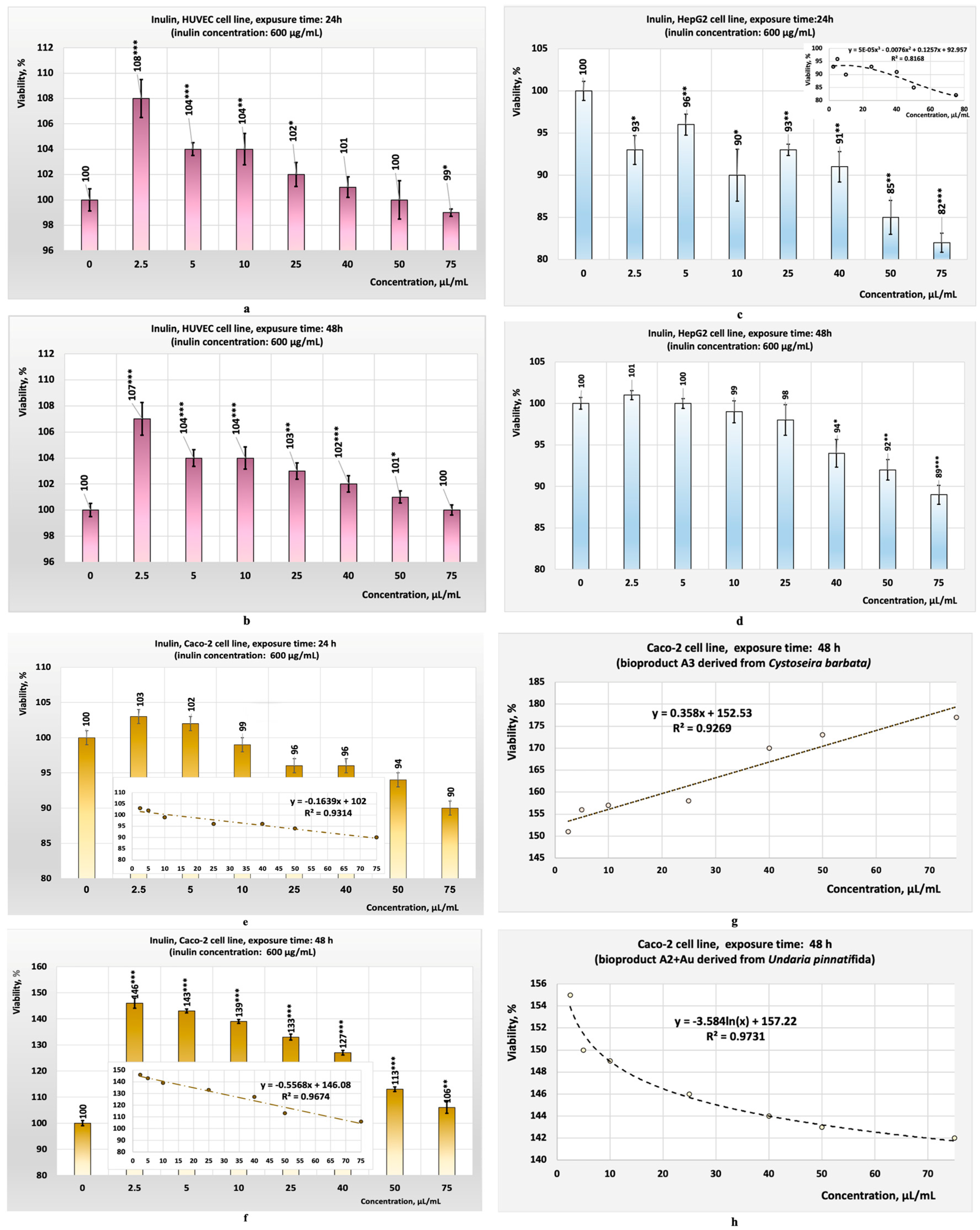
  • The HUVEC cell line consists of primary endothelial cells derived from human umbilical veins; they are not transformed or cancer cells. This characteristic recommends them for preliminary in vitro studies aimed at evaluating the cytotoxic effects of specific chemical compounds or bioproducts on normal human cells [59,60,61]. The HUVEC cell line is sensitive to toxic effects such as oxidative stress, apoptosis, and necrosis, making it suitable for detecting subtle cytotoxic effects. Since HUVECs are representative of vascular endothelial cells, they are useful for studying the potential cytotoxicity of a chemical compound or a new bioproduct on normal and healthy endothelial cells [62,63]. Evaluating cytotoxicity on these types of cell lines helps to predict how some specific chemical compounds or some bioproducts might affect normal and healthy tissues in the human body. The response of this cell line to various substances added to the culture medium provides a more accurate model than non-human cell lines for predicting human-specific toxicological effects [60,61]. The results obtained during this study from the test performed on this cell line showed that the eight bioproducts tested did not exhibit cytotoxicity for this cell line (Figure 4a–d). Similarly, the tests performed in parallel with inulin on the same cell line yielded comparable results (Figure 7a–f);
  • The Caco-2 and HepG2 cell lines were chosen because they are widely used in cytotoxicity studies. These cell lines can model the physiological processes of the intestinal or liver epithelium, which occur upon their exposure to different compounds. The preliminary results obtained are used to evaluate in vitro the toxicity of specific chemical compounds or bioproducts [64]. The Caco-2 tumour cell line is derived from human colon adenocarcinoma and develops properties similar to differentiated enterocytes. This characteristic underlies the use of this cell line in preliminary in vitro studies to evaluate the cytotoxic effects of ingested substances on the intestinal tract as well as to evaluate the cytotoxic effects on colon tumour cells [64,65,66]. The HepG2 cell line is also used in preliminary in vitro studies aimed at evaluating the toxic effects on liver tumour cells. Both cell lines are complementary and can provide a more comprehensive understanding of the effects of a chemical compound or a bioproducts on intestinal barriers and hepatic metabolism. Studies have shown that their combined use can generate relevant data on the safety of specific chemical compounds or new bioproducts by analysing their effects on cell viability, DNA, or the production of reactive oxygen species in response to the toxicity of a chemical compound or bioproduct [58,64,67]. The results obtained during this study, from the tests performed in vitro on the Caco-2 cell line (all with statistical significance, p < 0.05), showed that the three crude algal bioproducts tested (A1; A2; A4) exhibited cytotoxicity for this cell line (Figure 5a) after 24 h of exposure. Despite this, the tests performed in parallel on the HepG2 cell line suggest that, in the case of the crude bioproduct A3, cytotoxic effects may occur at concentrations higher than 50 µL/mL (Figure 6a) after 24 h of exposure.
Regarding the effects of the bioproducts with gold, only A4+Au (derived from Chlorella sp. crude aqueous extract) appears to exhibit cytotoxic effects after 24 h of exposure. In this last case, at all tested concentration levels, the viability of the Caco-2 cell line remains below 70%. The main values obtained for the Caco-2 cell line after 24 h of exposure to both bioproduct A4 and A4+Au are statistically significant (p ≤ 0.001). The cytotoxic effects observed for the Caco-2 cell line exposed to bioproducts A4 and A4+Au are most likely due to specific water-soluble phenolic compounds from Chlorella sp., which are found both in the crude bioproduct as well in bioproducts with gold, such as gallic acid [68,69].
As for the antitumour effects on Caco-2 cells of extract A4, after 24 h of exposure, the data obtained align with the results reported by Andrade and colleagues. Their studies, performed with two exopolysaccharides isolated from the culture medium used for the growth of Chlorella zofingiensis and Chlorella vulgaris, revealed that the polysaccharides tested reduced the viability of the human colon cancer cell line HCT-8 by approximately 20–30% at concentrations of (0.15–0.6) mg/mL in the culture medium. The viability of tumour cells after treatment with 0.6 mg/mL of exopolysaccharide in the culture medium was 70% and 80%, respectively [52]. Lemieszek and Rzeski tested bioproducts obtained by lyophilisation of an extract derived from the dried biomass of Chlorella pyrenoidosa in an aqueous medium, incubated for 24 h at room temperature, on the two standardised human cell lines: a colon adenocarcinoma tumour cell line (HT-29) and a normal colon epithelial cell line (CCD841CoN) [54]. Results obtained after 96 h of exposure to crude extract concentrations ranging from 10 to 1000 µg/mL showed that the bioproduct does not affect normal colon epithelial cells [54]. However, it significantly reduces the viability of colon adenocarcinoma tumour cells, lowering their viability to 50% at 10 µg/mL and 20% at 1000 µg/mL, respectively [54].
Yusof et al. conducted in vitro tests on normal liver cell lines (WRL68) and tumour liver cell lines (HepG2), with a bioproduct obtained by lyophilising the supernatant resulting from centrifugation of the solution produced by boiling dried microalgae in an aqueous media. The obtained results indicated that this bioproduct inhibits cell growth, at relatively similar IC50 values (IC50 = 1.6 mg/mL for the HepG2 tumour cell line and IC50 = 1.7 mg/mL for the normal liver cell line WRL68). Exposing the two cell lines to 2 mg bioproduct/mL for two hours induces apoptosis in the tumour liver cell lines. Under these conditions, apoptosis was 70% in tumour liver cells and 15% in normal liver cells. The mechanism of apoptosis induction in the above-studied tumour liver cells involves the destruction of tumour cell DNA and arresting the cell cycle in the G1 or G2 phases. The bioproduct obtained by extraction in an aqueous media mediates apoptosis by increasing the expression of Bax proteins and decreasing the expression of Bcl-2 proteins in a time-dependent manner [25].
The cytotoxic effect of extract A3 on the HepG3 cell line is likely due to soluble polysaccharides such as fucoidan and laminarin, which exhibit antitumour properties [4]. The mechanism involves cell cycle arrest, mitochondrial membrane depolarisation, and the production of nitric oxide [17]. The cytotoxic effect of bioproduct A3 observed for the HepG2 tumour cell line after 24 h of exposure may be attributed to its high polyphenolic compound content (714.17 ± 1.26 mg GAE/L), which is significantly higher compared to bioproduct A2 (19.70 ± 0.13 mg GAE/L) (Table 2). Regarding the behaviour of the two tumour cell lines under the action of algal polysaccharides, the results obtained were compared with those for inulin (Figure 7a–h) for the same time of exposure (the tests were performed in parallel with algal polysaccharides for comparison). In the case of inulin, the results obtained after 24 h of exposure generally showed a decrease in cell viability with increasing inulin concentration, except for the HepG2 cell line (Figure 7a,c,e). The behaviour of the Caco-2 cell line under the influence of bioproducts A1, A2, and A3 at concentrations greater than 10 μL/mL suggests that the mechanism of action on the Caco-2 cell line is different. By increasing the exposure time to 48 h, the stimulatory effect becomes more pronounced for both inulin and algal polysaccharides (Figure 7b,c,h), except for bioproduct A3 (Figure 7g), where the discrepancies obtained can be due to the experimental errors or other mechanisms.
Math modelling of the antitumour activities of bioproduct A4 and bioproduct A3, using three parameters—extract concentration in cell culture media, residual values of prooxidant or antioxidant activities, and cytotoxicity (Figure 8a,b)—revealed an IC50 value for A4 of 329 µL/mL (i.e., 18.24 µg A4/mL) (Figure 6a), and for bioproduct A3, an IC50 of 70 µL/mL (i.e., 12.34 µg A3/mL) (Figure 8b).
The results obtained after 24 h of exposure may be attributed to the algal polysaccharides and phlorotannins (algal polyphenols identified in the crude extract through analysis) present in the culture media. Due to their water solubility (as observed in Undaria pinnatifida and Cystoseira barbata [70,71], these compounds are found in the crude extracts obtained from aqueous solutions and are released into the aqueous medium. This process is also due to existing interactions between phlorotannins and polysaccharides via van der Waals and hydrogen bonding [72,73,74]. Metallic cations, acquired from the environment and integrated within algal structures, bond with polysaccharides and phlorotannins through interactions involving glycosyl groups and/or hydroxyl groups in the phlorotannins structure [75,76,77,78].
Regarding the cytotoxic activity of the bioproducts (A1+Au) and (A4+Au) at 24 h (Figure 5b), these are generally comparable to bioproducts that contain only algal polysaccharides. However, further studies are needed to explain this behaviour, as in both cases, the concentration of polysaccharides from each system containing gold decreases, and in the case of the bioproduct (A1+Au), the gold is present in the form of nanoparticles with a size of 33.96 nm (Figure 3a).
After 48 h of exposure, tumour cell proliferation increases, likely due to the monosaccharides/polysaccharides present in the system, which may modulate tumour cell line proliferation, although to a lesser extent compared to polysaccharides without gold.
Regarding the results obtained after 48 h of exposure to the analysed bioproducts, the observed effects may be attributed to the consumption of active compounds (fucoidans, laminarins [79,80], and phlorotannins [81,82,83]) and monosaccharides from the culture medium, which can promote the proliferation of the remaining tumour cells [84,85,86]. Algal polysaccharides, under specific conditions, might paradoxically promote tumour cell proliferation. This can occur due to several reasons:
1.
Certain monosaccharides or polysaccharides (such as fucose, glucose, fucoidan, alginate, or laminarin) may mimic or enhance the activity of growth factors, thereby promoting tumour cell growth through mechanisms that may include direct binding to growth factor receptors, stabilising receptor–ligand interactions, or extracellular matrix modification to promote signalling, or modulating responses that support tumour growth [84,85].
2.
Crude extracts of algal polysaccharides may contain other water-soluble compounds (proteins, peptides, or other small molecules) that might interact with tumour cell receptors, modulating signalling pathways in tumour cells, which promote their proliferation [86].
The results obtained during this preliminary study highlight the benefits of algal use in human food and open new research possibilities for elucidating the mechanisms of action of bioproducts A1, A2, and A4 on the Caco-2 cell line in a concentration range of (2.5–10) µL/mL and 24 h of exposure.
Similarly, further studies are required to elucidate the mechanism of action of the bioproduct (A4+Au) on Caco-2 cell lines in the concentration range of (2.5–25 µL/mL) after 24 h of exposure. Intriguing properties were observed for the action of bioproduct A3 on the HepG2 cell line when used at concentrations ranging from (25–75) µL/mL; however, more extensive studies are necessary to confirm or infirm these findings.
The novelty of this study lies in the protective effect of the polysaccharides isolated from the four algal species on tumourigenesis processes that may occur in the digestive system, where foods containing algae or algal polysaccharides come into direct contact with it.
Our preliminary studies indicated, in the case of these products, potential selective cytotoxicity. The studied algae-based bioproducts, obtained by extraction from aqueous media, do not exert a cytotoxic effect on the HUVEC cell line (considered a normal cell line) but appear to exhibit selective cytotoxicity on the Caco-2 cell line. If future research confirms the existence of a selective cytotoxicity, these types of bioproducts could represent valuable raw materials for developing new nutraceuticals or dietary supplements with antitumour properties. Highlighting the cytotoxic effects on CaCo-2 cells (a standardised colorectal cancer cell line) provides a preliminary indication that these algae extracts may contain specific bioactive compounds with anticancer properties. Although the algae polysaccharides are known for their biological effects (anti-inflammatory, and immunomodulatory properties), their use as anticancer agents is still in the early stages of research. Recent studies show that they can affect the viability of tumour cells through mechanisms such as the induction of apoptosis (programmed cell death, generation of oxidative stress in cancer cells, and inhibition of some signalling pathways involved in tumour cell survival [65]. Marine algae, such as Undaria pinnatifida, Porphyra umbilicalis, Cystoseira barbata, and Chlorella sp., produce unique water-soluble secondary metabolites, such as polysaccharides, proteins, peptides, and free amino acids, and certain pigments like phycoerythrin or hydrophilic phenolic compounds, which can be found in aqueous crude extracts obtained in this study [87,88]. The preliminary results obtained during this study will generate future research regarding the molecular mechanisms involved in cytotoxicity (apoptosis induction, oxidative stress, or cell cycle arrest), which can lead to the development of new, less toxic cancer treatments. The study highlights the pharmaceutical potential of these underexplored resources, with marine algae representing a renewable and sustainable resource for new drug discovery and production.
These effects are highlighted by exposing the Caco-2 tumour cell line to the bioproducts A1, A2, and A4 for 24 h. In the case of the HepG2 cell line, only bioproduct A3 seems to have a beneficial effect, but at concentrations starting from 75 µL A3/mL. In this context, further studies are needed to evaluate the mechanism of action of bioproducts A1, A2, and A4 on the Caco-2 tumour cell line at 24 h, as well for bioproduct A3 on the HepG2 tumour cell line at 24 and 48 h. No additional studies have been performed regarding the size of gold particles formed in the bioproducts (A1+Au), (A2+Au), (A3+Au), and (A4+Au), because these do not exhibit cytotoxic effects on the studied cell lines (cell viability was generally above 70%). However, in the products (A1+Au) and (A2+Au), the analyses performed showed the presence of nanoparticles with sizes below 34 nm. These bioproducts could be used in the development of new electrochemical sensors.

5. Conclusions

Crude bioproducts enriched in polysaccharides can be obtained through extraction in hot aqueous media from four algae species: Porphyra umbilicalis, Undaria pinnatifida, Cystoseira barbata, and Chlorella sp. By adding gold ions to the crude solutions containing algal polysaccharides, biomaterials are formed in which gold is reduced to its metallic form as nanoparticles smaller than 34 nm, particularly in the case of polysaccharide extracts obtained from Porphyra umbilicalis or Undaria pinnatifida.
The proliferation tests performed in vitro showed that the eight bioproducts did not exhibit cytotoxic effects on a normal cell line-type HUVEC. Upon exposing the Caco-2 tumour cell line to the eight bioproducts for 24 h, at certain concentrations of the crude extract in the system (c = 10 µL/mL), only the bioproducts containing polysaccharidic fractions derived from Porphyra umbilicalis, Undaria pinnatifida, and Chlorella sp. exhibited cytotoxic effects. The studies performed on the standardised HepG2 tumour cell line with the eight bioproducts indicated that, after 24 h of exposure, none of the bioproducts studied exhibited cytotoxic effects on the HepG2 tumour cell line, except for the polysaccharidic extract derived from Cystoseira barbata, which reduced the viability of the HepG2 cells to 54% at c = 5 µL/mL.

Author Contributions

Conceptualisation, N.R.; methodology, N.R. and L.C.P.; software, N.R.; validation, N.R.; formal analysis, M.B.; investigation, T.M., N.R., C.L.N., M.B., M.M. and M.V.; resources, N.R. and M.B.; data curation, N.R. and M.B.; writing—original draft preparation, N.R.; writing—review and editing, N.R.; visualisation, M.B. and L.C.P.; supervision, N.R.; funding acquisition, N.R., M.B. and M.M. All authors have read and agreed to the published version of the manuscript.

Funding

The research was funded by the Doctoral School of Biotechnology of the University of Agronomic Sciences and Veterinary Medicine of Bucharest, Romania.

Institutional Review Board Statement

Not applicable.

Informed Consent Statement

Not applicable.

Data Availability Statement

The raw data supporting the conclusions of this article will be made available by the authors upon request.

Acknowledgments

This research was made as a part of the current activity of the Doctoral School of Biotechnology from USAMV Bucharest, Romania.

Conflicts of Interest

The authors declare no conflicts of interest.

References

  1. Matos, J.; Cardoso, C.; Serralheiro, M.L.; Bandarra, N.M.; Afonso, C. Seaweed bioactives potential as nutraceuticals and functional ingredients: A review. J. Food Compos. Anal. 2024, 133, 106453. [Google Scholar] [CrossRef]
  2. Taboada, C.; Millan, R.; Miguez, I. Evaluation of marine algae Undaria pinnatifida and Porphyra purpurea as a food supplement: Composition, nutritional value and effect of intake on intestinal, hepatic and renal enzyme activities in rats. J. Sci. Food Agric. 2023, 93, 1863–1868. [Google Scholar] [CrossRef]
  3. Alves, C.; Silva, J.; Pinteus, S.; Gaspar, H.; Alpoim, M.C.; Botana, L.M.; Pedrosa, R. From Marine Origin to Therapeutics: The Antitumor Potential of Marine Algae-Derived Compounds. Front. Pharmacol. 2018, 9, 777. [Google Scholar] [CrossRef]
  4. Dragan, A.M.L.; Sirbu, R.; Cadar, E. Brown Seaweeds from Black Sea Coast as an Important Source of Bioactive Compounds of Interest for Human Health Brown Seaweeds from Black Sea Coast as an Important Source of Bioactive Compounds of Interest for Human Health. Eur. J. Nat. Sci. Med. 2023, 6, 98–110. [Google Scholar] [CrossRef]
  5. Mutlu-Durak, H.; Arikan-Algul, Y.; Bayram, E.; Haznedaroglu, B.Z.; Kutman, U.B.; Kutman, B.Y. Various extracts of the brown seaweed Cystoseira barbata with different compositions exert biostimulant effects on seedling growth of wheat. Physiol. Plant 2024, 176, e14503. [Google Scholar] [CrossRef]
  6. Baghel, R.S.; Choudhary, B.; Pandey, S.; Pathak, P.K.; Patel, M.K.; Mishra, A. Rehashing Our Insight of Seaweeds as a Potential Source of Foods, Nutraceuticals, and Pharmaceuticals. Foods 2023, 12, 3642. [Google Scholar] [CrossRef]
  7. Cho, T.J.; Rhee, M.S. Health Functionality and Quality Control of Laver (Porphyra, Pyropia): Current Issues and Future Perspectives as an Edible Seaweed. Mar. Drugs 2020, 18, 14. [Google Scholar] [CrossRef]
  8. Gamero-Vega, G.; Palacios-Palacios, M.; Quitral, V. Nutritional Composition and Bioactive Compounds of Red Seaweed: A Mini-Review. J. Food Nutr. Res. 2020, 8, 431–440. [Google Scholar] [CrossRef]
  9. Salomone, V.N.; Riera, M. Proximal Composition of Undaria pinnatifida from San Jorge Gulf (Patagonia, Argentina). Biol. Trace Elem. Res. 2020, 196, 252–261. [Google Scholar] [CrossRef]
  10. Martinez, M.A.; Becherucci, M.E. Study of the potential use of the invasive marine algae Undaria pinnatifida in the preliminary development of a functional textile. J. Ind.Text. 2022, 51, 8127S–8141S. [Google Scholar] [CrossRef]
  11. Park, J.-S.; Han, J.-M.; Park, S.-W.; Kim, J.-W.; Choi, M.-S.; Lee, S.-M.; Haq, M.; Zhang, W.; Chun, B.-S. Subcritical Water Extraction of Undaria pinnatifida: Comparative Study of the Chemical Properties and Biological Activities across Different Parts. Mar. Drugs 2024, 22, 344. [Google Scholar] [CrossRef] [PubMed]
  12. Kolb, N.; Vallorani, L.; Milanović, N.; Stocchi, V. Evaluation of Marine Algae Wakame (Undaria pinnatifida) and Kombu (Laminaria digitata japonica) as Food Supplements. Food Technol. Biotechnol. 2024, 42, 57–61. [Google Scholar]
  13. Council Directive 92/43/EEC of 21 May 1992 on the Conservation of Natural Habitats and of Wild Fauna and Flora—Consolidated Version 01/01/2007. Available online: https://environment.ec.europa.eu/topics/nature-and-biodiversity_en (accessed on 3 December 2024).
  14. Manev, Z.; Iliev, A.; Vachkova, V. Chemical characterization of brown seaweed—Cystoseira barbata. Bulg. J. Agric. Sci. 2013, 19, 12–15. [Google Scholar]
  15. Cadar, E.; Sirbu, R.; Ibram, A.; Ionescu, A.M. Evaluation of Total Phenolic Content in Relation to Antioxidant Activity of Brown Algae Cystoseira barbata from Black Sea. Rev. Chim. 2019, 70, 2684–2689. [Google Scholar] [CrossRef]
  16. Sellimi, S.; Kadri, N.; Barragan-Montero, V.; Laouer, H.; Hajji, M.; Nasri, M. Fucans from a Tunisian brown seaweed Cystoseira barbata: Structural characteristics and antioxidant activity. Int. J. Biol. Macromol. 2014, 66, 281–288. [Google Scholar] [CrossRef]
  17. Nigam, S.; Singh, R.; Bhardwaj, S.K.; Sami, R.; Nikolova, M.P.; Chavali, M.; Sinha, S. Perspective on the Therapeutic Applications of Algal Polysaccharides. J. Polym. Environ. 2022, 30, 785–809. [Google Scholar] [CrossRef]
  18. Dobrinčić, A.; Balbino, S.; Zorić, Z.; Pedisić, S.; Bursać Kovačević, D.; Elez Garofulić, I.; Dragović-Uzelac, V. Advanced Technologies for the Extraction of Marine Brown Algal Polysaccharides. Mar. Drugs 2020, 18, 168. [Google Scholar] [CrossRef]
  19. Montero, L.; Herrero, M.; Ibáñez, E.; Cifuentes, A. Separation and characterization of phlorotannins from brown algae Cystoseira abies-marina by comprehensive two-dimensional liquid chromatography. Electrophoresis 2014, 35, 1644–1651. [Google Scholar] [CrossRef]
  20. Türkmen, M.; Duran, K. The Effect of Brown Seaweed and Cattle Manure Combinations on The Properties of Eisenia fetida’s Organic Fertilizer. Turk. J. Agric.-Food Sci. Technol. 2021, 9, 1070–1075. [Google Scholar] [CrossRef]
  21. Ramandani, A.A.; Sun, Y.-M.; Lan, J.C.W.; Lim, J.W.; Chang, J.S.; Srinuanpan, S.; Khoo, K.S. Upcycling food waste as a low-cost cultivation medium for Chlorella sp. microalgae. J. Sci. Food Agric. 2024. early view. [Google Scholar] [CrossRef]
  22. Zakaria, M.; Kamal, S.M.M.; Harun, M.R.; Omar, R.; Siajam, S.I. Extraction of antioxidants from Chlorella sp. using subcritical water treatment. In IOP Conference Series: Materials Science and Engineering, Volume 206, 29th Symposium of Malaysian Chemical Engineers (SOMChE) 2016, Miri, Sarawak, Malaysia, 1–3 December 2016; IOP Publishing: Bristol, UK, 2016; Available online: https://iopscience.iop.org/article/10.1088/1757-899X/206/1/012035 (accessed on 30 November 2024).
  23. Abreu, A.P.; Martins, R.; Nunes, J. Emerging Applications of Chlorella sp. and Spirulina (Arthrospira sp.). Bioengineering 2023, 10, 955. [Google Scholar] [CrossRef] [PubMed]
  24. Toshkova-Yotova, T.; Sulikovska, I.; Djeliova, V.; Petrova, Z.; Ognyanov, M.; Denev, P.; Toshkova, R.; Georgieva, A. Exopolysaccharides from the Green Microalga Strain Coelastrella sp. BGV-Isolation, Characterization, and Assessment of Anticancer Potential. Curr. Issues Mol. Biol. 2024, 46, 10312–10334. [Google Scholar] [CrossRef]
  25. Yusof, Y.A.; Saad, S.M.; Makpol, S.; Shamaan, N.A.; Ngah, W.Z. Hot water extract of Chlorella vulgaris induced DNA damage and apoptosis. Clinics 2010, 65, 1371–1377. [Google Scholar] [CrossRef]
  26. El-Naggar, N.E.A.; Hussein, M.H.; Shaaban-Dessuuki, S.A. Production, extraction and characterization of Chlorella vulgaris soluble polysaccharides and their applications in AgNPs biosynthesis and biostimulation of plant growth. Sci. Rep. 2020, 10, 3011. [Google Scholar] [CrossRef]
  27. Ramakrishna, M.; Rajesh Babu, D.; Gengan, R.M.; Chandra, S.; Rao, G.N. Green synthesis of gold nanoparticles using marine algae and evaluation of their catalytic activity. J. Nanostruct. Chem. 2016, 6, 1–13. [Google Scholar] [CrossRef]
  28. Senthilkumar, P.; Surendran, L.; Sudhagar, B.; Kumar, D.S.R.S. Facile green synthesis of gold nanoparticles from marine algae Gelidiella acerosa and evaluation of its biological Potential. SN Appl. Sci. 2019, 1, 284. [Google Scholar] [CrossRef]
  29. He, R.; Zhou, D.; Xiao, L.; Li, Y. Chlorella vulgaris Extract-Decorated Gold Nanoparticle Hybridized Antimicrobial Hydrogel as a Potential Dressing. Gels 2023, 9, 11. [Google Scholar] [CrossRef]
  30. González-Ballesteros, N.; Maietta, I.; Rey-Méndez, R.; Rodríguez-Argüelles, M.C.; Lastra-Valdor, M.; Cavazza, A.; Grimaldi, M.; Bigi, F.; Simón-Vázquez, R. Gold Nanoparticles Synthesized by an Aqueous Extract of Codium tomentosum as Potential Antitumoral Enhancers of Gemcitabine. Mar. Drugs 2023, 21, 20. [Google Scholar] [CrossRef]
  31. Hamouda, R.A.; Abd El Maksoud, A.I.; Wageed, M.; Alotaibi, A.S.; Elebeedy, D.; Khalil, H.; Hassan, A.; Abdella, A. Characterization and Anticancer Activity of Biosynthesized Au/Cellulose Nanocomposite from Chlorella vulgaris. Polymers 2021, 13, 3340. [Google Scholar] [CrossRef]
  32. Gürsoy, N.; Öztürk, B.Y.; Dağ, İ. Synthesis of intracellular and extracellular gold nanoparticles with a green machine and its antifungal activity. Turk. J. Biol. 2021, 45, 196–213. [Google Scholar] [CrossRef]
  33. Radu, N.; Voicescu, M.; Radu, E.; Ciprian Tanasescu, C. Biomaterial with antioxidant and antifungal activities, obtained from Romanian indigenous plants. Mol. Cryst. Liq. Cryst. 2017, 655, 243–249. [Google Scholar] [CrossRef]
  34. Radu, N.; Ghita, I.; Rau, I. Therapeutic Effect of Polysaccharides from Plantago Species. Mol. Cryst. Liq. Cryst. 2010, 523, 236–246. [Google Scholar] [CrossRef]
  35. Zaharie, M.G.O.; Radu, N.; Pirvu, L.; Bostan, M.; Voicescu, M.; Begea, M.; Constantin, M.; Voaides, C.; Babeanu, N.; Roman, V. Studies Regarding the Pharmaceutical Potential of Derivative Products from Plantain. Plants 2022, 11, 1827. [Google Scholar] [CrossRef] [PubMed]
  36. Ioan, D.-C.; Rău, I.; Albu Kaya, M.G.; Radu, N.; Bostan, M.; Zgârian, R.G.; Tihan, G.T.; Dinu-Pîrvu, C.E.; Lupuliasa, A.; Ghica, M.V. Ciprofloxacin-Collagen-Based Materials with Potential Oral Surgical Applications. Polymer 2020, 12, 1915. [Google Scholar] [CrossRef]
  37. Rezayian, M.; Niknam, V.; Ebrahimzadeh, H. Oxidative damage and antioxidative system in algae. Toxicol. Rep. 2019, 6, 1309–1313. [Google Scholar] [CrossRef]
  38. Nowak, M.; Tryniszewski, W.; Sarniak, A.; Wlodarczyk, A.; Nowak, P.J.; Nowak, D. Concentration Dependence of Anti- and Pro-Oxidant Activity of Polyphenols as Evaluated with a Light-Emitting Fe2+-Egta-H2O2 system. Molecules 2022, 27, 3453. [Google Scholar] [CrossRef]
  39. Eghbaliferiz, I. Prooxidant Activity of Polyphenols, Flavonoids, Anthocyanins and Carotenoids: Updated Review of Mechanisms and Catalyzing Metals. Phytother. Res. 2016, 30, 1379–1397. [Google Scholar] [CrossRef]
  40. Cao, G.; Sofic, E.; Prior, R.L. Antioxidant and prooxidant behavior of flavonoids: Structure-activity relationships. Free Radic. Biol. Med. 1997, 22, 749–760. [Google Scholar] [CrossRef]
  41. Vignaud, J.; Loiseau, C.; Hérault, J.; Mayer, C.; Côme, M.; Martin, I.; Ulmann, L. Microalgae Produce Antioxidant Molecules with Potential Preventive Effects on Mitochondrial Functions and Skeletal Muscular Oxidative Stress. Antioxidants 2023, 12, 1050. [Google Scholar] [CrossRef]
  42. Ribeiro, D.; Freitas, M.; Silva, A.M.S.; Carvalho, F.; Fernandes, E. Antioxidant and pro-oxidant activities of carotenoids and their oxidation products. Food Chem. Toxicol. 2018, 120, 681–699. [Google Scholar] [CrossRef]
  43. Talbot, G. Food and Beverage Stability and Shelf Life. In The Stability and Shelf Life of Fats and Oils; Woodhead Publishing Limited: Cambridge, UK, 2011; pp. 683–715. [Google Scholar] [CrossRef]
  44. Sotler, R.; Poljšak, B.; Dahmane, R.; Jukić, T.; Pavan Jukić, D.; Rotim, C.; Trebše, P.; Starc, A. Prooxidant activities of antioxidants and their impact on health. Acta Clin. Croat. 2019, 58, 726–736. [Google Scholar] [CrossRef] [PubMed]
  45. Neshovska, H.; Manev, I.; Kirov, V. Heavy metal levels in water, brown algae (Cystoseira barbata), and eelgrass (Zostera marina) from the Southern Black Sea coast of Bulgaria. Int. J. Vet. Sci. Anim. Husb. 2021, 6, 15–18. [Google Scholar] [CrossRef]
  46. Cadar, E.; Negreanu Pîrjol, T.; Tomescu, A.; Paris, S.; Erimia, C.L.; Bogdan, N.S. Heavy Metals Existing in the Seaweed from the Romanian Coast of the Black Sea. Eur. J. Med. Nat. Sci. 2019, 2, 14–21. [Google Scholar] [CrossRef]
  47. Rzymski, P.; Budzulak, J.; Niedzielski, P.; Klimaszyk, P.; Proch, J.; Kozak, L.; Poniedziałek, B. Essential and toxic elements in commercial microalgal food supplements. J. Appl. Phycol. 2019, 31, 3567–3579. [Google Scholar] [CrossRef]
  48. Available online: https://www.ceva-algues.com/wp-content/uploads/2021/04/EN-Undaria-pinnatifida-1.pdf (accessed on 5 December 2024).
  49. van Groenigen, J.; Derksen, G.C.H.; Timmermans, K.R. Review of Presence, Induction and Isolation of Major Cellular Constituents From Porphyra Sensu Lato (Rhodophyceae), Including Mycosporine-Like Amino Acids (MAA’s). J. Mar. Biol. Aquac. Res. 2022, 4, 30–46. [Google Scholar] [CrossRef]
  50. Maliar, T.; Maliarová, M.; Blažková, M.; Kunštek, M.; Uváčková, Ľ.; Viskupičová, J.; Purdešová, A.; Beňovič, P. Simultaneously Determined Antioxidant and Pro-Oxidant Activity of Randomly Selected Plant Secondary Metabolites and Plant Extracts. Molecules 2023, 28, 6890. [Google Scholar] [CrossRef]
  51. Le Tutour, B.; Benslimane, F.M.; Gouleau, M.; Gouygou, J.P.; Saadan, B.; Quéméneur, F. Antioxidant and pro-oxidant activities of the brown algae, Laminaria digitata, Himanthalia elongata, Fucus vesiculosus, Fucus serratus and Ascophyllum nodosum. J. Appl. Phycol. 1998, 10, 121–129. [Google Scholar] [CrossRef]
  52. Andrade, L.M.; Andrade, C.J.; Dias, M.; Nascimento, C.A.O.; Mendes, M.A. Chlorella and spirulina microalgae as sources of functional foods, nutraceuticals, and food supplements; an overview. MOJ Food Process. Technol. 2018, 6, 45–58. [Google Scholar] [CrossRef]
  53. Zhang, J.; Liu, L.; Chen, F. Production and characterization of exopolysaccharides from Chlorella zofingiensis and Chlorella vulgaris with anti-colorectal cancer activity. Int. J. Biol. Macromol. 2019, 134, 976–983. [Google Scholar] [CrossRef]
  54. Lemieszek, M.K.; Rzeski, W. Enhancement of chemopreventive properties of young green barley and Chlorella extracts used together against colon cancer cells. Ann. Agric. Environ. Med. AAEM 2020, 27, 591–598. [Google Scholar] [CrossRef]
  55. Mosmann, T. Rapid colorimetric assay for cellular growth and survival: Application to proliferation and cytotoxicity assays. J. Immunol. Methods 1983, 65, 55–63. [Google Scholar] [CrossRef] [PubMed]
  56. Fotakis, G.; Timbrell, J.A. In vitro cytotoxicity assays: Comparison of LDH, neutral red, MTT, and protein assay in hepatoma cell lines and primary hepatocytes cultured on collagen and on standard plastic plates. Toxicol. Lett. 2006, 160, 171–177. [Google Scholar] [CrossRef] [PubMed]
  57. Babeanu, N.; Radu, N.; Enascuta, C.-E.; Alexandrescu, E.; Ganciarov, M.; Mohammed, M.S.O.; Suica-Bunghez, I.R.; Senin, R.; Ursu, M.; Bostan, M. Obtaining and Characterizing Composite Biomaterials of Animal Resources with Potential Applications in Regenerative Medicine. Polymers 2022, 14, 3544. [Google Scholar] [CrossRef]
  58. Ciric, A.; Radu, N.; Zaharie, M.G.O.; Neagu, G.; Pirvu, L.C.; Begea, M.; Stefaniu, A. Potential Antitumor Effect of Functional Yogurts Formulated with Prebiotics from Cereals and a Consortium of Probiotic Bacteria. Foods 2023, 12, 1250. [Google Scholar] [CrossRef]
  59. Fletcher, D.; Mullins, R. Cell mechanics and the cytoskeleton. Nature 2010, 463, 485–492. [Google Scholar] [CrossRef]
  60. Kowanetz, M.; Ferrara, N. Vascular endothelial growth factor signaling pathways: Therapeutic perspective. Clin. Cancer Res. 2006, 12, 5018–5022. [Google Scholar] [CrossRef]
  61. Carmeliet, P.; Jain, R. Molecular mechanisms and clinical applications of angiogenesis. Nature 2011, 473, 298–307. [Google Scholar] [CrossRef]
  62. Pfuhler, S.; Fellows, M.; van Benthem, J.; Corvi, R.; Curren, R.; Dearfield, K.; Fowler, P.; Frötschl, R.; Elhajouji, A.; Le Hégarat, L.; et al. In vitro genotoxicity test approaches with better predictivity: Summary of an IWGT workshop. Mutat. Res./Genet. Toxicol. Environ. Mutagen. 2011, 723, 101–107. [Google Scholar] [CrossRef]
  63. Cao, Y.; Gong, Y.; Liu, L.; Zhou, Y.; Fang, X.; Zhang, C.; Li, Y.; Li, J. The use of human umbilical vein endothelial cells (HUVECs) as an in vitro model to assess the toxicity of nanoparticles to endothelium: A review. J. Appl. Toxicol. 2017, 37, 1359–1369. [Google Scholar] [CrossRef]
  64. Scheers, N.M.; Almgren, A.B.; Sandberg, A.S. Proposing a Caco-2/HepG2 cell model for in vitro iron absorption studies. J. Nutr. Biochem. 2014, 25, 710–715. [Google Scholar] [CrossRef]
  65. Mora-Navarro, C.; Méndez-Vega, J.; Caraballo-León, J.; Lee, M.; Palecek, S. Hydrophobicity of Antifungal β-Peptides is Associated with Their Cytotoxic Effect on In Vitro Human Colon Caco-2 and Liver HepG2 Cells. PLoS ONE 2016, 11, e0157025. [Google Scholar]
  66. Roursgaard, M.; Rothmann, M.H.; Schulte, J.; Karadimou, I.; Marinelli, E.; Møller, P. Genotoxicity of Particles From Grinded Plastic Items in Caco-2 and HepG2, Cells. Front. Public Health 2022, 10, 906430. [Google Scholar] [CrossRef] [PubMed]
  67. O’Brien, P.J. High-content analysis in toxicology: Screening substances for human toxicity potential, elucidating subcellular mechanisms and in vivo use as translational safety biomarkers. Basic Clin. Pharmacol. Toxicol. 2014, 115, 4–17. [Google Scholar] [CrossRef]
  68. Sawasdee, N.; Jantakee, K.; Wathikthinnakon, M.; Panwong, S.; Pekkoh, J.; Duangjan, K.; Yenchitsomanus, P.; Panya, A. Microalga Chlorella sp. extract induced apoptotic cell death of cholangiocarcinoma via AKT/mTOR signaling pathway. Biomed. Pharmacother. 2023, 160, 114306. [Google Scholar] [CrossRef]
  69. Celep, A.G.S.; Demirkaya, A.; Solak, E.K. Antioxidant and Anticancer Activities of Gallic Acid Loaded Sodium Alginate Microspheres on Colon Cancer. Curr. Appl. Phys. 2022, 40, 30–42. [Google Scholar] [CrossRef]
  70. Koivikko, R. Brown Algal Phlorotannins Improving and Applying Chemical Methods. Doctoral Thesis, University of Turku, Turku, Finland, 2008. Available online: https://urn.fi/URN:ISBN:978-951-29-3503-1 (accessed on 23 December 2024).
  71. Fernandes, P.A.R.; Le Bourvellec, C.; Renard, C.M.G.C.; Wessel, D.F.; Cardoso, S.M.; Coimbra, M.A. Interactions of arabinan-rich pectic polysaccharides with polyphenols. Carbohydr. Polym. 2020, 230, 115644. [Google Scholar] [CrossRef]
  72. Le Bourvellec, C.; Guyot, S.; Renard, C. Interactions between apple (Malus x domestica borkh.) polyphenols and cell walls modulate the extractability of polysaccharides. Carbohydr. Polym. 2009, 75, 251–261. [Google Scholar] [CrossRef]
  73. Fernandes, P.A.; Manuel, A.; Coimbra, A. The antioxidant activity of polysaccharides: A structure-function relationship overview. Carbohydr. Polym. 2023, 314, 120965. [Google Scholar] [CrossRef]
  74. Zdziebłowska, S.; Czarnecki, M.; Ciosek-Skibińska, P.; Ruzik, L. The microalgae’s ability to accumulate selected trace elements studied by ICP-MS/MS and chemometric methods. J. Trace Elem. Med. Biol. 2024, 81, 127351. [Google Scholar] [CrossRef]
  75. Hau, L.; Robertson, J.; White, W. Metals in New Zealand Undaria pinnatifida (Wakame). Open J. Mar. Sci. 2014, 4, 163–173. [Google Scholar] [CrossRef]
  76. Jing, Y.; Zhang, S.; Li, M.; Zhang, R.; Zhang, H.; Zheng, Y.; Zhang, D.; Wu, L. Structural characterization and biological activities of polysaccharide iron complex synthesized by plant polysaccharides: A review. Front. Nutr. 2022, 9, 1013067. [Google Scholar] [CrossRef] [PubMed]
  77. Zeng, F.-S.; Yao, Y.-F.; Wang, L.-F.; Li, W.-J. Polysaccharides as antioxidants and prooxidants in managing the double-edged sword of reactive oxygen species. Biomed. Pharmacother. 2023, 159, 114221. [Google Scholar] [CrossRef]
  78. Kiselevskiy, M.V.; Anisimova, N.Y.; Ustyuzhanina, N.E.; Vinnitskiy, D.Z.; Tokatly, A.I.; Reshetnikova, V.V.; Chikileva, I.O.; Shubina, I.Z.; Kirgizov, K.I.; Nifantiev, N.E. Perspectives for the Use of Fucoidans in Clinical Oncology. Int. J. Mol. Sci. 2022, 23, 11821. [Google Scholar] [CrossRef] [PubMed]
  79. Anisimova, N.Y.; Ustyuzhanina, N.E.; Bilan, M.I.; Morozevich, G.E.; Usov, A.I.; Nifantiev, N.E.; Kiselevskiy, M.V. Anti-angiogenic properties of sulfated polysaccharides fucoidans and their analogs. Russ. Chem. Bull. 2022, 71, 2505–2514. [Google Scholar] [CrossRef]
  80. Catarino, M.D.; Fernandes, I.; Oliveira, H.; Carrascal, M.; Ferreira, R.; Silva, A.M.S.; Cruz, M.T.; Mateus, N.; Cardoso, S.M. Antitumor Activity of Fucus vesiculosus-Derived Phlorotannins through Activation of Apoptotic Signals in Gastric and Colorectal Tumor Cell Lines. Int. J. Mol. Sci. 2021, 22, 7604. [Google Scholar] [CrossRef]
  81. Dutot, M.; Olivier, E.; Fouyet, S.; Magny, R.; Hammad, K.; Roulland, E.; Rat, P.; Fagon, R. In Vitro Chemopreventive Potential of Phlorotannins-Rich Extract from Brown Algae by Inhibition of Benzo[a]pyrene-Induced P2X7 Activation and Toxic Effects. Mar. Drugs 2021, 19, 34. [Google Scholar] [CrossRef]
  82. Zheng, H.; Zhao, Y.; Guo, L. A Bioactive Substance Derived from Brown Seaweeds: Phlorotannins. Mar. Drugs 2022, 20, 742. [Google Scholar] [CrossRef]
  83. Shrestha, S.; Zhang, W.; Smid, S.D. Phlorotannins: A review on biosynthesis, chemistry and bioactivity. Food Biosci. 2021, 39, 100832. [Google Scholar] [CrossRef]
  84. Ohtsubo, K.; Marth, J.D. Glycosylation in cellular mechanisms of health and disease. Cell 2006, 126, 855–867. [Google Scholar] [CrossRef]
  85. Fitton, J.H.; Stringer, D.N.; Karpiniec, S.S. Therapies from Fucoidan: An Update. Mar. Drugs 2015, 13, 5920–5946. [Google Scholar] [CrossRef]
  86. Roca-Lema, D.; Martinez-Iglesias, O.; Portela, C.F.d.A.; Rodríguez-Blanco, A.; Valladares-Ayerbes, M.; Díaz-Díaz, A.; Casas-Pais, A.; Prego, C.; Figueroa, A. In Vitro Anti-proliferative and Anti-invasive Effect of Polysaccharide-rich Extracts from Trametes Versicolor and Grifola Frondosa in Colon Cancer Cells. Int. J. Med. Sci. 2019, 16, 231–240. [Google Scholar] [CrossRef] [PubMed]
  87. Salehi, B.; Sharifi-Rad, J.; Seca, A.M.L.; Pinto, D.C.G.A.; Michalak, I.; Trincone, A.; Mishra, A.P.; Nigam, M.; Zam, W.; Martins, N. Current Trends on Seaweeds: Looking at Chemical Composition, Phytopharmacology, and Cosmetic Applications. Molecules 2019, 24, 4182. [Google Scholar] [CrossRef] [PubMed]
  88. Espinosa-Ramírez, J.; Mondragón-Portocarrero, A.C.; Rodríguez, J.A.; Lorenzo, J.M.; Santos, E.M. Algae as a potential source of protein meat alternatives. Front. Nutr. 2023, 10, 1254300. [Google Scholar] [CrossRef] [PubMed]
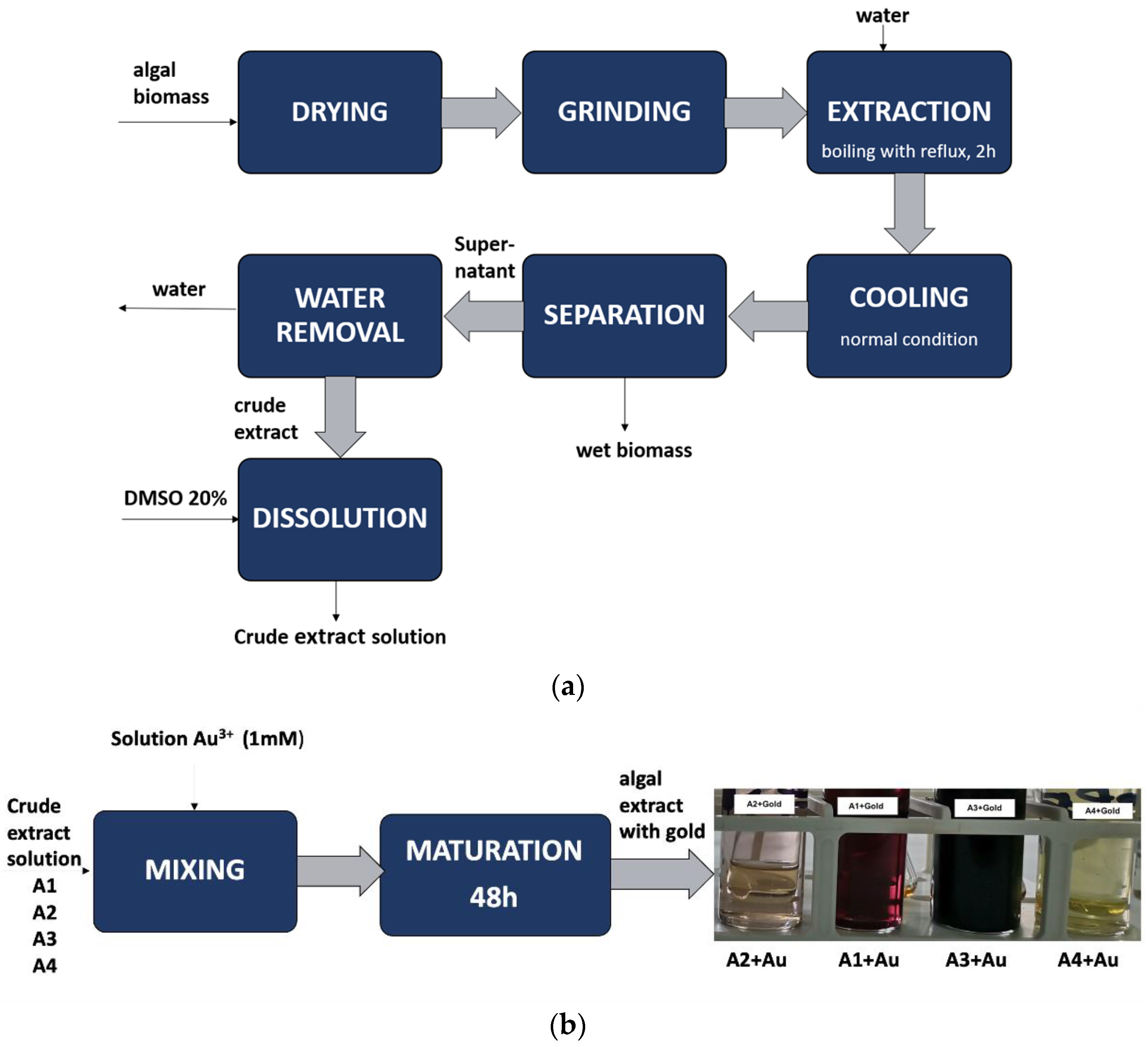
Figure 1. The methodology used to obtain algal bioproducts (a) aqueous polysaccharides extract (b) bioproduct derived from aqueous algal extract and Au3+.
Jmse 13 00318 g001
Figure 2. Antioxidant/prooxidant properties of agal bioproducts obtained by aqueous extraction (a) prooxidant properties of crude bioproduct obtained from Phorpyra umbilicalis; (b) prooxidant properties of crude bioproduct obtained from Undaria pinnatifida; (c) antioxidant properties of crude bioproduct obtained from Cystoseira barbata; (d) prooxidant properties of crude bioproduct obtained from Chlorella sp.
Jmse 13 00318 g002
Figure 3. Characterisation of the bioproducts obtained from gold and each aqueous extracts of algae: (a) A1+Au3+ (crude extract solution of Porphyra umbilicalis and Au3+); (b) A2+Au3+ (crude extract solution of Undaria pinnatifida and Au3+); (c) A3+Au3+ (crude extract solution of Cystoseira barbata and Au3+); (d) A4+Au3+ (crude extract solution of Chlorella sp. and Au3+).
Jmse 13 00318 g003
Figure 4. The effect of bioproducts obtained from algae on HUVEC cell line proliferation: (a) the effect of exposure at 24 h of the HUVEC cell line to bioproducts A1, A2, A3, and A4; (b) the effect of exposure at 24 h of the HUVEC cell line to bioproducts A1+Au, A2+Au, A3+Au, and A4+Au; (c) the effect of exposure at 48 h of the HUVEC cell line to bioproducts A1, A2, A3, and A4; (d) the effect of exposure at 48 h of the HUVEC cell line to bioproducts A1+Au, A2+Au, A3+Au, and A4+Au.
Jmse 13 00318 g004
Figure 5. The effect of bioproducts obtained from algae on Caco-2 cell line proliferation: (a) the effect of exposure at 24 h of the Caco-2 cell line to bioproducts A1, A2, A3, and A4; (b) the effect of exposure at 24 h of the Caco-2 cell line to bioproducts A1+Au, A2+Au, A3+Au, and A4+Au; (c) the effect of exposure at 48 h of the Caco-2 cell line to bioproducts A1, A2, A3, and A4; (d) the effect of exposure at 48 h of the CaCo2 cell line to bioproducts A1+Au, A2+Au, A3+Au, and A4+Au.
Jmse 13 00318 g005
Figure 6. The effect of bioproducts obtained from algae on HepG2 cell line proliferation: (a) the effect of exposure at 24 h of the HepG2 cell line to bioproducts A1, A2, A3, and A4; (b) the effect of exposure at 24 h of the HepG2 cell line to bioproducts A1+Au, A2+Au, A3+Au, and A4+Au; (c) the effect of exposure at 48 h of the HepG2 cell line to bioproducts A1, A2, A3, and A4; (d) the effect of exposure at 48 h of the HepG2 cell line to bioproducts A1+Au, A2+Au, A3+Au, and A4+Au.
Jmse 13 00318 g006
Figure 7. Comparative effect of polysaccharides on the investigated cell line: (a) the inulin effect on HUVEC cell line after 24 h of exposure; (b) the inulin effect on HUVEC cell line after 48 of exposure; (c) the inulin effect on HepG2 cell line after 24 h of exposure; (d) the inulin effect on HepG2 cell line after 48 h of exposure; (e) the inulin effect on Caco-2 cell line after 24 h of exposure; (f) the inulin effect on Caco-2 cell line after 48 h of exposure; (g) the A3 bioproduct effect on Caco-2 cell line after 48 h of exposure; (h) the (A2+Au) bioproduct effect on Caco-2 cell line after 48 h of exposure.
Jmse 13 00318 g007
Figure 8. Mathematical modelling of the antitumoural activities exhibited by bioproducts A4 and A3 as a function of three parameters: (a) mathematical modelling of antitumoural activity as a function of the A4 bioproduct concentration in cell culture media, the residual value of prooxidant activities measured at 140 s, and corresponding cytotoxicity; (b) mathematical modelling of antitumoural activity as a function of the A3 bioproduct concentration in cell culture media, the residual value of prooxidant activities measured at 140 s, and corresponding cytotoxicity.
Jmse 13 00318 g008
Table 1. Raw materials used for obtaining algal polysaccharide extracts.
NameSourcePoly-
Saccharides
Bioproduct
Codification
Extraction ConditionsConcentration, in Crude
Extract
mg/mL ± STDEV
Bioproduct with Gold
Codification
Maturation TimeVolume
Between
A1:Au3+
Voucher Specimen
Deposited at INCDCF Bucharest, Romania
Porphyra umbilicalisRomanian market
EAN 13 code:
8858960303134
A1t = 95 °C
Time = 2 h
27.27 ± 0.01A1+Au48 h1:4.5Poum19-fractionate
Undaria pinnatifidaRomanian market
EAN 13 code:
8717703617535
A2t = 95 °C
time: 2 h
60 ± 0.10A2+Au48 h1:4.5Unpi19-fractionate
Cystoseira barbataBlack SeaA3t = 95 °C
time:2 h
176.4 ± 0.05A3+Au48 h1:4.55Cyba19-fractionate
Chlorella sp.Photobioreactor multiplicationA4t = 95 °C
time: 2 h
55.55 ± 0.10A4+Au48 h1:4.5Chl19-fractionate
Table 2. Algal bioproducts characterisation.
Bioproduct with Poly-SaccharidesSourcePolyphenols Content,
mgGAE/L ± STDEV
Bioproduct with GoldPolyphenols Content
mgGAE/L ± STDEV
The Total Content of Gold in the Bioproduct,
mM ± STDEV
A1Porphyra umbilicalis19.84 ± 0.10A1+Au3.67 ± 0.020.812 ± 0.008
A2Undaria pinnatifida19.70 ± 0.13A2+Au3.03 ± 0.010.849 ± 0.004
A3Cystoseira barbata714.17 ± 1.26A3+Au85.70 ± 0.150.876 ± 0.010
A4Chlorella sp.54.72 ± 0.11A4+Au8.42 ± 0.030.832 ±0.041
Disclaimer/Publisher’s Note: The statements, opinions and data contained in all publications are solely those of the individual author(s) and contributor(s) and not of MDPI and/or the editor(s). MDPI and/or the editor(s) disclaim responsibility for any injury to people or property resulting from any ideas, methods, instructions or products referred to in the content.