Article

Quantitative Real-Time Polymerase Chain Reaction (PCR) Assay for Rapid Monitoring of the Harmful Algal Bloom Species Cochlodinium polykrikoides

1
Library of Marine Samples, Korea Institute of Ocean Science & Technology, Geoje 53201, Republic of Korea
2
Department of Oceanography and Marine Research Institute, Pusan National University, Busan 46241, Republic of Korea
3
Department of Ocean Science, University of Science & Technology, Daejeon 34113, Republic of Korea
4
Sea Power Reinforcement·Security Researcher Department, Korea Institute of Ocean Science & Technology, Busan 49111, Republic of Korea
5
Ecological Risk Research Department, Korea Institute of Ocean Science & Technology, Geoje 53201, Republic of Korea
6
Department of Science, Sainte-Anne University, Church Point, NS B0W 1M0, Canada
7
Marine Biotechnology & Bioresource Research Department, Korea Institute of Ocean Science & Technology, Busan 49111, Republic of Korea
*
Authors to whom correspondence should be addressed.
J. Mar. Sci. Eng. 2025, 13(2), 277; https://doi.org/10.3390/jmse13020277
Submission received: 10 January 2025 / Revised: 24 January 2025 / Accepted: 30 January 2025 / Published: 31 January 2025

Abstract

:
Harmful blooms of the dinoflagellate Cochlodinium polykrikoides (Margalefidinium polykrikoides) had detrimental aquacultural and economic effects globally, and to reduce the damage caused by these blooms, early biomonitoring and quantitative analysis of C. polykrikoides are of the utmost importance. Here, for the detection of C. polykrikoides using quantitative real-time polymerase chain reactions, we developed specific primers targeting the large subunit ribosomal DNA (LSU rDNA) and evaluated their applicability in the field during the occurrence of a C. polykrikoides bloom. The specific primers not only accurately detected C. polykirkoides but also had a detection performance comparable with that obtained using microscopic observations. Accordingly, we developed a system that can be used in the field and applied when red tides occur, with accurate results being obtained more than five times more rapidly than those obtained based on microscopic analysis. Collectively, our findings indicate that the C. polykrikoides bloom detection system developed in this study can be applied to rapidly detect and accurately quantify C. polykrikoides in environmental samples. Data obtained using this system could be used as a basis for developing prompt monitoring and warning systems for the early detection of C. polykrikoides blooms in the field.

1. Introduction

Harmful algal blooms (HABs) have become a major environmental concern globally for aquatic ecosystems, public health, fishery resources, and economies. The determinantal effects of these blooms can be attributed to the high biomass of accumulated algae, which can lead to anoxia, toxin accumulation, and damage to the gills of fish [1,2]. In numerous countries, considerable time and effort were expended in attempts to stem the economic losses attributable to harmful algae and climate change-induced HABs [3]. Since 1980, coastal water pollution has become a serious problem in Republic of Korea as a consequence of the widespread development of coastal industries, and the frequency of HABs correspondingly increased in response to prevalent eutrophication [4]. Among these HAB species, the dinoflagellate Cochlodinium polykrikoides causes extensive damage to aquaculture organisms in the South Sea. In 1995, damage to the aquaculture industry caused by C. polykrikoides blooms resulted in an overall loss of approximately USD 81 million [5]. Consequently, studies focusing on monitoring and predicting the occurrence of C. polykrikoides in coastal seas are essential for protecting both public health and aquatic ecosystems [6,7]. Therefore, accurate and rapid detection of the presence of HABs in conjunction with quantitative analysis is of particular importance for understanding the occurrence and prevalence of HABs and facilitating the reduction or prevention of their impacts on aquatic ecosystems [8].
To date, microscopic analysis has been the traditionally used approach for identifying HAB species and associated quantitative analysis [9,10]. However, as well as being time-consuming, given the low density of harmful algae during the early stages of bloom development, these analyses tend to be limited in terms of identifying HAB species [11,12]. As more viable alternatives, molecular biological techniques, such as quantitative real-time polymerase chain reaction (qPCR), digital PCR (ddPCR), high-throughput sequencing (HTS), and isothermal amplification using environmental DNA, were developed and used to meet the need for rapid detection and accurate quantification of harmful algae [13,14,15]. In addition to recent molecular techniques, remote sensing using absorbance spectra, remote sensing using reflectance, short-term and long-term movement monitoring using the Geostationary Ocean Color Imager (GOCI; Korea Institute of Ocean Science & Technology, Republic of Korea), and content analysis methods for HAB species using High-performance Liquid Chromatography (HPLC) [16,17,18]. Among the techniques, qPCR facilitates the economic, rapid, and highly sensitive quantification of harmful algae, particularly in the case of field samples [19,20]. qPCR is widely adopted as a more accurate and reliable alternative molecular method to those of microscopic analysis for the quantification of HABs [21,22]. Here, to evaluate the feasibility of direct monitoring, we developed specific primers for amplifying the large subunit ribosomal DNA (LSU rDNA) and applied these for qPCR-based detection of C. polykrikoides in the field when blooms of this alga occur along the southern coast of Republic of Korea.

2. Materials and Methods

2.1. Isolation and Culture of Cochlodinium polykrikoides

As a target dianoflagellate for developing a qPCR analysis method, we used a strain of C. polykrikoides isolated from the coastal waters off Tongyeong (34°46′29.3″ N 128°23′24.2″ E) in Republic of Korea (Figure 1a,b).
The isolated strain was cultured in a sterile F/2 medium at 20 °C under a 12:12 light/dark cycle, with a light intensity of approximately 80 µmol photons m−2 s−1. The strain was morphologically identified as C. polykrikoides based on Axioplan light microscope (Carl Zeiss, Oberkochen, Germany) observations and scanning electron microscopy (JSM7600F; Jeol, Tokyo, Japan) using a previously described preparatory method [23].

2.2. Design of Cochlodinium polykrikoides-Specific Primers

Primers targeting the large subunit ribosomal DNA (LSU rDNA) were designed to specifically detect C. polykrikoides. The LSU rDNA sequence of C. polykrikoides was obtained through a BLAST search of the GenBank nucleotide collection and the sequences of HAB species frequently occurring in Republic of Korean coastal waters, including Heterosigma akashiwo, Akashiwo sanguinea, Gyrodinium dominans, and Gymnodinium mikomotoi, were obtained for comparison when PCR was performed using the C. polykrikoides-specific primers. The forward and reverse primers targeting the D2 domain expansion region of LSU were designated as CPF (5ʹ-TGGGTCATTGGTGATTGG-3′) and CPR (5ʹ-TGGTCGTAGACGTGTGTCAG-3′), respectively. The specificity of the primers was confirmed by performing 1000 bootstraps based on ClustalW Multiple alignments using BioEdit version 7.2 (https://bioedit.software.informer.com/7.2, accessed on 1 April 2024).

2.3. Development of LSU Standard Materials for Cochlodinium polykrikoides

Having concentrated the cultured C. polykrikoides to 1 × 10⁵ cells/mL, genomic DNA was extracted using an i-genomic Plant DNA Extraction Mini Kit (iNtRON, Seongnam, Republic of Korea), following the manufacturer’s protocol. The LSU gene of C. polykrikoides was amplified using the CPF and CPR primers in conjunction with Takara EX Taq DNA polymerase (Takara Mirus Bio, Madison, WI, USA), with the following PCR conditions: pre-denaturation at 94.8 °C for 2 min; followed by 32 cycles of denaturation at 94.8 °C for 1 min, annealing at 56.8 °C for 1.5 min, and extension at 72.8 °C for 1 min; with a final extension at 72.8 °C for 5 min. The amplified products were visualized by loading on 2% agarose gels, and the identified bands were purified using QIAquick PCR purification columns (Qiagen, Hilden, Germany). The purified C. polykrikoides LSU was ligated into a pGEMT vector and the resulting construct was used to transform E. coli DH5 α, with blue/white screening used to identify transformants. The sequence of the product was subsequently confirmed via PCR and sequencing. Plasmids were harbored by a transformant in which the LSU gene sequence was confirmed and extracted using a QIAwave Plasmid Miniprep Kit (Qiagen) and used as a standard material.

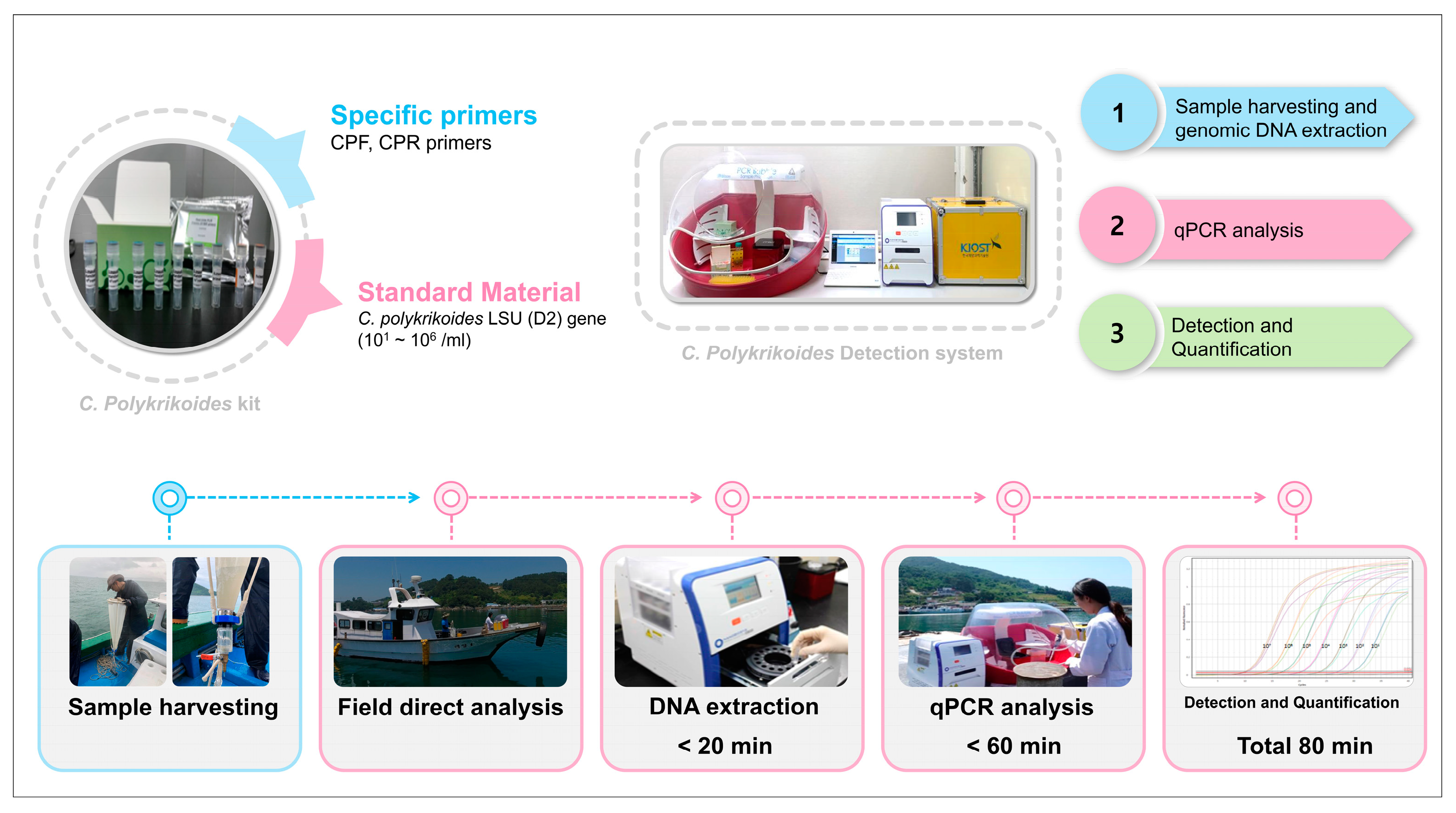
2.4. Development of a Cochlodinium polykrikoides Detection System

For a rapid evaluation of the occurrence of C. polykrikoides when blooms occur in coastal waters, we developed an HAB species detection system based on qPCR for quantitative and qualitative analyses. This system consists of three steps, in the first of which, HAB samples are collected, and genomic DNA (gDNA) is extracted. In the second step, the extracted DNA is amplified using qPCR, and in the final step, C. polykrikoides is enumerated. The collection step involves the concentration of 10 L of algal-colonized seawater to 50 mL using a Kitahara plankton net (mesh size: 10 µm). A pellet of the HAB species was obtained from the concentrated seawater, and an average of 100 ng/μL (total volume of 50 µL) of gDNA was extracted using UltraFast Sample Prep G2-16TU (NANOBIOSYS INC., Seoul, Republic of Korea). The qPCR kit consisted of a positive control, primer mix, nuclease-free water, and real-time PCR master mix (SYBR green). The primer mix consisted of the C. polykrikoides LSU gene-specific primer pair (CPF and CPR). The positive control consisted of 101–106 copy numbers of the C. polykrikoides LSU standard gene. The evaluation method for field verification of C. polykrikoides blooms using the C. polykrikoides detection kit is as follows. The qPCR reaction mixture consists of 5 μL of the template and 2 μL of the primer mix and was adjusted to a total volume of 20 μL with nuclease-free water in a real-time PCR master mix (SYBR green) tube. Using the prepared qPCR reaction mixture, amplification was performed using the following conditions: an initial denaturation step at 95 °C for 5 min, followed by 45 cycles of denaturation at 95 °C for 10 s and annealing and extension at 60 °C for 40 s. The final step involved establishing a standard curve for C. polykrikoides based on the qPCR results. A standard curve was established to determine copy numbers by preparing triplicate samples using positive controls. C. polykrikoides were used to establish a standard curve according to cell number and were collected during the exponential phase of the growth phase, and the concentration of cells was quantitatively analyzed using an Axioplan optical microscope (Carl Zeiss, Jena, Germany). DNA was extracted from each prepared sample at the same dilution as previously described. The cycle threshold (Ct) values for each sample were calculated using qPCR. The normality of the data was verified using the Kolmogorov–Smirnov test and a quantile–quantile plot [24,25]. The standard material and cell count templates were used to construct a linear regression line (standard curve) between the log10 and Ct values. Reproducibility was confirmed by performing qPCR on samples with low and high concentrations, and low and high cell counts.

2.5. Field Application of the Cochlodinium polykrikoides Detection System

On 4 September 2019, C. polykrikoides samples were collected from blooms developed along the Yeosu coast of Republic of Korea. Twenty-two sampling sites along the Yeosu coastline were selected to investigate the distribution patterns of C. polykrikoides. HAB samples were collected using a Kitahara plankton net, and gDNA was immediately extracted for C. polykrikoides detection. The gDNA thus obtained was subjected to qPCR analysis using a Mic qPCR Cycler (Bio Molecular Systems, Upper Coomera Australia) and a C. polykrikoides detection kit. To verify the detection capacity of the qPCR, C. polykrikoides cells were identified and counted under a light microscope [7].

3. Results and Discussion

3.1. Evaluation of the Cochlodinium polykrikoides-Specific Primers

Molecular techniques, including qPCR, used to detect C. polykrikoides vegetative cells and cysts in sediments [26,27], highlighted the importance of using specific primers for detection [27]. Previous studies reported that HAB algae can be differentiated at the species level based on differences in the LSU rDNA (D1-D2) region [27]. However, the previously used primers have limitations in specifically detecting C. polykrikoides. For example, the universal LSUBs (D2 domain) primers are disadvantageous in that they bind to an LSU region conserved in most HAB species, including C. polykrikoides [27,28]. Additionally, primers known to bind specifically to the LSU (D1-D2 domain) in C. polykrikoides do not specifically bind in some C. polykrikoides strains [27]. Therefore, it was necessary to design new primers that could bind specifically to the LSU region of C. polykrikoides.
In this study, based on multiple alignments, we confirmed the specificity of CPF and CPR primer regions using the isolated C. polykrikoides (Figure 1c). Specifically, compared with that of the 22 LSU sequences obtained from other HAB species, the CPF and CPR primers exclusively amplified the D2 domain of C. polykrikoides LSU.
To detect and quantify C. polykrikoides, we prepared a standard curve based on the Ct values obtained from cell counting and gene copy numbers based on qPCR (Figure 1d,e). The correlation coefficients of the standard curves revealed highly significant correlations with the numbers of cells (R2 = 0.959) and gene copy numbers (R2 = 0.980). These findings thus provide evidence to indicate that the C. polykrikoides-specific primers (CPF and CPR) targeting the LSU rDNA (D2 domain) region are very specific and can accordingly be for the accurate detection and quantification of C. polykrikoides. Furthermore, this gene region could also be a suitable target for the identification of species in eukaryotic groups [29].

3.2. Verification Through Field Application

Kim et al. [7] reported that a bloom of C. polykrikoides was initially detected on 20 August 2019, in waters off the Yeosu coast, and that by 4 September, extensive red tides (maximum cell density of 12,000 cells/mL) developed. For the early detection of such red tides, we developed a C. polykrikoides quality and quantity detection system (Figure 2), the field application of which involves the three steps of sample harvesting and DNA extraction, qPCR analysis, and detection and quantification, and can yield results within approximately 80 min (Figure 2). Given that the entire analytic procedure can be conducted in the field, this HAB species detection system has the advantage of highly accurate rapid detection of multiple samples. Accordingly, this HAB species detection system is prominently time-efficient when applied in the field.
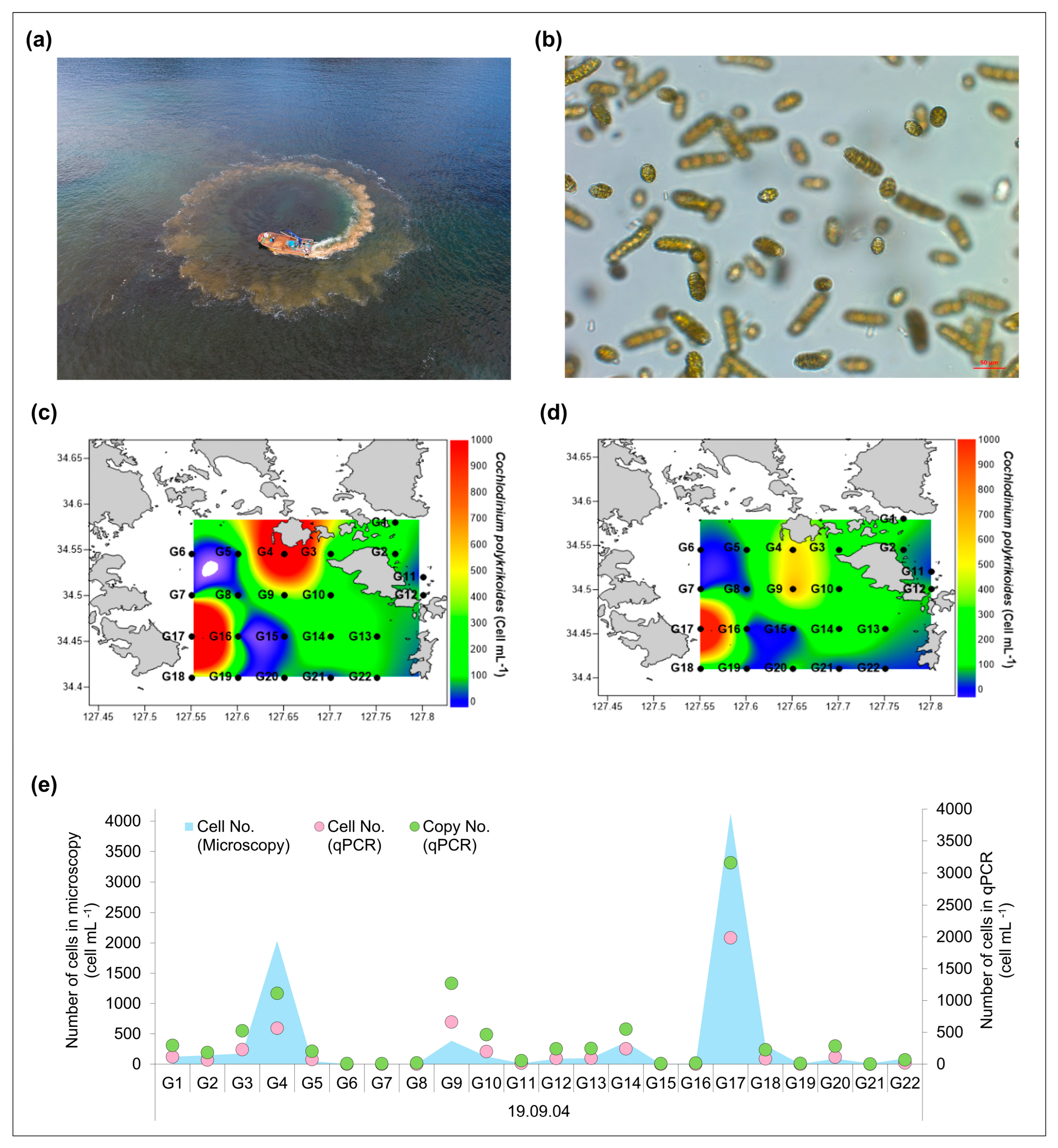
The C. polykrikoides detection system was applied on 4 September 2019 to analyze samples collected from the algal blooms developed along the Yeosu coast (Figure 3a,b). Hence, a comparison of microscopy and qPCR data were considered essential for establishing whether the detection system could accurately detect and quantify C. polykrikoides under field conditions. We accordingly found that the microscopic observations and qPCR results were significantly and highly correlated (R2 = 0.938) (Figure 3c–e). Other studies similarly reported comparable qPCR quantitative results and microscopic observations (approximately R2 = 0.74) when detection systems were applied in the field [27,30]. Our findings in the current study thus indicate that the CPF and CPR primer pair used in the detection system are more specific for C. polykrikoides detection than that for the primers previously described. Additionally, the quantitative consistency of the microscopic and detection systems results provided further evidence to confirm the high specificity of these primers (Figure 3e). However, we did obtain disparate results for samples collected at the G9 sampling site, for which C. polykrikoides was detected at 385 cells/mL and 695 cells/mL using microscopy and the qPCR system, respectively, which thus tends to indicate an overestimation using the qPCR approach (Figure 3c,d). To account for this apparent disparity in findings, we propose the following two explanations. First, gene copy numbers can vary depending on the stage of the C. polykrikoides growth cycle or DNA fragmentation [31], and second, during red tides, the vegetative cells of C. polykrikoides can take different forms depending on the stage of the life cycle. Alternatively, it is conceivable that the microscopic observation, in this case, provided an underestimate of cell numbers that can occur in the presence of divided cells, which may be difficult to distinguish when viewed under a microscope [32,33]. In this regard, it was proposed that if the complete life cycle is elucidated and abundant different forms of vegetative cells can be obtained in the laboratory, a more accurate determination of vegetative cell counts can be achieved, thus enabling the effective monitoring of red tide occurrences. Conversely, at the G4 site, which was characterized by the densest bloom development (2035 cells/mL), as determined using microscopy, the detection system appeared to show an underestimate of 593 cells/mL (Figure 3c,d). We speculate that this disparity could be ascribed to the fact that the amount of polymerase used to amplify DNA in qPCR is fixed and high concentrations of gDNA extracted from high-density cells may exceed the polymerase amplification capacity [34]. This, however, can readily be rectified by diluting the gDNA [35]. The detection limit and accuracy of qPCR are influenced by the copy number of the target gene, which is considered necessary for the reliability of the qPCR results [35]. The copy number results showed a pattern similar to that of the cell numbers, which thereby indicates that these findings can be useful for an early warning of C. polykrikoides red tide development.
In addition to qPCR, ddPCR and HTS analysis methods are widely applied to HAB analysis [26,30,31,36]. HTS has the advantage of being able to analyze various species, but the composition ratio of the clusters was found to be significantly different compared to microscopic analysis [36]. ddPCR has the advantage of not requiring a standard curve, recognizing even a tiny number of cells, but it has the disadvantage of being more expensive than qPCR [26,37]. On the other hand, qPCR has the advantage of showing similar results to microscopy and being relatively inexpensive [37].
However, in most cases, samples collected in the field are analyzed in the laboratory, and comparatively few studies have directly analyzed samples in the field. Samples collected in external environments may, nevertheless, be susceptible to contamination if analyzed immediately, and equipment vulnerable to shaking or vibrations, such as that caused by waves, cannot be reliably used [14]. Nonetheless, immediate on-site analysis has numerous advantages, and to facilitate such analyses, the corresponding detection systems should ideally have the properties of portability, high sample throughput, ease of use, and accuracy [38]. Given their purpose of rapidly conveying information from the field [39], such systems must be compatible with unprocessed samples and provide fully interpreted results for multiple samples within a short period of time [40]. Therefore, by combining automatic nucleic acid extraction and miniaturized qPCR equipment, we were able to perform sample analysis within approximately 80 min. Additionally, it is beneficial to use portable and conveniently used equipment for field applications. Accordingly, the automation of nucleic acid analysis contributes to minimizing hands-on time, and small-sized qPCR equipment maximizes portability. Finally, given that during the period of red tide development, HABs such as C. polykrikoides occur as vegetative cells, division and decomposition continue to occur from the time of sample collection [33]. Consequently, the most significant advantage of immediate on-site analysis is that accurate analysis is possible without substantial changes in sample characteristics [39]. The C. polykrikoides detection system developed in this study enables immediate on-site monitoring of C. polykrikoides and provides an approach for rapid early warning during subsequent mass bloom periods.

4. Conclusions

Conclusively, having initially developed C. polykrikoides-specific primers, we used these as a basis for constructing a C. polykrikoides detection system that can be applied for rapid analysis in marine environments. This system can be used to predict algal blooming at a level of accuracy comparable to that predicted based on microscopy. Furthermore, we demonstrate that the quantitative detection of C. polykrikoides is possible through sample dilution during the early stages of red tide bloom development (low-cell-density stage), as well as during the vegetative cell stage, and even during the high-cell-density stage. This system can thus be applied to facilitate field monitoring and thereby enable the establishment of early warning systems for C. polykrikoides bloom development.

Author Contributions

M.-J.K.: Writing—original draft and visualization. H.-J.K.: data curation and formal analysis. J.S.P., D.K., S.C., H.K., S.H.B. and J.J.C.P.: data curation. K.E.K.: data curation. J.H. and S.W.J.: conceptualization, writing—original draft, and funding acquisition. All authors have read and agreed to the published version of the manuscript.

Funding

This research was financially supported by the Korea Institute of Ocean Science and Technology (PEA0312), and the Korea Institute of Marine Science and Technology Promotion (KIMST), funded by the Ministry of Oceans and Fisheries, Republic of Korea (RS-2021-KS211475).

Institutional Review Board Statement

Not applicable.

Informed Consent Statement

Not applicable.

Data Availability Statement

Data will be made available on request.

Acknowledgments

The genomic DNA samples were stored in the Library of Marine Samples of the Korea Institute of Ocean Science and Technology (KIOST), Republic of Korea.

Conflicts of Interest

The authors declare no conflicts of interest.

References

  1. Ternon, E.; Pavaux, A.-S.; Marro, S.; Thomas, O.P.; Lemée, R. Allelopathic interactions between the benthic toxic dinoflagellate Ostreopsis cf. ovata and a co-occurring diatom. Harmful Algae 2018, 75, 35–44. [Google Scholar] [CrossRef] [PubMed]
  2. Gobler, C.J. Climate change and harmful algal blooms: Insights and perspective. Harmful Algae 2020, 91, 101731. [Google Scholar] [CrossRef] [PubMed]
  3. Heil, C.A.; Muni-Morgan, A.L. Florida’s harmful algal bloom (HAB) problem: Escalating risks to human, environmental and economic health with climate change. Front Ecol. Evol. 2021, 9, 646080. [Google Scholar] [CrossRef]
  4. Lee, C.-K.; Park, T.-G.; Park, Y.-T.; Lim, W.-A. Monitoring and trends in harmful algal blooms and red tides in Korean coastal waters, with emphasis on Cochlodinium polykrikoides. Harmful Algae 2013, 30, S3–S14. [Google Scholar] [CrossRef]
  5. Park, T.G.; Lim, W.A.; Park, Y.T.; Lee, C.K.; Jeong, H.J. Economic impact, management and mitigation of red tides in Korea. Harmful Algae 2013, 30, S131–S143. [Google Scholar] [CrossRef]
  6. Lee, Y.-C.; Jin, E.; Jung, S.W.; Kim, Y.-M.; Chang, K.S.; Yang, J.-W.; Kim, S.-W.; Kim, Y.-O.; Shin, H.-J. Utilizing the algicidal activity of aminoclay as a practical treatment for toxic red tides. Sci. Rep. 2013, 3, 1292. [Google Scholar] [CrossRef] [PubMed]
  7. Kim, H.-J.; Jeoung, G.; Kim, K.E.; Park, J.S.; Kang, D.; Baek, S.H.; Lee, C.Y.; Kim, H.; Cho, S.; Lee, T.-K.; et al. Co-variance between free-living bacteria and Cochlodinium polykrikoides (Dinophyta) harmful algal blooms, South Korea. Harmful Algae 2023, 122, 102371. [Google Scholar] [CrossRef]
  8. Kang, D.; Kim, B.K.; Jung, S.W.; Baek, S.H.; Choi, J.-Y.; Cho, H.-Y.; Lee, S.-J.; Kim, H. Development and application of an integrated system for the detection and prediction of harmful algal blooms in Korea. J. Mar. Sci. Eng. 2023, 11, 2207. [Google Scholar] [CrossRef]
  9. LeGresley, M.; McDermott, G. Counting Chamber Methods for Quantitative Phytoplankton Analysis-Haemocytometer, Palmer-Maloney Cell and Sedgewick-Rafter Cell. In United Nations Educational, Scientific and Cultural Organization (IOC Manuals and Guides); UNESCO: Paris, France, 2010; Volume 55, pp. 25–30. [Google Scholar]
  10. Antonella, P.; Luca, G. The quantitative real-time PCR applications in the monitoring of marine harmful algal bloom (HAB) species. Environ. Sci. Pollut. Res. 2013, 20, 6851–6862. [Google Scholar] [CrossRef] [PubMed]
  11. Karlson, B.; Cusack, C.; Bresnan, E. (Eds.) Microscopic and Molecular Methods for Quantitative Phytoplankton Analysis; UNESCO: Paris, France, 2010; p. 110. [Google Scholar]
  12. Medlin, L. Molecular tools for monitoring harmful algal blooms. Environ. Sci. Pollut. Res. Int. 2013, 20, 6683–6685. [Google Scholar] [CrossRef]
  13. Lee, H.-G.; Kim, H.M.; Min, J.; Park, C.; Jeong, H.J.; Lee, K.; Kim, K.Y. Quantification of the paralytic shellfish poisoning dinoflagellate Alexandrium species using a digital PCR. Harmful Algae 2020, 92, 101726. [Google Scholar] [CrossRef] [PubMed]
  14. Feist, S.M.; Lance, R.F. Genetic detection of freshwater harmful algal blooms: A review focused on the use of environmental DNA (eDNA) in Microcystis aeruginosa and Prymnesium parvum. Harmful Algae 2021, 110, 102124. [Google Scholar] [CrossRef] [PubMed]
  15. Wang, J.; Wang, Y.; Lai, J.; Li, J.; Yu, K. Improvement and application of qPCR assay revealed new insight on early warning of Phaeocystis globosa bloom. Water Res. 2023, 229, 119439. [Google Scholar] [CrossRef]
  16. Kim, W.; Han, T.; Jung, S.; Kang, D. Analysis on the Optical Absorption Property of Sea Waters Dominated by Alexandrium affine in Coastal Waters off Tongyeong, 2017. Photogramm. Cartogr. 2019, 37, 563–570. [Google Scholar]
  17. Choi, J.K.; Min, J.E.; Noh, J.H.; Han, T.H.; Yoon, S.; Park, Y.J.; Park, J.H. Harmful algal bloom (HAB) in the East Sea identified by the Geostationary Ocean Color Imager (GOCI). Harmful Algae 2014, 39, 295–302. [Google Scholar] [CrossRef]
  18. Dizaji, S.Z.; Fariman, G.A.; Zahedi, M.M. Pigment content analysis in two HAB forming dinoflagellate species during the growth period. J. Appl. Phycol. 2021, 33, 807–817. [Google Scholar] [CrossRef]
  19. Medlin, L.K.; Orozco, J. Molecular techniques for the detection of organisms in aquatic environments, with emphasis on harmful algal bloom species. Sensors 2017, 17, 1184. [Google Scholar] [CrossRef]
  20. Hernández-López, E.L.; Gasperin, J.; Bernáldez-Sarabia, J.; Licea-Navarro, A.F.; Guerrero, A.; Lizárraga-Partida, M.L. Detection of Alcanivorax spp., Cycloclasticus spp., and Methanomicrobiales in water column and sediment samples in the Gulf of Mexico by qPCR. Environ. Sci. Pollut. Res. Int. 2019, 26, 35131–35139. [Google Scholar] [CrossRef]
  21. Engesmo, A.; Strand, D.; Gran-Stadniczeñko, S.; Edvardsen, B.; Medlin, L.K.; Eikrem, W. Development of a qPCR assay to detect and quantify ichthyotoxic flagellates along the Norwegian coast, and the first Norwegian record of Fibrocapsa japonica (Raphidophyceae). Harmful Algae 2018, 75, 105–117. [Google Scholar] [CrossRef] [PubMed]
  22. Hatfield, R.G.; Bean, T.; Turner, A.D.; Lees, D.N.; Lowther, J.; Lewis, A.; Baker-Austin, C. Development of a TaqMan qPCR assay for detection of Alexandrium spp. and application to harmful algal bloom monitoring. Toxicon X 2019, 2, 100011. [Google Scholar] [CrossRef]
  23. Jung, S.W.; Joo, H.M.; Park, J.S.; Lee, J.H. Development of a rapid and effective method for preparing delicate dinoflagellates for scanning electron microscopy. J. Appl. Phycol. 2010, 22, 313–317. [Google Scholar] [CrossRef]
  24. Joanes, D.N.; Gill, C.A. Comparing measures of sample skewness and kurtosis Soc Ser D-Stat 47(1). J. R. Stat. Soc. Ser. D 1998, 47, 183–189. [Google Scholar] [CrossRef]
  25. Dhar, S.S.; Chakraborty, B.; Chaudhuri, P. Comparison of multivariate distributions using quantile–quantile plots and related tests. Bernoulli 2014, 20, 1484–1506. [Google Scholar] [CrossRef]
  26. Jung, S.W.; Kang, D.; Kim, H.-J.; Shin, H.H.; Park, J.S.; Park, S.Y.; Lee, T.-K. Mapping distribution of cysts of recent dinoflagellate and Cochlodinium polykrikoides using next-generation sequencing and morphological approaches in South Sea, Korea. Sci. Rep. 2018, 8, 7011. [Google Scholar] [CrossRef] [PubMed]
  27. Park, T.G.; Kim, J.J.; Kim, W.J.; Won, K.M. Development of real-time RT-PCR for detecting viable Cochlodinium polykrikoides (Dinophyceae) cysts in sediment. Harmful Algae 2016, 60, 36–44. [Google Scholar] [CrossRef] [PubMed]
  28. Litaker, R.W.; Vandersea, M.W.; Kibler, S.R.; Reece, K.S.; Stokes, N.A.; Steidinger, K.A.; Millie, D.F.; Bendis, B.J.; Pigg, R.J.; Tester, P.A. Identification of Pfiesteria piscicida (Dinophyceae) and Pfiesteria-like organisms using internal transcribed spacer-specific pcr assays 1. J. Phycol. 2003, 39, 754–761. [Google Scholar] [CrossRef]
  29. Sonnenberg, R.; Nolte, A.W.; Tautz, D. An evaluation of LSU rDNA D1-D2 sequences for their use in species identification. Front. Zool. 2007, 4, 6. [Google Scholar] [CrossRef] [PubMed]
  30. Park, T.G.; Park, Y.T.; Lee, Y. Development of a SYTO9 based real-time PCR probe for detection and quantification of toxic dinoflagellate Karlodinium veneficum (Dinophyceae) in environmental samples. Phycologia 2009, 48, 32–43. [Google Scholar] [CrossRef]
  31. Handy, S.M.; Demir, E.; Hutchins, D.A.; Portune, K.J.; Whereat, E.B.; Hare, C.E.; Rose, J.M.; Warner, M.; Farestad, M.; Cary, S.C.; et al. Using quantitative real-time PCR to study competition and community dynamics among Delaware Inland Bays harmful algae in field and laboratory studies. Harmful Algae 2008, 7, 599–613. [Google Scholar] [CrossRef]
  32. Kim, C.-J.; Kim, H.-G.; Kim, C.-H.; Oh, H.-M. Life cycle of the ichthyotoxic dinoflagellate Cochlodinium polykrikoides in Korean coastal waters. Harmful Algae 2007, 6, 104–111. [Google Scholar] [CrossRef]
  33. Shin, H.H.; Li, Z.; Yoon, Y.H.; Oh, S.J.; Lim, W.A. Formation and germination of temporary cysts of Cochlodinium polykrikoides Margalef (Dinophyceae) and their ecological role in dense blooms. Harmful Algae 2017, 66, 57–64. [Google Scholar] [CrossRef] [PubMed]
  34. Wang, D.; Wang, S.; Du, X.; He, Q.; Liu, Y.; Wang, Z.; Feng, K.; Li, Y.; Deng, Y. ddPCR surpasses classical qPCR technology in quantitating bacteria and fungi in the environment. Mol. Ecol. Resour. 2022, 22, 2587–2598. [Google Scholar] [CrossRef] [PubMed]
  35. Hu, X.-K.; Lin, Z.-R.; Zhang, Q.-C.; Kong, F.-Z.; Cen, J.-Y.; Zeng, Y.-L.; Yu, R.-C. Detection of bloom-forming dinoflagellates Karenia mikimotoi and Prorocentrum donghaiense using qPCR assays. J. Appl. Phycol. 2022, 34, 1483–1496. [Google Scholar] [CrossRef]
  36. Gałuszka, A.; Migaszewski, Z.M.; Namieśnik, J. Moving your laboratories to the field–Advantages and limitations of the use of field portable instruments in environmental sample analysis. Environ. Res. 2015, 140, 593–603. [Google Scholar] [CrossRef] [PubMed]
  37. Lee, H.G.; Kim, H.M.; Min, J.; Kim, K.; Park, M.G.; Jeong, H.J.; Kim, K.Y. An advanced tool, droplet digital PCR (ddPCR), for absolute quantification of the red-tide dinoflagellate, Cochlodinium polykrikoides Margalef (Dinophyceae). Algae 2017, 32, 189–197. [Google Scholar] [CrossRef]
  38. Zowawi, H.M.; Alenazi, T.H.; AlOmaim, W.S.; Wazzan, A.; Alsufayan, A.; Hasanain, R.A.; Aldibasi, O.S.; Althawadi, S.; Altamimi, S.A.; Mutabagani, M.; et al. Portable RT-PCR system: A rapid and scalable diagnostic tool for COVID-19 testing. J. Clin. Microbiol. 2021, 59, 10.1128. [Google Scholar] [CrossRef]
  39. Spikmans, V. The evolution of environmental forensics: From laboratory to field analysis. WIREs Forensic Sci. 2019, 1, e1334. [Google Scholar] [CrossRef]
  40. Lepej, S.Z.; Poljak, M. Portable molecular diagnostic instruments in microbiology: Current status. Clin. Microbiol. Infect. 2020, 26, 411–420. [Google Scholar] [CrossRef] [PubMed]
Figure 1. Cochlodinium polykrikoides morphology, specific primer design, and standard curve construction. (a) Light microscopic and (b) scanning electron microscopic micrographs of C. polykrikoides, (c) C. polykrikoides-specific primer target sites, (d) a cell number-based standard curve, and (e) a copy number-based standard curve.
Jmse 13 00277 g001
Figure 2. Schematic diagram of the Cochlodinium polykrikoides field detection system.
Jmse 13 00277 g002
Figure 3. Field application of a qPCR monitoring system for Cochlodinium polykrikoides red tides. (a,b) Photographs of C. polykrikoides mitigation using natural yellow clay. Contour charts of C. polykrikoides red tide occurrence based on microscopic (c) and qPCR (d) analyses. (e) Comparison of C. polykrikoides red tide cell counts in the field using microscopic analysis and qPCR methods.
Jmse 13 00277 g003
Disclaimer/Publisher’s Note: The statements, opinions and data contained in all publications are solely those of the individual author(s) and contributor(s) and not of MDPI and/or the editor(s). MDPI and/or the editor(s) disclaim responsibility for any injury to people or property resulting from any ideas, methods, instructions or products referred to in the content.