Article

Abundance and Diversity of Several Bacterial Genera in the Mariculture Environment

1
State Key Laboratory of Tropical Oceanography, South China Sea Institute of Oceanology, Chinese Academy of Sciences, Guangzhou 510000, China
2
Daya Bay Marine Biology Research Station, Chinese Academy of Sciences, Shenzhen 518000, China
3
Sanya Institute of Ocean Eco-Environmental Engineering, Sanya 572000, China
4
Putian Institute of Aquaculture Science of Fujian Province, Putian 351100, China
*
Author to whom correspondence should be addressed.
These authors contributed equally to this work.
J. Mar. Sci. Eng. 2024, 12(2), 209; https://doi.org/10.3390/jmse12020209
Submission received: 31 October 2023 / Revised: 19 November 2023 / Accepted: 22 November 2023 / Published: 24 January 2024
(This article belongs to the Section Marine Aquaculture)

Abstract

:
Although there have been some studies on pathogenic bacteria and their pathogenicity in animals, few studies have assessed the effects of aquaculture on the diversity of potentially pathogenic bacteria. This study used Illumina sequencing and quantitative PCR to explore the diversity of several bacterial genera containing pathogenic bacteria in the mariculture environment and the intestines of different cultured animals. These bacterial genera can be divided into two categories: The first category (14 genera) had high abundances and a low coefficient of variation among similar samples were significantly correlated with the total number of bacteria (r2 > 0.7, p ≈ 0). The other category (7 genera) with low abundances and a high coefficient of variation had no significant relationship with bacterial abundance. These results indicated that these bacterial genera had different responses and adaptation mechanisms to the aquaculture environment. Principal component analysis (PCA) showed that a high abundance of genera was closely related to the pond environment. The abundance of these bacterial genera in the animals’ intestines was much higher than source water, especially for Mycoplasma, Pseudoalteromonas, Vibrio, and Enterococcus, suggesting the aquaculture promoted the high abundance of these bacteria. This study provides a theoretical basis for sediment-associated pathogens acting as a potential pathogen source in the aquaculture environment. This study provides a strategy for disease prevention and control according to the characteristics of potential pathogens in the cultural process.

1. Introduction

In recent decades, the escalating demand for seafood has outpaced the capacity of wild fisheries to meet it, leading to the rapid expansion of the offshore aquaculture industry. This industry has experienced significant growth in terms of both the number of species cultivated and the quantity of aquaculture production, predominantly fish, crab, and shellfish. According to the Food and Agriculture Organization of the United Nations (FAO), the world’s food and feed supply need to grow by 70% by 2050 to support an expanding population [1]. As such, the output of seafood from aquaculture has far surpassed that of offshore fishing. Consequently, large-scale aquaculture has emerged as the most extensively distributed offshore system.
As the aquaculture industry rapidly expands in scale, so too do the associated safety requirements. In contrast to fishing, cultured seafood is more vulnerable to potential pathogens [2]. This susceptibility is evidenced by the increasing frequency and intensity of recorded disease outbreaks in many marine taxa worldwide [2]. The recent uptick in such outbreaks [3] may be attributed to the introduction of new pathogens or environmental changes [2,4,5,6,7]. Bacteria, fungi, viruses, oomycetes, amoebas, and other ectoparasites have emerged as causal agents of several diseases that limit fish and shellfish production [8]. For example, Vibrio is a dominant bacterium recorded in both animal intestines and cultivation environments [4,9,10,11]. Pathogenic Vibrio can cause significant mortality in aquaculture, resulting in major losses to both population and revenue [12].
The composition of gut microbiota is closely related to the development, nutritional status, immune response, and host resistance to diseases [13,14], which is a necessary factor for maintaining animal health. The composition of gut microbiota is determined not only by host genetics and diet, but also by microbiota present in the ambient water and sediment [4,15,16], highlighting the successful transfer of microorganisms from sediment and water to animal intestines [4,9]. The occurrence of aquaculture diseases is closely related to the composition of the surrounding environmental microbiota. There was a significant relationship between potential pathogenic bacteria and nutrient levels in the aquaculture environment [9]. Bacteria in the aquatic environment play a decisive role in the formation of intestinal bacteria in animals. The composition of gut microbiota in adult animals is still influenced by the environment [17]. The aquaculture environment was the main source of bacteria in the intestine of culture animals [4,9]. If there is a change in the internal or external environment of the gut, the stability of the gut bacterial community structure may be disrupted, allowing pathogenic bacteria to invade the digestive system and trigger infections [18]. The quantity and type of pathogenic microbes present in the culture environment are key factors that influence the production of high-quality seafood, and are directly impacted by environmental factors. Moreover, they are closely linked to disease outbreaks [19,20,21]. Thus, a higher abundance of these bacteria in the aquatic environment may indicate an increased risk of pathogenicity in cultured animals. However, a comprehensive assessment of the species and migration of these bacteria in the aquaculture environment has yet to be conducted.
The aim of this study is to evaluate the differences in bacterial abundance between the water source and aquaculture ponds (water and sediment), analyze the abundance and changes in diversity of specific bacterial genera from the water source to the aquaculture ponds using Illumina sequencing and qPCR, and investigate the distribution patterns of different bacterial genera in the water source, aquaculture pond, and animal intestine. This study will reveal the distribution patterns of specific bacterial genera in aquaculture environments, providing a theoretical basis for the scientific management of water quality and the prevention and control of diseases.

2. Materials and Methods

2.1. Samples Collection

The samples were obtained from a seawater reclamation area in Putian city, where the ponds are supplied with seawater from a common source that enters the intensive aquaculture system via an inlet. The water inlet and outlet are distanced from each other as a measure to prevent pollution. There are 35 culture ponds in this reclamation area. Each culture pond has an area of approximately 66,600 m2 and an average water depth of 1 m. The bottom of the pond is mud and sand in a ratio of 81:19.
Samples of source water (n = 6) and pond water (n = 35) were collected from the different culture ponds or sites without disturbance using a glass water hydrophore in different sites or ponds. A 1000 mL sample of water was extracted at a depth of 0.5 m and filtered through a 0.22 µm polycarbonate membrane (EMD Millipore, Billerica, MA, USA), which was then placed into a 1.5 mL sterile centrifuge tube. Sediment samples (n = 19) were collected from a depth of 0–2 cm using a core sampler in different ponds. Culture animals (crab, shrimp, and cram) were collected from the different culture ponds or at different points in the water inlet. Eight individuals of each organism were selected, and their intestines were carefully dissected using conventional aseptic techniques. Subsequently, their contents were gently extracted under sterile laboratory conditions and transferred into a sterile 1.5 mL centrifuge tube. All samples were stored in liquid nitrogen until further DNA extraction.

2.2. Illumina MiSeq Sequencing of 16S rRNA

The Illumina Sequencing method, as described in a previous study [4], was employed in this investigation. Total DNA from filter membranes, sediment samples, and animal intestines (1 g) was extracted using a DNA Extraction Kit (OMEGA, Norwalk, CT, USA). The 16S rRNA gene was targeted using the V3 and V4 regions with the primers 319F (5′-ACTCCTACG GGAGGCAGCAG-3′) and 806R (GGACTACHVGGTATCTAAT) with the barcode. A PCR reaction volume of 25 μL was used, consisting of 5× FastPfu buffer (4 μL), 2.5 mmol/L dNTPs (2 μL), 5.0 μmol/L primer (1.0 μL), 5 U/μL FastPfu polymerase (0.5 μL), and 10 ng DNA template. The amplified products were purified and quantified using the QuantiFluorTM fluorometer (Promega, Milan, Italy). The libraries were generated with a TruSeq® DNA PCR-Free sample preparation kit (Illumina, San Diego, CA, USA), and sequenced on an Illumina MiSeq platform (Illumina, San Diego, CA, USA) following standard protocols.

2.3. Data Analysis of High-Throughput Sequencing

For paired-end reads obtained through Illumina MiSeq sequencing, barcode and primer sequences were trimmed from the paired-end sequences. Subsequently, assembly was performed using FLASH (version 1.2.11) [22]. Low-quality reads were removed, and the Chimera Check was applied to eliminate chimeric sequences. VSEARCH (version 1.9.6) was employed to cluster high-quality sequences into operational taxonomic units (OTUs) with a sequence identity threshold of 97%. The Ribosomal Database Project (RDP) classifier was utilized to assign taxonomic categories to all OTUs with a confidence threshold of 0.8. BLAST was employed to assign representative sequences for each OTU (the most abundant) against the Silva_123 16S rRNA database (https://www.arb-silva.de/, accessed on 21 November 2023). Differences in bacterial community among source water, pond water, and pond sediment were conducted by performing principal component analysis (PCA) using CANOCO 4.5 software (CANOCO, Ithaca, NY, USA).

2.4. 16S rRNA Quantiative PCR (qPCR)

The quantification of 16S rRNA genes was conducted through qPCR employing the Applied Biosystems StepOnePlusTM Real-Time System (Applied Biosystems, Carlsbad, CA, USA) with 341F CCTACGG-GAGGCAGCAG and 518R ATTACCGCGGCTGCTGG primers [23]. Each sample was subjected to triplicate qPCR runs, and quantification was based on the amplification-dependent increase in fluorescence intensity of the SYBR Green dye. qPCR standards were generated from PCR products that were amplified from environmental samples using the pMD19-T cloning kit (TaKaRa, Shiga, Japan), and the plasmid DNA was purified using the Plasmid Purification kit (TaKaRa, Shiga, Japan). The concentrations of qPCR standard products were determined at 260/280 nm using the ND-2000 spectrophotometer (NanoDrop, Wilmington, DE, USA). The real-time PCR assay was performed in a 20 μL reaction volume with SYBR Premix Ex Taq II (TaKaRa, Shiga, Japan), following the specified qPCR conditions: 95 °C for 5 min, 35 cycles at 95 °C for 30 s, annealing at 58 °C for 30 s, and extension at 72 °C for 30 s, 72 °C for 5 min. Triplicate runs of tenfold serially diluted standards and no-template controls were included in each reaction.

3. Results

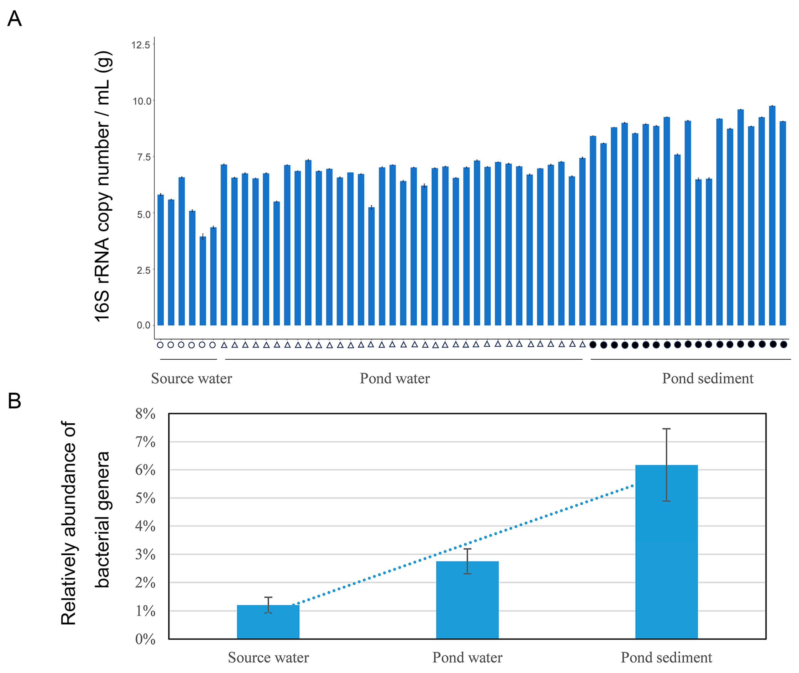
3.1. Abundance of 16S rRNA Genes in Culture Environment Identified through qPCR

This study compared changes in the copy numbers of bacterial 16S rRNA genes in the water source, pond water, and pond sediment using qPCR. The total number of bacteria in water and sediment samples was expressed as copy number/mL of water and copy number/g of sediment (Figure 1). As there was a large difference in bacterial copy number between water and sediment samples, a log10 transformation was used to represent the data. The average copy numbers of bacteria were lowest in source water (8.29 × 10⁵), higher in pond water (9.56 × 10⁶), and highest in sediment (1.17 × 10⁹).

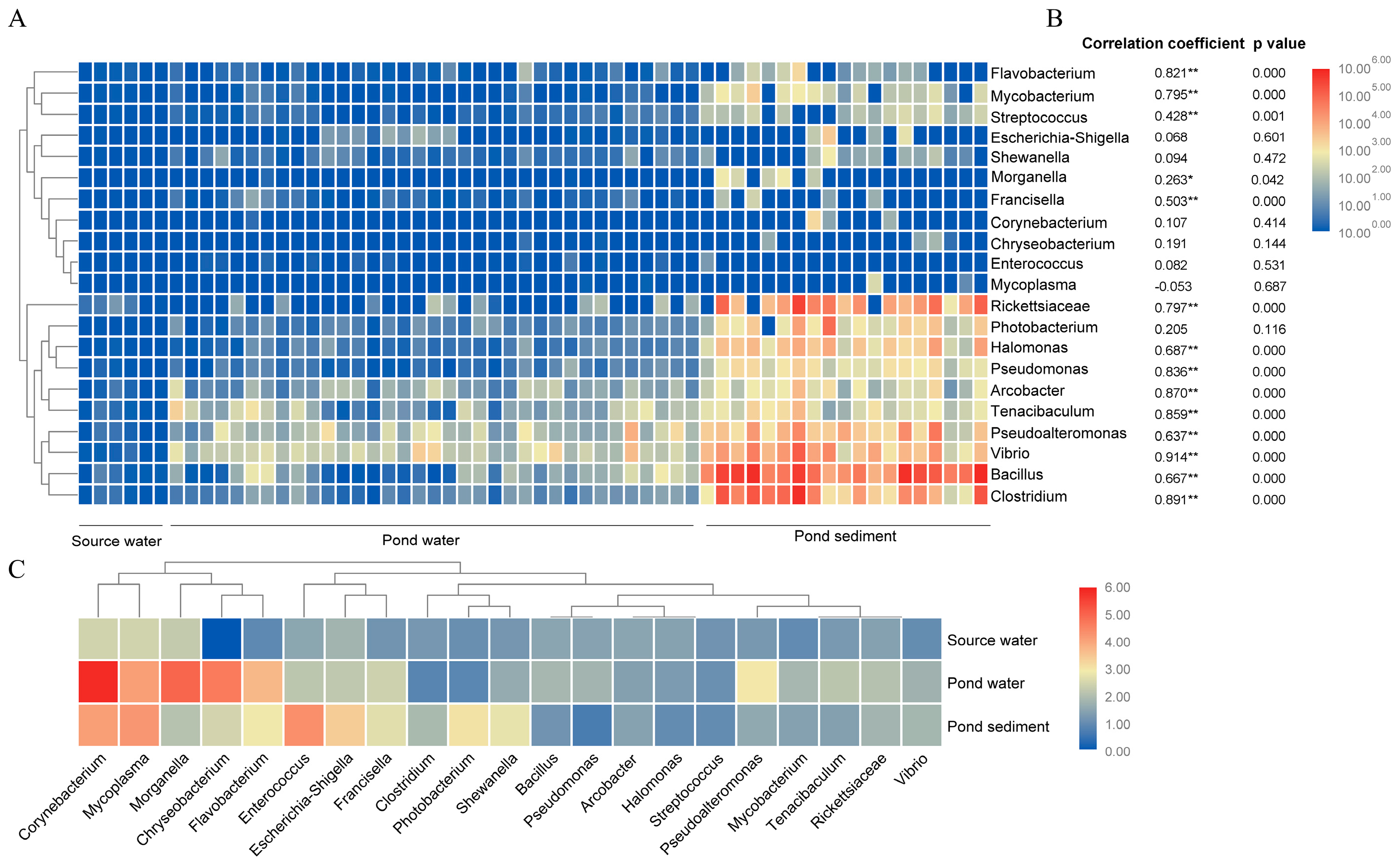
3.2. Diversity and Abundance of 21 Identified Bacteria Genera by Combining qPCR and High throughout Sequencing Analysis

In all samples, consisting of source water, pond water, and sediment, a total of 7,565,354 sequences were identified. Through a literature review, we detected 21 genera containing pathogenic bacteria (Table S1) that potentially pose a pathogenic threat to marine organisms, including fish, invertebrates, and mammals [24]. The composition of bacterial communities in both the source water and aquaculture ponds was analyzed, and the relative abundance of bacteria for the 21 identified genera was standardized by performing qPCR and normalizing to the total bacteria count in each sample. Using high throughput sequencing of 16S rRNA and qPCR, we calculated changes in the abundance of the 21 genera in source water (copy number/mL), pond water (copy number/mL), and pond sediment (copy number/g). The results showed that the relative abundance of bacterial genera was much higher in the aquaculture area than in the source water. Overall, the abundance of these genera in aquaculture water was 2.7 times higher (2.75% ± 0.44%) than in the inlet water (1.20% ± 0.28%). Furthermore, the abundance of these bacterial genera in pond sediment was 6.2 times higher (6.17% ± 1.28%) than in inlet water (Figure 1B).
The results showed that the abundance of these bacteria in the aquaculture area was significantly higher than in the inlet. The composition of dominant genera in pond water was consistent with pond sediment; however, their abundance increased sharply in pond sediment (Figure 2A, Table S1). The dominant genera in pond water were Vibrio, Pseudoalteromonas, Tenacibaculum, Bacillus, Arcobacter, Rickettsiaceae, and Clostridium. The dominant genera in pond sediment were Bacillus, Clostridium, Rickettsiaceae, Vibrio, Pseudoalteromonas, Photobacterium, Halomonas, Pseudomonas, Tenacibaculum, and Arcobacter.
The correlation between the abundance of these bacterial genera and the total bacterial abundance was analyzed (Figure 2B). The results showed that among the 21 genera investigated, 14 genera were positively correlated with the total number of bacteria (r2 > 0.7, p ≈ 0), especially for the dominant genera in water and sediment, such as Rickettsia, Halomonas, Arcobacter, and Tenaciulum and Pseudomonas, Vibrio, and Trilosdium, respectively. At the same time, these bacteria had a low coefficient of variation among different samples (0.73–1.95, Figure 2C).
The remaining seven genera with lower abundances, Escherichia, Shewanella, Corynebacterium, Chryseobacterium, Mycoplasma, Enterococcus, and Photobacterium, had a high coefficient of variation among samples (2.09–5.85, Figure 2C). Correlation analysis showed that they had a very low correlation with total bacterial abundance. According to the analysis results, 21 bacterial genera in aquaculture can be clustered into two groups: some genera present in the aquaculture areas with low coefficient of variation, and those present in the aquaculture areas with high coefficient of variation.

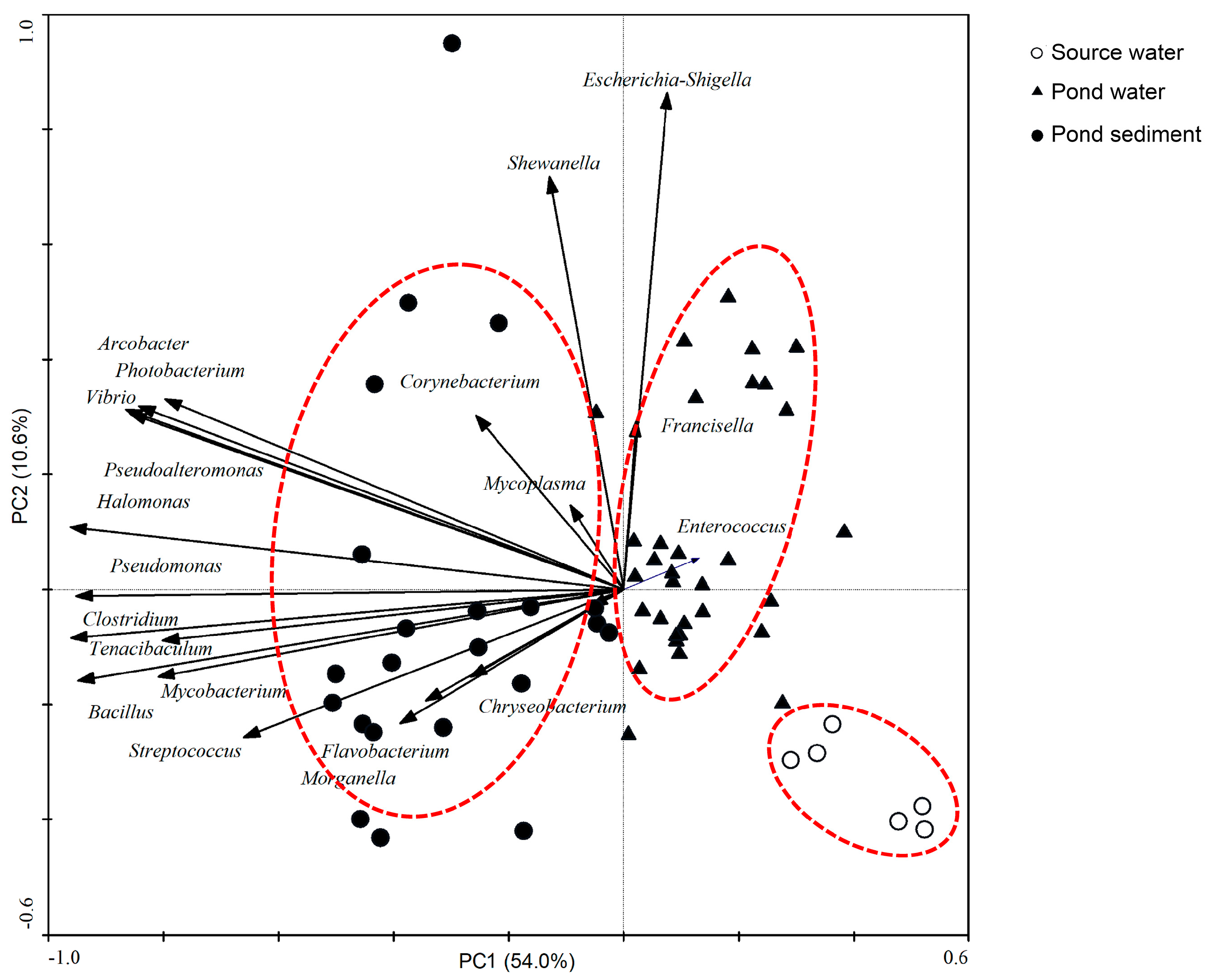
3.3. Principal Component Analysis (PCA) of the Distribution of Bacterial Genera between Different Environments

PCA was conducted to identify the possible associations between 21 bacterial genera and the sample characteristics (Figure 3). Assemblages of these bacterial genera (assessed by composition and relative abundance) formed three clear groups (cumulative percentage variance 76.1) and differed significantly between source water, pond water, and pond sediment (ANOVA, p < 0.01). The separations between the groups were associated with the higher relative abundance of Rickettsia, Halomonas, Pseudomonas, Arcobacter, Tenacibaculum, Vibrio, and Clostridium in the culture environment.

3.4. Bacterial Genera in the Intestines of Animals in Source Water and Pond Water Based on Illumina Sequencing

The study compared the abundance characteristics of above bacterial genera in the aquatic animals’ intestine in the water source and culture ponds. Results indicated that the abundance of the most bacterial genera of animals was obviously higher in the aquaculture areas than in the source water (Figure 4). The composition of dominant genera in pond animals was consistent with source water animals. The dominant genera in source water were Photobacterium (8.60%), Vibrio (4.65%), Mycoplasma (0.61%), Pseudomonas (0.19%), and Shewanella (0.10%). The dominant genera in pond animals were Vibrio (15.34%), Photobacterium (7.41%), Pseudoalteromonas (6.58%), Mycoplasma (3.11%), Streptococcus (0.26%), and Enterococcus (0.19%). The abundance of bacterial genera, especially Mycoplasma, Pseudoalteromonas, Vibrio, and Enterococcus, was much higher in pond animals than that in the source water.

4. Discussion

Studying the relationship between potential pathogenic bacteria and their habitats is crucial for understanding the formation of gut microbiota and its impact on host health. In this study, we investigated the differences in the composition of specific bacterial genera containing potentially pathogenic bacteria in water sources and aquaculture environments and analyzed the potential impact of the environment on them.
Aquaculture environments may increase the abundance and diversity of potentially pathogenic bacteria. For example, Vibrio species are naturally occurring bacteria found in marine aquatic environments, and most human infections are acquired through exposure to these environments or through consumption of food derived from them [25,26,27,28]. Vibrio levels often correlate with other bacterial pathogens commonly found in coastal and aquaculture areas, and can cause disease in animals [29]. The results of this study showed that the total number of bacteria increased in the aquaculture environment. This is likely due to the fact that feedstuffs are added to aquaculture water, which increases the nutrient concentration and promotes the growth of total microbial biomass [30]. It can be speculated that the abundance of bacteria known as potentially pathogenic bacteria benefited from the culture conditions and proliferated with the increase in total bacteria, which was influenced by nutrient levels in the culture environment. Previous studies have reported that the abundance and diversity of pathogenic microbes in the culture environment are key factors in the production of high-quality seafood and are directly affected by environmental factors [4,31,32]. The findings of this study suggest that high abundances of potentially pathogenic bacteria had a low coefficient of variation among different samples, indicating that changes in abundance were uniform and easily affected by the culture environment.
Correlation analysis showed that bacterial genera in this study with low abundances had a very low correlation with the total bacterial number, indicating that the abundance of these bacteria did not change with the total abundance of bacteria and was less affected by the culture environment. Indicators and pathogens such as Escherichia and Enterococcus have been reported to be positively associated with urban land cover [33]. According to our results, bacterial genera in this study in aquaculture can be divided into two groups: bacteria that are naturally present in the aquaculture environment and respond rapidly to environmental changes, and those that are present as a result of contamination with human or animal feces or respond slowly to the environment. This suggests that these two types of bacterial genera have different response mechanisms to the aquaculture environment.
The dominant genera in this study were aerobic bacteria, which could quickly adapt to the environment and increase in abundance [4,34,35]. These bacteria are also one of the main factors causing the outbreak of breeding diseases, such as Vibrio. Some bacterial genera, such as Halomonas, Pseudomonas, and Arcobacter, which have high metabolic activity and the best growing strain, can grow and reproduce rapidly [36,37,38]. Previous studies on canonical correlation analysis have found that bacterial genera including Alteromonas, Arcobacter, Halomonas, Vibrio, Tenacibaculum, etc., showed a positive correlation with the levels of nitrite and nitrate in the sorting axis [4]. Most of these bacteria are heterotrophic, indicating that they are primarily regulated by nutrient levels and are prone to rapid changes in response to environmental influences. The level of inorganic nutrients in culture environment was significantly correlated with the presence of these bacterial genera [4], which partly explains the reason why they had the ability to grow rapidly in aquaculture ponds. These species pose a threat to the health of farmed animals in the environment of physiological imbalance [39,40]. The low abundance of bacterial genera in this study showed that they grew and metabolized slowly in the aquaculture environment. Escherichia and Enterococcus, for example, were mainly adapted to the intestinal environment of animals and were also used as indicators of water pollution [8,31]. Shewanella, Escherichia, Corynebacterium, and Photobacterium were facultative anaerobes [41], which grew slowly under aerobic conditions. In addition to the significant impact of Vibrio on aquaculture, other potential pathogenic bacteria also cause significant losses. For example, some species in the Flavobacterium genus have pathogenic effects on marine fish, amphibians, and other aquatic animals [42]. Infected fish exhibit symptoms such as gill rot, skin ulcers, tissue necrosis, and mortality rates can reach 100%. The economic losses caused by this disease are extremely serious worldwide. Tenacibaculum sp. is a pathogenic bacteria that causes ulcerative diseases known as tenacibaculosis, which can lead to high mortality rates in aquaculture animals and also increase susceptibility to other pathogens [43]. Some species in Rickettsia, such as Piscirickettsia, are pathogens of fish (salmon and sea bass) [44], and infected fish become lethargic and lose their appetite.
PCA analysis showed that bacteria genera with high abundance and low coefficient of variation were closely related in pond water and sediment, indicating that these bacteria contributed most to the aquaculture environment. Pond sediment acted as a reservoir for these bacterial genera, and they have the potential to be released into the water from the sediment environment. The influence of sediment on the intestinal bacteria of cultured animals was greater than that of feeding water. Previous studies have also shown that sediment is a major contributor to the gut microbes of the major farmed animals (shrimp, fish, crabs, shellfish, and clams) in mariculture ecosystems [4,10,11]. The feeding behavior of benthic organisms does indeed make their gut microbiota more susceptible to the influence of sediment in their habitat, as the sediments they ingest contain a large population of microorganisms [9,45]. Additionally, aquatic animals ingest various microorganisms in the water, some of which can grow and reproduce in the gut and are often closely related to the gut health of the host [46,47]. The ability of bacteria, such as Vibrio and Aeromonas, can quickly colonize the gut, suggesting that these bacteria possess strong adaptability and competitiveness [48]. Therefore, the composition of microorganisms and potentially pathogenic bacteria in environment has an important impact on animal health. This observation of increased levels of potentially pathogenic bacteria in culture ponds provides further support for promoting the development of these bacterial genera in farmed environments. Although determining the mechanisms involved will require further research, the characteristics of the aquaculture environment play an important role in shaping the diversity of potential pathogens. In recent years, studies have reported that physical and chemical factors in the habitat environment and microbial communities have significant impacts on the gut microbiota of aquatic animals [4,49].

5. Conclusions

In the current study, the composition of specific bacterial genera was affected by the culture environment. Many types of intestinal pathogens also exist in the environment, and the culture environment played a decisive role in the composition of intestinal pathogens. Therefore, environmental conditions are closely related to the health of cultured animals, and improving the culture environment will help to reduce the risk of animal disease. The balance of gut microbiota in aquaculture animals is easily influenced by the water environment. If there are changes in the internal and external environments of the gut, the stability of the gut microbiota may be disrupted. Potential pathogen may invade the digestive system and cause infections, which can affect the gut function of aquatic animals. In this study, it was concluded that sediment-associated bacteria have been identified as a potential source of potential pathogen in aquaculture. To sum up, the current investigation has delineated the importance of optimizing the aquaculture environment to prevent potential breeding disease microorganisms and the onset of animal disease.

Supplementary Materials

The following supporting information can be downloaded at: https://www.mdpi.com/article/10.3390/jmse12020209/s1, Table S1: Abundance of bacterial genera in samples which determined by high-throughput sequencing and qPCR analysis.

Author Contributions

Conceptualization, F.S. and Z.X.; methodology, F.S.; software, Z.X.; investigation, F.S and C.W.; writing—original draft preparation, F.S.; writing—review and editing, F.S.; visualization, F.S. and Z.X.; supervision, Z.X. and C.W.; funding acquisition, F.S. and Z.X. All authors have read and agreed to the published version of the manuscript.

Funding

The present study was supported financially by the Key Research and Development Program of Hainan Province (ZDYF2021XDNY131), the Natural Science Foundation of Guangdong Province (2023A1515012004), and the Project of Fujian Science and Technology Department (2021N3001).

Institutional Review Board Statement

Not applicable.

Informed Consent Statement

Not applicable.

Data Availability Statement

The data presented in this study are available on request.

Conflicts of Interest

The authors declare no conflicts of interest.

References

  1. FAO. How to Feed the World: 2050; Food and Agriculture Organization of the United Nations: Rome, Italy, 2009. [Google Scholar]
  2. Burge, C.A.; Eakin, C.M.; Friedman, C.S.; Froelich, B.; Hershberger, P.K.; Hofmann, E.E.; Petes, L.E.; Prager, K.C.; Weil, E.; Willis, B.L.; et al. Climate Change Influences on Marine Infectious Diseases: Implications for Management and Society. Annu. Rev. Mar. Sci. 2014, 6, 249–277. [Google Scholar] [CrossRef] [PubMed]
  3. Ward, J.R.; Lafferty, K.D. The Elusive Baseline of Marine Disease: Are Diseases in Ocean Ecosystems Increasing? PLoS Biol. 2004, 2, e120. [Google Scholar] [CrossRef] [PubMed]
  4. Sun, F.; Wang, Y.; Wang, C.; Zhang, L.; Tu, K.; Zheng, Z. Insights into the intestinal microbiota of several aquatic organisms and association with the surrounding environment. Aquaculture 2019, 507, 196–202. [Google Scholar] [CrossRef]
  5. El-Sayed, M.R.; Emam, A.M.; Osman, A.E.; Abd El-Galil, M.A.E.-A.A.; Sayed, H.H. Detection and description of a novel Psychrobacter glacincola infection in some Red Sea marine fishes in Hurghada, Egypt. BMC Vet. Res. 2023, 19, 23. [Google Scholar] [CrossRef]
  6. Duman, M.; Mulet, M.; Altun, S.; Saticioglu, I.B.; Ozdemir, B.; Ajmi, N.; Lalucat, J.; García-Valdés, E. The diversity of Pseudomonas species isolated from fish farms in Turkey. Aquaculture 2021, 535, 736369. [Google Scholar] [CrossRef]
  7. Umasuthan, N.; Valderrama, K.; Vasquez, I.; Segovia, C.; Hossain, A.; Cao, T.; Gnanagobal, H.; Monk, J.; Boyce, D.; Santander, J. A novel marine pathogen isolated from wild cunners (Tautogolabrus adspersus): Comparative genomics and transcriptome profiling of Pseudomonas sp. Strain J380. Microorganisms 2021, 9, 812. [Google Scholar] [CrossRef] [PubMed]
  8. de Bruijn, I.; Liu, Y.; Wiegertjes, G.F.; Raaijmakers, J.M. Exploring fish microbial communities to mitigate emerging diseases in aquaculture. FEMS Microbiol. Ecol. 2017, 94, fix161. [Google Scholar] [CrossRef]
  9. Sun, F.; Wang, C.; Chen, L.; Weng, G.; Zheng, Z. The intestinal bacterial community of healthy and diseased animals and its association with the aquaculture environment. Appl. Microbiol. Biot. 2020, 104, 775–783. [Google Scholar] [CrossRef]
  10. Sun, F.; Wang, C.; Chen, X. Bacterial community in Sinonovacula constricta intestine and its relationship with culture environment. Appl. Microbiol. Biotechnol. 2022, 106, 5211–5220. [Google Scholar] [CrossRef]
  11. Sun, F.; Xu, Z. Significant differences in intestinal microbial communities in aquatic animals from an aquaculture area. J. Mar. Sci. Eng. 2021, 9, 104. [Google Scholar] [CrossRef]
  12. Romalde, J.L. Photobacterium damselae subsp. piscicida: An integrated view of a bacterial fish pathogen. Int. Microbiol. 2002, 5, 3–9. [Google Scholar] [CrossRef] [PubMed]
  13. Austin, B. The bacterial microflora of fish, revised. Sci. World J. 2006, 6, 931–945. [Google Scholar] [CrossRef] [PubMed]
  14. Round, J.L.; Mazmanian, S.K. The gut microbiota shapes intestinal immune responses during health and disease. Nat. Rev. Immunol. 2009, 9, 313–323. [Google Scholar] [CrossRef] [PubMed]
  15. Sullam, K.; Essinger, S.; Lozupone, C.; O’connor, M.P.; Rosen, G.L.; Knight, R.; Kilham, S.S.; Russell, J.A. Environmental and ecological factors that shape the gut bacterial communities of fish: A meta-analysis. Mol. Ecol. 2012, 21, 3363–3378. [Google Scholar] [CrossRef]
  16. Giatsis, C.; Sipkema, D.; Smidt, H.; Heilig, H.; Benvenuti, G.; Verreth, J.; Verdegem, M. The impact of rearing environment on the development of gut microbiota in tilapia larvae. Sci. Rep. 2015, 5, 18206. [Google Scholar] [CrossRef] [PubMed]
  17. Chaiyapechara, S.; Rungrassamee, W.; Suriyachay, I.; Kuncharin, Y.; Klanchui, A.; Karoonuthaisiri, N.; Jiravanichpaisal, P. Bacterial Community Associated with the Intestinal Tract of P. monodon in Commercial Farms. Microb. Ecol. 2012, 63, 938–953. [Google Scholar] [CrossRef] [PubMed]
  18. Soonthornchai, W.; Rungrassamee, W.; Karoonuthaisiri, N.; Jarayabhand, P.; Klinbunga, S.; Soderhall, K.; Jiravanichpaisal, P. Expression of immune-related genes in the digestive organ of shrimp, Penaeus monodon, after an oral infection by Vibrio harveyi. Dev. Comp. Immunol. 2010, 34, 19–28. [Google Scholar] [CrossRef]
  19. Lavilla-Pitogo, C.R.; Leaño, E.M.; Paner, M.G. Mortalities of pond-cultured juvenile shrimp, Penaeus monodon, associated with dominance of luminescent vibrios in the rearing environment. Aquaculture 1998, 164, 337–349. [Google Scholar] [CrossRef]
  20. Sung, H.-H.; Hsu, S.-F.; Chen, C.-K.; Ting, Y.-Y.; Chao, W.-L. Relationships between disease outbreak in cultured tiger shrimp (Penaeus monodon) and the composition of Vibrio communities in pond water and shrimp hepatopancreas during cultivation. Aquaculture 2001, 192, 101–110. [Google Scholar] [CrossRef]
  21. Xiong, J.; Wang, K.; Wu, J.; Qiuqian, L.; Yang, K.; Qian, Y.; Zhang, D. Changes in intestinal bacterial communities are closely associated with shrimp disease severity. Appl. Microbiol. Biot. 2015, 99, 6911–6919. [Google Scholar] [CrossRef]
  22. Magoč, T.; Salzberg, S.L. FLASH: Fast length adjustment of short reads to improve genome assemblies. Bioinformatics 2011, 27, 2957–2963. [Google Scholar] [CrossRef] [PubMed]
  23. Muyzer, G.; de Waal, E.C.; Uitterlinden, A.G. Profiling of complex microbial populations by denaturing gradient gel electrophoresis analysis of polymerase chain reaction-amplified genes coding for 16S rRNA. Appl. Environ. Microbiol. 1993, 59, 695–700. [Google Scholar] [CrossRef] [PubMed]
  24. Lamb, J.B.; van de Water, J.A.J.M.; Bourne, D.G.; Altier, C.; Hein, M.Y.; Fiorenza, E.A.; Abu, N.; Jompa, J.; Harvell, C.D. Seagrass ecosystems reduce exposure to bacterial pathogens of humans, fishes, and invertebrates. Science 2017, 355, 731–733. [Google Scholar] [CrossRef] [PubMed]
  25. Dalsgaard, A. The occurrence of human pathogenic Vibrio spp. and Salmonella in aquaculture. Int. J. Food Sci. Technol. 1998, 33, 127–138. [Google Scholar] [CrossRef]
  26. Feldhusen, F. The role of seafood in bacterialfoodborne diseases. Microbes Infect. 2000, 2, 1651–1660. [Google Scholar] [CrossRef]
  27. Tao, Z.; Larsen, A.M.; Bullard, S.A.; Wright, A.C.; Arias, C.R. Prevalence and population structure of Vibrio vulnificus on fishes from the northern gulf of Mexico. Appl. Environ. Microbiol. 2012, 78, 7611–7618. [Google Scholar] [CrossRef] [PubMed]
  28. Duman, M.; Buján, N.; Altun, S.; Romalde, J.L.; Saticioglu, I.B. Population genetic and evolution analysis of Vibrio isolated from Turkish fish farms. Aquaculture 2023, 562, 738728. [Google Scholar] [CrossRef]
  29. Fleming, L.E.; Broad, K.; Clement, A.; Dewailly, E.; Elmir, S.; Knap, A.; Pomponi, S.A.; Smith, S.; Solo Gabriele, H.; Walsh, P. Oceans and human health: Emerging public health risks in the marine environment. Mar. Pollut. Bull. 2006, 53, 545–560. [Google Scholar] [CrossRef]
  30. Ganesh, E.A.; Das, S.; Chandrasekar, K.; Arun, G.; Balamurugan, S. Monitoring of total heterotrophic bacteria and Vibrio spp. in an aquaculture pond. Curr. Res. J. Biol. Sci. 2010, 2, 48–52. [Google Scholar]
  31. Engering, A.; Hogerwerf, L.; Slingenbergh, J. Pathogen–host–environment interplay and disease emergence. Emerg. Microbes Infect. 2013, 2, e5. [Google Scholar] [CrossRef]
  32. Caruso, G.; Maimone, G.; Mancuso, M.; Modica, A.; Genovese, L. Microbiological controls across the productive cycle of Dicentrarchus labrax L. and Sparus aurata L.: A study from the environment to the final product. Aquac. Res. 2004, 35, 184–193. [Google Scholar] [CrossRef]
  33. Walters, S.P.; Thebo, A.L.; Boehm, A.B. Impact of urbanization and agriculture on the occurrence of bacterial pathogens and stx genes in coastal waterbodies of central California. Water Res. 2011, 45, 1752–1762. [Google Scholar] [CrossRef] [PubMed]
  34. Kaspar, C.W.; Tamplin, M.L. Effects of temperature and salinity on the survival of Vibrio vulnificus in seawater and shellfish. Appl. Environ. Microbiol. 1993, 59, 2425–2429. [Google Scholar] [CrossRef] [PubMed]
  35. Vezzulli, L.; Pezzati, E.; Brettar, I.; Höfle, M.; Pruzzo, C. Effects of global Warming on Vibrio ecology. Microbiol. Spectr. 2015, 3. [Google Scholar] [CrossRef] [PubMed]
  36. Avendaño-Herrera, R.E.; Riquelme, C.E. Production of a diatom-bacteria biofilm in a photobioreactor for aquaculture applications. Aquac. Eng. 2007, 36, 97–104. [Google Scholar] [CrossRef]
  37. Doménech, A.; Fernández-Garayzábal, J.F.; Lawson, P.; García, J.A.; Cutuli, M.T.; Blanco, M.; Gibello, A.; Moreno, M.A.; Collins, M.D.; Domínguez, L. Winter disease outbreak in sea-bream (Sparus aurata) associated with Pseudomonas anguilliseptica infection. Aquaculture 1997, 156, 317–326. [Google Scholar] [CrossRef]
  38. Vandenberg, O.; Dediste, A.; Houf, K.; Ibekwem, S.; Souayah, H.; Cadranel, S.; Douat, N.; Zissis, G.; Butzler, J.P.; Vandamme, P. Arcobacter species in humans. Emerg. Infect. Dis. 2004, 10, 1863–1867. [Google Scholar] [CrossRef]
  39. Rungrassamee, W.; Klanchui, A.; Maibunkaew, S.; Chaiyapechara, S.; Jiravanichpaisal, P.; Karoonuthaisiri, N. Characterization of intestinal bacteria in wild and domesticated adult black tiger shrimp (Penaeus monodon). PLoS ONE 2014, 9, e91853. [Google Scholar] [CrossRef]
  40. Gainza, O.; Ramírez, C.; Ramos, A.S.; Romero, J. Intestinal microbiota of white shrimp Penaeus vannamei under intensive cultivation conditions in ecuador. Microb. Ecol. 2018, 75, 562–568. [Google Scholar] [CrossRef]
  41. Ryan, K.J.; Ray, C.G. Medical microbiology. McGraw Hill 2004, 4, 370. [Google Scholar]
  42. Bernardet, J.-F. Order I. Flavobacteriales ord. nov. In Bergey’s Manual of Systematics of Archaea and Bacteria; Krieg, N.R., Staley, J.T., Brown, D.R., Hedlund, B.P., Paster, B.J., Ward, N.L., Ludwig, W., Whitman, W.B., Eds.; Wiley: Hoboken, NJ, USA, 2011; p. 105. [Google Scholar]
  43. Avendaño-Herrera, R.; Toranzo, A.E.; Magariños, B. Tenacibaculosis infection in marine fish caused by Tenacibaculum maritimum: A review. Dis. Aquat. Organ. 2006, 71, 255–266. [Google Scholar] [CrossRef] [PubMed]
  44. McCarthy, U.; Steiropoulos, N.A.; Thompson, K.D.; Adams, A.; Ellis, A.E.; Ferguson, H.W. Confirmation of Piscirickettsia salmonis as a pathogen in European sea bass Dicentrarchus labrax and phylogenetic comparison with salmonid strains. Dis. Aquat. Organ. 2005, 64, 107–119. [Google Scholar] [CrossRef] [PubMed]
  45. Plante, C.J.; Jumars, P.A.; Baross, J.A. Digestive associations between marine detritivores and bacteria. Annu. Rev. Ecol. Syst. 1990, 21, 93–127. [Google Scholar] [CrossRef]
  46. Bøgwald, J.; Dalmo, R.A. Gastrointestinal pathogenesis in aquatic animals. In Aquaculture Nutrition; Wiley: Hoboken, NJ, USA, 2014; pp. 53–74. [Google Scholar] [CrossRef]
  47. Li, X.; Ringø, E.; Hoseinifar, S.H.; Lauzon, H.L.; Birkbeck, H.; Yang, D. The adherence and colonization of microorganisms in fish gastrointestinal tract. Rev. Aquac. 2019, 11, 603–618. [Google Scholar] [CrossRef]
  48. Hansen, G.H.; Olafsen, J.A. Bacterial interactions in early life stages of marine cold water fish. Microb. Ecol. 1999, 38, 1–26. [Google Scholar] [CrossRef]
  49. Yukgehnaish, K.; Kumar, P.; Sivachandran, P.; Marimuthu, K.; Arshad, A.; Paray, B.A.; Arockiaraj, J. Gut microbiota metagenomics in aquaculture: Factors influencing gut microbiome and its physiological role in fish. Rev. Aquac. 2020, 12, 1903–1927. [Google Scholar] [CrossRef]
Figure 1. qPCR analysis of bacterial 16S rRNA in source water and culture ponds (A); total abundance of 21 identified genera in samples based on high throughout sequencing (B).
Jmse 12 00209 g001
Figure 2. Abundance distribution of 21 bacterial genera in samples calculated based on high-throughput sequencing and qPCR analysis (A); correlation analysis between bacteria genera and total bacteria abundance, significance test (p < 0.05 or p < 0.01) was marked by ** and * (B); coefficient of variation analysis of differential genera in source water, pond water, and pond sediment (C).
Jmse 12 00209 g002
Figure 3. PCA analysis based on the relative abundance of 21 bacterial genera in source water, pond water, and pond sediment. The red areas represent collections of samples of different properties (source water, pond water, and pond sediment).
Jmse 12 00209 g003
Figure 4. Relative abundance of bacterial genera in the intestine of cultured animals in source water and pond water. Comparison the average abundance of these bacterial genera in samples in the figure right.
Jmse 12 00209 g004
Disclaimer/Publisher’s Note: The statements, opinions and data contained in all publications are solely those of the individual author(s) and contributor(s) and not of MDPI and/or the editor(s). MDPI and/or the editor(s) disclaim responsibility for any injury to people or property resulting from any ideas, methods, instructions or products referred to in the content.

