
Myco-Barriers as Sustainable Tool for Port Seawater Decontamination from Metals


Abstract
1. Introduction
2. Materials and Methods
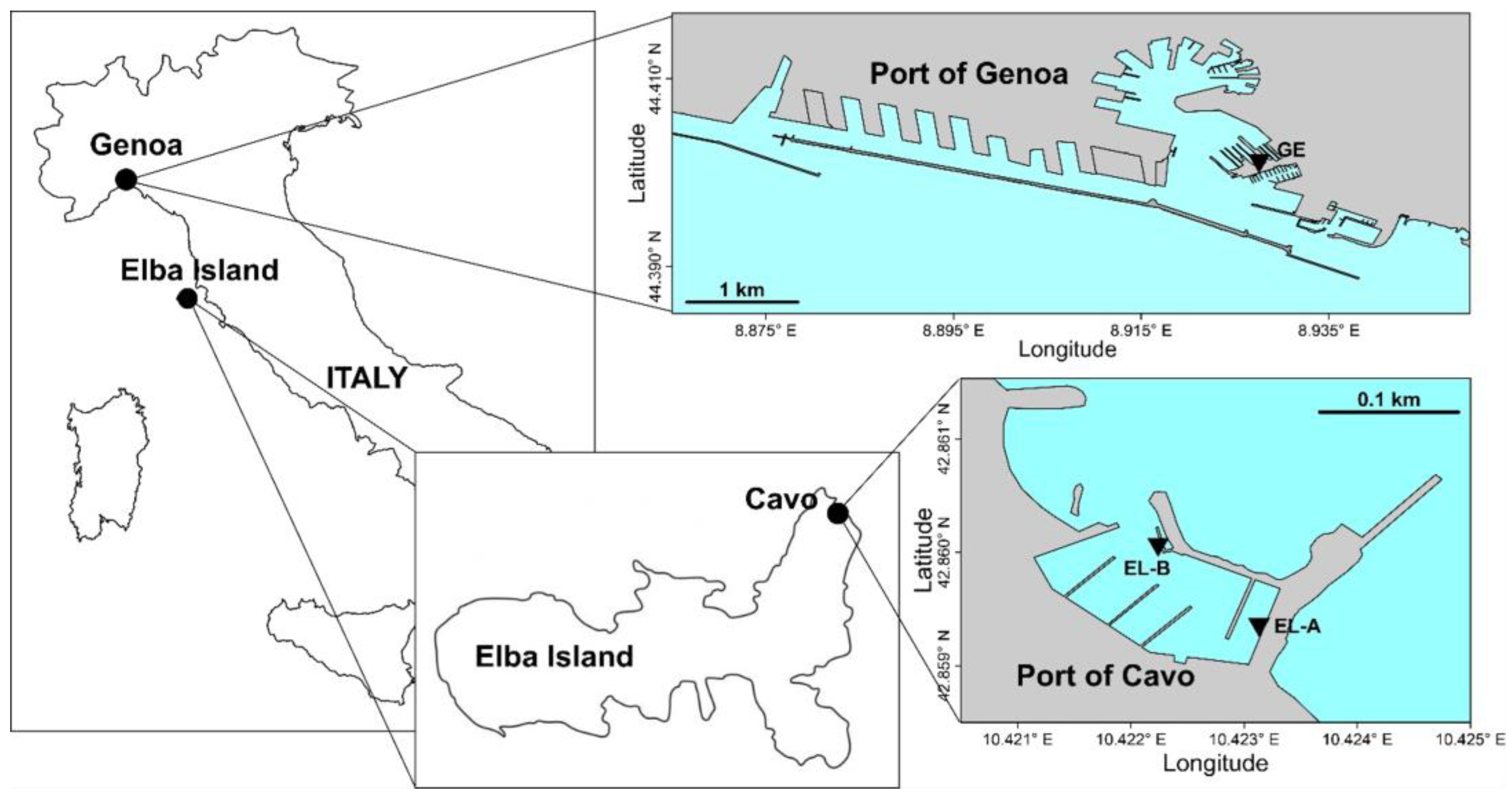
2.1. Study Areas
2.2. Fungal Selection
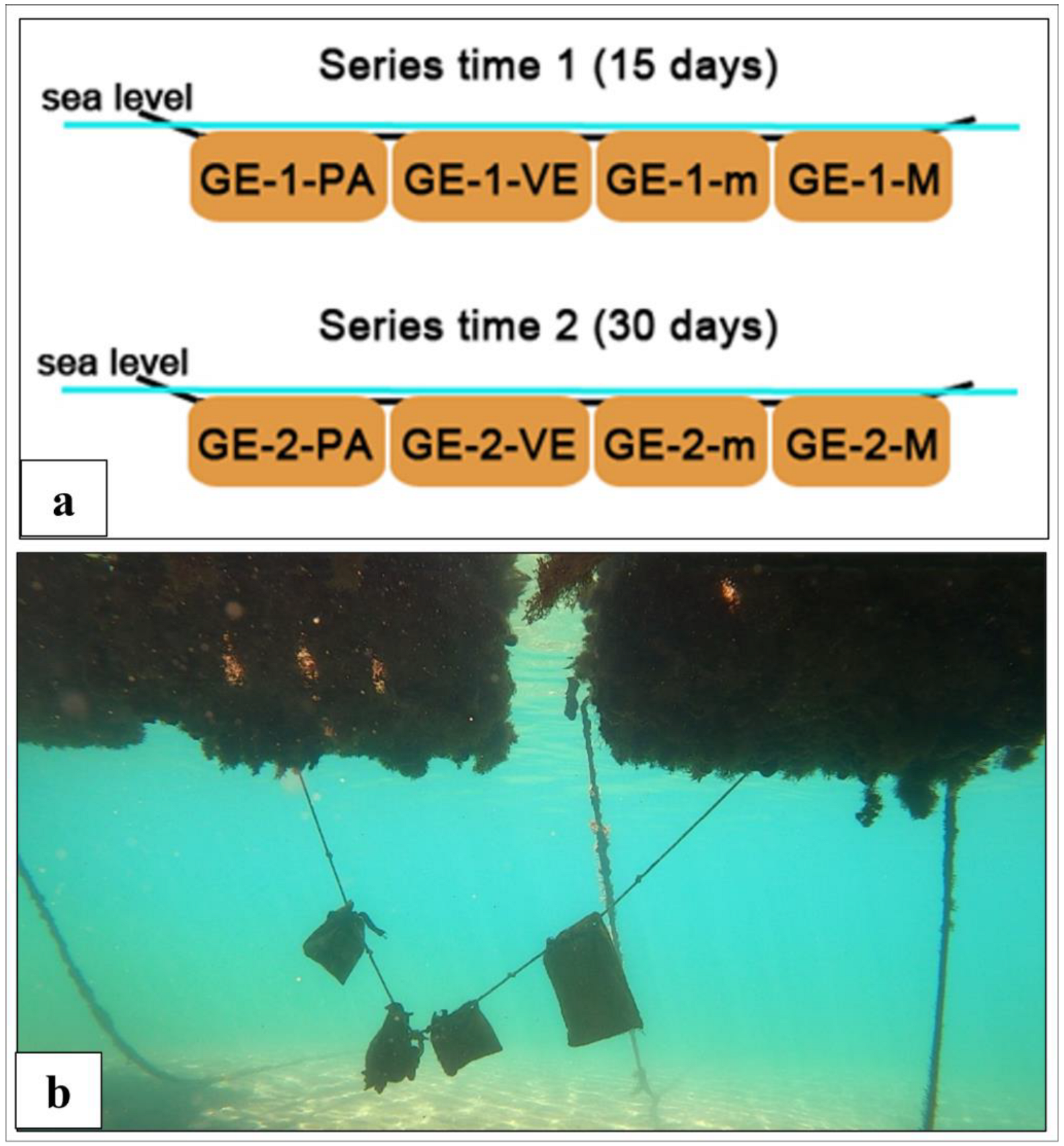
2.3. Myco-Barriers Development and Positioning in the Ports
2.4. Sampling Program and Analyses of Myco-Barriers Efficiency
2.5. Statistical Analysis
3. Results
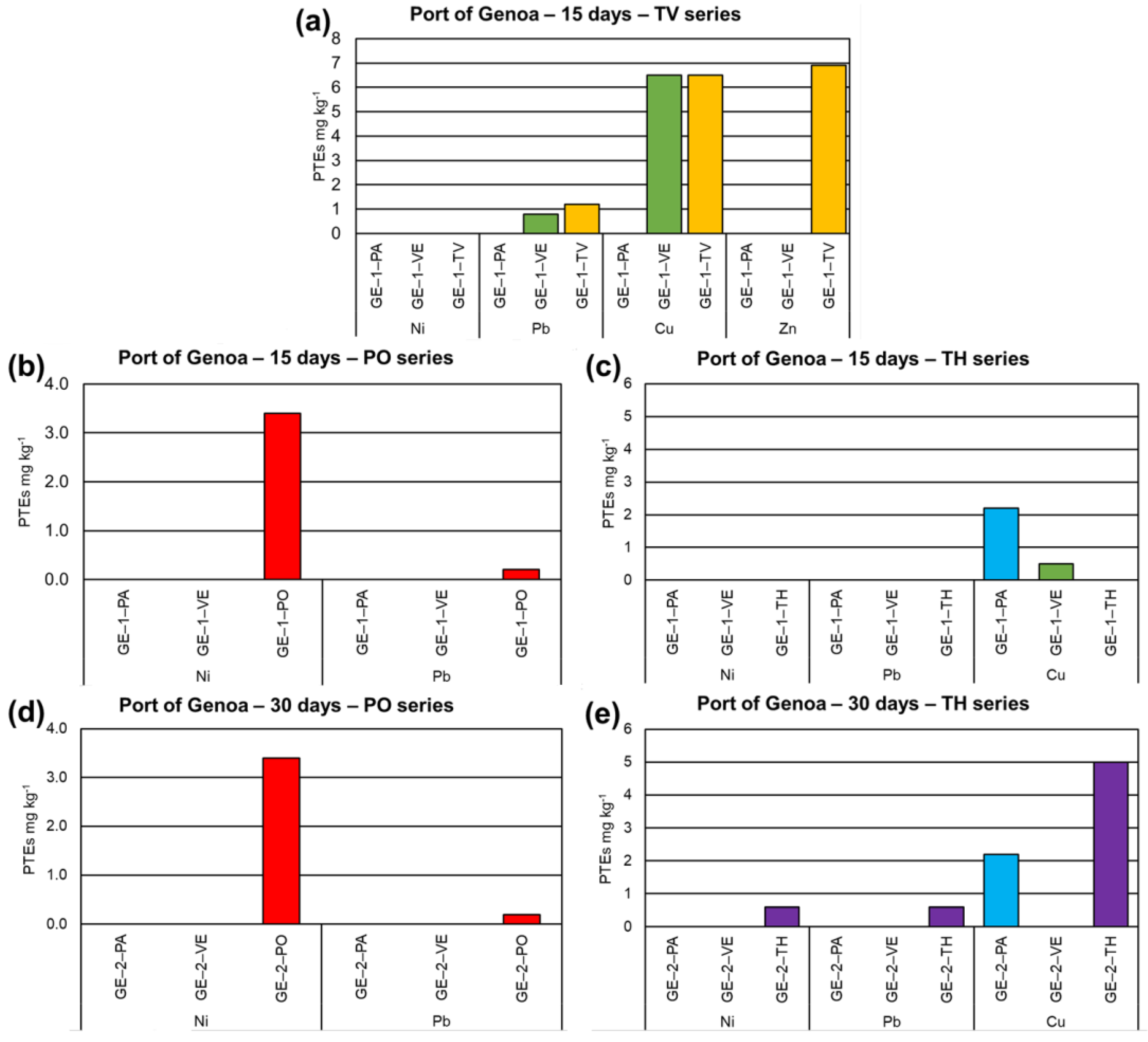
3.1. Port of Genoa
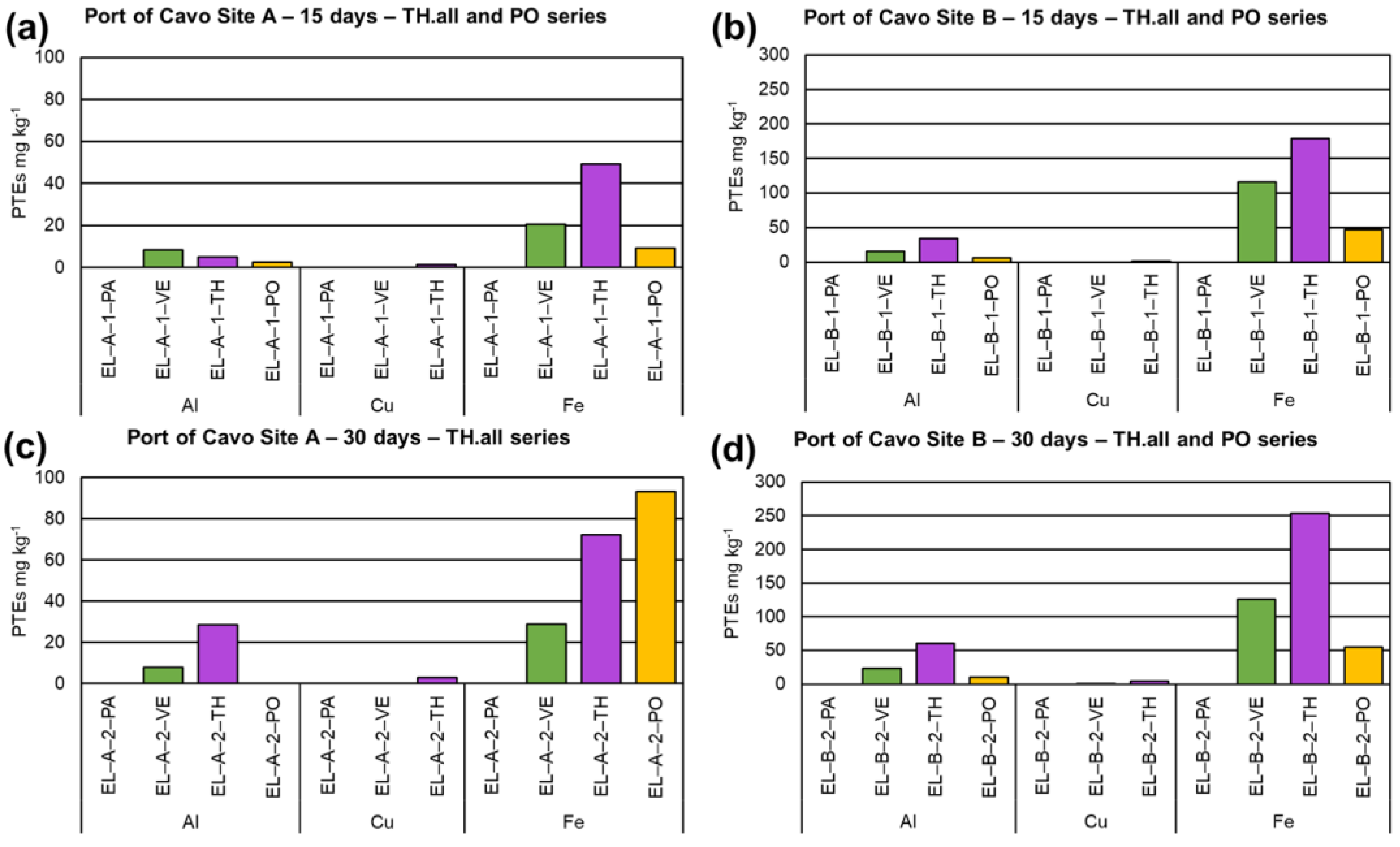
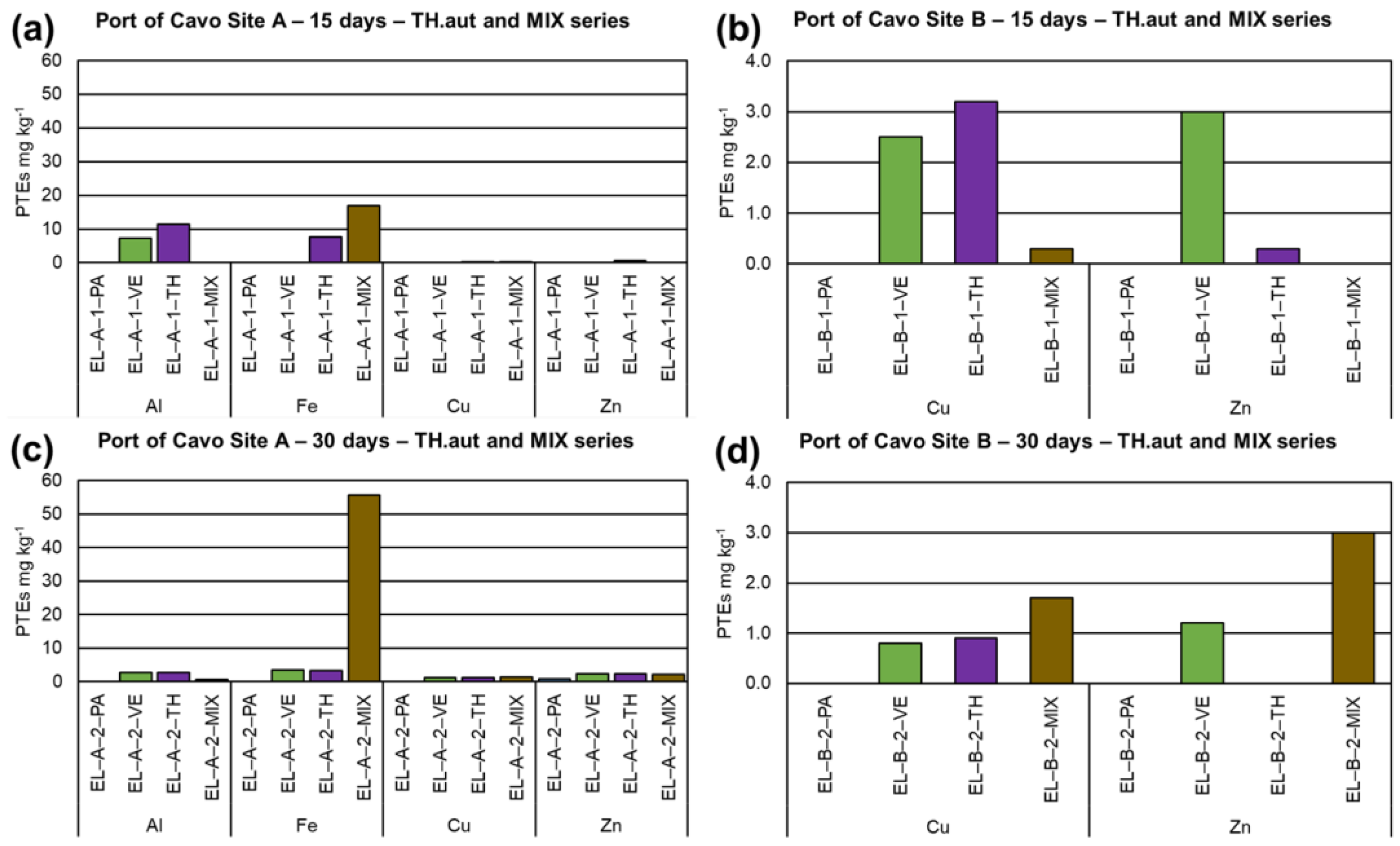
3.2. Port of Cavo
4. Discussion
5. Conclusions
Supplementary Materials
Author Contributions
Funding
Institutional Review Board Statement
Informed Consent Statement
Data Availability Statement
Acknowledgments
Conflicts of Interest
References
- Spagnoli, F.; De Marco, R.; Dinelli, E.; Frapiccini, E.; Frontolani, F.; Giordano, P. Sources and metal pollution of sediments from a coastal area of the Central Western Adriatic Sea (Southern Marche Region, Italy). Appl. Sci. 2021, 11, 1118. [Google Scholar] [CrossRef]
- Tornero, V.; Hank, G. Chemical contaminants entering the marine environment from sea-based sources: A review with a focus on European seas. Mar. Pollut. Bull. 2016, 112, 17–38. [Google Scholar] [CrossRef]
- Chen, Q.; Bao, B.; Li, Y.; Liu, M.; Zhu, B.; Mu, J.; Chen, Z. Effects of marine oil pollution on microbial diversity in coastal waters and stimulating indigenous microorganism bioremediation with nutrients. Reg. Stud. Mar. Sci. 2020, 39, 101395. [Google Scholar] [CrossRef]
- Merhaby, D.; Ouddane, B.; Net, S.; Halwani, J. Assessment of trace metals contamination in surficial sediments along Lebanese Coastal Zone. Mar. Pollut. Bull. 2018, 133, 881–890. [Google Scholar] [CrossRef]
- Monte, C.N.; Rodrigues, A.P.C.; de-Freitas, A.R.; Freire, A.S.; Santelli, R.E.; Braz, B.F.; Machado, W. Dredging impact on trace metal behavior in a polluted estuary: A discussion about sampling design. Braz. J. Oceanogr. 2019, 67, e19227. [Google Scholar] [CrossRef]
- Shree, B.V.; Nishikant, G. Examining the heavy metal contents of an estuarine ecosystem: Case study from Maharashtra, India. J. Coast. Conserv. 2019, 23, 977–984. [Google Scholar]
- Wang, X.; Zhao, L.; Xu, H.; Zhang, X. Spatial and seasonal characteristics of dissolved heavy metals in the surface seawater of the Yellow River Estuary, China. Mar. Pollut. Bull. 2018, 137, 465–473. [Google Scholar] [CrossRef]
- Zaynab, M.; Al-Yahyai, R.; Ameen, A.; Sharif, Y.; Ali, L.; Fatima, M.; Khan, K.A.; Li, S. Health and environmental effects of heavy metals. J. King Saud Univ. Sci. 2022, 34, 101653. [Google Scholar] [CrossRef]
- Ali, H.; Khan, E. Environmental chemistry and ecotoxicology of hazardous heavy metals: Environmental persistence, toxicity, and bioaccumulation. J. Chem. 2019, 2019, 6730305. [Google Scholar] [CrossRef]
- Szynkowska, M.I.; Pawlaczyk, A.; Mackiewicz, E. Bioaccumulation and biomagnification of trace elements in the environment. In Recent Advances in Trace Elements; Chojnacka, K., Saeid, A., Eds.; John Wiley & Sons Ltd.: Hoboken, NJ, USA, 2018. [Google Scholar]
- Dave, D.; Ghaly, A.E. Remediation technologies for marine oil spills: A critical review and comparative analysis. Am. J. Environ. Sci. 2011, 7, 423–440. [Google Scholar] [CrossRef]
- Malhas, R.; Al-Ibrahim, Y.; Al-Meraj, A.; Abdullah, H.; Alshatti, A. Application of magnetic separation for oil spill remediation and recovery in Kuwait sea water. Desal. Water Treat. 2021, 209, 114–120. [Google Scholar] [CrossRef]
- Tewari, S.; Sirvaiya, A. Oil spill remediation and tis regulation. Int. J. Res. Sci. Engin. 2015, 1, 1–7. [Google Scholar]
- Fiorati, A.; Grassi, G.; Graziano, A.; Liberatori, G.; Pastori, N.; Melone, L.; Bonciani, L.; Pontorno, L.; Punta, C.; Corsi, I. Eco-design of nanostructured cellulose sponges for sea-water decontamination from heavy metal ions. J. Clean. Product. 2020, 246, 119009. [Google Scholar] [CrossRef]
- Dewi, E.R.S.; Nuravivah, R. Potential of microalgae Chlorella vulgaris as bioremediation agents of heavy metal Pb (Lead) On Culture Media. In E3S Web of Conferences 31; EDP Sciences: Les Ulis, France, 2018; p. 05010. [Google Scholar] [CrossRef]
- Ferrante, M.; Vassallo, M.; Mazzola, A.; Brundo, M.V.; Pecoraro, R.; Grasso, A.; Copat, C. In vivo exposure of the marine sponge Chondrilla nucula Schmidt, 1862 to cadmium (Cd), copper (Cu) and lead (Pb) and its potential use for bioremediation purposes. Chemosphere 2018, 193, 1049–1057. [Google Scholar] [CrossRef]
- Seidel, H.; Löser, C.; Zehnsdorf, A.; Hoffmann, P.; Schmerold, R. Bioremediation process for sediments contaminated by heavy metals: Feasibility study on a pilot scale. Environ. Sci. Technol. 2004, 38, 1582–1588. [Google Scholar] [CrossRef]
- Dixit, R.; Malaviya, D.; Pandiyan, K.; Singh, U.B.; Sahu, A.; Shukla, R.; Paul, D. Bioremediation of heavy metals from soil and aquatic environment: An overview of principles and criteria of fundamental processes. Sustainability 2015, 7, 2189–2212. [Google Scholar] [CrossRef]
- Consorzio Italbiotec. Available online: www.italbiotec.it (accessed on 30 April 2023).
- Gunyar, O.A.; Uztan, A.H. Environmental mycobiotechnology in special reference to fungal bioremediation. In Nanotechnology Applications in Health and Environmental Sciences; Saglam, N., Korkusuz, F., Prasad, R., Eds.; Nanotechnology in the Life Sciences; Springer: Cham, Switzerland, 2021; pp. 361–383. [Google Scholar]
- Kumar, V.; Shahi, S.K.; Singh, S. Bioremediation: An eco-sustainable approach for restoration of contaminated sites. In Microbial Bioprospecting for Sustainable Development; Singh, J., Sharma, D., Kumar, G., Sharma, N., Eds.; Springer: Singapore, 2018; pp. 115–136. [Google Scholar]
- Fulke, A.B.; Kotian, A.; Giripunje, M.D. Marine microbial response to heavy metals: Mechanism, implications and future prospect. Bull. Environ. Contam. Toxicol. 2020, 105, 182–197. [Google Scholar] [CrossRef]
- Ceci, A.; Pinzari, F.; Russo, F.; Persiani, A.M.; Gadd, G.M. Roles of saprotrophic fungi in biodegradation or transformation of organic and inorganic pollutants in co-contaminated sites. Appl. Microbiol. Biotechnol. 2019, 103, 53–68. [Google Scholar] [CrossRef]
- Kumar, A.; Yadav, A.N.; Mondal, R.; Kour, D.; Subrahmanyam, G.; Shabnam, A.A.; Khan, S.A.; Yadav, K.K.; Sharma, G.K.; Cabral-Pinto, M.; et al. Myco-remediation: A mechanistic understanding of contaminants alleviation from natural environment and future prospect. Chemosphere 2021, 284, 131325. [Google Scholar] [CrossRef]
- Gadd, G.M. Geomycology: Biogeochemical transformations of rocks, minerals, metals and radionuclides by fungi, bioweathering and bioremediation. Mycol. Res. 2007, 111, 3–49. [Google Scholar] [CrossRef]
- Areco, M.M.; Salomone, V.N.; dos Santos Afonso, M. Ulva lactuca: A bioindicator for anthropogenic contamination and its environmental remediation capacity. Mar. Environ. Res. 2021, 171, 105468. [Google Scholar] [CrossRef] [PubMed]
- Priyadarshanee, M.; Das, S. Bioremediation potential of biofilm forming multi-metal resistant marine bacterium Pseudomonas chengduensis PPSS-4 isolated from contaminated site of Paradip Port, Odisha. J. Earth Syst. Sci. 2021, 130, 1–17. [Google Scholar] [CrossRef]
- Singh, J.K.; Yadav, A.K.; Gupta, S.; Verma, R. Heavy metal pollution in coastal Environment and its remediation using mangroves. An eco-sustainable approach. In Wetlands Conservation: Current Challenges and Future Strategies; Sharma, S., Singh, P., Eds.; John Wiley & Sons Ltd.: Hoboken, NJ, USA, 2021; pp. 201–228. [Google Scholar]
- Cecchi, G.; Cutroneo, L.; Di Piazza, S.; Besio, G.; Capello, M.; Zotti, M. Port Sediments: Problem or Resource? A Review Concerning the Treatment and Decontamination of Port Sediments by Fungi and Bacteria. Microorganisms 2021, 9, 1279. [Google Scholar] [CrossRef] [PubMed]
- Dell’Anno, A.; Beolchini, F.; Corinaldesi, C.; Amato, A.; Becci, A.; Rastelli, E.; Hekeu, M.; Regoli, F.; Astarita, E.; Greco, S.; et al. Assessing the efficiency and eco-sustainability of bioremediation strategies for the reclamation of highly contaminated marine sediments. Mar. Environ. Res. 2020, 162, 105101. [Google Scholar] [CrossRef]
- Negrin, V.L.; Gironés, L.; Serra, A.V. Eco-friendly strategies of remediation in the marine system: Bioremediation and phytoremediation. In Coastal and Deep Ocean Pollution; Arias, A.H., Botté, S.E., Eds.; CRC Press: Boca Raton, FL, USA, 2020; pp. 184–214. [Google Scholar]
- Chen, Y.; Liu, Q.; Xu, M.; Wang, Z. Inter-annual variability of heavy metals pollution in surface sediments of Jiangsu coastal region, China: Case study of the Dafeng Port. Mar. Pollut. Bull. 2020, 150, 110720. [Google Scholar] [CrossRef] [PubMed]
- Cutroneo, L.; Carbone, C.; Consani, S.; Vagge, G.; Canepa, G.; Capello, M. Environmental complexity of a port: Evidence from circulation of the water masses, and composition and contamination of bottom. Mar. Pollut. Bull. 2017, 119, 184–194. [Google Scholar] [CrossRef]
- Autorità di Sistema Portuale del Mar Tirreno Settentrionale. Available online: https://www.portialtotirreno.it/i-porti/pontile-di-cavo/ (accessed on 30 April 2023).
- QUALIPORTI Project. Rapport Résultats et Comparaisons Entre les Zones Enquêtes. Available online: https://interreg-maritime.eu/documents/780767/0/QUALIPORTI_T2.2.2_Synthe%CC%80se+transfrontalie%CC%80re+du+monitorage+de+la+qualite%CC%81+des+eaux.pdf/a2594384-ae2a-9fc9-c6b0-480ec48a3a1b?t=1664287497081 (accessed on 30 April 2023).
- Cecchi, G.; Cutroneo, L.; Di Piazza, S.; Capello, M.; Zotti, M. Culturable fungi from dredged and marine sediments from six ports studied in the framework of the SEDITERRA Project. J. Soils Sed. 2021, 21, 1563–1573. [Google Scholar] [CrossRef]
- Lalitha, N.; Patil, P.K.; Rajesh, R.; Muralidhar, M. Usage of Pleurotus ostreatus for Degradation of Oxytetracycline in Varying Water Salinities in Brackishwater Aquaculture System. J. Coast Res. 2019, 86, 138–141. [Google Scholar] [CrossRef]
- Mori, T.; Sudo, S.; Kawagishi, H.; Hirai, H. Biodegradation of diuron in artificially contaminated water and seawater by wood colonized with the white-rot fungus Trametes versicolor. J. Wood Sci. 2018, 64, 690–696. [Google Scholar] [CrossRef]
- Ayad, F.; Matallah-Boutiba, A.; Rouane–Hacene, O.; Bouderbala, M.; Boutiba, Z. Tolerance of Trichoderma sp. to heavy metals and its antifungal activity in Algerian marine environment. J. Pure Appl. Microbiol. 2018, 12, 855–870. [Google Scholar] [CrossRef]
- De Padua, J.C.; dela Cruz, T.E.E. Isolation and characterization of nickel-tolerant Trichoderma strains from marine and terrestrial environments. J. Fungi 2021, 7, 591. [Google Scholar] [CrossRef]
- Hussain, D.F.; Mutlag, N.H. Assessment the ability of Trichoderma harzianum Fungi in Bioremediation of some of Heavy Metals in Waste Water. In IOP Conference Series: Earth and Environmental Science; IOP Publishing: Bristol, UK, 2021; Volume 790, p. 012087. [Google Scholar]
- Liu, X.; Xing, X.; Dong, Q.; Liu, W.; Li, W. Efficient removal of nitrogen/phosphorous by mix-cultivation of Haematococcus pluvialis and Simplicillium lanosoniveum in wastewater supplemented with NaHCO3. Biochem. Eng. J. 2022, 182, 108433. [Google Scholar] [CrossRef]
- Wei, D.P.; Wanasinghe, D.N.; Hyde, K.D.; Mortimer, P.E.; Xu, J.; Xiao, Y.P.; Bhunjun, C.S.; To-Anun, C. The genus Simplicillium. MycoKeys 2019, 60, 69–92. [Google Scholar] [CrossRef] [PubMed]
- Gao, Y.; Zhou, C.; Gaulier, C.; Bratkic, A.; Galceran, J.; Puy, J.; Baeyens, W. Labile trace metal concentration measurements in marine environments: From coastal to open ocean areas. Trends Analyt. Chem. 2019, 116, 92–101. [Google Scholar] [CrossRef]
- Gams, W.; Van der Aa, H.A.; Van der Plaats-Niterink, A.J.; Samson, R.A.; Stalpers, J.A. CBS Course of Mycology; Centraalbureau voor Schimmelcultures: Baarn, The Netherlands, 1975; p. 104. [Google Scholar]
- Domsch, K.H.; Gams, W.; Anderson, T.H. Compendium of Soil Fungi, 2nd ed.; IHW-Verlag: Eching, Germany, 2007; p. 672. [Google Scholar]
- Hawksworth, D.L.; Sridhar, K.R.; Deshmukh, S.K. The macrofungal resource. In Advances in Macrofungi: Diversity, Ecology and Biotechnology; CRC Press: Boca Raton, FL, USA, 2019; pp. 1–9. [Google Scholar]
- Haider, K.; Trojanowski, J. A comparison of the degradation of 14C-labeled DHP and corn stalk lignins by micro-and macrofungi and bacteria. In Lignin Biodegradation: Microbiology, Chemistry, and Potential Applications; CRC Press: Boca Raton, FL, USA, 2018; pp. 111–134. [Google Scholar]
- Văcar, C.L.; Covaci, E.; Chakraborty, S.; Li, B.; Weindorf, D.C.; Frențiu, T.; Podar, D. Heavy metal-resistant filamentous fungi as potential mercury bioremediators. J. Fungi 2021, 7, 386. [Google Scholar] [CrossRef] [PubMed]


Disclaimer/Publisher’s Note: The statements, opinions and data contained in all publications are solely those of the individual author(s) and contributor(s) and not of MDPI and/or the editor(s). MDPI and/or the editor(s) disclaim responsibility for any injury to people or property resulting from any ideas, methods, instructions or products referred to in the content. |
© 2023 by the authors. Licensee MDPI, Basel, Switzerland. This article is an open access article distributed under the terms and conditions of the Creative Commons Attribution (CC BY) license (https://creativecommons.org/licenses/by/4.0/).
Share and Cite
Cecchi, G.; Cutroneo, L.; Di Piazza, S.; Rosa, E.; Zotti, M.; Capello, M. Myco-Barriers as Sustainable Tool for Port Seawater Decontamination from Metals. J. Mar. Sci. Eng. 2023, 11, 1117. https://doi.org/10.3390/jmse11061117
Cecchi G, Cutroneo L, Di Piazza S, Rosa E, Zotti M, Capello M. Myco-Barriers as Sustainable Tool for Port Seawater Decontamination from Metals. Journal of Marine Science and Engineering. 2023; 11(6):1117. https://doi.org/10.3390/jmse11061117
Chicago/Turabian StyleCecchi, Grazia, Laura Cutroneo, Simone Di Piazza, Ester Rosa, Mirca Zotti, and Marco Capello. 2023. "Myco-Barriers as Sustainable Tool for Port Seawater Decontamination from Metals" Journal of Marine Science and Engineering 11, no. 6: 1117. https://doi.org/10.3390/jmse11061117
APA StyleCecchi, G., Cutroneo, L., Di Piazza, S., Rosa, E., Zotti, M., & Capello, M. (2023). Myco-Barriers as Sustainable Tool for Port Seawater Decontamination from Metals. Journal of Marine Science and Engineering, 11(6), 1117. https://doi.org/10.3390/jmse11061117