Article

Long-Term Ultraviolet Treatment for Macrofouling Control in Northern and Southern Hemispheres

1
School of Natural and Environmental Sciences, Newcastle University, Newcastle upon Tyne NE1 7RU, UK
2
Defense Science and Technology Group (DSTG), Melbourne 3207, Australia
3
Defense Science and Technology Laboratory (DSTL), Porton Down SP4 0JQ, UK
4
School of Life Sciences, University of Essex, Colchester CO4 3SQ, UK
*
Author to whom correspondence should be addressed.
J. Mar. Sci. Eng. 2023, 11(12), 2211; https://doi.org/10.3390/jmse11122211
Submission received: 25 October 2023 / Revised: 12 November 2023 / Accepted: 14 November 2023 / Published: 21 November 2023
(This article belongs to the Special Issue Marine Environmentally-Friendly Antifouling Technology)

Abstract

:
The biofouling of marine structures must be controlled if crippling operational and maintenance costs are to be avoided and biological invasions prevented. However, traditional methods of biofouling control typically involve the use of toxic chemicals, which have their own drawbacks, both financial and environmental. For ships, the hull is the largest surface requiring a fouling-control coating; however, there are other so-called ‘niche’ areas (up to 10% of the total wetted area) that typically cannot be, or are not routinely, treated to prevent biofouling accumulation. The use of UV light is a tried and tested sterilization method that has been shown to also work underwater. However, the speed with which UV can be applied to large-scale biofouling control will be determined by the engineering challenges involved and the lack of basic understanding of the biological mode of action. The former is essential for the effective translation of this established technology into a high-performance, industrially useful fouling-control system. The latter will be important for environmental regulation and safe use as well as performance optimisation. Here, we developed two bespoke flow-through systems to replicate ship niche areas and deployed them in Melbourne, Australia, and North East England. We demonstrated a 40–90% reduction in biofouling coverage on silicone tiles embedded with UV-emitting LEDs, even as the LED output waned (after ~8000 h). Image analysis and amplicon sequencing of 18S genes provided complementary information about the taxonomic composition of the fouling communities and highlighted some taxa, for example, ascidians and diatoms, which may have, or in the future develop, UV resistance. Interestingly, the UV treatment far exceeded performance estimates based on the predicted attenuation distance of UV in seawater. Overall, while it is clear that UV treatment works in terms of its efficacy against the vast majority of observed fouling species, technical challenges remain, as do knowledge gaps surrounding the biological and ecological effects of widespread use.

1. Introduction

Marine biofouling is a long-standing global challenge for the maritime industry with interconnected economic, environmental and ecological aspects [1]. Contemporary fouling control technologies are designed to kill, inhibit, or reduce the settlement/adhesion of fouling organisms such as barnacles, mussels, microbial biofilms and seaweeds [2,3,4,5,6,7,8]. Typically, fouling-control technologies are designed for the ship’s hull and not for so-called ‘niche areas’. Niche areas are parts of the vessel that experience a different hydrodynamic regime or are otherwise protected and include sea chests, pipework, intake grates, propellors and thrusters. Together, these can account for 10% of the wetted area of a ship [9,10]. They are also the most problematic areas to clean and can harbour non-indigenous species (NIS). Niche areas tend to be treated with standard protective coatings, perhaps combined with indirect methods such as copper dosing or electrochlorination. Biocidal coatings typically underperform in niche areas due to the lower shear stresses within these locations and the reduced water turnover relative to the hull [11,12,13].
It is well-established that shipping can translocate NIS globally via growth in niche spaces, with potential negative impacts on ecosystems [14,15,16,17]. The International Maritime Organisation considers that up to 69% of introductions may have occurred via biofouling and recommends specific inspection of niche areas where colonisation by sessile species, and retention of mobile species, is far more likely than on the hull surface. Damage or failure to vessel systems is also inevitable if water intake/outflow pipes and heat exchangers are not maintained free from biofouling. Manual cleaning, often by divers, remains a costly and laborious process. The fouling of niche areas is, therefore, a matter of great concern, with implications for ecosystems, port access and the global economy [14]. At present, there are no completely effective, economical and environmentally friendly solutions.
While macrofouling communities can be distinguished via visual inspection to a high taxonomic classification, identification to family level and lower can be problematic. Further, determining microbial species within biofilms was historically performed through the use of microscopy, which is laborious and impractical for large surfaces and sample sizes. Modern molecular techniques, based on the sequencing of amplicons of taxon-specific genes, have become far more cost-effective in recent years [18]. Databases of, e.g., 18S ribosomal RNA genes of eukaryotic taxa have improved, and the technique can be somewhat quantitative. The application of this method to the identification of macrofouling communities has been explored in numerous studies [19,20,21,22,23,24,25,26]. But, due to the uneven quantities of representative DNA from taxa in the sample and the amplification required for next-generation sequencing, the abundance of reads in sequencing datasets can be a misleading proxy for organism abundance. Amplicon sequencing of macrofouling communities is, therefore, best coupled with conventional image analysis. When based on a combination of these techniques, community composition analysis at a high taxonomic level can provide a detailed comparison of community structure [27,28].
This study aimed to marry an emerging fouling control technology, the use of Ultraviolet (UV)-emitting LEDs, with the requirement for a bespoke (commissioned to a unique specification) solution for ship niche areas. UV is a form of electromagnetic radiation that can be categorized according to wavelength: UVA (315–400 nm), UVB (280–315 nm), UVC (200–280 nm) and vacuum UV (100–200 nm) [29]. Exposure to UV can have lasting impacts on organisms ranging from prokaryotes to mammals [30]. UVB and UVC wavelengths cause lesions in DNA that can lead to mutations, impeding cell division and growth [30]. UV treatment has been implemented for surface, device and equipment sterilization as well as water treatment and in human medicine, where it can target the intestinal parasites Cryptosporidium parvum and Giardia lamblia [31,32,33,34]. The high susceptibility of microorganisms to UVC, combined with the high photoreactivation in eukaryote cells, has led to the successful use of UVC in the treatment of infected wounds [25] and, more recently, marine biofouling control [35,36,37,38,39,40,41].
UV-emitting LEDs have advanced substantially in the past decade and, in so doing, expanded the application space for the technology beyond the limits of less practical UV sources (e.g., fluorescent tubes). Biofouling control was previously investigated by [42] using a tile design incorporating UVC LEDs in a UV-transparent silicone, allowing continual surface irradiance. The bactericidal effect of UVC could, it has been proposed, also prevent initial bacterial colonisation of surfaces that can influence subsequent biofouling growth. UV irradiation for marine sensors and experimental equipment is commercially available and is being developed and tested by companies such as AML Oceanographic [41,43,44]. Duty cycling rates of 1:20 are effective for the control of diatoms, barnacles, bryozoans and tunicates, with a smaller effect on mussels [39,45]. Duty cycles as low as 1 min/week have effected reductions in macrofouling growth [37]. In addition to the direct effects of UV, there may be indirect effects on water chemistry, environmental conditions or other factors that could impact community structure. UV treatment induces chemical reactions that produce reactive oxygen species (ROS), for example [46]. These ROS can interact with solutes within the water, causing chemical shifts. Elevated dissolved oxygen (DO) levels can interact with ROS, reducing the disinfection efficiency of UV [47]. Moreover, low dissolved oxygen levels can have unexpected effects, such as promoting the settlement of Aurelia aurita planula larvae [48].
Several studies have worked towards defining the operational parameters of UV-emitting sources in the context of marine biofouling [36,37,39,49]. However, long-term effects on community structure have not been thoroughly investigated. This experiment incorporated two different field locations in opposite hemispheres: Hartlepool in the UK and Melbourne in Australia, into which were deployed custom-designed flow-through systems replicating niche areas of ships in terms of dimensions and flow regime (Figure 1). These housed specifically designed UV-emitting LED-embedded silicone (polydimethylsiloxane) panels and commercial fouling control (fluoropolymer) coatings and were subjected to two different flow regimes in each location. To determine the effectiveness of UV against different taxonomic groups, control and irradiated surfaces were analysed for differences via image analysis and amplicon sequencing (at one location) at monthly intervals over 12 months (March 2021–February 2022). Geographical location, water chemistry, environmental conditions, flow dynamics, and biofilm composition can all interact with the recruitment of taxa [17,50,51]; therefore, by comparing community assemblages between different operational profiles, the degree of fouling inhibition by UV could be determined, both on the emitting surfaces and other surfaces within the test chambers. For some organisms, such as tunicates, polychaetes and bryozoans, there is evidence of UV tolerance, which may enable them to mitigate UV damage [37,52,53]. If irradiance levels are below the minimum effective level, this could result in the restructuring of communities towards resilience. There is also the clear possibility of evolved resistance at sub-lethal levels, analogous to the rise of antimicrobial-resistant (AMR) bacterial strains. As the technology progresses towards commercialisation, field testing will be essential to address these possibilities. The genetic diversity of wild-type organisms enables them to adapt to environmental conditions and cope more readily with changes compared to laboratory-cultured organisms [54]. Additionally, organisms associated with in situ experiments do not always respond similarly to wild-type organisms [55,56,57], further justifying the need for field evaluation.
In the medium term, fouling-control coatings will remain the first line of defence for marine shipping, but their typical use fully protects, at best, 90% of the ship’s wetted surface area. The remaining 10% can disproportionally inflate the maintenance and repair costs of vessels as well as the risk of biological invasions. An effective UV-based protection technology would eliminate these risks at low cost to the operator and thereby also reduce reliance on chemically toxic fouling-control methods. This study aims to support the timely development of such technology by elaborating on the long-term efficacy of the technique in replicated operational conditions.

2. Materials and Methods

2.1. Test Chamber and Substrate Design

Test chambers (Figure 1) were constructed and deployed in Hartlepool Marina, UK (54.69 N, −1.19 E) and Melbourne, Australia (37.51 S, 144.53 E). Two chambers were constructed at each location: one as a control and one for UV-irradiated exposure. Each system included a fast and slow flow chamber, and these were constructed using glass-reinforced plastic one-piece tanks (45 L—560 mm × 410 mm × 340 mm, Direct Water Tanks Ltd. (Retford, UK): SKU SC10) in Hartlepool, and a custom-manufactured PVC tank (120 L—450 mm × 1800 mm × 400 mm W × L × D) in Melbourne. The Hartlepool and Melbourne chambers were coated internally with IntersleekTM 1100SR (International Paint, Gateshead, UK) and included an internal baffle system to prevent direct flow between the input and output (Figure 1). Water was supplied from the surrounding marina using an Argonaut AV200-3DN-S seawater pump (Plastica Ltd, St. Leonards On Sea, UK). Flow rates were monitored for each exposure directly, before entering each test chamber, using FlowVis flow meters (H2flow Controls, Inc., Sylvania, OH, USA) in Hartlepool and three Siemens SITRANS MagFlo flowmeters (Siemens, Munich, Germany) in Melbourne. Speeds were adjusted using flow control levers to produce a 9:1 flow division between fast:slow flow chambers. The control chambers each had two 350 mm × 350 mm × 7 mm Lumisol 903 silicone tiles (four in total) attached internally within removable sheathes. In irradiated chambers, the Lumisol 903 silicone tiles measured 350 mm × 350 mm × 18 mm (length × width × depth) and contained 16 UV-B/C emitting LEDs (TY-SMD-275 nm-3535-B-20 mm; UVTauYan, Shenzhen City, China). This was a further development of the 2 × 2 array described by Whitworth et al. (2022) [39]. The LEDs were embedded in each tile, evenly spaced in a 4 × 4 matrix to allow for optimal surface irradiation (hence increased tile thickness). Each chamber in each location, both irradiated and control, included two IntersleekTM 1100SR surfaces facing 12 cm from the surface of the tile. IntersleekTM 1100SR surfaces were included as a standard fouling-release (FR) coating to permit analysis of UV irradiance in combination with such coating systems. An experiment was performed in the laboratory to measure attenuation of UV light from individual LEDs over distance in solutions of varying turbidity. This informed the choice of stand-off distance (12 cm) over which, in coastal seawater, attenuation should be complete (Supplementary Figure S1).
At the start of the experiment, and at monthly intervals thereafter, UV irradiances were measured for each of the 16 LEDs on the four tiles at both locations (eight tiles and 128 LEDs in total). Measurements at Melbourne were taken using a Solar Light IP60 radiometer (PMA2120) and, at Hartlepool, an ILT 950 spectroradiometer and a SXL 55 radiometer were used.

2.2. Environmental Sampling

The test chambers were deployed in February 2021. They were sampled after 2 weeks and then monthly thereafter for 12 months. At each sampling, environmental conditions were recorded, the tiles were photographed, and swabs were taken for molecular analysis of community composition. Environmental data for the chamber and surrounding seawater were recorded using a Horiba U52G. At Hartlepool, tiles were extracted within their sheaths and photographed before 50 mm × 50 mm of the community was swabbed at random positions using TS/15-B hygiene sponge sampling kits. More substantial communities required physical biomass removal and swabbing to achieve full 50 mm × 50 mm sampling. IntersleekTM and tile surfaces from the four experimental chambers were swabbed three times, totalling 24 swabs and 16 photographs per sampling month. DNA was extracted from swab samples, which had 5 mm × 7 mm × 35 mm sections removed for analysis. The QIAGEN DNeasy biofilm standard operating procedure (SOP) was followed with three changes:
  • During step 5.B.2., the swabs were found to reabsorb the supernatant after centrifugation, requiring an additional step to release the lysed DNA. The silicate membrane was removed from the sterile spin columns and spin columns were placed within clean collection tubes. The sponges were added to the spin column and centrifuged at 13,000× g for 1 min. This allowed all lysed material to be spun down through the spin column into the collection tube below while the sponge remained in the upper spin column.
  • Initial extractions were difficult to quantify via NanoDrop, owing to high salt contamination. To resolve this, steps 12 and 13 were repeated and the solutions were used at −20 °C.
  • Step 16 was split into two elutions of 50 µL rather than one 100 µL elution. This provided higher DNA yields.
Purified DNA was frozen and stored for sequencing of 18S ribosomal RNA gene amplicons (hereafter, 18S sequencing) using an Illumina MiSeq platform. Only samples from Hartlepool were subjected to 18S sequencing and this was carried out at Northumbria University’s NU-OMICs facility. Library preparation and sequencing followed the Schloss wet lab protocol [58] using [59] primer sets for eukaryotic 18S rRNA gene amplification. The only variation within these protocols was the substitution of polymerase from the Invitrogen Platinum Taq kit, which was changed to Kapa 2G robust.
Amplified DNA was standardised for quantity and sequenced using an Illumina MiSeq [58]. Sequencing data were checked for quality, trimmed, aligned, assessed for chimeric content and filtered using Mothur (version 1.48). Each extraction had a blank extraction conducted and any taxa identified in blank extractions were removed from sample comparisons. Count data were compared between irradiated and control exposures for each month, on each surface and the two flow speeds.

2.3. Image Capture and Analysis

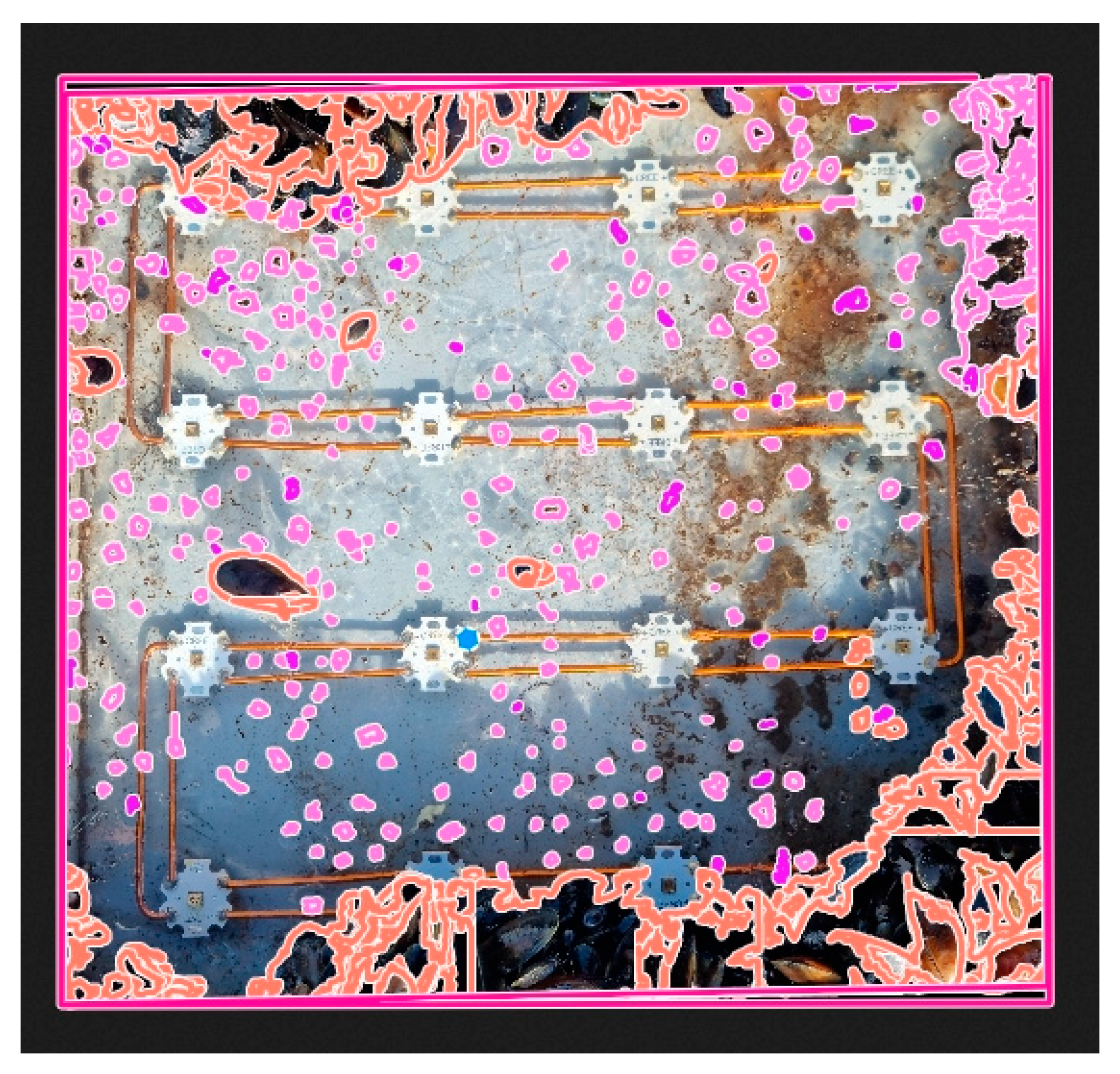
The images of each surface and condition were taken at each sample collection throughout the deployment (February 2021 to February 2022). The images were cropped using ImageJ (version 1.53t), and then uploaded and annotated for percentage coverage using the online software BIIGLE (version 2.0) [60]. Background overlap was annotated on each image and removed from the total image pixel count for accurate calculation of coverage (Figure 2). Hartlepool images had nine labels and the Melbourne images had twelve labels defined. The labels represented visually identifiable taxonomic groups. The labels included Amphipoda, amphipod nests, biofilm, Bryozoa, Hydrozoa, Bivalvia, Annelida, Tunicata and unknown. At Melbourne, Asteroidea and Porifera were also present. Images were individually annotated using BIIGLE’s ‘Lawnmower’ feature at a magnification allowing 16 windows of annotation per image. Annotations were exported as an area coverage Excel document and pixel coverage was converted into percentage coverage. Percentage coverage was then plotted within R version 4.1.1 using the ggplot2 and ggpubr packages [61].

3. Results

3.1. Environmental Conditions at the Test Sites

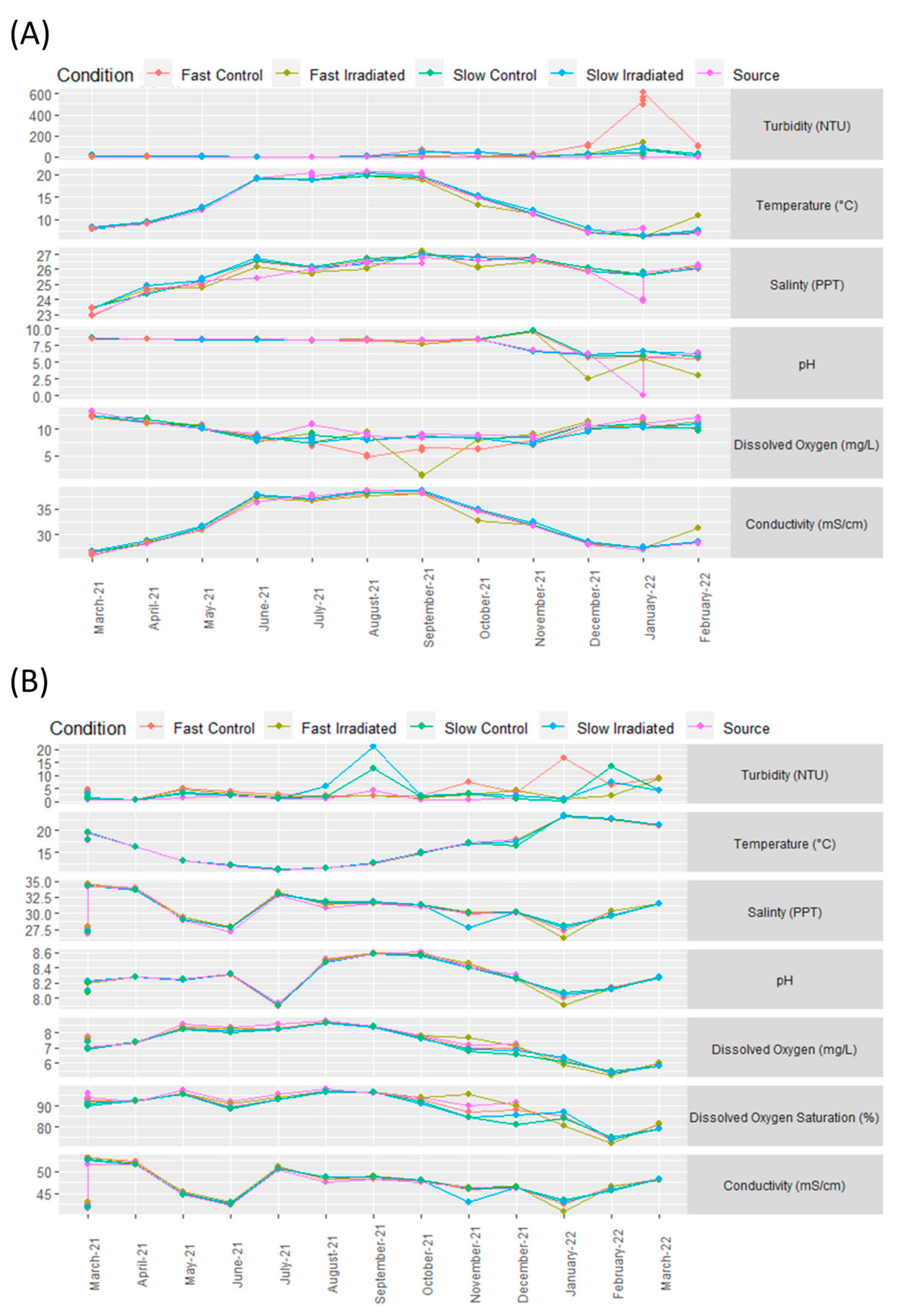
Seasonal changes were recorded across all environmental metrics. Small differences were also observed between experimental chambers. Seawater at Hartlepool had a peak conductivity of 38 mS/cm in July and August, with the lowest conductivity (24 mS/cm) recorded in January. Dissolved oxygen (DO) was lowest in August and September (7 and 9 mg/L for control and irradiated chambers, respectively). DO was highest in March, at 12 mg/L for all conditions. The seawater pH remained stable throughout the year, however recording ceased in October due to equipment failure. Salinity peaked in September and October at 27 ppt and was lowest in March at 23 ppt, in all chambers. The highest temperature of 21 °C was recorded in July and August in both flow and irradiance conditions, and the lowest temperature of 4 °C appeared between January and March with only the slow flow control chamber varying up to 8 °C in March. Turbidity remained below 10 Nephelometric Turbidity Units (NTUs) until October. Turbidity in the fast flow control tank increased up to ~200 NTUs in December and February, with a peak of 600 NTUs in January. In January, small fluctuations were recorded in the other chambers, with highs of 55, 76 and 98 NTUs for fast flow irradiated, slow flow irradiated and slow flow control chambers, respectively.
At Melbourne, turbidity in all conditions remained at 0–5 NTUs between March and August, after which the slow flow chambers increased to 21 and 11 NTUs for irradiated and control chambers, respectively. The fast flow control chambers peaked at 16 NTU in January 2022, whilst readings in all the other chambers remained low. In February and March 2022, more turbid conditions were evident in all chambers. No significant differences in temperature were identified between chambers (ANOVA, p > 0.05), ranging from 10 °C in July 2021 to 25 °C in January 2022. Salinity, pH, DO, and conductivity did not show any significant variation between chambers (Figure 3).
Seawater flow rates were maintained at 140 and 14 L/min over the experimental period for fast and slow flow chambers, respectively. A gradual reduction in irradiance output was observed from all UV-emitting tiles at Hartlepool, with tiles in fast flow conditions degrading more quickly than those in slow flow. Tile irradiance reduced by 97%, 96%, 65% and 65% for fast flow left, fast flow right, slow flow left and slow flow right, respectively. Of the 64 LEDs in Hartlepool, 35 had reduced to <10% output (32 fast flow, 3 slow flow) and 16 gave off <10 µW/cm2 (15 fast flow, 1 slow flow) by the end of the experiment.
In Melbourne, tiles in the fast chambers had a 50% reduction in irradiance after 10 months of deployment, however slow flow chambers were still operating at 50% after 12 months. At the end of the experiment, the overall tile irradiances in Melbourne had reduced by 75%, 63%, 49% and 50% for fast flow A, fast flow B, slow flow A, and slow flow B tiles, respectively. Of the 64 LEDs operating over 12 months in Melbourne, forty-four were reduced by >50%, nine were reduced to <10% (eight fast flow, one slow flow) and eleven were reduced to <10 µW/cm2 (eight fast flow, three slow flow).

3.2. Imaging-Based Analysis of Fouling Communities

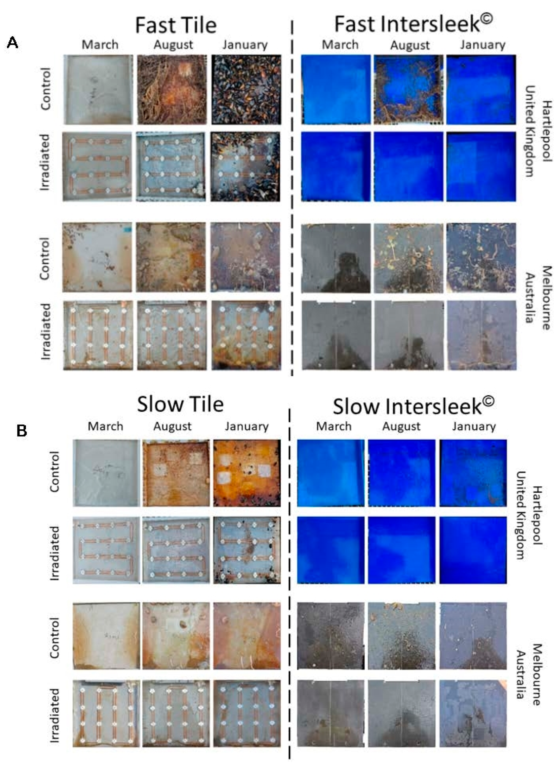
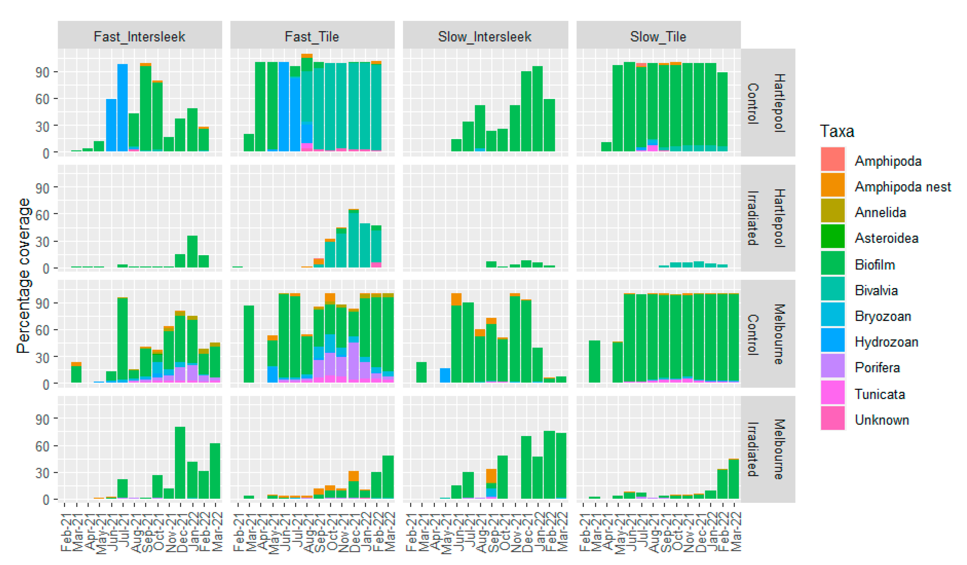
Image analysis of the entire community assemblage was conducted on control, IntersleekTM and irradiated surfaces, in fast and slow flow conditions at Hartlepool and Melbourne, for each monthly observation. At both locations, irradiated surfaces were fouled less than the controls (Figure 4 and Figure 5). Both locations had low recruitment initially, with colonisation increasing from September onwards. The slow flow irradiated samples from Hartlepool had the lowest continuous biofouling presence in December and January, with minimal coverage, mainly consisting of Bivalvia and byssal threads, (Figure 6). The biofilm was better developed on the Hartlepool control surfaces under slow flow compared to fast flow. However, surfaces in the fast flow chambers had more macrofouling coverage. In the fast flow chambers, the initial biofilm transitioned to Hydrozoa between May and June. The control IntersleekTM under fast flow then returned to predominately biofilm coverage between July and August. The control tile surface community also shifted from mainly biofilm to hydrozoan domination between May and June. However, from August, those surfaces were dominated by bivalves. The irradiated chambers suppressed fouling throughout the study, attracting significantly lower (p < 0.05) fouling than control chambers.
There was significantly lower coverage on irradiated surfaces compared to controls at both Hartlepool and Melbourne (Figure 6). Seasonal community shifts were, however, different at Melbourne compared to Hartlepool. Both surface types were initially dominated by biofilm, then sponges and finally bryozoans appeared in the fast flow chambers at Melbourne between September and February. The shift in community structure to sponges and bryozoans did not occur on the surfaces in the slow flow chambers at Melbourne which, instead, remained dominated by biofilm from May onwards. Irradiated surfaces had less micro- and macrofouling than the controls in both locations and at both flow speeds. The community composition on the IntersleekTM control surfaces was similar to the tile controls, however there was higher coverage on the latter. All irradiated surfaces experienced gradual increases in coverage over the final 6 months of field exposure.

3.3. Community and Environmental Correlations

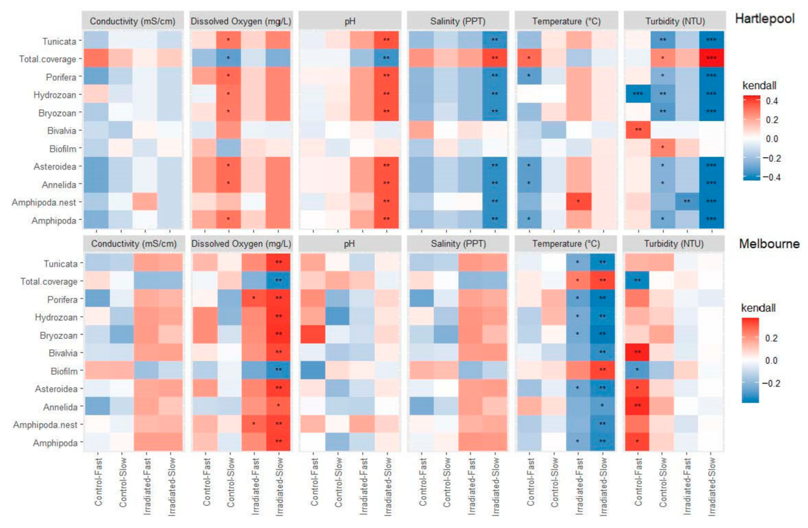
Kendall correlations were calculated between surface coverage, estimated via image analysis, and environmental conditions (Figure 7). Total coverage and biofilm coverage had opposite responses to the correlations observed between individual taxonomic groups and specific environmental parameters. Irradiated slow flow chambers at Hartlepool had positive correlations between all taxonomic groups and pH. Taxa in Irradiated slow flow chambers at Hartlepool had negative correlations with turbidity and salinity. Control slow flow chambers at Hartlepool had positive correlations with DO but negative correlations with turbidity. Most taxa in control fast flow chambers had negative correlations with temperature. Irradiated slow flow chambers at Melbourne presented strong positive correlations between DO and taxonomic groups. Positive correlations were detected between taxa and turbidity on control fast flow surfaces. The effect of temperature was strongly negatively correlated with taxonomic profile for both fast and slow flow irradiated surfaces in Melbourne.

3.4. Amplicon-Based Analysis of Biofouling Communities at Hartlepool

High-throughput sequencing of 18S ribosomal RNA gene amplicons from community samples collected at Hartlepool produced a total of 8.5 million reads. After quality controls, chimera detection and removal, the library was reduced to 4,775,605 reads. A total of 61,972 operational taxonomic units (OTUs) was identified and categorised within the 18S dataset. The sample mean was 13,013 counts with a range from 1 to 46,444 counts. Data were standardised by subsampling to the lowest count without sacrificing data, determined via rarefaction analysis to be 1415 counts. Subsampling removed 13 samples below this threshold of which seven were blank extractions. A total of 495,250 individual counts remained within 14,950 OTUs, covering 76–99% of the community depending on the sample. One hundred and nineteen classes were identified with over 90% of the abundance being represented by 10 classes. These enriched taxa were: ‘Eukaryota_unclassified’, ‘Bivalvia’, ‘Ascidiacea’, ‘Intramacronucleata’, ‘Diatomea’, ‘Arthropoda_unclassified’, ‘Dinophyceae’, ‘Maxillopoda’, ‘Scyphozoa’, and ‘Phaeophyceae’, listed from most to least abundant.

3.5. Seasonal Fouling Community Differences

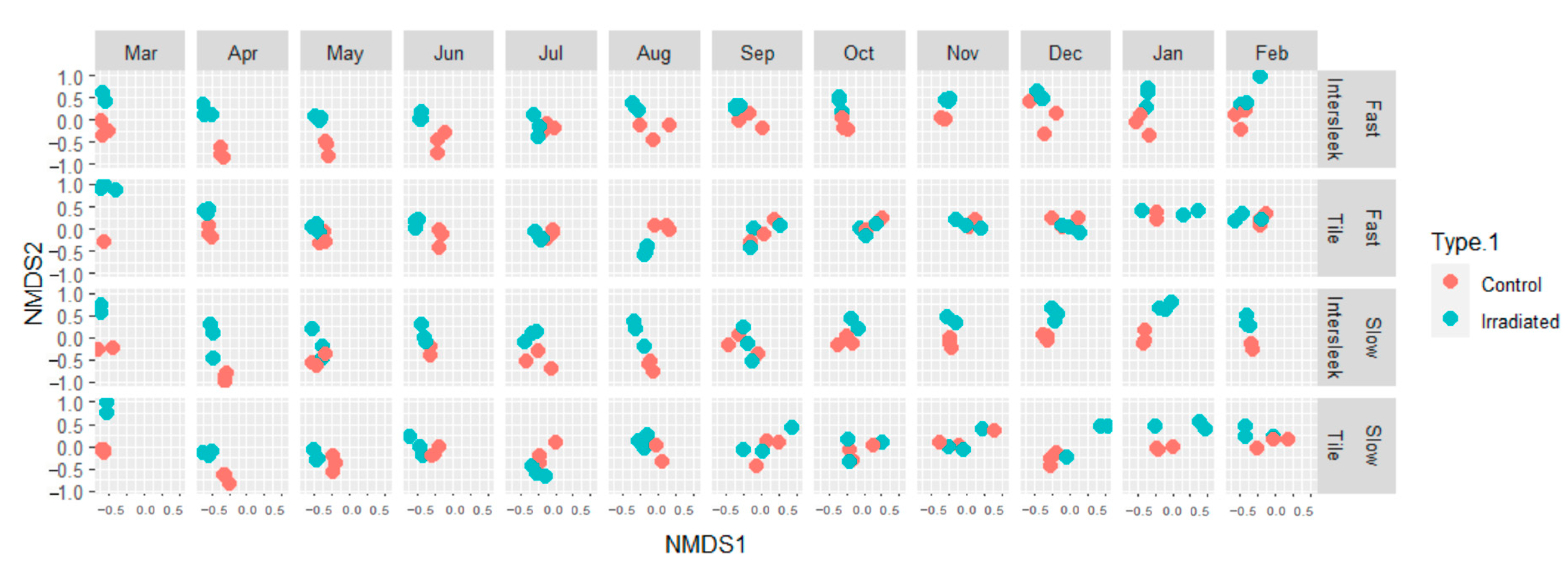
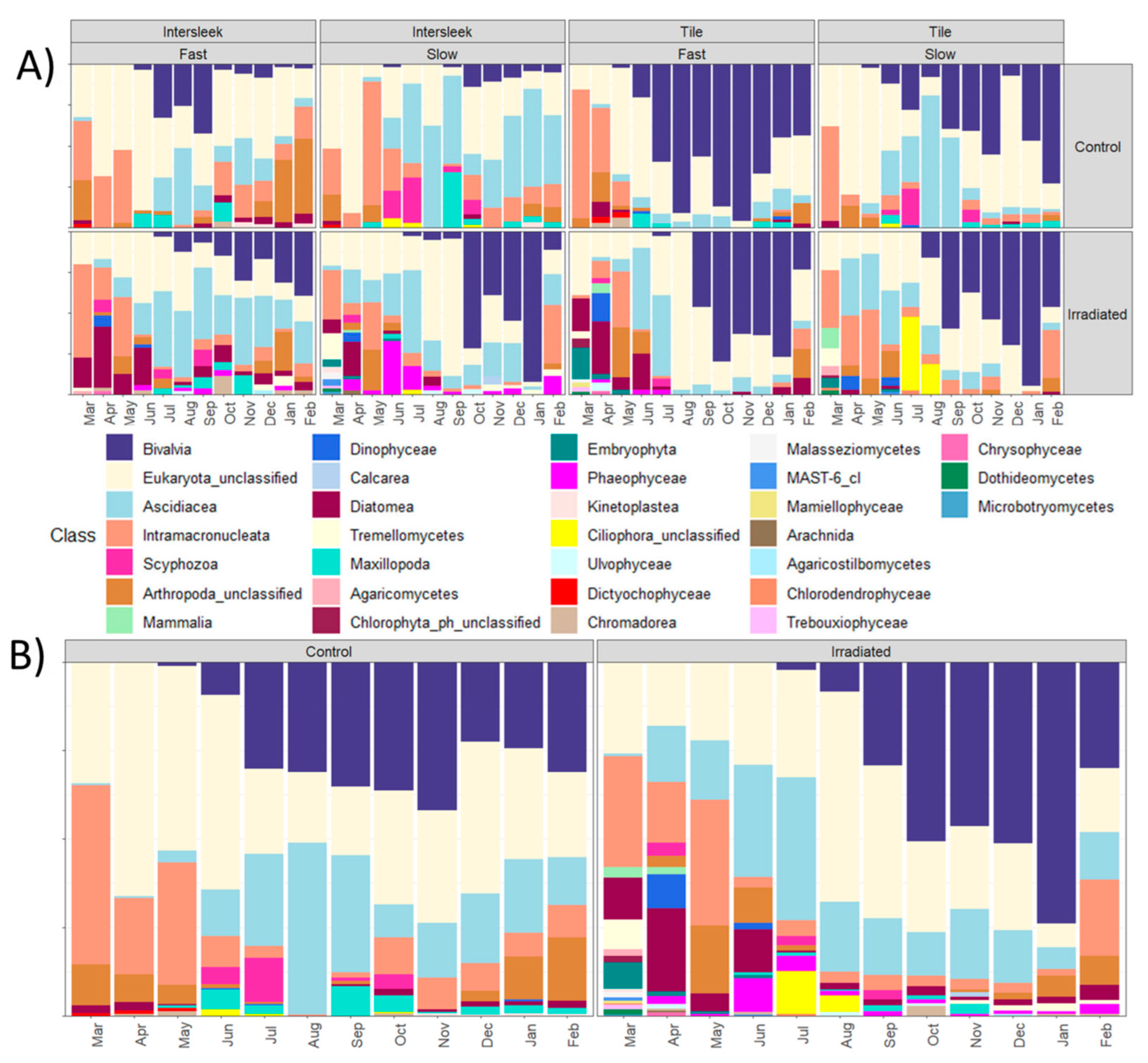
Eukaryotic community assemblages varied between irradiated and control exposures and displayed different community dynamics over time (Figure 8). Control and irradiated samples were significantly different (p < 0.05) based on Adonis and Anosim PERMANOVA analysis. The seasonal community shifts in control chambers were different to those identified in irradiated chambers. Eukaryota_unclassified represented between 5 and 60% of the community but there was no difference between control and irradiated chambers (Figure 9). Arthropoda_unclassified and Maxillopoda had a higher proportional abundance in controls than in irradiated chambers. Bivalvia were detected in May in controls but not until July in irradiated chambers. Ascidiacea were detected in April in irradiated chambers but not until May/June in controls. Diatomea were more abundant in the irradiated chamber than in the control. Dinophycea and Tremellomycetes were only prominent in irradiated chambers.
Sobs, Chao and Ace diversity indices for all conditions indicated a rise in richness towards the end of the experimental period. Initial richness suggested that irradiated samples were more diverse than controls. However, from October to February the controls had higher richness. Water samples remained constant throughout and did not vary between control and irradiated exposures (Supplementary Materials).

4. Discussion

Ultraviolet (UV) irradiance has long been employed for the sterilization of laboratory and clinical settings and has recently emerged as a viable technology for marine biofouling control. While the developing literature suggests great potential, the ocean presents many challenges that must be overcome, including fluctuations in temperature and turbidity, operational conditions, corrosive seawater ingress, the lifespan of hardware including LEDs, the non-uniform toxicity effects on marine fouling organisms and the potential harm to non-target species and/or promotion of evolved resistance.
In this study, UV irradiance prevented biofouling at two field locations: Hartlepool Marina, UK, and Melbourne, Australia. Visual and molecular techniques were used to describe the taxonomic composition of fouling communities in a range of flow, surface and illumination conditions over the course of a year. Beyond demonstrating the effectiveness of the approach in preventing growth and delaying the recruitment of fouling species, this enabled the identification of species that generate UV-resilient compounds and may therefore become resistant to the treatment. Interestingly, the effects of irradiation were measured well beyond the predicted attenuation distance of UV light in seawater which is highly variable in relation to turbidity.

4.1. UV Effects on Community Ecology

Community assembly varied between irradiated and control exposures for all surface types, chamber speeds and between geographical locations. The fouling that occurred on surfaces in the slow flow chambers mainly included biofilm and soft fouling organisms, whereas hard macrofoulers were abundant in the fast flow chambers (Figure 6). Initially, fast flow chamber communities were composed of biofilms, before shifting towards hydrozoans and then to bivalves, or reverting to biofilm. There were no hydrozoans, sponges or bryozoans on the irradiated tiles or irradiated IntersleekTM surfaces, but bivalves were able to colonise from October 2021 to February 2022 at Hartlepool and polychaetes from September 2021 to March 2022 at Melbourne. Both periods were when the LED irradiance was waning, with significant reductions in output across all LEDs. Determining why the LED degradation was restricted to fast flow chambers needs to be investigated further.
Based solely on the data obtained through image analysis, fast flow chambers had a similar level of fouling to slow flow chambers because both produced similar metrics of surface coverage. It is therefore important to note that although surfaces under fast flow appeared similar to the corresponding surfaces in slow flow by image analysis, the larger macrofouling organisms in the former would have a far greater impact on hydrodynamics. Had biomass been measured, fast flow chambers would have far outweighed the slow flow chambers emphasising the limitations of a 2-D assessment. The colonisation of control and irradiated IntersleekTM 1100SR surfaces was taxonomically different, and differences in surface coverage were apparent between the treatments in the two conditions (Figure 6).
Environmental factors clearly influenced the taxa present on the surfaces. Dissolved oxygen (DO), pH, salinity, temperature, and turbidity were all correlated, either positively or negatively, with certain taxonomic groups. In all conditions, biofilm and total fouling coverage were linked by significant correlations. The biofilm and total coverage abundances were inverse to other correlations. This is because biofilm comprised the majority of the initial coverage. The reduction in ‘measured’ biofilm coverage does not mean that actual biofilm coverage reduced, only that it was obscured by other taxa creating an apparent negative correlation. The correlations between taxonomic groups and environmental conditions are not expected to be dependent on this relative reduction in biofilm coverage, but to result from genuine community transitions.

4.2. Indirect and Environmental Effects

Of course, the use of UV could have both direct and indirect effects on fouling communities. The direct effect is DNA damage, whereas indirect effects can be alterations to water conditions, which have implications for larval settlement. Small changes in environmental conditions can influence biofouling community composition [62]. Chemical availability, temperature, light levels and substrate texture are a few variables affecting biofilm and microbiota settlement and growth [63,64,65]. Environmental conditions remained consistent between control and irradiated chambers at each site, demonstrating that the UV treatment itself did not alter water conditions. Although fouling coverage was correlated with environmental conditions in some chambers, these correlations were not attributable to UV exposure, but to differences in ecology, water flow and surface type.
Coverage in slow flow chambers at Hartlepool was negatively correlated with the turbidity of the water. More turbid water typically contains higher dissolved organic matter (DOM), which is often used as a nutrient source by fouling organisms [66,67,68]. The higher numbers of filter-feeding taxa, such as bivalves, tunicates and tube worms, would consume these nutrients within the chambers. Negative correlations against salinity could be attributed to the relatively high starting salinity in March 2021, when the tiles were introduced, and fouling pressure was comparatively low. The salinity in Hartlepool Marina varied substantially, which is likely attributable to precipitation and a partially closed system of lock gates at the Hartlepool site. Fouling organisms in marinas are typically tolerant to changing conditions and can occupy locations with high salinity shifts [69,70,71,72,73]. At the start of the fouling season and towards the end of the field exposure, salinity was lower. Fouling coverage increased from May to December 2021 and coincided with the salinity gradient.
In Melbourne, a negative correlation was evident between taxonomic richness and temperature in irradiated chambers, however, the controls had no significant correlation between richness and temperature. Higher temperatures increase the metabolic rate of fouling organisms, which could leave them more susceptible to UV treatment and lead to reduced survival [74,75,76,77,78]. A positive correlation with coverage and dissolved oxygen (DO) in the slow flow irradiated chambers at Melbourne could be due to denitrification. Photosynthetic communities in low-flow environments can create alterations in DO levels [79]. High DO levels can impact the production of extracellular polymeric substances by microbes and increase membrane permeability, resulting in different microbial communities [48,79]. Temperature was inversely linked to DO levels in the same chambers. Temperature could, in turn, affect the DO levels and have impacts on the macrofouling community composition [80].
The flow of 140 L/min promoted earlier fouling in the fast flow chambers compared to the slow flow chambers (14 L/min) for both control and irradiated treated chambers. Although the supply of potential recruits to the slow flow chambers would have been lower, conditions of reduced flow also may have favoured colonisation. Larvae capable of attaching in fast-flow environments have been shown to also detach more frequently than those recruited in slower flow conditions [81,82,83]. Faster flow creates turbulent eddies that increase shear stresses on settling larvae [50,84,85]. Additionally, rapid flow can reduce the exploratory period of some organisms [86,87,88]. Balanus amphitrite larvae, however, have been noted to display greater surface exploration within dynamic rather than static conditions [86]. Therefore, although higher flow can reduce settlement, this was likely offset by an associated increase in nutrient and larval supply in the fast flow chambers.
The 18S analysis at Hartlepool provided an understanding of the community composition that was not detected via imaging alone. Taxa were identified to class level to allow association with image analysis data. Distinct community compositions were detected, however the 10 most abundant classes accounted for >90% of the community. This represented the core community with a large overlap between conditions (Figure 8 and Figure 9). Unclassified eukaryotes were dominant at the class level; however, this was an accumulation of 9065 different unidentifiable OTUs. Unclassified eukaryotes represented >60% of the OTUs subsampled and could not be aligned to a specific phylum or class. Nevertheless, the remaining taxa detected via 18S sequencing were similar to the taxa observed through the use of image analysis. Irradiated surfaces had distinct community differences compared to the control surfaces over the duration of the experiment. UV irradiation suppressed colonisation by some organisms, for example ‘Bivalvia’ increased in abundance two months later than under control conditions and ‘Porifera’ were not observed at all. Once established, abundance estimated from 18S amplicons was similar to the controls, which contradicted the image analysis results but may be a consequence of the limited quantitative power of the sequencing technique. Amplicon sequencing can be problematic in its reproducibility and reliability when taken alone, as some organisms’ DNA can be extracted more readily than others, and some organisms have proportionally more DNA [89,90]. The extracted DNA provides no direct information about the live or dead status of the donor, or whether it is important to the ecosystem [91]. Additionally, organisms shed DNA into the local environment, which can lead to skewed proportions [92].

4.3. Specific Effects of UV on Different Taxonomic Groups

A range of UV-absorbing compounds may be found in bacteria, fungi, and algae [93,94]. Tremellomycetes are fungi known for their yeast states that can form hyphae, basidia, and basidiospores and are widespread through the marine environment as pathogens of marine mammals [95]. Tremellomycetes were present in all irradiated Hartlepool samples at the start of the experiment and within the last 4 months, but not in any of the controls. Some species of Tremellomycetes can produce mycosporine-like amino acids (MAAs) and some can produce UV-absorbing mycosporine-glutamicol-glucoside (MGG) [96,97,98,99]. Tremellomycetes are found in the marine environment with yeasts rich in carotenoid pigments and are known to ferment sugars as a component of the biofilm [100]. Intramacronucleata were present on both control and irradiated surfaces throughout the experiment. Intramacronucleata is a subphylum of ciliates with some of the species capable of swimming freely and others are sessile components of biofilms [101]. Some ciliates can produce MAAs whilst others can form hypericin or hypericin-like pigments for protection against UV-B damage [102,103].
Diatomea were found on all surfaces in both exposures but were more prominent in the irradiated chambers. Diatomea is a large group of microalgae that produce 20–25% of global primary production and are often dominant in biofilm formation [104]. Unique to this clade of marine algae is the silicate frustule that can reflect and/or absorb UV rays [104]. These organisms were all constituents of the core community found within the experimental chambers. All of these microorganisms are associated in some form with UV resilience and can form biofilms [20,105,106].
The Mytilidae (mussels) are motile organisms that use collagenous threads (byssus) to anchor to a surface [107]. Mytilidae colonised the back and side of the acrylic sheath holding the UV tiles in place, which were protected from direct UV exposure. Towards the end of the experiment, observation of the chambers revealed that the increase in mussel coverage was from the periphery of the tiles rather than from the tile centre. Abandoned byssal threads were also visible on the irradiated surfaces, which validates the conclusions of the 18S analysis and supports the likelihood that, over time, mussels sought refuge from irradiation.
Remarkably, Ascidiacea (sea squirts) appeared two months earlier on irradiated surfaces than on the controls. This is the opposite trend to Bivalvia and potentially indicates the resilience of Ascidiacea to UV exposure. Ascidiacea are a subphylum of Tunicata that are filter feeders that can host algal symbionts for energy production. Prochloron sp. is a symbiotic algal species of the Ascidiacea that produces UV-absorbing mycosporine-like amino acids (MAAs): mycosporine–glycine, palythine, shinorine, and porphyra-334 [108]. A broad range of Ascidiacea contain MAAs from their symbionts but are able to synthesise MAAs de novo [94,109]. Ascidiacea containing MAAs would be able to tolerate some level of UV irradiation and be more likely to colonise the substrate [94]. Their initial absence in the control chambers could indicate that Ascidiacea were outcompeted by other taxa or required other taxa for settlement. Image analysis determined that Ascidiacea were present in Hartlepool from July only on the control surfaces. From February, they were also identified on a single irradiated surface. The 18S amplicon sequencing detected the presence of Ascidiacea at an earlier point than the image analysis, and that they occurred on all surfaces. This discrepancy could be due to the collection of DNA from settled larvae, which would not be visible to the image analysis.
Scyphozoa were found in both control and irradiated chambers at Hartlepool, periodically throughout field exposures. Scyphozoa are a class of the phylum Cnidaria that have a pelagic medusoid stage and can have a benthic polyp stage [110,111]. Cnidaria are also able to bioaccumulate MAAs as a protection against UV through the ingestion of algae [112,113]. The transparent planula larvae, which are generally <1.5 mm, may have attempted to colonise the irradiated surfaces and, similar to Ascidiacea, remained undetected via image analysis [114]. Although some Scyphozoa can survive UV exposure [112], they were clearly unable to thrive under continuous UV exposures here.
Maxillopoda is a diverse class of the phylum Arthropoda with jointed appendages [115]. Maxillopoda and Arthopoda_unclassified were both present in all chambers throughout the experiment according to metagenetic analysis of Hartlepool samples. Image analysis assigned these as Amphipoda, and further identification was outside the scope of this study. The hard carapace that surrounds these organisms is composed of chitin, remicilin and proteins that can block up to 80% of UVB [116,117]. These organisms are free-swimming but colonise surfaces by building nests [118,119], which are formed from benthic detritus and silt, and in some cases, amphipod-produced silk [120,121]. The nests protect amphipods inside from external predation; however, the nest bases are not fully enclosed, enabling irradiation from beneath. Additionally, the shells of Mytilidae block UV and allow areas in which the Maxillopoda could find protection. High turnover and the free-swimming nature of these organisms could explain the higher predicted presence in the 18S analysis compared to imaging.

4.4. Hardware Considerations

UV irradiance levels dropped substantially towards the end of the field experiments. Although all tiles showed a fall in irradiance, not all LEDs degraded at the same rate. The suppliers predicted that irradiance levels would reduce by half (L50) after 10,000 h. The actual L50 observed was after 10–12 months or 7465–8880 h. This discrepancy could explain the presence of fouling organisms on irradiated tiles in the final months of exposure. Nevertheless, biofouling was greatly reduced (40–90%) compared to controls. The level of fouling on the irradiated tiles was proportional to the irradiance loss within specific chambers. Fast flow chambers experienced the largest UV reduction and the highest fouling pressure, with the opposite observed for slow flow chambers. Extending the longevity of LEDs’ life span is imperative to achieve continuous fouling control. Since the lifespan of light sources depends on the time the light is powered on, optimising intermittent powering (on:off periods) for biofouling control is important to extend LED lifespans [122,123]. Duty cycling can be detrimental to traditional iridescent and halogen light sources [124], however, LEDs are not so sensitive. LED longevity can be extended via pulsing or irradiance output manipulation and still achieve effective fouling prevention [36,39,49].
One of the predicted advantages of UV as a fouling-control technology is the highly localised effect. The tile design aimed to prevent organisms from colonising the surface of the tiles without impacting organisms in the surrounding water. Transmission measurements through relatively clear water (0.1 NTU) indicated high attenuation (>90%) within 5 cm of the emitting surface and irradiance rates were negligible at 14 cm from the tile surface in clear media (Supplementary Figure S1). Higher NTU levels drastically reduced the transmission of light and saw linear reduction with distance. The average turbidity of the North Sea ranges from 15 to 60 NTU at the coast to 7 NTU offshore [125]. Under these conditions, the irradiance would penetrate to <10 cm in coastal environments and <14 cm when offshore. Thus, the effect of UV on colonisation of the IntersleekTM 1100SR surface positioned 12 cm from the UV-emitting tile in the test chambers was expected to be negligible. This was not the case and, in fact, effects of the UV emission in test chambers could be observed across the entire interior surface of the chamber (Figure 5). This could be due to acute toxicity to propagules passing close to the UV tile in a chamber that was designed to exhibit significant internal circulation. Or it could be due to the additive effect of multiple overlapping LED emissions at increasing distance from the tile. Additionally, chemical composition within the water column can be impacted by UV and result in a variety of free radicals which could have had impacted upon the study. However, as chemical conditions (Figure 3) did not vary across the test chambers this could not be quantified.

5. Conclusions

While this study raises important questions regarding the differential effects of UV on different colonising taxa, the potential for evolved resistance in microbial and metazoan taxa, and off-target effects beyond the predicted range of the LEDs, it is nevertheless clear that the technology works. The surfaces of the UV-emitting tiles remained effectively clean and their presence in large replica sea chests was sufficient to maintain a fouling-free environment beyond expectations. Future work will be needed in two key areas to fully implement this potentially transformative approach: (1) The technology: can better, longer-lasting systems be designed, and optimisation of their performance be better understood? (2) The biology: what are the physiological effects on the organisms that are exposed, what are the direct and indirect effects on recruitment, and how will off-target effects be legally regulated?

Supplementary Materials

The following supporting information can be downloaded at: https://www.mdpi.com/article/10.3390/jmse11122211/s1, Figure S1: Sobs diversity indices of OTUs identified from 18S metagenetic analysis. Samples were deployed for 12 months in Hartlepool. UK; Figure S2: ACE diversity indices of OTUs identified from 18S metagenetic analysis. Samples were deployed for 12 months in Hartlepool. UK; Figure S3: Chao diversity indices of OTUs identified from 18S metagenetic analysis. Samples were deployed for 12 months in Hartlepool. UK.

Author Contributions

P.W. undertook the design and implementation of the experiments, field sampling, laboratory and results analysis and manuscript construction. N.A. provided supervisory support and assisted in the design of the experiments and manuscript construction. A.S.C. provided supervisory support and assisted in the design of the experiments and manuscript construction. J.A.F. assisted in field sampling, laboratory analysis and manuscript construction. J.P. provided supervisory support and financial investment and aided in manuscript construction. R.F.P. undertook the design and implementation of experiments, field sampling and manuscript construction. All authors have read and agreed to the published version of the manuscript.

Funding

This project was funded by an award from the Materials for Strategic Advantage Programme of the Defence Science and Technology Laboratory (DSTLX-1000129048) and Newcastle University, which provided studentship funding (PW) for one year. Diatom cultures were supported by a US ONR award (N00014-20-1-2248) and AkzoNobel provided materials and support for UV-LED tile preparation.

Institutional Review Board Statement

Not applicable.

Informed Consent Statement

Not applicable.

Data Availability Statement

All data are available at DOI: 10.25405/data.ncl.24549112 (accessed on 10 October 2023).

Acknowledgments

We would like to thank Jessica Clarke and Peter Allen for their help in field sampling in Hartlepool and Clare Granidon, Mark Ciacic and Jim Dimas from DSTG for design and construction of field testing units and sample collection in Melbourne. Kevin Reynolds from AkzoNobel was instrumental in aiding in the tile design and construction.

Conflicts of Interest

The authors declare that the research was conducted in the absence of any commercial or financial relationships that could be construed as a potential conflict of interest.

References

  1. Readman, J.W. Development, Occurrence and Regulation of Antifouling Paint Biocides: Historical Review and Future Trends. In Antifouling Paint Biocides; Springer: Berlin/Heidelberg, Germany, 2006; pp. 1–15. [Google Scholar]
  2. Qin, L.; Lu, S.; Liu, J.; Wu, Y.; Ma, Z.; Mawignon, F.J.; Dong, G. Bionic Non-Smooth Epoxy Resin Coating with Corrosion Inhibitor for Drag-Reduction and Durability. Prog. Org. Coat. 2022, 173, 107176. [Google Scholar] [CrossRef]
  3. Yang, H.; Qin, L.; Zhao, W.; Mawignon, F.J.; Guo, H.; Wu, Y.; Zhang, Y.; Dong, G. Eco-Friendly Polysaccharide Coatings for Antifouling and Drag-Reduction and Potential Application for Marine Devices. Friction 2023, 12, 1–19. [Google Scholar] [CrossRef]
  4. Almeida, E.; Diamantino, T.C.; de Sousa, O. Marine Paints: The Particular Case of Antifouling Paints. Prog. Org. Coat. 2007, 59, 2–20. [Google Scholar] [CrossRef]
  5. Munk, T.; Kane, D.; Yebra, D.M. The Effects of Corrosion and Fouling on the Performance of Ocean-Going Vessels: A Naval Architectural Perspective. In Advances in Marine Antifouling Coatings and Technologies; Woodhead Publishing: Sawston, UK, 2009; pp. 148–176. [Google Scholar] [CrossRef]
  6. Liu, M.; Li, S.; Wang, H.; Jiang, R.; Zhou, X. Research Progress of Environmentally Friendly Marine Antifouling Coatings. Polym. Chem. 2021, 12, 3702–3720. [Google Scholar] [CrossRef]
  7. Finlay, J.A.; Franco, S.C.; Daniels, J.; Hadfield, M.G.; Stafslien, S.J.; Webster, D.C.; Wendt, D.E.; Brewer, L.; Clare, A.S.; Teo, S.L.M. Fouling-Release Performance of Silicone Oil-Modified SiloxanePolyurethane Coatings. ACS Appl. Mater. Interfaces 2016, 8, 29025–29036. [Google Scholar]
  8. Clare, A.S.; Rittschof, D.; Gerhart, D.J.; Maki, J.S. Molecular Approaches to Nontoxic Antifouling. Invertebr. Reprod. Dev. 1992, 22, 67–76. [Google Scholar] [CrossRef]
  9. Davidson, I.; Ashton, G.; Ruiz, G.; Scianni, C.; Brown, C.; Pagenkopp-Lohan, K.; Fleischer, R. Richness, Extent, Condition, Reproductive Status, and Parasitism of Fouling Communities on Commercial Vessels; Report to the California State Lands Commission Marine Invasive Species Program; Portland State University: Portland, OR, USA, 2013. [Google Scholar]
  10. Growcott, A.; Kluza, D.; Georgiades, E.; Science, B. Technical Advice: Evaluation of in-Water Systems to Reactively Treat or Remove Biofouling within Vessel Internal Niche Areas; Ministry for Primary Industries: Wellington, New Zealand, 2019; Volume 4, ISBN 9781988594194.
  11. Rahman, S. Implementation of Ballast Water Management Plan in Ships through Ballast Water Exchange System. Procedia Eng. 2017, 194, 323–329. [Google Scholar] [CrossRef]
  12. Coutts, A.D.M.; Taylor, M.D. A Preliminary Investigation of Biosecurity Risks Associated with Biofouling on Merchant Vessels in New Zealand. New Zeal. J. Mar. Freshw. Res. 2004, 38, 215–229. [Google Scholar] [CrossRef]
  13. Frey, M.A.; Simard, N.; Robichaud, D.D.; Martin, J.L.; Therriault, T.W. Fouling around: Vessel Sea-Chests as a Vector for the Introduction and Spread of Aquatic Invasive Species. Manag. Biol. Invasions 2014, 5, 21–30. [Google Scholar] [CrossRef]
  14. Fernandes, J.A.; Santos, L.; Vance, T.; Fileman, T.; Smith, D.; Bishop, J.D.D.; Viard, F.; Queirós, A.M.; Merino, G.; Buisman, E.; et al. Costs and Benefits to European Shipping of Ballast-Water and Hull-Fouling Treatment: Impacts of Native and Non-Indigenous Species. Mar. Policy 2016, 64, 148–155. [Google Scholar] [CrossRef]
  15. Sylvester, F.; MacIsaac, H.J. Is Vessel Hull Fouling an Invasion Threat to the Great Lakes? Divers. Distrib. 2010, 16, 132–143. [Google Scholar] [CrossRef]
  16. Georgiades, E.; Kluza, D.; Bates, T.; Lubarsky, K.; Brunton, J.; Growcott, A.; Smith, T.; McDonald, S.; Gould, B.; Parker, N.; et al. Regulating Vessel Biofouling to Support New Zealand’s Marine Biosecurity System—A Blue Print for Evidence-Based Decision Making. Front. Mar. Sci. 2020, 7, 390. [Google Scholar] [CrossRef]
  17. Cacabelos, E.; Ramalhosa, P.; Canning-Clode, J.; Troncoso, J.S.; Olabarria, C.; Delgado, C.; Dobretsov, S.; Gestoso, I. The Role of Biofilms Developed under Different Anthropogenic Pressure on Recruitment of Macro-Invertebrates. Int. J. Mol. Sci. 2020, 21, 2030. [Google Scholar] [CrossRef]
  18. Grant, K.R. Next-Generation Amplicon Sequencing: A Cost-Effective Method for Exploring Microbial Biodiversity. In Molecular Genetics and Genomics Tools in Biodiversity Conservation; Springer: Berlin/Heidelberg, Germany, 2022; pp. 203–236. [Google Scholar]
  19. Comeau, A.M.; Lagunas, M.G.; Scarcella, K.; Varela, D.E.; Lovejoy, C. Nitrate Consumers in Arctic Marine Eukaryotic Communities: Comparative Diversities of 18S RRNA, 18S RRNA Genes, and Nitrate Reductase Genes. Appl. Environ. Microbiol. 2019, 85, e00247-19. [Google Scholar] [CrossRef]
  20. Weber, S.D.; Hofmann, A.; Pilhofer, M.; Wanner, G.; Agerer, R.; Ludwig, W.; Schleifer, K.-H.; Fried, J. The Diversity of Fungi in Aerobic Sewage Granules Assessed by 18S RRNA Gene and ITS Sequence Analyses. FEMS Microbiol. Ecol. 2009, 68, 246–254. [Google Scholar] [CrossRef]
  21. Chen, C.L.; Maki, J.S.; Rittschof, D.; Teo, S.L.M. Early Marine Bacterial Biofilm on a Copper-Based Antifouling Paint. Int. Biodeterior. Biodegrad. 2013, 83, 71–76. [Google Scholar] [CrossRef]
  22. de Vries, H.J.; Kleibusch, E.; Hermes, G.D.A.; van den Brink, P.; Plugge, C.M. Biofouling Control: The Impact of Biofilm Dispersal and Membrane Flushing. Water Res. 2021, 198, 117163. [Google Scholar] [CrossRef]
  23. Angelova, A.G.; Ellis, G.A.; Wijesekera, H.W.; Vora, G.J. Microbial Composition and Variability of Natural Marine Planktonic and Biofouling Communities from the Bay of Bengal. Front. Microbiol. 2019, 10, 2738. [Google Scholar] [CrossRef]
  24. Takada, K.; Shiba, T.; Yamaguchi, T.; Akane, Y.; Nakayama, Y.; Soda, S.; Inoue, D.; Ike, M. Cake Layer Bacterial Communities during Different Biofouling Stages in Full-Scale Membrane Bioreactors. Bioresour. Technol. 2018, 259, 259–267. [Google Scholar] [CrossRef]
  25. Von Ammon, U.; Wood, S.A.; Laroche, O.; Zaiko, A.; Tait, L.; Lavery, S.; Inglis, G.J.; Pochon, X. Combining Morpho-Taxonomy and Metabarcoding Enhances the Detection of Non-Indigenous Marine Pests in Biofouling Communities. Sci. Rep. 2018, 8, 16290. [Google Scholar] [CrossRef]
  26. Chiellini, C.; Iannelli, R.; Modeo, L.; Bianchi, V.; Petroni, G. Biofouling of Reverse Osmosis Membranes Used in River Water Purification for Drinking Purposes: Analysis of Microbial Populations. Biofouling 2012, 28, 969–984. [Google Scholar] [CrossRef]
  27. Chessa, D.; Murgia, M.; Sias, E.; Deligios, M.; Mazzarello, V.; Fiamma, M.; Rovina, D.; Carenti, G.; Ganau, G.; Pintore, E. Metagenomics and Microscope Revealed T. Trichiura and Other Intestinal Parasites in a Cesspit of an Italian Nineteenth Century Aristocratic Palace. Sci. Rep. 2020, 10, 12656. [Google Scholar] [CrossRef]
  28. Satjarak, A.; Graham, L.E.; Piotrowski, M.J.; Trest, M.T.; Wilcox, L.W.; Cook, M.E.; Knack, J.J.; Arancibia-Avila, P. Shotgun Metagenomics and Microscopy Indicate Diverse Cyanophytes, Other Bacteria, and Microeukaryotes in the Epimicrobiota of a Northern Chilean Wetland Nostoc (Cyanobacteria). J. Phycol. 2021, 57, 39–50. [Google Scholar] [CrossRef]
  29. Cheng, N.; Moe, P. Inactivation of Enveloped Viruses (Coronavirus, H5N1 Virus) and Disinfection of the Air with Legionella-X100 via Ultraviolet Germicidal Irradiation (UVGI); Academia.Edu: San Francisco, CA, USA, 2020; pp. 1–18. [Google Scholar]
  30. Rastogi, R.P.; Kumar, A.; Tyagi, M.B.; Sinha, R.P. Molecular Mechanisms of Ultraviolet Radiation-Induced DNA Damage and Repair. J. Nucleic Acids 2010, 2010, 592980. [Google Scholar] [CrossRef]
  31. Sarathy, S.R.; Mohseni, M. An Overview of UV-Based Advanced Oxidation Processes for Drinking Water Treatment. IUVA News 2006, 8, 16–27. [Google Scholar]
  32. Bukhari, Z.; Hargy, T.M.; Bolton, J.R.; Dussert, B.; Clancy, J.L. Medium-pressure UV for Oocyst Inactivation. J. Am. Water Work. Assoc. 1999, 91, 86–94. [Google Scholar] [CrossRef]
  33. Craik, S.A.; Weldon, D.; Finch, G.R.; Bolton, J.R.; Belosevic, M. Inactivation of Cryptosporidium Parvum Oocysts Using Medium-and Low-Pressure Ultraviolet Radiation. Water Res. 2001, 35, 1387–1398. [Google Scholar] [CrossRef]
  34. Dai, T.; Vrahas, M.S.; Murray, C.K.; Hamblin, M.R. Ultraviolet C Irradiation: An Alternative Antimicrobial Approach to Localized Infections? Expert. Rev. Anti. Infect. Ther. 2012, 10, 185–195. [Google Scholar] [CrossRef]
  35. Braga, C.; Hunsucker, K.; Erdogan, C.; Gardner, H.; Swain, G. The Use of a Uvc Lamp Incorporated with an ROV to Prevent Biofouling: A Proof-of-Concept Study. Mar. Technol. Soc. J. 2020, 54, 76–83. [Google Scholar] [CrossRef]
  36. Hunsucker, K.Z.; Braga, C.; Gardner, H.; Jongerius, M.; Hietbrink, R.; Salters, B.; Swain, G. Using Ultraviolet Light for Improved Antifouling Performance on Ship Hull Coatings. Biofouling 2019, 35, 658–668. [Google Scholar] [CrossRef]
  37. Richard, K.N.; Hunsucker, K.Z.; Gardner, H.; Hickman, K.; Swain, G. The Application of UVC Used in Synergy with Surface Material to Prevent Marine Biofouling. J. Mar. Sci. Eng. 2021, 9, 662. [Google Scholar] [CrossRef]
  38. Dobretsov, S.V.; Gosselin, L.; Qian, P.Y. Effects of Solar PAR and UV Radiation on Tropical Biofouling Communities. Mar. Ecol. Prog. Ser. 2010, 402, 31–43. [Google Scholar] [CrossRef]
  39. Whitworth, P.; Aldred, N.; Reynolds, K.J.; Plummer, J.; Duke, P.; Clare, A.S. Importance of Duration, Duty-Cycling and Thresholds for the Implementation of Ultraviolet C in Marine Biofouling Control. Front. Mar. Sci. 2022, 8, 809011. [Google Scholar] [CrossRef]
  40. Lakretz, A.; Ron, E.Z.; Mamane, H. Biofouling Control in Water by Various UVC Wavelengths and Doses. Biofouling 2010, 26, 257–267. [Google Scholar] [CrossRef]
  41. Pinnacle, F. Foul Free for over Two Years-Presentation of Results from AML Oceanographic UV•Xchange Biofouling Control Experiment at Folger Pinnacle. In Proceedings of the OCEANS 2016 MTS/IEEE Monterey, Monterey, CA, USA, 19–23 September 2016; pp. 19–23. [Google Scholar]
  42. Piola, R.; Salters, B.; Grandison, C.; Ciacic, M.; Hietbrink, R. Assessing the Use of Low Voltage UV-Light Emitting Miniature LEDs for Marine Biofouling Control; DST-Group-TR-3266; Australian Government Department of Defense: Canberra, Australia, 2016; pp. 2–28.
  43. Bueley, C. UV Irradiation: A New Kind of Antifoulant. Ocean News & Technology, 4 March 2014. [Google Scholar]
  44. Bueley, C.; Olender, D.; Brocking, B. The (in)Fluence of Light. J. Ocean. Technol. 2014, 9, 48–67. [Google Scholar]
  45. MacKenzie, A.F.; Maltby, E.A.; Harper, N.; Bueley, C.; Olender, D.; Wyeth, R.C. Periodic Ultraviolet-C Illumination for Marine Sensor Antifouling. Biofouling 2019, 35, 483–493. [Google Scholar] [CrossRef]
  46. Phukan, T.; Rai, A.N.; Syiem, M.B. Dose Dependent Variance in UV-C Radiation Induced Effects on Carbon and Nitrogen Metabolism in the Cyanobacterium Nostoc muscorum Meg1. Ecotoxicol. Environ. Saf. 2018, 155, 171–179. [Google Scholar] [CrossRef]
  47. Meunier, S.M.; Todorovic, B.; Dare, E.V.; Begum, A.; Guillemette, S.; Wenger, A.; Saxena, P.; Campbell, J.L.; Sasges, M.; Aucoin, M.G. Impact of Dissolved Oxygen during UV-Irradiation on the Chemical Composition and Function of CHO Cell Culture Media. PLoS ONE 2016, 11, e0150957. [Google Scholar] [CrossRef]
  48. Ishii, H.; Ohba, T.; Kobayashi, T. Effects of Low Dissolved Oxygen on Planula Settlement, Polyp Growth and Asexual Reproduction of Aurelia aurita. Plankt. Benthos Res. 2008, 3, 107–113. [Google Scholar] [CrossRef]
  49. Braga, C.R. Lighting a Path to Biofouling Prevention: Investigating the Effect of Ultraviolet Light on Biofouling. Photosynthetica 2018, 2, 1–13. [Google Scholar]
  50. Crimaldi, J.P.; Thompson, J.K.; Rosman, J.H.; Lowe, R.J.; Koseff, J.R. Hydrodynamics of Larval Settlement: The Influence of Turbulent Stress Events at Potential Recruitment Sites. Limnol. Oceanogr. 2002, 47, 1137–1151. [Google Scholar] [CrossRef]
  51. Dobretsov, S. Expected Effect of Climate Change on Fouling Communities and Its Impact on Antifouling Research. In Advances in Marine Antifouling Coatings and Technologies; Woodhead Publishing: Sawston, UK, 2009; pp. 222–239. [Google Scholar] [CrossRef]
  52. Raikow, D.F.; Reid, D.F.; Blatchley, E.R.; Jacobs, G.; Landrum, P.F. Effects of Proposed Physical Ballast Tank Treatments on Aquatic Invertebrate Resting Eggs. Environ. Toxicol. Chem. 2007, 26, 717–725. [Google Scholar] [CrossRef]
  53. Hung, O.S.; Thiyagarajan, V.; Wu, R.S.S.; Qian, P.Y. Effect of Ultraviolet Radiation on Biofilms and Subsequent Larval Settlement of Hydroides Elegans. Mar. Ecol. Prog. Ser. 2005, 304, 155–166. [Google Scholar] [CrossRef]
  54. Montero, C.I.; Johnson, M.R.; Chou, C.-J.; Conners, S.B.; Geouge, S.G.; Tachdjian, S.; Nichols, J.D.; Kelly, R.M. Responses of Wild-Type and Resistant Strains of the Hyperthermophilic Bacterium Thermotoga maritima to Chloramphenicol Challenge. Appl. Environ. Microbiol. 2007, 73, 5058–5065. [Google Scholar] [CrossRef]
  55. Habeebu, S.S.; Liu, J.; Liu, Y.; Klaassen, C.D. Metallothionein-Null Mice Are More Susceptible than Wild-Type Mice to Chronic CdCl2-Induced Bone Injury. Toxicol. Sci. 2000, 56, 211–219. [Google Scholar] [CrossRef]
  56. Huesemann, M.H.; Hausmann, T.S.; Bartha, R.; Aksoy, M.; Weissman, J.C.; Benemann, J.R. Biomass Productivities in Wild Type and Pigment Mutant of Cyclotella sp.(Diatom). Appl. Biochem. Biotechnol. 2009, 157, 507–526. [Google Scholar] [CrossRef]
  57. Bonhivers, M.; Carbrey, J.M.; Gould, S.J.; Agre, P. Aquaporins in Saccharomyces: Genetic and Functional Distinctions between Laboratory and Wild-Type Strains. J. Biol. Chem. 1998, 273, 27565–27572. [Google Scholar] [CrossRef]
  58. Kozich, J.; Schloss, P.; Baxter, N.; Jenior, M.; Koumpouras, C.; Bishop, L. 16S RRNA Sequencing with the Illumina MiSeq: Library Generation, QC, & Sequencing; Novogene: Guangzhou, China, 2013. [Google Scholar]
  59. Amaral-Zettler, L.A.; Bauer, M.; Berg-Lyons, D.; Betley, J.; Caporaso, J.G.; Ducklow, W.; Walters, W.A. EMP 18S Illumina Amplicon Protocol. Protocols.io. 2018. Available online: https://www.protocols.io/view/emp-18s-illumina-amplicon-protocol-ewov1b6pgr24/v1?version_warning=no (accessed on 7 September 2021).
  60. Langenkämper, D.; Zurowietz, M.; Schoening, T.; Nattkemper, T.W. BIIGLE 2.0—Browsing and Annotating Large Marine Image Collections. Front. Mar. Sci. 2017, 4, 83. [Google Scholar] [CrossRef]
  61. Wickham, H.; Navarro, D.; Pedersen, T.L. Elegant Graphics for Data Analysis (Ggplot2); Springer: New York, NY, USA, 2016. [Google Scholar]
  62. Rivero, N.K.; Dafforn, K.A.; Coleman, M.A.; Johnston, E.L. Environmental and Ecological Changes Associated with a Marina. Biofouling 2013, 29, 803–815. [Google Scholar] [CrossRef]
  63. Floerl, O.; Pool, T.K.; Inglis, G.J. Positive Interactions between Nonindigenous Species Facilitate Transport by Human Vectors. Ecol. Appl. 2004, 14, 1724–1736. [Google Scholar] [CrossRef]
  64. Mineur, F.; Cook, E.J.; Minchin, D.; Bohn, K.; MacLeod, A.; Maggs, C.A. Changing Coasts: Marine Aliens and Artifcial Structures. In Oceanography and Marine Biology; CRC Press: Boca Raton, FL, USA, 2012; pp. 198–243. ISBN 0429109385. [Google Scholar]
  65. Tait, L.; Inglis, G.; Seaward, K.; Spong, K.; Wilkens, S. Optimising Settlement Arrays for Surveillance of Non-Indigenous Biofouling Species; MPI Information—Paper No: 2016/71; Ministry for Primary Industries: Wellington, New Zealand, 2016; p. 74.
  66. Pierri, C.; Colangelo, P.; Del Pasqua, M.; Longo, C.; Giangrande, A. Consequences of the Experimental Removal of Sabella Spallanzanii (Gmelin, 1791) from the Fouling Assemblage of a Mediterranean Harbour. Mediterr. Mar. Sci. 2019, 20, 476–486. [Google Scholar] [CrossRef]
  67. Trani, R.; Corriero, G.; de Pinto, M.C.; Mercurio, M.; Pazzani, C.; Pierri, C.; Scrascia, M.; Longo, C. Filtering Activity and Nutrient Release by the Keratose Sponge Sarcotragus spinosulus Schmidt, 1862 (Porifera, Demospongiae) at the Laboratory Scale. J. Mar. Sci. Eng. 2021, 9, 178. [Google Scholar] [CrossRef]
  68. Charles, F.; Grémare, A.; Amouroux, J.-M.; Cahet, G. Filtration of the Enteric Bacteria Escherichia Coli by Two Filter-Feeding Bivalves, Venus verrucosa and Mytilus galloprovincialis. Mar. Biol. 1992, 113, 117–124. [Google Scholar] [CrossRef]
  69. Morton, B. The Biology and Functional Morphology of Mytilopsis sallei (Recluz)(Bivalvia: Dreissenacea) Fouling Visakhapatnam Harbour, Andhra Pradesh, India. J. Molluscan Stud. 1981, 47, 25–42. [Google Scholar] [CrossRef]
  70. Nair, P.S.R.; Krishnamurthy, K.; Mawatari, S.F. Salinity Tolerance in Four Estuarine Species of Bryozoa. Mar. Fouling 1992, 9, 15–20. [Google Scholar] [CrossRef]
  71. Mann, R.; Harding, J.M. Salinity Tolerance of Larval Rapana venosa: Implications for Dispersal and Establishment of an Invading Predatory Gastropod on the North American Atlantic Coast. Biol. Bull. 2003, 204, 96–103. [Google Scholar] [CrossRef]
  72. Astudillo, J.C.; Bonebrake, T.C.; Leung, K.M.Y. The Recently Introduced Bivalve Xenostrobus securis Has Higher Thermal and Salinity Tolerance than the Native Brachidontes variabilis and Established Mytilopsis sallei. Mar. Pollut. Bull. 2017, 118, 229–236. [Google Scholar] [CrossRef]
  73. Lowe, A.J. Microcosmus Sqamiger, a Solitary Ascidian Introduced to Southern California Harbors and Marinas: Salinity Tolerance and Phylogenetic Analysis; California State University: Fullerton, CA, USA, 2002. [Google Scholar]
  74. Lesser, M.P. Oxidative Stress Causes Coral Bleaching during Exposure to Elevated Temperatures. Coral Reefs 1997, 16, 187–192. [Google Scholar] [CrossRef]
  75. Garcia-Corral, L.S.; Martinez-Ayala, J.; Duarte, C.M.; Agusti, S. Experimental Assessment of Cumulative Temperature and UV-B Radiation Effects on Mediterranean Plankton Metabolism. Front. Mar. Sci. 2015, 2, 48. [Google Scholar] [CrossRef]
  76. Icoglu Aksakal, F.; Ciltas, A. The Impact of Ultraviolet B (UV-B) Radiation in Combination with Different Temperatures in the Early Life Stage of Zebrafish (Danio rerio). Photochem. Photobiol. Sci. 2018, 17, 35–41. [Google Scholar] [CrossRef]
  77. Clarke, A.; Fraser, K.P.P. Why Does Metabolism Scale with Temperature? Funct. Ecol. 2004, 18, 243–251. [Google Scholar] [CrossRef]
  78. Vernberg, W.B.; Vernberg, F.J. The Synergistic Effects of Temperature, Salinity, and Mercury on Survival and Metabolism of the Adult Fiddler Crab, Uca pugilator. Fish. Bull. 1972, 70, 415–420. [Google Scholar]
  79. Wang, J.; Rong, H.; Zhang, C. Evaluation of the Impact of Dissolved Oxygen Concentration on Biofilm Microbial Community in Sequencing Batch Biofilm Reactor. J. Biosci. Bioeng. 2018, 125, 532–542. [Google Scholar] [CrossRef]
  80. Skolimowski, M.; Nielsen, M.W.; Emnéus, J.; Molin, S.; Taboryski, R.; Sternberg, C.; Dufva, M.; Geschke, O. Microfluidic Dissolved Oxygen Gradient Generator Biochip as a Useful Tool in Bacterial Biofilm Studies. Lab. Chip 2010, 10, 2162–2169. [Google Scholar] [CrossRef]
  81. Mullineaux, L.S.; Butman, C.A. Initial Contact, Exploration and Attachment of Barnacle (Balanus amphitrite) Cyprids Settling in Flow. Mar. Biol. 1991, 110, 93–103. [Google Scholar] [CrossRef]
  82. Jonsson, P.R.; Berntsson, K.M.; Larsson, A.I. Linking Larval Supply to Recruitment: Flow-mediated Control of Initial Adhesion of Barnacle Larvae. Ecology 2004, 85, 2850–2859. [Google Scholar] [CrossRef]
  83. Larsson, A.I.; Jonsson, P.R. Barnacle Larvae Actively Select Flow Environments Supporting Post-settlement Growth and Survival. Ecology 2006, 87, 1960–1966. [Google Scholar] [CrossRef]
  84. Eckman, J.E.; Savidge, W.B.; Gross, T.F. Relationship between Duration of Cyprid Attachment and Drag Forces Associated with Detachment of Balanus amphitrite Cyprids. Mar. Biol. 1990, 107, 111–118. [Google Scholar] [CrossRef]
  85. Abelson, A.; Denny, M. Settlement of Marine Organisms in Flow. Annu. Rev. Ecol. Syst. 1997, 28, 317–339. [Google Scholar] [CrossRef]
  86. Walters, L.J.; Miron, G.; Bourget, E. Endoscopic Observations of Invertebrate Larval Substratum Exploration and Settlement. Mar. Ecol. Prog. Ser. 1999, 182, 95–108. [Google Scholar] [CrossRef]
  87. Pernet, F.; Tremblay, R.; Bourget, E. Settlement Success, Spatial Pattern and Behavior of Mussel Larvae Mytilus spp. in Experimental’downwelling’systems of Varying Velocity and Turbulence. Mar. Ecol. Prog. Ser. 2003, 260, 125–140. [Google Scholar] [CrossRef]
  88. Lindegarth, M.; Jonsson, P.R.; André, C. Physical and Numerical Modeling of the Role of Hydrodynamic Processes on Adult-Larval Interactions of a Suspension-Feeding Bivalve. J. Mar. Res. 2002, 60, 499–516. [Google Scholar] [CrossRef]
  89. Zhou, J.; Wu, L.; Deng, Y.; Zhi, X.; Jiang, Y.-H.; Tu, Q.; Xie, J.; Van Nostrand, J.D.; He, Z.; Yang, Y. Reproducibility and Quantitation of Amplicon Sequencing-Based Detection. ISME J. 2011, 5, 1303–1313. [Google Scholar] [CrossRef]
  90. Gregory, T.R. Coincidence, Coevolution, or Causation? DNA Content, Cell Size, and the C-Value Enigma. Biol. Rev. 2001, 76, 65–101. [Google Scholar] [CrossRef]
  91. Nocker, A.; Cheung, C.-Y.; Camper, A.K. Comparison of Propidium Monoazide with Ethidium Monoazide for Differentiation of Live vs. Dead Bacteria by Selective Removal of DNA from Dead Cells. J. Microbiol. Methods 2006, 67, 310–320. [Google Scholar] [CrossRef]
  92. Andruszkiewicz Allan, E.; Zhang, W.G.; C Lavery, A.; F Govindarajan, A. Environmental DNA Shedding and Decay Rates from Diverse Animal Forms and Thermal Regimes. Environ. DNA 2021, 3, 492–514. [Google Scholar] [CrossRef]
  93. Shick, W.; Dunlap, J.M. Mycosporine-like Amino Acids and Related Gradusols: Biosynthesis, Accumulation, and UV-Protective Functions in Aquatic Organisms. Annu. Rev. Physiol. 2002, 64, 223–262. [Google Scholar] [CrossRef]
  94. Hirose, E.; Ohtsuka, K.; Ishikura, M.; Maruyama, T. Ultraviolet Absorption in Ascidian Tunic and Ascidian-Prochloron Symbiosis. J. Mar. Biol. Assoc. UK 2004, 84, 789–794. [Google Scholar] [CrossRef]
  95. Miller, M.A.; Gardner, I.A.; Kreuder, C.; Paradies, D.M.; Worcester, K.R.; Jessup, D.A.; Dodd, E.; Harris, M.D.; Ames, J.A.; Packham, A.E. Coastal Freshwater Runoff Is a Risk Factor for Toxoplasma gondii Infection of Southern Sea Otters (Enhydra lutris nereis). Int. J. Parasitol. 2002, 32, 997–1006. [Google Scholar] [CrossRef]
  96. Bellora, N.; Moliné, M.; David-Palma, M.; Coelho, M.A.; Hittinger, C.T.; Sampaio, J.P.; Gonçalves, P.; Libkind, D. Comparative Genomics Provides New Insights into the Diversity, Physiology, and Sexuality of the Only Industrially Exploited Tremellomycete: Phaffia rhodozyma. BMC Genom. 2016, 17, 901. [Google Scholar] [CrossRef]
  97. Chang, C.F.; Lee, C.F.; Liu, S.M. Cystobasidium keelungensis sp. Nov., a Novel Mycosporine Producing Carotenogenic Yeast Isolated from the Sea Surface Microlayer in Taiwan. Arch. Microbiol. 2019, 201, 27–33. [Google Scholar] [CrossRef]
  98. Sayed, A.M.; Hassan, M.H.A.; Alhadrami, H.A.; Hassan, H.M.; Goodfellow, M.; Rateb, M.E. Extreme Environments: Microbiology Leading to Specialized Metabolites. J. Appl. Microbiol. 2020, 128, 630–657. [Google Scholar] [CrossRef]
  99. Thomas, A. Microorganisms in Sea Ice Melt Pools as a Source of Ultraviolet Radiation Absorbing Metabolites. Master’s Thesis, University of Prince Edward Island, Charlottetown, PE, Canada, 2016. [Google Scholar]
  100. Jebaraj, C.S.; Forster, D.; Kauff, F.; Stoeck, T. Molecular Diversity of Fungi from Marine Oxygen-Deficient Environments (ODEs). In Biology of Marine Fungi; Springer: Berlin/Heidelberg, Germany, 2012; pp. 189–208. [Google Scholar]
  101. Lobban, C.S.; Hallam, S.J.; Mukherjee, P.; Petrich, J.W. Photophysics and Multifunctionality of Hypericin-like Pigments in Heterotrich Ciliates: A Phylogenetic Perspective. Photochem. Photobiol. 2007, 83, 1074–1094. [Google Scholar] [CrossRef]
  102. Buonanno, F.; Saltalamacchia, P.; Miyake, A. Defence Function of Pigmentocysts in the Karyorelictid Ciliate Loxodes striatus. Eur. J. Protistol. 2005, 41, 151–158. [Google Scholar] [CrossRef]
  103. Miyake, A.; Harumoto, T.; Salvi, B.; Rivola, V. Defensive Function of Pigment Granules in Blepharisma japonicum. Eur. J. Protistol. 1990, 25, 310–315. [Google Scholar] [CrossRef]
  104. Ellegaard, M.; Lenau, T.; Lundholm, N.; Maibohm, C.; Friis, S.M.M.; Rottwitt, K.; Su, Y. The Fascinating Diatom Frustule—Can It Play a Role for Attenuation of UV Radiation? J. Appl. Phycol. 2016, 28, 3295–3306. [Google Scholar] [CrossRef]
  105. Dahms, H.-U.; Dobretsov, S.; Qian, P.-Y. The Effect of Bacterial and Diatom Biofilms on the Settlement of the Bryozoan Bugula neritina. J. Exp. Mar. Bio. Ecol. 2004, 313, 191–209. [Google Scholar] [CrossRef]
  106. Kirstein, I.V.; Wichels, A.; Krohne, G.; Gerdts, G. Mature Biofilm Communities on Synthetic Polymers in Seawater-Specific or General? Mar. Environ. Res. 2018, 142, 147–154. [Google Scholar] [CrossRef]
  107. Seed, R.; Richardson, C.A. Evolutionary Traits in Perna viridis (Linnaeus) and Septifer virgatus (Wiegmann) (Bivalvia: Mytilidae). J. Exp. Mar. Bio. Ecol. 1999, 239, 273–287. [Google Scholar] [CrossRef]
  108. Karentz, D. Chemical Defenses of Marine Organisms against Solar Radiation Exposure: UV-Absorbing Mycosporine-like Amino Acids and Scytonemim. In Marine Chemical Ecololgy; CRC Press: Boca Raton, FL, USA, 2001; pp. 481–520. [Google Scholar]
  109. Shick, J.M.; Dunlap, W.C.; Pearse, J.S.; Pearse, V.B. Mycosporine-like Amino Acid Content in Four Species of Sea Anemones in the Genus Anthopleura Reflects Phylogenetic but Not Environmental or Symbiotic Relationships. Biol. Bull. 2002, 203, 315–330. [Google Scholar] [CrossRef]
  110. Kingsford, M.J.; Pitt, K.A.; Gillanders, B.M. Management of Jellyfish Fisheries, with Special Reference to the Order Rhizostomeae. Oceanogr. Mar. Biol. Annu. Rev. 2000, 38, 85–156. [Google Scholar]
  111. Pitt, K.A.; Kingsford, M.J. Reproductive Biology of the Edible Jellyfish Catostylus mosaicus (Rhizostomeae). Mar. Biol. 2000, 137, 791–799. [Google Scholar] [CrossRef]
  112. Klein, S.G.; Pitt, K.A.; Carroll, A.R. Surviving but Not Thriving: Inconsistent Responses of Zooxanthellate Jellyfish Polyps to Ocean Warming and Future UV-B Scenarios. Sci. Rep. 2016, 6, 28859. [Google Scholar] [CrossRef]
  113. Carroll, A.K.; Shick, J.M. Dietary Accumulation of UV-Absorbing Mycosporine-like Amino Acids (MAAs) by the Green Sea Urchin (Strongylocentrotus droebachiensis). Mar. Biol. 1996, 124, 561–569. [Google Scholar] [CrossRef]
  114. Isomura, N.; Nishihira, M. Size Variation of Planulae and Its Effect on the Lifetime of Planulae in Three Pocilloporid Corals. Coral Reefs 2001, 20, 309–315. [Google Scholar] [CrossRef]
  115. Tiemeyer, M.; Selleck, S.B.; Esko, J.D. Arthropoda. In Essentials of Glycobiology, 2nd ed.; Cold Spring Harbor Laboratory Press: Cold Spring Harbor, NY, USA, 2009. [Google Scholar]
  116. van den Broecke, L.; Martens, K.; Pieri, V.; Schön, I. Ostracod Valves as Efficient UV Protection. J. Limnol. 2012, 71, 119–124. [Google Scholar] [CrossRef]
  117. Smith, R.J.; Facts, A. The Ostracod Carapace. Available online: https://www.biwahaku.jp/smith/ostracod_carapace.html (accessed on 27 July 2022).
  118. Barnard, J.L.; Thomas, J.D.; Sandved, K.B. Behavior of Gammaridean Amphipoda: Corophium, Grandidierella, Podocerus, and Gibberosus (American Megaluropus) in Florida. Crustac. Suppl. 1988, 13, 234–244. [Google Scholar]
  119. Poore, A.G.B.; Steinberg, P.D. Preference–Performance Relationships and Effects of Host Plant Choice in an Herbivorous Marine Amphipod. Ecol. Monogr. 1999, 69, 443–464. [Google Scholar]
  120. Barnard, J.L.; Sandved, K.; Thomas, J.D. Tube-Building Behavior in Grandidierella, and Two Species of Cerapus. Hydrobiologia 1991, 223, 239–254. [Google Scholar] [CrossRef]
  121. Fernandez-Leborans, G.; Fernandez-Gonzalez, V.; Sanchez-Jerez, P.; Roura, A. Epibiontic Associations between Apostomid Ciliates Conidophrys Spp. and Amphipods Associated with Fish Farms Fouling in the Western Mediterranean Sea. Helgol. Mar. Res. 2016, 70, 12. [Google Scholar] [CrossRef]
  122. Song, K.; Mohseni, M.; Taghipour, F. Application of Ultraviolet Light-Emitting Diodes (UV-LEDs) for Water Disinfection: A Review. Water Res. 2016, 94, 341–349. [Google Scholar] [CrossRef]
  123. Song, L.; Li, W.; He, J.; Li, L.; Li, T.; Gu, D.; Tang, H. Development of a Pulsed Xenon Ultraviolet Disinfection Device for Real-Time Air Disinfection in Ambulances. J. Healthc. Eng. 2020, 2020, 6053065. [Google Scholar] [CrossRef]
  124. Tonzani, S. Time to Change the Bulb. Nature 2009, 459, 312. [Google Scholar] [CrossRef]
  125. Vansteenbrugge, L.; Ampe, B.; De Troch, M.; Vincx, M.; Hostens, K. On the Distribution and Population Dynamics of the Ctenophore Mnemiopsis leidyi in the Belgian Part of the North Sea and Westerschelde Estuary. Mar. Environ. Res. 2015, 110, 33–44. [Google Scholar] [CrossRef]
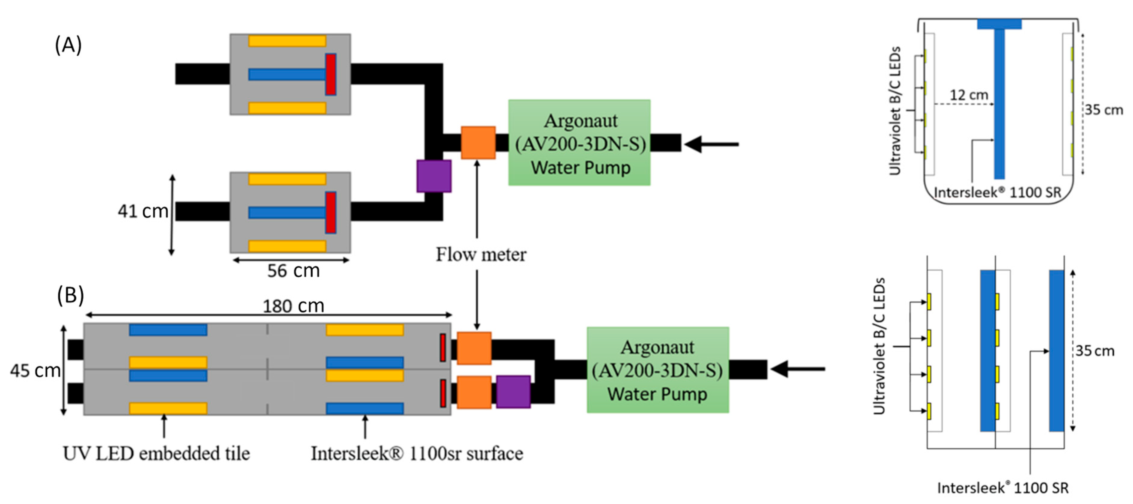
Figure 1. Test-chamber schematic for (A) Hartlepool and (B) Melbourne. Each location had two chambers in operation, one with a UV irradiated tile and one with a blank tile as control. Chamber speed was distributed at a ratio of 9:1 for fast: slow flow chambers using flow control levers and based on flow meter readings. Grey = experimental chamber, yellow = UV/control tile, blue = IntersleekTM 1100 SR, red = baffle dispersal, orange = flow meter, purple = flow control, green = water pump, black = pipe work. Cross view of the chamber’s internal setup displayed to the right and a top-down schematic to the left.
Jmse 11 02211 g001
Figure 2. Image analysis of a deployed silicone tile with embedded UV-emitting LEDs, conducted by highlighting broad taxonomic groups and determining percentage coverages. Highlights indicate mussels, amphipod nests and amphipods (pink, orange and blue, respectively). Brown areas that were not highlighted were located on the rear of the tile and did not contribute to the surface community.
Jmse 11 02211 g002
Figure 3. Environmental data from experimental chambers and surrounding waters from March 2021 to March 2022 in (A) Hartlepool, UK and (B) Melbourne, Australia.
Jmse 11 02211 g003
Figure 4. (A) Imaging of biofouling after 1, 6 and 11 months from different surfaces in a fast flow experimental chamber. (B) Imaging of biofouling after 1, 6 and 11 months from different surfaces in a slow flow experimental chamber.
Jmse 11 02211 g004
Figure 5. Biofouling in experimental chambers after deployment for 6 months at Hartlepool, UK.
Jmse 11 02211 g005
Figure 6. Taxonomic abundance expressed as percentage coverage from images of UV irradiated and control surfaces over a 12-month deployment in Hartlepool, UK and Melbourne, Australia.
Jmse 11 02211 g006
Figure 7. Positive and negative Kendall correlations between environmental conditions and taxa from four sampling conditions in Hartlepool and Melbourne. Red = Positive correlation, Blue = Negative correlation. Statistically significant correlations indicated by * = p < 0.05, ** = p < 0.01, *** = p < 0.001.
Jmse 11 02211 g007
Figure 8. Bray–Curtis dissimilarity index representing control and UV-irradiated eukaryote communities at Hartlepool, UK. Non-metric multi-dimensional scale plot for each condition and surface over the experimental duration. Points represent overall community assemblage, and distance between points symbolizes community disparity. The closer the points are, the more similar their community assemblage and vice versa.
Jmse 11 02211 g008
Figure 9. Proportional abundances of the top 31 classes identified at Hartlepool using 18s rRNA sequencing. (A) All data separated into experimental conditions over 12 months (March 2021–February 2022) at Hartlepool, UK. (B) All data separated into control and irradiated exposures over 12 months. Taxa not classifiable to class-level were labelled as unclassified.
Jmse 11 02211 g009
Disclaimer/Publisher’s Note: The statements, opinions and data contained in all publications are solely those of the individual author(s) and contributor(s) and not of MDPI and/or the editor(s). MDPI and/or the editor(s) disclaim responsibility for any injury to people or property resulting from any ideas, methods, instructions or products referred to in the content.