Review

Complex Plastids and the Evolution of the Marine Phytoplankton

1
Institute of Parasitology, Biology Centre, Czech Academy of Sciences, 370 05 České Budějovice, Czech Republic
2
Faculty of Science, University of South Bohemia, 370 05 České Budějovice, Czech Republic
3
Marine Biological Association of the UK, The Citadel, Plymouth PL1 2PB, UK
*
Author to whom correspondence should be addressed.
J. Mar. Sci. Eng. 2023, 11(10), 1903; https://doi.org/10.3390/jmse11101903
Submission received: 27 July 2023 / Revised: 13 September 2023 / Accepted: 14 September 2023 / Published: 30 September 2023
(This article belongs to the Special Issue Marine Phytoplankton and Their Evolution)

Abstract

:
Photosynthesis allows for the formation of biomass from inorganic carbon and therefore greatly enhances the amount of organic material on planet Earth. Especially, oxygenic photosynthesis removed a major bottleneck in the formation of biomass by utilising ubiquitous water (H2O) and CO2 molecules as raw materials for organic molecules. This, over billions of years, shaped the world into the form we know today, with an oxygen-containing atmosphere, largely oxygenated water bodies and landmasses consisting of sediment rocks. Oxygenic photosynthesis furthermore enabled the evolution of aerobic energy metabolism, and it would be very difficult to imagine animal (including human) life in the absence of molecular oxygen as an electron acceptor. Oxygenic photosynthesis first, and exclusively, evolved in cyanobacteria. However, eukaryotes also learned to photosynthesise, albeit with a trick, which is the integration of formerly free-living cyanobacteria into the eukaryotic cell. There, the former bacteria became endosymbionts, and from these endosymbionts, the photosynthetic organelles (termed plastids) evolved. In almost all major groups of eukaryotes, plastid-containing members are found. At the same time, plastid-related features also indicate that these plastids form a monophyletic group. This can be explained by the transfer of plastids between the eukaryotic super-groups, leading to plastids being found in groups that are otherwise non-photosynthetic. In this chapter, we discuss the evolutionary origin of plastids, with a special emphasis on the evolution of plankton algae, such as diatoms or dinoflagellates, who acquired their plastids from other photosynthetic eukaryotes.

1. Origin of Photosynthesis

Photosynthesis is a process in which carbohydrates (glucose) are synthesised from water and carbon dioxide in a redox reaction, utilising the energy from sunlight. Photosynthesis is considered to be the most important biological process for the existence of life on Earth. There are two kinds of photosynthesis: viz., oxygenic or anoxygenic photosynthesis, of which the latter is done by anaerobic green and sulphur bacteria [1], which have photosynthetic pigments called bacteriochlorophylls. Instead of using water to reduce carbon dioxide to carbohydrates, green and purple sulphur bacteria use hydrogen sulfide and no oxygen is released. Green plants, algae and cyanobacteria are the organisms responsible for oxygenic photosynthesis, which uses water to reduce carbon dioxide. The critical difference between them is that molecular oxygen is generated during glucose synthesis in oxygenic photosynthesis, whereas none is released in anoxygenic photosynthesis of the bacteria. Oxygenic photosynthesis is most important because the production of oxygen from photosynthesis has transformed our primitive anoxygenic atmosphere on Earth into the oxygen-rich atmosphere of today, although this did not happen immediately [2,3]. But supposedly this happened exactly once—in the last common ancestor of extant cyanobacteria, which are the only prokaryotes that perform oxygenic photosynthesis. Eukaryotes, which emerged at 1.78–1.68 Gyr ago, are predated by these photosynthetic prokaryotes to 2.15 Gyr ago [4,5]. Geider et al. [6] proposed that 258 billion tons of carbon dioxide are converted annually by plants and algae into biomass through photosynthesis, sustaining virtually all life on the planet.
Scientists had thought that anoxygenic photosynthesis appeared before oxygenic photosynthesis; they also thought that the earth’s atmosphere was devoid of oxygen until about 2.4 to 3 Byr ago [7] (Figure 1). However, a newer study by Cardona [8] suggests that oxygenic photosynthesis may have originated a billion years earlier, which implies complex life had to be present earlier [8,9]. Cardona’s study examined the enzyme called photosystem I (PSI). If the sequences of PSI enzymes involved in the two types of photosynthesis are compared, it is easy to see that the core of the enzyme is different. Cardona calculated how long ago the two genes diverged. He concluded that oxidative photosynthesis first occurred possibly more than 3.4 Byr ago. Cardona postulated that a gene duplication event led to the heterodimerisation of PSI, which occurred after the evolution of water oxidation and was driven by the presence of oxygen. Cardona has suggested that the evolution of Type I reaction centres is inconsistent with a hypothesis that cyanobacteria may have acquired photosynthesis via horizontal gene transfer (HGT) from anoxygenic photosynthetic bacteria [8] (also see [10,11,12]). Most plastid-encoded genes and endosymbiont genes that have been moved to the host nucleus are cyanobacterial in origin, likely from a heterocyst-forming one [13]. In contrast, some genes have high similarity to homologs from Chlamydia, a pathogen. Thus, early eukaryotes possibly contained a Chlamydia-like pathogen that transferred some genes to the host nucleus, in addition to genes acquired from cyanobacteria (see discussion and references in [14]). In contrast, Domman et al. [15] argue that there is no evidence for a chlamydial partner in the establishment of primary plastid endosymbiosis.

2. Origin of Plastids

In photosynthetic eukaryotes, photosynthesis, as well as other important metabolic pathways, take place in organelles called plastids (or chloroplasts) [16]. Plastids evolved from once free-living photosynthetic cyanobacteria, which were engulfed by a non-photosynthetic host (Figure 2), the so-called primary endosymbiosis. Plastid evolution, which led to the evolution of all eukaryotic algae and land plants, has been perhaps the single most important event leading to the diversification of life on earth [17]. Even though organelle evolution by endosymbiosis is often described as an “event” in the context of the origin and diversification of eukaryotes, it rather has to be seen as a continuous process in which the partners are integrated with each other to varying degrees [18,19]. McFadden [14] has summarised these features as follows: The host and symbiont are intimately integrated, morphologically, genetically and metabolically. Nevertheless, the endosymbionts remain partially autonomous because they can still encode and express a number of genes that act in concert with products from genes transferred to the host. In this way, the hosts have greater control over their endosymbionts. This also makes the symbiont non-autonomous, which would keep it from exiting the symbiosis and becoming free-living again. Such a close-knit interaction would be required for selection to favour the host–symbiont partnership over solo existences. Plastids are passed down vertically with each successive generation. Thus, plastids must replicate before the host cell divides. The synchrony of the important feature of the host and the endosymbiont is critical to establish the endosymbiont as a permanent feature of the host cell. If the endosymbionts replicate too quickly, they could take over and possibly kill the host; if too slow, then the host might divide and form daughter cells missing symbionts.
There are some points to thinking of organelles as distinct species in a very special habitat [20], and it is generally not easy to draw a line between endosymbionts and organelles [19]; however, one possible criterion for the status of a host/endosymbiont system is the state of sexual integration: if genetic recombination solely depends on the life- and cell-cycles of the host, then the former symbiont can be seen as a cellular feature of the host, in other words, an organelle [18].
Primary, secondary and tertiary plastids are the three types of plastids known today (Figure 2). They are contained in different types of hosts, with different types of endosymbionts, different amounts of gene transfer from the endosymbiont to the host and different morphologies relative to the number of membranes surrounding the plastid, which correspond to the type of endosymbiosis it represents (Figure 2).

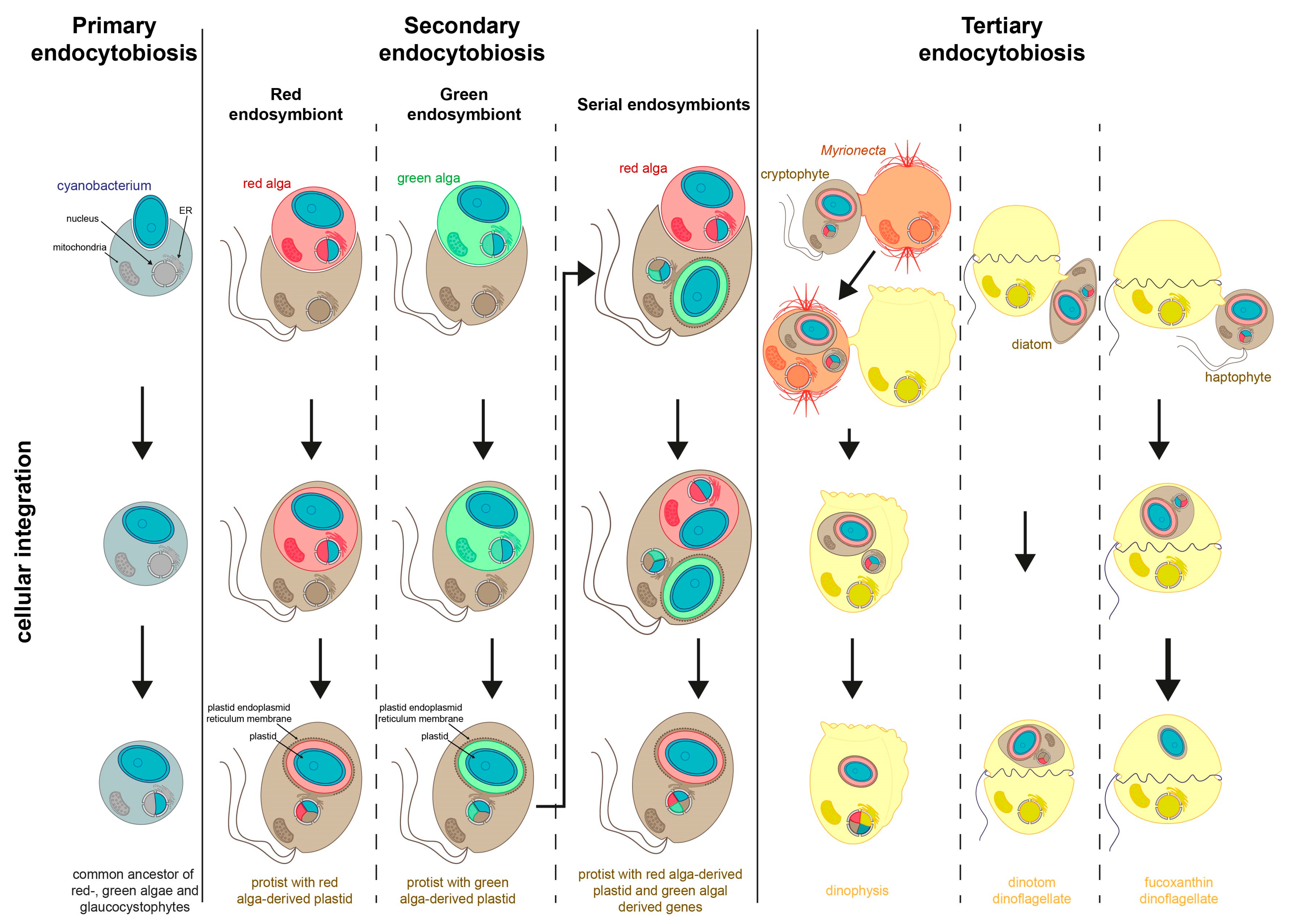
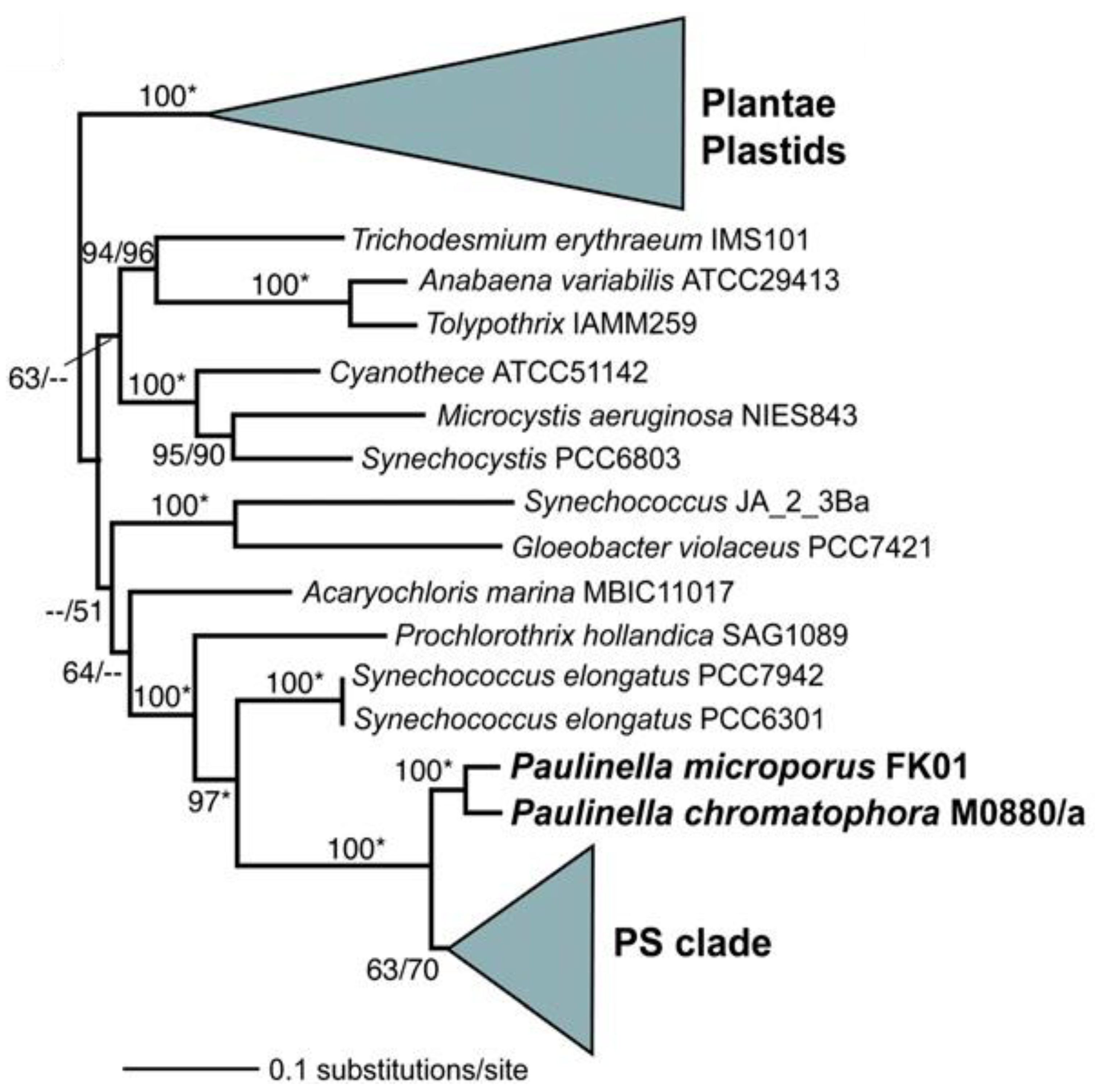
2.1. Primary Endosymbiosis

Primary plastids evolved directly from cyanobacteria by uptake into a eukaryotic cell (Figure 2 and Figure 3). All known primary plastids are surrounded by two membranes, which already puts a question mark on the origin of these membranes; cyanobacteria, like all Gram-negative bacteria, are surrounded by two membranes, and the uptake of the cyanobacteria is commonly thought to have occurred by phagocytosis similar to food uptake, which would add a third membrane to the nascent organelle. If the uptake was via endocytosis, then one of these membranes must have been lost. The interesting question is, of course, which of the membranes have been retained and which have been lost. There are two components of plastids and bacteria that could answer this question: the membrane lipids and the integral membrane proteins. With respect to lipid composition, the outer membrane of Gram-negative bacteria is asymmetric; while the inner leaflet consists of phospholipids, the outer leaflet in addition contains a high proportion of lipopolysaccharides (LPS) [21]. LPS are generally not found in eukaryotes; hence, they are also not present in the outer plastid membrane, leaving the question of the origin of this membrane unanswered. However, with respect to membrane protein content, there is a striking similarity between plastid outer envelopes and bacterial outer membranes, and that is the presence of beta-barrel proteins. Membrane-spanning beta-barrel proteins only occur in these two types of membranes, a strong sign that the outer envelope membrane originated from the outer membrane of the bacterial ancestor of the plastid. Beta-barrel proteins, during their insertion into the membrane, depend on the presence of pre-existing beta-barrel proteins in the target membrane [22]. Cavalier-Smith [23] therefore has argued that the plastid (and mitochondrial) outer membranes are distinctly inherited membranes, which can only be synthesised in the presence of a pre-existing template of the same kind. This would mean that it must have been the membrane of the endocytotic food vacuole that was lost. However, it should be noted that the outer plastid membrane does interact with the eukaryotic endomembrane system in some way, as is evident from the targeting of nucleus-encoded proteins to the plastids via a non-canonical pathway that involves transport through the endoplasmic reticulum [24,25]. Based on this, and on the absence of LPS, the plastid outer membrane is best described as combining features of bacterial outer membranes and eukaryotic endomembranes.
An important feature of cyanobacteria is the cell wall consisting of peptidoglycan, which surrounds the cell in the bacterial periplasm (the space between the inner and outer bacterial membranes) [21]. Primary plastids generally are not surrounded by peptidoglycan layers, with some notable exceptions. One exception is the archaeplastidal group of Glaucocystophyta [26], which therefore for a long time has been regarded as basal among the Archaeplastida, but this view had to be corrected because of the identification of peptidoglycan layers in the moss Physcomitrella patens [27], a member of the green algae and land plant clade of Archaeplastida.
Primary plastids are present in three groups: land plants and green algae (which contain chlorophylls a and b pigments), the red algae, which contain chlorophyll a and phycobiliproteins, and the glaucophyte algae, which contain chlorophyll a and phycobiliproteins [17]. These groups of organisms, as well as their plastids, are considered to be monophyletic. For a long time, the evolution of primary plastids has been believed to have happened only once; however, recent studies of the cercozoan testate amoeba Paulinella chromatophora, which was described by Lauterborn in 1895 [28], show that the chromatophores of this organism (also termed cyanelles), might qualify for organelle status [29] (Figure 3). These discoveries have markedly changed our knowledge of the the evolution of plastid endosymbiosis. In Paulinella, there are photosynthetic symbionts that resemble free-living cyanobacteria because they retain the cyanobacterial peptidoglycan wall [30], and are not bound by a vacuolar membrane. The amount of reduction in the chromatophore genome [31] and also the presence of protein targeting of nucleus-encoded proteins to the chromatophore [32] show that although initially called cyanelles, the chromatophores are really plastids, but with a separate origin: a parallel primary endosymbiotic event [14,29]. There are several striking similarities between the primary plastids of Archaeplastida and the Paulinella chromatophores; however, they must have evolved convergently, which might point to more general mechanisms of plastid evolution. The first similarity is that Archaeplastida, as well as the photosynthetic Paulinella species, largely lost the ability to phagocytise (with only few exceptions among Archaeplastida [33,34]). The second similarity between primary plastids of Archaeplastida and the Paulinella chromatophores is their monophyly; in both cases, speciations occurred that included the organelle/former symbiont [35,36], clearly pointing to sexual integration of the plastids/chromatophores and to the rarity of plastid acquisition compared to speciation. A third similarity is the fact that primary plastids of Archaeplastida and the Paulinella chromatophores are not within the endomembrane system of the host cell but reside freely in the cytosol without the former phagotrophic vacuole (if one was present). Also the cyanobacterial peptidoglycan layer has been retained in Paulinella chromatophores, albeit in reduced thickness as compared to free-living cyanobacteria. In canonical primary plastids, remnants of peptidoglycan cell walls are only found in a few groups (Glaucocystophyta and mosses [27,37]), which implies that the peptidoglycan layer persisted for a long time during the evolution of primary plastids.
Figure 3. Phylogenetic relationship of plastids and cyanobacteria. Note that the rhizarioid Paulinella plastid sequences fall within the cyanobacteria as another primary endosymbiosis. The main primary endosymbiosis organisms are in the clade labelled Plantae Plastids. The numbers at the nodes show support values derived from a RAxML bootstrap analysis followed by those from a PhyML analysis. Asterisks (*) mark 100 % Bootstrap support in both analyses. Only bootstrap values >50 % are shown, when a node is not resolved with a method, this is denoted with dashes (--). Reprinted with permission from Chan et al. [38].
Jmse 11 01903 g003

2.2. Geologic and Atmospheric Chemistry Context of Primary Endosymbiosis

Photosynthesis has greatly influenced geochemical cycles and has contributed three times more energy to geochemical cycles than Earth’s internal heat engine. Rosing et al. [39] have hypothesised that the energy from photosynthesis modified Earth’s geochemical cycles to such an extent that vast amounts of granite were produced during the earliest Archaean through weathering. Granite had led to the initial stabilization of the continents on Earth. Stable continents did not form during the earliest part of Earth’s history. Rosing et al. maintain that biological forcing of weathering and diagenetic rock alteration caused the rise of the continents. Photosynthetic energy fixation provided the power to drive this biological forcing.
Photosynthesis produces an ocean–atmosphere system that is not in equilibrium with Earth’s crust, and this forces weathering of the crust. Without photosynthesis, life cannot influence Earth’s carbon cycle in any significant way. Rosing et al. [39] suggest that evidence for extensive bioactivity and management of the carbon cycle by life through the 3800 Myr of the geologic record is a reflection of how far back photosynthesis can be documented in the fossil record and how far back it influenced the formation of the continents (see discussion and references in [39]).

2.3. Geochemical Consequences of Primary Endosymbiosis

The air and the oceans were anoxic before photosynthesis, and oxygen was toxic to all organisms living before the evolution of free oxygen [7]. After cyanobacterial oxygenic photosynthesis evolved, several million years passed before the oxygen-rich atmosphere of today was stable [40]. Initially, oxygenic photosynthesis was coupled to the carbon cycle through the burial efficiency of organic matter in the lithosphere. Then, the nitrogen cycle was fundamentally altered, allowing ammonium to be oxidised to nitrate and subsequently denitrified [41]. Once this was established, more complex life forms evolved.
Because photosynthesis, coupled to respiration, regulates the carbon cycle, this cycle has had profound effects for the control of Earth’s surface temperature and the budgets of atmospheric carbon dioxide and methane, which in turn established global temperatures, weather patterns and likely caused major glacial events [7]. Different forms of primitive photosynthesis have affected climate features [7]. Bendall et al. [7] discussed that a hybrid ecosystem of H2-based and Fe2+-based anoxygenic photoautotrophs-organisms that perform photosynthesis without producing oxygen gives rise to a strong nonlinear amplification of Earth’s methane (CH4) cycle and would thus have represented a critical component of Earth’s early climate system before the advent of oxygenic photosynthesis.
Ozaki et al. [40] used a global redox balance model to investigate the impacts of different primitive photosynthesis systems on biogeochemical cycles, which in turn affected the early climate of Earth. They found a hybrid ecosystem with both H2-based and Fe2+-based anoxygenic photoautotrophs. These organisms could increase Earth’s methane (CH4) concentration, which would increase this compound in Earth’s early climate system before the advent of oxygenic photosynthesis. They concluded that a hybrid photosynthetic biosphere increased the range of geochemical conditions that favoured warmer climates well in excess of either system on its own. Their results suggest that the Earth’s early climate was governed by many factors, most of them as yet unknown, that linked the anoxic biosphere to H, C, and Fe cycles so that habitable areas for primitive photosynthetic life were increased. As oxygenic photosynthetic productivity increased, it gained control of the budgets of atmospheric carbon dioxide and, eventually, methane too. These controlled global temperatures, weather patterns and likely caused some of the major glacial events, when much of the Earth was covered in ice.

2.4. Genome Reduction of Symbiont, Transfer of Genes to the Nucleus and Retention of Genes in the Plastid

The cyanobacterial endosymbiont introduced a vast amount of new genetic material into the host. Larkum et al. [42] have suggested that hosts have apparently picked through this new genetic material like a “bargain hunter at a trash and treasure outlet”. Some choices were strategic acquisitions, whereas others were more unconventional [13,43]. About 100–200 genes are found in modern plastids in contrast to the several thousand genes found in free-living cyanobacteria [43]. Although most endosymbiont genes were lost, many genes were transferred to the host nucleus (see references in [43]) and increased metabolic and genetic integration of the endosymbiont and host cell. Of the many genes lost from the symbiont genome during evolution, about ca. 1500 of the genes have been transferred to the host nucleus by endosymbiotic gene transfer (EGT). These genes are translated in the host cytosol, but their products (proteins) are targeted to the plastid, where they are needed for photosynthesis and other plastid functions [43,44].
Reasons why the transfer of genes to the host nucleus were advantageous to the host cell include (1) the endosymbionts became a pure clonal line with no opportunity for genetic exchange, (2) those genes transferred to the host nucleus, however, became diploid and could recombine according to host genetics, and (3) those genes transferred to the nucleus, as a result of spatial separation, were likely protected from damage by reactive oxygen species produced during photosynthesis [45]. How DNA is released from the endosymbiont and then integrated into the host nucleus is unknown, but it is generally assumed to have occurred with the rupture of the symbiont and random integration of pieces of DNA into the host nuclei by nonhomologous ends joining into chromosome breaks. The reverse movement of DNA from host into plastid is very rare (see discussion and references in [14]).
Overall, the gene products of essential plastid-encoded genes that were lost from the plastid genome had to be provided by pre-existing nuclear encoded counterparts, which also implies that a protein-targeting system for such proteins existed [14,46]. The existence of genomic redundancy and functional protein targeting leads to a ratchet-like mechanism recognised by Doolittle [47], which over time leads to more and more genes being transferred from a symbiont/organelle genome to the nucleus. This leads to the question of what limits the gene transfer from organelle to nuclear genomes. This question can be answered on two levels: One level is with respect to the probability of the actual gene transfer, which mechanistically requires multiple symbionts/organelles per host cell in order to ensure survival of the organelles in case of lysis of one organelle in the cell. In most groups of algae, however, the number of plastids is highly regulated and synchronised with the cell cycle, drastically reducing the number of gene transfers to the nucleus compared to unsynchronised symbionts (the “monoplastidic bottleneck”) [48]. The other level on which the question of what limits organelles to nucleus gene transfer can be answered is related to the function of the gene product of the transferred gene; this can be caused by constraints in targeting but most prominently in the requirement of redox-regulated gene expression; genes which require tight regulation would lose the regulatory mechanisms for their transcription and translation once they are transferred to the nucleus [45,49].

2.5. Theories for the Relationship between Host and Symbiont

In the conversion of the cyanobacterium to a plastid, either the host (outsider model) or the endosymbiont (insider model) can drive the evolution of the plastid [50]. The cyanobacterial forerunner of plastids probably secreted products from photosynthesis into the cytosol of the host, which were then used for its nutrition [51]. Therefore, for the host to survive, it was critical to maintain the fitness of the cyanobacterium. Gross and Bhattacharya [50] propose that a nucleus-to-organelle flow of biogenetic information was established through the progressive ability to direct nuclear-encoded proteins synthesised in the host cytosol to specific subcellular locations within the plastid or endosymbiont; this is the outside model. Thus, the progressive transformation of the free-living cyanobacterium endosymbiont into an organelle means that the host nucleus must have increased control over the function of the cyanobacterium. A discussion of some of steps needed to manage this control can be found in Chan et al. [38].

2.6. Ecological and Diversity Consequences of Endosymbiosis

Nowack and Melkonian [52] note that endosymbiosis is an evolutionary strategy of hosts, which were initially heterotrophic, to acquire novel biochemical functions and thus is an important source of genetic innovation that enables the host to diversify into new niches. Hengeveld and Fedonkin [53] suggest that evolution progresses modularly, such that existing processes are not changed but are used as interchangeable modules of newly formed processes. In this manner, the process can enable the host to invade new ecological niches and modify the original set of modular processes as needed. While a large number of hypotheses/theories for the explanation of eukaryote evolution by endosymbiosis have been suggested [54], it is important to test these hypotheses independent of individual gene phylogenies, for example by considering homologies in protein import mechanisms across the cell/organelle membrane [55].
Photosynthetic eukaryotes present an exceptional diversity of primary producers that provide most of their biomass to the other organisms on Earth. This can also be seen in the extensive exploitation of temporary plastid acquisition by animals and heterotrophic protists, which also instantly provides photosynthate to an enormous range of organisms, from protists to corals to molluscs [56,57].

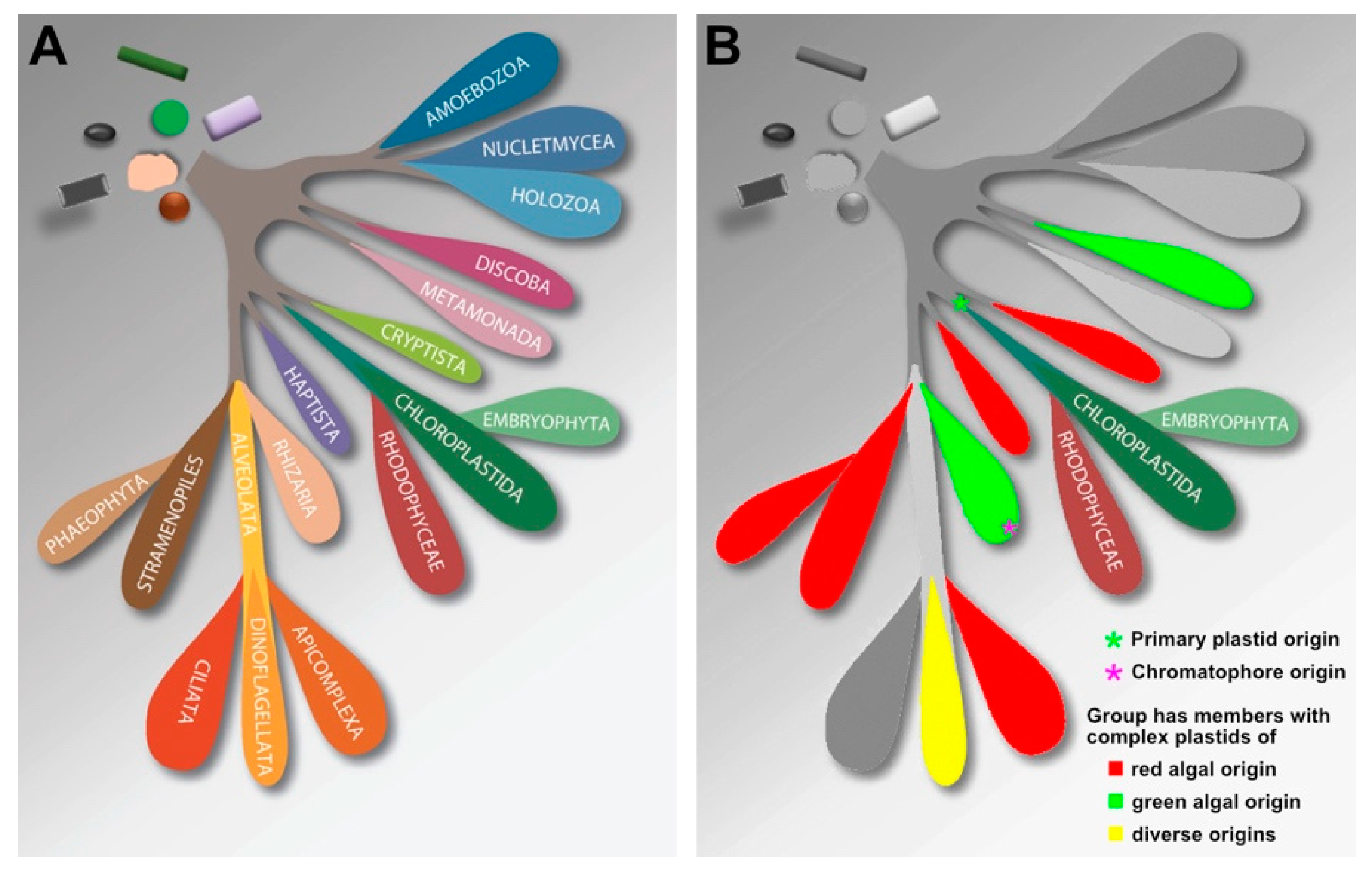
2.7. Diversification of Archaeplastida and Relationship to Other Eukaryotic Groups

In the evolution of Archaeplastida from green algae to bryophytes, ferns and higher plants, there is a progression to evolve more elaborate carbon-concentrating mechanisms (CCM), such as the pyrenoid with its high concentration of RUBISCO [58,59] and C4 type of photosynthesis in some land plants, to bring CO2 closer to the photosynthesis reaction centre. C4 photosynthesis replaced the CCMs in those plants that have modified their morphology to perform C4 photosynthesis. Land plants also evolved to package and display their photosynthetic units in a wide array of structures combined with a diversity of mechanisms for capturing other limiting resources [60].
Members of the “green” plastid lineage are much more closely related by plastid phylogeny and photosynthetic physiology than are the host cells, although there is a strong phylogenetic signal for monophyly of the red + green lineages. This phylogenetic signal is four-fold stronger when multiple rhodophytes whole genomes are included in the phylogenetic analyses [38] By adding 60,000 novel genes from Porphyridium cruentum and Calliarthron tuberculosum to the analyses, it has been shown that ca. 50% of the red algal genes are shared with other eukaryotes and prokaryotes. It is most likely that these shared genes have been introduced by EGT, and their duplication was the means for extending their biological functions [53].

2.8. Ecological Niches and Roles of Red vs Green

One important evolutionary process that generates biodiversity is adaptive radiation, through which a species lineage diversifies rapidly to occupy available niches within a habitat [61]. Algae are composed of organisms with deep branches in the eukaryotic lineage corresponding to an evolutionary time span of roughly 1.5 billion years [62]. Also, unlike land plants, which are a monophyletic clade, algae are polyphyletic, being distributed throughout the eukaryotic tree of life. They do not have a single common ancestor in the traditional sense but are related through endosymbiosis that resulted in the transfer of plastids and genes to various eukaryotic hosts and created distinct lineages of algae outside of Archaeplastida (the phylogenetic clade to which photosynthetic eukaryotes with primary plastids reside). Many algae are more closely related to non-photosynthetic protists than they are to other algae [62]. The benthic red macroalgae today occupy the intertidal to great depths, and the planktonic red microalgae are mainly oceanic in their distribution. The green macroalgae are also intertidal but usually occur in the high intertidal and not at depth like the red macroalgae. Green microalgae are more or less limited to coastal near-shore environments in the modern ocean today but were the dominant member of the phytoplankton before the Permian/Triassic extinction (see Section 3.4).

3. Secondary Endosymbiosis

Secondary plastids are bounded by more than two membranes and represent the acquisition of a photosynthetic eukaryote (either a green or a red alga) by a non-photosynthetic eukaryotic host [62,63,64,65,66] (Figure 2). Each of these new eukaryote cells has three to four membranes around the plastid, each one representing a different membrane in the endosymbiosis ranging from the two membranes of the primary plastid and its cell membrane to two additional membranes of the host representing its cell membrane and the phagosomal vacuolar membrane [14] (Figure 2 and Figure 4). There can be a reduction to three membranes surrounding the secondary plastid, and there is normally a loss of the photosynthetic eukaryote nucleus [17]. Also, Melkonian [67] suggested that the outer membrane of complex plastids was originated from an autophagosomal membrane. Gould et al. [68] hypothesised that the outer two membranes were derived from an ER membrane. These lineages today include the haptophytes; the stramenopiles of which the diatoms are a major component of the marine phytoplankton; the cryptophytes; the dinoflagellates, in which the engulfed cell was a red alga; and the euglenophytes and the chlorarachniophytes, where it was a green algal cell (Figure 3). In the cryptophytes and the chlorarachniophytes, the photosynthetic eukaryotic nucleus (of the endosymbiont) is reduced and retained as a nucleomorph between two outer membranes surrounding the plastid [69,70]. Euglenophytes, chlorarachniophytes and the dinoflagellate genus Lepidodinium possess plastids that are derived from independent endosymbiotic relationships with a green alga [71].

3.1. Diversification of Secondary Plastids

The red algal primary endosymbiosis lineage (the rhodophytes) includes many features of the cyanobacterial pigmentation; however, the diverse set of phytoplankton whose plastids are evolutionarily derived from the endosymbiosis of a red alga [14,17,62,63] (Figure 2 and Figure 4), utilizes chlorophyll c and its derivatives as accessory photosynthetic pigments [72]. Of the eight major eukaryotic phytoplankton taxa in the modern ocean, all but euglenophytes and the chlorarchniophytes possess “red” plastids; however, these two lineages are not very numerous in the plankton, being mostly benthic intertidal. Thus, the red algal secondary endosymbiosis gave rise to the stramenopiles, of which the diatoms are a major phytoplankton group; the haptophytes; the dinoflagellates; and the cryptomonads. These groups are the major members of the modern marine microphytoplankton. The plastid genome is a circular genome in all plastids, with most photosynthetic genes transferred to the nucleus. In the peridinin dinoflagellates, the circular genome is broken up into minicircles of DNA containing only a few genes (see in-depth discussion and references in [71,73]).
Figure 4. Details of the membranes associated with the various stages of endosymbioses and the transfer of genetic material from the endosymbiont as well as from external organisms. (A) primary endosymbiosis; (B) secondary endosymbiosis; (C) tertiary endosymbiosis. Reprinted from Archibald [74] with permission.
Jmse 11 01903 g004

3.2. Discussion of Number and Timing of Secondary Endosymbiosis

A good resolution of the phylogeny of the red algal-derived plastid with chlorophyll c has been difficult to obtain. This supergroup, termed Chromalveolata by Cavalier-Smith [75], contains the alveolate (ciliates, apicomplexans, dinoflagellates) and chromist (stramenopiles, cryptophytes, haptophytes) protist lineages. Cavalier-Smith [75] hypothesised that these taxa shared a single, ancient red algal secondary endosymbiosis because the genomic changes to convert a eukaryotic endosymbiont to a plastid were deemed to be too complex for it to have happened more than once. Consequently, ciliates and the basal, plastid-lacking, cryptophyte lineage Goniomonas must have secondarily lost their plastids. The monophyly of the Chromalveolata has been repeatedly tested by many groups with initial multigene analyses that support it, albeit weakly in the host phylogeny (see discussion in [63]), but there are several studies that dispute its monophyly [76,77,78]. Whereas the chromealveolates hypothesis appears reasonable at first sight, the biggest challenge to it comes not from molecular phylogenies but from the existence of various heterotrophic sister groups to all the major groups of photosynthesisers that were assigned to the group of chromealveolates, which would also imply that if the plastids resulting from secondary endosymbiosis were monophyletic, then all the non-plastidic and heterotrophic sister groups must have lost their plastids independently (Figure 4). The question of chromalveolate monophyly, the branching order of its major clades, and the history of plastid endosymbiosis in its photosynthetic members has been discussed in greater detail recently [35,63]. Here, we would just like to highlight two lines of evidence that convincingly argue against chromealveolates monophyly: (i) Baurain et al. [76] concluded that if the chromalveolate hypothesis were true, the phylogenetic signal in support of it should be similarly strong across the nuclear, plastid and mitochondrial genomes. This however is not the case, pointing to more complex scenarios involving transfers of plastids between eukaryotic groups [76]; (ii) Strassert el al. [79] showed in molecular clock studies that the branching between the different groups of algae in which plastids of red algal origin are found occurred earlier than the diversification of the Archaeplastida and the formation of red algae, which would make an endosymbiotic acquisition of a red algal plastid in a common ancestor impossible because of the non-existence of red algae at that time in which their later hosts diversified [79].
Taken together, it appears certain that complex plastids of red algal origin were, further to their initial acquisition by secondary endosymbiosis, horizontally transferred between eukaryotic phyla in higher-order endosymbioses, including serial replacements of plastids. The exact path of all these plastids, however, remains an unresolved problem at this time.

3.3. Ancient Green Endosymbiosis

To complicate further the endosymbiosis story, there is evidence that some of the lineages with the red algal plastid may have originally had a green plastid, viz., the diatoms, which was later exchanged for a red algal plastid [80]. Analysis of the completed genomes of the diatoms, Thalassiosira pseudonana and Phaeodactylum tricornutum, shows hundreds of genes to be derived from green algae [80] (in particular prasinophytes, which are the group of microalgae most commonly found in neritic nanoplankton). These results complicate possible scenarios of diatom evolution because it implies that an ancestral cell contained a green algal endosymbiont before the current red algal plastid (Figure 2). These green algal genes coming from endosymbiotic or horizontal gene transfer serve as a footprint of this ancient event [81]. A similar finding has now been reported for the haptophytes [80]. But to complicate matters even more, the chloroarachniophyte Bigelowiella natans contains many green algal-derived nuclear genes as well as many red algal-derived, plastid-targeted proteins [81]. This would suggest that both plastids co-existed for a time and that in this microalgal lineage, the red plastid was lost and the green one retained. Similarly in the green alga-derived plastids found in the dinoflagellate Lepidodinium chloroporum, there are nuclear genes that encode plastid-targeted proteins from multiple different algal lineages [82]. These data support the hypothesis that secondary endosymbioses happened more than once and that endosymbiotic horizontal gene transfer is more common than expected.
Assuming that the chromalveolate hypothesis of a single endosymbiosis leading to all red-algal derived plastids is true, several studies have tried to estimate the time this event occurred in the history of Earth [80]. Bhattacharya and Medlin [83] have noted that the early timing of these secondary endosymbiosis events (at 1 Ga, see references in [83]) does not match the first appearance in the fossil record of the phytoplankton that are the modern components of the red algal secondary endosymbiotic event but is obtained if fossil dates are used from outside of the phytoplankton, e.g., the red algae. Using these fossil dates from the phytoplankton to constrain the molecular clock for the diatoms, the haptophytes and the dinoflagellates, Bhattacharya and Medlin [83] suggested that there was a radiation of these groups at the P/T boundary (250 MYA). It was likely at this time that the green plastid now predicted to have been initially present in these microalgae was replaced by a red alga because of the adaptive advantage that the plastid type conferred on the host cell [62]. Medlin [84] also noted that the green plastid endosymbiosis in the “chromalveolate” host lineages must be older than the red plastid endosymbiosis because there are four times more green genes retained in the host nucleus than there are red genes [80].
Chan and Bhattacharya [85] question the advantage of independent endosymbiotic events. They state that the least complex and therefore most parsimonious explanation for an observation is generally to be preferred in biology. Accepting multiple endosymbiosis events avoids a complicated explanation of organelle and gene losses to explain the random plastid distribution among non-photosynthetic chromalveolates.

3.4. Permian/Triassic (P/T) Extinction and the Green vs. Red Switch in the Ocean

With the volcanic eruptions at the P/T boundary, ocean chemistry changed, making Fe more abundant. Falkowski and Oliver [86] have hypothesised that this water chemistry difference favoured the radiation of the phytoplankton, with a red algal plastid replacing those cells with a green algal plastid, making the red algal plastid lineages the dominant algal groups in the marine phytoplankton. Fe-containing cytochrome c6 is present in the red plastid-bearing microalgae, whereas green plastid-bearing algae have Cu-containing plastocyanin.
Trace-element chemistry is driven by hypoxic conditions, which selected for red plastids in the nascent host cells. Green plastid lineages contain large iron, zinc and copper quotas. Red algae (rhodophytes) and all algae with complex plastids of red algal origin (viz., red plastid–containing dinoflagellates, coccolithophorids and diatoms) have higher quotas for manganese, cobalt and cadmium. These trace metal preferences resulted in a niche partitioning between the green and red plastid lineages, with the former more successful offshore and the latter flourishing in coastal benthic habitats with their anoxic conditions. The genes responsible for most trace metabolism are no longer found in extant plastid genomes but were transferred to the host-cell nuclear genome early in endosymbiosis and are related to the redox history of the ocean [62,84,86].

3.5. Eukaryotic Nuclei Reduction and Gene Transfer

There are many theories that have tried to resolve why some organellar genes are transferred to the nucleus and others remain. Allen and Raven [87], Martin and Herrmann [44] and others have suggested that mutation by oxygen-free radicals and Muller’s ratchet effect of nonrecombining genomes are the most logical explanations. Core subunits of the photosystem, cytochrome b6f, and ATP synthase complexes (atpA, atpB, petB, petD, psaA, psaB, psbA–E, psbI) are the few genes that remain in the plastid. Their retention in the plastids supports the CORR hypothesis of Allen [45,49]. His hypothesis suggests that core subunits of the photosystem remain encoded in the plastid, close to the functional site of the proteins, which allows the organism to maintain close control of the plastid’s redox potential so that the cell can respond quickly to changes, maximize efficiency and minimize the creation of harmful free radicals.

3.6. Genome Evolution Consequences of Higher Order Endosymbioses

In secondary and higher order endosymbioses leading to organisms with complex plastids, the original nucleus of the symbiont, along with the original symbiont mitochondria, disappears (except for organisms that retain a nucleomorph). The transfer of nucleus- encoded symbiont genes to the nucleus of the host cell is in principle easier than the transfer of primary plastid/bacterial genes because the genes already come with features of eukaryotic genes, including promoters and terminators. However, one particular part of the coding sequence becomes useless (in the best case) or potentially harmful: the targeting signal [63,88]. Upon transfer to the host nucleus, translation of the transferred gene is initiated in the cytosol, where most targeting signals cannot be processed in the same way as in the symbiont cell. The acquisition of a targeting signal therefore in many case leads to rearrangements in the cellular metabolism, which may then be shuffled between cellular compartments [63,88]. Examples of such rearrangements are the presence of a partial glycolysis in the mitochondria of stramenopiles (including non-photosynthetic ones) [89], the presence of an Entner Doudoroff pathway in diatom mitochondria [90], or the import of de-novo synthesised nucleotides to the plastids of diatoms [91].
Because the loss of a symbiont gene (the second part of gene transfer between nuclei) requires a pre-existing replacement, algae with complex plastids seem to be masters of acquiring genes from other sources than the symbiont [88]. Accumulation of horizontally acquired genes from other algal groups than the plastid progenitors has been observed not only in diatoms (see above) but also in cryptophytes and chlorarachniophytes [92].

3.7. Convergent Evolution of Nucleomorph Structure in Chloroarachniophytes and Cryptophytes

Nucleomorphs are remnants of algal nuclei within the periplastidal membranes of both cryptophyte and chloroarachniophytes plastids [93]. These plastids have four membranes, which are the result of the endosymbiosis of a red or green algal ancestor, respectively (Figure 2). Just as in a normal nucleus, the nucleomorph has linear chromosomes and is surrounded by a nuclear envelope with pores. Their genomes are much reduced (ca.<1 Mbp}, making them the smallest nuclear genomes known. A ‘core set’ of housekeeping genes is present in these very compact and gene dense organelles, making up for the majority of the encoded genes. Strikingly, there are only 17–30 plastid-associated genes, with all others having been transferred to the host nucleus during the original endosymbiosis, or lost. Thus, in these two plastids, gene content and genome structure are similar despite having evolved independently, one with a red plastid and the other with a green one. In the dinoflagellate lineages that converted a cryptophyte into a kleptoplastid, the nucleomorph is lost [71].

3.8. Relationship of Membrane Number to Protein Targeting

About 200 proteins are encoded in the plastid, whereas a thousand or more proteins are nuclear-encoded, of which many but not all are clearly cyanobacterial in origin [43,44]. These are needed to maintain a fully functional plastid. A dedicated protein import apparatus moves the proteins translated on the cytoplasmic ribosomes into the plastid. This system must be established for the endosymbiont to be completely transformed into an organelle. Therefore the progressive transformation of the endosymbiont into an organelle includes an increasing governance of the host nucleus over cyanobacterial functions [43,44]. In cells with complex plastids of red algal origin, the targeting signals and mechanisms are remarkably similar between the different groups [16,94,95]. Generally, complex plastids are part of the endomembrane system of the cell. In the case of four membrane- bound plastids of red algal origin, the first transport step for nuclear-encoded plastids is usually co-translational import into the endoplasmic reticulum (ER), mediated by a signal peptide that is exchangeable with the signal peptide of other ER proteins [96,97]. The following steps of plastid protein import require a second N-terminal targeting domain immediately downstream of the signal peptide, which is thought to correspond to the chloroplast transit peptide of the original symbiont [96,97]. Such targeting signals are therefore often referred to as bipartite targeting signals [94]. A special role comes to the very N-terminus of this downstream transit peptide, in the +1 position of the signal peptide cleavage site. The amino acid residues in this position are conserved in a way that only phenylalanine, tryptophan, tyrosine or leucine can be found in this position in diatoms [97,98]. Similar motifs can be found in other groups of algae with complex red algal-derived plastids as well [95], and seem to have been inherited from the red algal plastid ancestors [37,95].
Following the co-translational insertion into the ER surrounding the plastid, the proteins must exit the ER through the second membrane surrounding the complex plastid, the periplastidial membrane, which corresponds to the cytoplasmic membrane of the former symbiont. It has been suggested that this is accomplished by re-purposing of the ERAD system, a system that in eukaryotic cells is responsible for the export of misfolded ER proteins for degradation in the cytosol [99]. There is increasing evidence for the involvement of an ERAD-derived transport machinery in protein transport to the complex plastids [100,101], and the sharing of phylogenetically traceable components of this transport system has been considered as support of the monophyly of the algae with complex plastids of red algal origin by Cavalier-Smith [102]. It should be noted that the genes for the protein transport system could also have been transferred horizontally together with the plastids via tertiary eukaryote–eukaryote endosymbioses. This possibility, however, is declared to be highly unlikely by Cavalier-Smith [102].
The remaining transport steps of nuclear-encoded plastid proteins en route to the four membrane-bound plastids of red algal origin are homologous to the plastid protein import into secondary plastids the two innermost envelope membranes contain homologues of the Toc/Tic (translocator of the outer/inner chloroplast membrane) system [16,101].

4. Tertiary Endosymbiosis

The dinoflagellates take endosymbiosis one step further. With five types of plastids in this group, each with its own evolutionary history, they are the winners of eukaryotic plastid endosymbiosis (Figure 2) [73]. In several genera, they have replaced the peridinin pigmented plastid, which is believed to be the original red algal endosymbiont and is surrounded by three membranes (the middle envelope membrane is believed to have been lost during evolution). The evolutionary origin of the peridinin plastid remains controversial, with one theory suggesting that it was a red alga (a secondary endosymbiosis [73] but another suggesting that it was a haptophyte, which would make it a tertiary endosymbiosis, not a secondary one [71].
Some dinoflagellates replaced this plastid with another eukaryotic cell (Figure 2), and the number of membranes around the plastid increased initially but in some cases decreased during the evolution of the tertiary plastid. Sometimes this replacement is permanent, and in others it is temporary (see 4.2), which is termed kleptoplasty. The permanent replacement plastids (see [71] for discussion and references) are a green algal plastid in Lepidodinium chlorophorum and Lepidodinium viride, a stramenopile plastid acquired by Kryptoperidinium foliaceum and Durinskia baltica, and a haptophyte-derived plastid in Karenia and Karlodinium spp. Furthermore, cryptophyte-derived plastids are temporary plastids that are continually renewed and can be found in Dinophysis.
In the haptophyte-derived plastids of the Kareniaceae, the plastid story is additionally complicated because the plastid is a combination of the earlier peridinin plastid (e.g., it retains the poly A tail and over 200 proteins) and the newly ingested haptophyte plastid [103]. The final plastid has likely involved extensive HGT and possibly serial endosymbiosis. The plastid donor came either from a chrysochromulinalean or a phaeocystalean cell [103].
In case of the stramenopile-derived plastids, dinoflagellates took up a diatom, forming an endosymbiotic consortium known as a dinotom [104,105,106]. In these cells, the entire diatom with its nucleus and mitochondria, minus its silica cell wall, is inside the dinoflagellate. Uptake of diatoms is via myzocytosis using a peduncle [105], and the endosymbiont’s cytosol is separated from the host by a single membrane, which may be the endosymbiont’s plasmalemma, but may also be a host derived phagosomal membrane. If it was the host’s phagosomal membrane, the endosymbiont could be at least partially digested, however, almost all of the diatom’s organelles are retained. Furthermore, the myzocytotic uptake of diatoms leaves “ghost” cells, consisting of the frustule, and possibly the diatom cell membrane [105]. Interestingly, within the dinotoms, several new aquisitions of endosymbionts took place, and at least one species has a temporary kleptoplastic relationship with its endosymbiont diatoms [105].

4.1. Theories for Relationships of Hosts and Symbionts (Symbiosis vs. Predation)

There are several different types of symbionts, which are defined based on the relationship between the two entities involved. A facultative endosymbiont retains all genes for its own proteins, and to function, it does not need any protein import from the host cell in which it resides. In contrast, an organelle retains only a small fraction of its original gene set. All other required genes are transferred into the host’s nucleus [107]. The proteins of these genes in the host nucleus are translated in the cytoplasm and then imported into the organelle/plastid. Targeting signals and import mechanisms in envelope membranes surrounding the organelle could only evolve in a long-lasting relationship, which then enables the conversion of the symbiont. If the organelles/plastids were the result of predation by the host, then they could not sustain their existence over such long periods of time.

4.2. Kleptoplasts as Intermediate States during Organelle Evolution

Numerous protists and metazoans have adapted functional nutritional modes in which they gained the capacity for phototrophy-mediated carbon acquisition by retaining organelles (plastids) by forming symbiotic associations with algae or predation followed by organelle retention. The latter process is fundamentally distinct from endosymbiosis because it involves the predatory capture of an alga and subsequent removal and temporary maintenance of one or more organelles, including their plastids. The term kleptoplastidy is sometimes used to describe the retention of a plastid from an alga, e.g., [108]; however, in many cases the number of organelles retained, as well as their functionality, has not been sufficiently tested to warrant a conceptual distinction. Many diverse lineages of eukaryotes (Figure 5), including the alveolata, katablepharidophyta, rhizeria, and metazoan, have temporarily acquired the ability to photosynthesise by endosymbiosis or organelle retention [109]. Although true endosymbiosis is by far recognised as a superior ecological and evolutionary process, the temporary enslavement of algal organelles imparts similar metabolic advantages to the hosts. Because the plastids remain functional for a long time, it has been suggested that photosynthetic sea slugs were sustaining their captured plastids, maybe even using proteins that were produced from the alga’s genes that the sea slugs had acquired [56,110,111].
This hypothesis is supported by the finding of plastid genes in the heterotrophic dinoflagellate Crypthecodinium cohnii, and Sanchez-Puerta et al. [113] have suggested that perhaps all colourless dinoflagellates may harbour undetected leucoplasts, as well as many genes for plastid-targeted proteins, that came from the original peridinin-containing plastid in Karlodinium micrum, a dinoflagellate with plastids derived from a secondary endosymbiont haptophyte host [114].

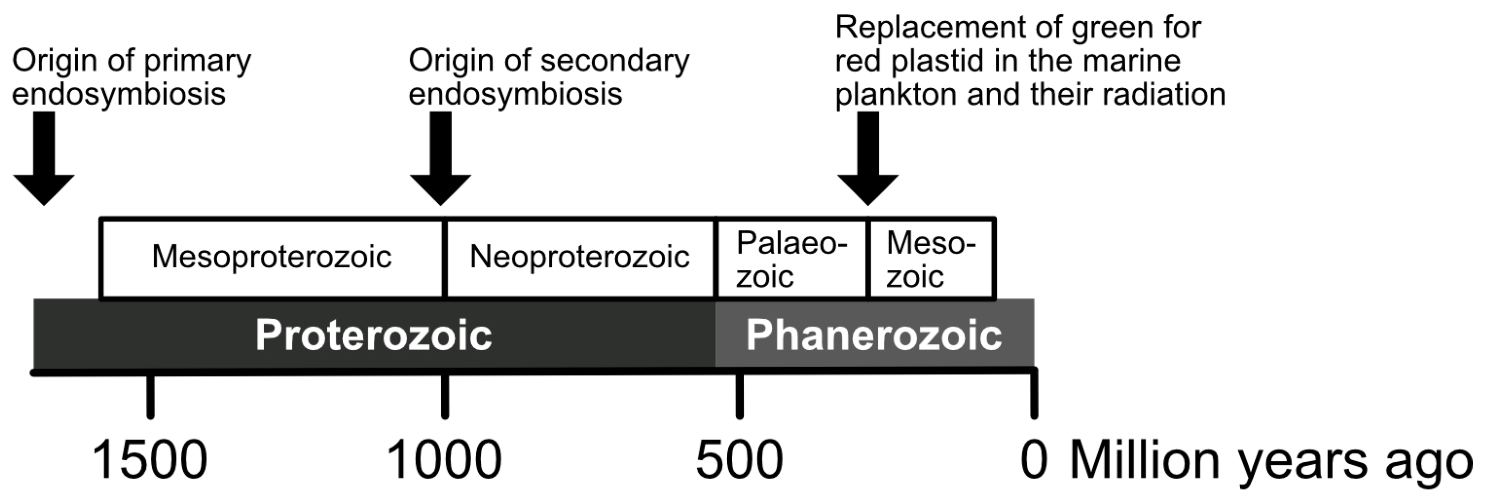
5. Molecular Clocks for the Timing and Diversification of Complex Plastids

Molecular clock approaches to dating plastid endosymbiosis are similarly controversial as the number and order of endosymbioses (Figure 1 and Figure 2). Fossil-dated phylogenies identify a window between 1.4 and 1.7 bya [115,116] for the last common ancestor of all plastids, whereas a more recent date of 0.9 bya has been estimated by the use of cross-calibrated Bayesian estimates for duplicated ATP synthase genes [117]. Unfortunately, only the most durable parts of cells (especially mineral cell walls or their parts) are preserved as fossils, and conclusions on the cell structure or genetics of fossil organisms are always indirect. Furthermore, the classification of cell walls is also challenging, as can be seen by the recent re-evaluation of the oldest diatom fossils that were previously considered diatoms of the Triassic–Jurassic boundary (ca. 200 Ma) and the Middle Jurassic age (174–163 Ma). These fossils turned out to not be remains of diatoms, thereby challenging the calibration of molecular phylogenies [118]. A timeline of the major events in the origins of the plastids is shown in Figure 1.
Most recently, dates based on fossil infochemicals, called protosterols, corroborate these early dates for the LCA of all eukaryotes [119]. These bio-markers were detected in deep and relatively shallow water environments, microbial mats and pelagic habitats, shales and carbonates, as well as marine and likely lacustrine basins, with an absence of or extreme scarcity of crown-group eukaryotes in open-water habitats. Thus, phytoplankton first evolved in near-shore environments and later expanded into oceanic habitats [119].
Phylogenies with both plastids and cyanobacteria have placed plastids either at the base of the cyanobacterial radiation [120] or sister to coccoid cyanobacteria [121], with the clade containing the heterocyst-forming, nitrogen-fixing Cyanothece [122], or with heterocyst-forming filamentous cyanobacteria [13] (Figure 4).
Dagan et al. [123] compare gene inventories and found that plastids share the most genes with filamentous, heterocyst-forming bacteria. They argue that nitrogen fixation, along with the alchemy or life perpetuation of photosynthesis, was an early driver for endosymbiosis but became less important to the partnership with increasing nitrate abundance [123]. An examination of the evolution of the various Rubisco proteins suggests that the first Rubisco may have evolved in a methanogen-like bacterial ancestor, and its function changed as it spread through the bacteria by lateral gene transfer [124]. The search for the closest relative to all plastids is compromised because there have been many lateral gene transfers among cyanobacteria after the primary endosymbiosis [14].
The End-Permian mass extinction marked a major transition in ocean ecosystem structure. Extensive anaerobic conditions were widespread during the extinction and lasted for several million years afterwards as evidence by the large carbon-isotope excursions and the deposition of extensive black shales in Early Triassic shelf sea settings [125,126]. What would have given the “red lineage” groups an adaptive advantage at this time to initiate their radiation? It was most likely the change in trace metal chemistry [62]. Mass extinctions likely provided ecological opportunities for the establishment of new clades [84]. Consequently, there must have been selective advantages for heterotrophic cells to acquire and retain a plastid. Most plastids largely (but not exclusively) descended from red algae and were retained by the new host cells once the oceans became oxic again. But note that the sterol profile of the chlorophyll c algae (= phytoplankton) more closely resembles that of green algae and not red ones [119].

6. Multigene Trees Showing How Many Groups Are in Plankton

Explanations of all gain and loss of plastids in all eukaryotes is still a controversial subject. The most widely accepted part is the origin of primary plastids via endosymbiosis from a heterotrophic host and a cyanobacterium. The origin of secondary plastids still yields contradictory results based on the organisms included, the type of analysis and the number of genes in the study (see discussion in [63]). Subsequent tertiary endosymbioses involving other free-living eukaryotes explain plastid origins in dinoflagellates lineages. What is clear is that the dominant members of the plankton changed after the P/T extinction, with the diatoms, dinoflagellates and coccolithophorids radiating through time to be the dominant members of the phytoplankton today and the green algae and cyanobacteria taking a minor role in selected niches. In the following chapters of this special issue, each important major phytoplankton group is discussed through time and space. Other features necessary to be a member of the phytoplankton also contribute to this special issue. This chapter hopefully sets the stage for describing the origin and evolution of the major groups of phytoplankton in the world’s ocean today.

Author Contributions

Conceptualization, L.K.M.; writing—original draft preparation, L.K.M.; writing—review and editing, L.K.M. and A.G.; visualisation, L.K.M. and A.G. All authors have read and agreed to the published version of the manuscript.

Funding

This research was supported by the Institute of Parasitology of the Czech Academy of Sciences and by the Grant Agency of the Czech Republic (21-26115S). Part of this work was carried out with the support of ELIXIR CZ Research Infrastructure (ID LM2023055, MEYS CR).

Institutional Review Board Statement

Not applicable.

Informed Consent Statement

Not applicable.

Data Availability Statement

Not applicable, no new data generated, review paper.

Acknowledgments

We thank Houda Ouns Maaroufi (Institute of Parasitology, Biology Centre, Czech Academy of Sciences) for drawing Figure 2.

Conflicts of Interest

The authors declare no conflict of interest.

References

  1. Hohmann-Marriott, M.F.; Blankenship, R.E. Evolution of photosynthesis. Annu. Rev. Plant Biol. 2011, 62, 515–548. [Google Scholar] [CrossRef] [PubMed]
  2. Fischer, W.W.; Hemp, J.; Johnson, J.E. Evolution of oxygenic photosynthesis. Annu. Rev. Earth Planet. Sci. 2016, 44, 647–683. [Google Scholar] [CrossRef]
  3. Holland, H.D. The oxygenation of the atmosphere and oceans. Philos. Trans. R. Soc. Lond. B Biol. Sci. 2006, 361, 903–915. [Google Scholar] [CrossRef] [PubMed]
  4. Koonin, E.V. The origin and early evolution of eukaryotes in the light of phylogenomics. Genome Biol. 2010, 11, 209. [Google Scholar] [CrossRef]
  5. Rasmussen, B.; Fletcher, I.R.; Brocks, J.J.; Kilburn, M.R. Reassessing the first appearance of eukaryotes and cyanobacteria. Nature 2008, 455, 1101–1104. [Google Scholar] [CrossRef]
  6. Geider, R.J.; Delucia, E.H.; Falkowski, P.G.; Finzi, A.C.; Grime, J.P.; Grace, J.; Kana, T.M.; La Roche, J.; Long, S.P.; Osborne, B.A.; et al. Primary productivity of planet earth: Biological determinants and physical constraints in terrestrial and aquatic habitats. Glob. Change Biol. 2001, 7, 849–882. [Google Scholar] [CrossRef]
  7. Bendall, D.S.; Howe, C.J.; Nisbet, E.G.; Nisbet, R.E. Photosynthetic and atmospheric evolution. Introduction. Philosophical transactions of the Royal Society of London. Series B, Biol. Sci. 2008, 363, 2625–2628. [Google Scholar] [CrossRef]
  8. Cardona, T. Early Archean origin of heterodimeric photosystem I. Heliyon 2018, 4, e00548. [Google Scholar] [CrossRef]
  9. Elsevier. Photosynthesis Originated a Billion Years Earlier than We Thought, Study Shows. Available online: www.sciencedaily.com/releases/2018/03/180306093304.htm (accessed on 14 July 2023).
  10. Baymann, F.; Brugna, M.; Mühlenhoff, U.; Nitschke, W. Daddy, where did (PS)I come from? Biochim. Et Biophys. Acta 2001, 1507, 291–310. [Google Scholar] [CrossRef]
  11. Raymond, J. The role of horizontal gene transfer in photosynthesis, oxygen production, and oxygen tolerance. Methods Mol. Biol. 2009, 532, 323–338. [Google Scholar] [CrossRef]
  12. Soo, R.M.; Hemp, J.; Parks, D.H.; Fischer, W.W.; Hugenholtz, P. On the origins of oxygenic photosynthesis and aerobic respiration in cyanobacteria. Science 2017, 355, 1436–1440. [Google Scholar] [CrossRef] [PubMed]
  13. Deusch, O.; Landan, G.; Roettger, M.; Gruenheit, N.; Kowallik, K.V.; Allen, J.F.; Martin, W.; Dagan, T. Genes of cyanobacterial origin in plant nuclear genomes point to a heterocyst-forming plastid ancestor. Mol. Biol. Evol. 2008, 25, 748–761. [Google Scholar] [CrossRef]
  14. McFadden, G.I. Origin and evolution of plastids and photosynthesis in eukaryotes. Cold Spring Harb. Perspect. Biol. 2014, 6, a016105. [Google Scholar] [CrossRef] [PubMed]
  15. Domman, D.; Horn, M.; Embley, T.M.; Williams, T.A. Plastid establishment did not require a chlamydial partner. Nat. Commununications 2015. [CrossRef] [PubMed]
  16. Gould, S.B.; Waller, R.F.; McFadden, G.I. Plastid evolution. Annu. Rev. Plant Biol. 2008, 59, 491–517. [Google Scholar] [CrossRef]
  17. Howe, C.J.; Barbrook, A.C.; Nisbet, R.E.; Lockhart, P.J.; Larkum, A.W. The origin of plastids. Philos. Trans. R. Soc. Lond. B Biol. Sci. 2008, 363, 2675–2685. [Google Scholar] [CrossRef]
  18. Gruber, A. What’s in a name? How organelles of endosymbiotic origin can be distinguished from endosymbionts. Microb. Cell 2019, 6, 123–133. [Google Scholar] [CrossRef]
  19. Keeling, P.J.; Archibald, J.M. Organelle evolution: what’s in a name? Curr. Biol. 2008, 18, R345–R347. [Google Scholar] [CrossRef]
  20. Oborník, M. In the beginning was the word: How terminology drives our understanding of endosymbiotic organelles. Microb. Cell 2019, 6, 134–141. [Google Scholar] [CrossRef]
  21. Sun, J.; Rutherford, S.T.; Silhavy, T.J.; Huang, K.C. Physical properties of the bacterial outer membrane. Nat. Rev. Microbiol. 2022, 20, 236–248. [Google Scholar] [CrossRef]
  22. Höhr, A.I.C.; Lindau, C.; Wirth, C.; Qiu, J.; Stroud, D.A.; Kutik, S.; Guiard, B.; Hunte, C.; Becker, T.; Pfanner, N.; et al. Membrane protein insertion through a mitochondrial β-barrel gate. Science 2018, 359, eaah6834. [Google Scholar] [CrossRef] [PubMed]
  23. Cavalier-Smith, T. Membrane heredity and early chloroplast evolution. Trends Plant Sci. 2000, 5, 174–182. [Google Scholar] [CrossRef] [PubMed]
  24. Chen, M.H.; Huang, L.F.; Li, H.M.; Chen, Y.R.; Yu, S.M. Signal peptide-dependent targeting of a rice alpha-amylase and cargo proteins to plastids and extracellular compartments of plant cells. Plant Physiol. 2004, 135, 1367–1377. [Google Scholar] [CrossRef] [PubMed]
  25. Villarejo, A.; Buren, S.; Larsson, S.; Dejardin, A.; Monne, M.; Rudhe, C.; Karlsson, J.; Jansson, S.; Lerouge, P.; Rolland, N.; et al. Evidence for a protein transported through the secretory pathway en route to the higher plant chloroplast. Nat. Cell Biol. 2005, 7, 1224–1231. [Google Scholar] [CrossRef] [PubMed]
  26. Price, D.C.; Chan, C.X.; Yoon, H.S.; Yang, E.C.; Qiu, H.; Weber, A.P.; Schwacke, R.; Gross, J.; Blouin, N.A.; Lane, C.; et al. Cyanophora paradoxa genome elucidates origin of photosynthesis in algae and plants. Science 2012, 335, 843–847. [Google Scholar] [CrossRef] [PubMed]
  27. Hirano, T.; Tanidokoro, K.; Shimizu, Y.; Kawarabayasi, Y.; Ohshima, T.; Sato, M.; Tadano, S.; Ishikawa, H.; Takio, S.; Takechi, K.; et al. Moss chloroplasts are surrounded by a peptidoglycan wall containing D-Amino acids. Plant Cell 2016, 28, 1521–1532. [Google Scholar] [CrossRef]
  28. Lauterborn, R. Protozoenstudien II. Paulinella chromatophora nov. gen., nov. spec., ein beschalter Rhizopode des Süßwassers mit blaugrünen chromatophorenartigen Einschlüssen. Z. Wiss. Zool. 1895, 59, 537–544. [Google Scholar]
  29. Marin, B.M.; Nowack, E.C.; Melkonian, M. A plastid in the making: evidence for a second primary endosymbiosis. Protist 2005, 156, 425–432. [Google Scholar] [CrossRef]
  30. Kies, L. Electron microscopical investigations on Paulinella chromatophora Lauterborn, a thecamoeba containing blue-green endosymbionts (Cyanelles). Protoplasma 1974, 80, 69–89. [Google Scholar] [CrossRef]
  31. Nowack, E.C.M.; Melkonian, M.; Glöckner, G. Chromatophore genome sequence of Paulinella sheds light on acquisition of photosynthesis by eukaryotes. Curr. Biol. 2008, 18, 410–418. [Google Scholar] [CrossRef]
  32. Nowack, E.C.M.; Grossman, A.R. Trafficking of protein into the recently established photosynthetic organelles of Paulinella chromatophora. Proc. Natl. Acad. Sci. USA 2012, 109, 5340–5345. [Google Scholar] [CrossRef] [PubMed]
  33. Maruyama, S.; Kim, E. A modern descendant of early green algal phagotrophs. Curr. Biol. 2013, 23, 1081–1084. [Google Scholar] [CrossRef] [PubMed]
  34. Gawryluk, R.M.R.; Tikhonenkov, D.V.; Hehenberger, E.; Husnik, F.; Mylnikov, A.P.; Keeling, P.J. Non-photosynthetic predators are sister to red algae. Nature 2019, 572, 240–243. [Google Scholar] [CrossRef]
  35. Irisarri, I.; Strassert, J.F.H.; Burki, F. Phylogenomic insights into the origin of primary plastids. Syst. Biol. 2021, 71, 105–120. [Google Scholar] [CrossRef] [PubMed]
  36. Yoon, H.S.; Nakayama, T.; Reyes-Prieto, A.; Andersen, R.A.; Boo, S.M.; Ishida, K.; Bhattacharya, D. A single origin of the photosynthetic organelle in different Paulinella lineages. BMC Evol. Biol. 2009, 9, 98. [Google Scholar] [CrossRef] [PubMed]
  37. Steiner, J.M.; Löffelhardt, W. Protein translocation into and within cyanelles (Review). Mol. Membr. Biol. 2005, 22, 123–132. [Google Scholar] [CrossRef]
  38. Chan, C.X.; Gross, J.; Yoon, H.S.; Bhattacharya, D. Plastid origin and evolution: New models provide insights into old problems. Plant Physiol. 2011, 155, 1552–1560. [Google Scholar] [CrossRef]
  39. Rosing, M.T.; Bird, D.K.; Sleep, N.H.; Glassley, W.; Albarede, F. The rise of continents—An essay on the geologic consequences of photosynthesis. Palaeogeogr. Palaeoclimatol. Palaeoecol. 2006, 232, 99–113. [Google Scholar] [CrossRef]
  40. Ozaki, K.; Tajika, E.; Hong, P.K.; Nakagawa, Y.; Reinhard, C.T. Effects of primitive photosynthesis on Earth’s early climate system. Nat. Geosci. 2018, 11, 55–59. [Google Scholar] [CrossRef]
  41. Falkowski, P.G.; Godfrey, L.V. Electrons, life and the evolution of Earth's oxygen cycle. Philos. Trans. R. Soc. B Biol. Sci. 2008, 363, 2705–2716. [Google Scholar] [CrossRef]
  42. Larkum, A.W.; Lockhart, P.J.; Howe, C.J. Shopping for plastids. Trends Plant Sci. 2007, 12, 189–195. [Google Scholar] [CrossRef] [PubMed]
  43. Martin, W. Gene transfer from organelles to the nucleus: Frequent and in big chunks. Proc. Natl. Acad. Sci. USA 2003, 100, 8612–8614. [Google Scholar] [CrossRef]
  44. Martin, W.; Herrmann, R.G. Gene transfer from organelles to the nucleus: How much, what happens, and Why? Plant Physiol. 1998, 118, 9–17. [Google Scholar] [CrossRef]
  45. Allen, J.F. The function of genomes in bioenergetic organelles. Philos. Trans. R. Soc. London Ser. B Biol. Sci. 2003, 358, 19–38. [Google Scholar] [CrossRef] [PubMed]
  46. McFadden, G.I. Plastids and protein targeting. J. Eukaryot. Microbiol. 1999, 46, 339–346. [Google Scholar] [CrossRef] [PubMed]
  47. Doolittle, W.F. You are what you eat: A gene transfer ratchet could account for bacterial genes in eukaryotic nuclear genomes. Trends Genet. TIG 1998, 14, 307–311. [Google Scholar] [CrossRef] [PubMed]
  48. de Vries, J.; Gould, S.B. The monoplastidic bottleneck in algae and plant evolution. J. Cell Sci. 2018, 131, jcs203414. [Google Scholar] [CrossRef]
  49. Allen, J.F. The CoRR hypothesis for genes in organelles. J. Theor. Biol. 2017, 434, 50–57. [Google Scholar] [CrossRef]
  50. Gross, J.; Bhattacharya, D. Endosymbiont or host: Who drove mitochondrial and plastid evolution? Biol. Direct 2011, 6, 12. [Google Scholar] [CrossRef]
  51. Johnson, M.D. The acquisition of phototrophy: Adaptive strategies of hosting endosymbionts and organelles. Photosynth. Res. 2011, 107, 117–132. [Google Scholar] [CrossRef]
  52. Nowack, E.C.; Melkonian, M. Endosymbiotic associations within protists. Philos. Trans. R. Soc. Lond. B Biol. Sci. 2010, 365, 699–712. [Google Scholar] [CrossRef] [PubMed]
  53. Hengeveld, R.; Fedonkin, M.A. Causes and consequences of eukaryotization through mutualistic endosymbiosis and compartmentalization. Acta Biotheor. 2004, 52, 105–154. [Google Scholar] [CrossRef] [PubMed]
  54. Martin, W.F.; Garg, S.; Zimorski, V. Endosymbiotic theories for eukaryote origin. Philosophical Transactions of the Royal Society of London. Ser. B Biol. Sci. 2015, 370, 20140330. [Google Scholar] [CrossRef]
  55. Zimorski, V.; Ku, C.; Martin, W.F.; Gould, S.B. Endosymbiotic theory for organelle origins. Curr. Opin. Microbiol. 2014, 22, 38–48. [Google Scholar] [CrossRef]
  56. Rumpho, M.E.; Pelletreau, K.N.; Moustafa, A.; Bhattacharya, D. The making of a photosynthetic animal. J. Exp. Biol. 2011, 214, 303–311. [Google Scholar] [CrossRef]
  57. Stoecker, D.K.; Johnson, M.D.; Vargas, C.d.; Not, F. Acquired phototrophy in aquatic protists. Aquat. Microb. Ecol. 2009, 57, 279–310. [Google Scholar]
  58. Mishra, S.; Joshi, B.; Dey, P.; Pathak, H.; Pandey, N.; Kohra, A. CCM in photosynthetic bacteria and marine alga. J. Pharmacogn. Phytochem. 2018, 7, 928–937. [Google Scholar]
  59. He, S.; Crans, V.L.; Jonikas, M.C. The pyrenoid: The eukaryotic CO2-concentrating organelle. Plant Cell 2023, 35, 3236–3259. [Google Scholar] [CrossRef] [PubMed]
  60. Woodward, F.I.; Lomas, M.R.; Kelly, C.K. Global climate and the distribution of plant biomes. Philos. Trans. R. Soc. London Ser. B Biol. Sci. 2004, 359, 1465–1476. [Google Scholar] [CrossRef]
  61. Tan, J.; Slattery, M.R.; Yang, X.; Jiang, L. Phylogenetic context determines the role of competition in adaptive radiation. Proc. Biol. Sci. 2016, 283, 20160241. [Google Scholar] [CrossRef]
  62. Falkowski, P.G.; Katz, M.E.; Knoll, A.H.; Quigg, A.; Raven, J.A.; Schofield, O.; Taylor, F.J. The evolution of modern eukaryotic phytoplankton. Science 2004, 305, 354–360. [Google Scholar] [CrossRef]
  63. Gruber, A.; Oborník, M. Evolution of plastids and mitochondria in diatoms. In Diatom Photosynthesis: From Primary Production to High Value; Goessling, J.W., Serodio, J., Lavaud, J., Eds.; Scrivener Publishing LLC: Berverly, CA, USA, 2023. [Google Scholar]
  64. Archibald, J.M. The puzzle of plastid evolution. Curr. Biol. 2009, 19, R81–R88. [Google Scholar] [CrossRef]
  65. Archibald, J.M. Secondary endosymbiosis. In Encyclopedia of Microbiology, 3rd ed.; Schaechter, M., Ed.; Academic Press: Oxford, UK, 2009; pp. 438–446. [Google Scholar] [CrossRef]
  66. Delwiche, C.F. Tracing the thread of plastid diversity through the tapestry of life. Am. Nat. 1999, 154, S164–S177. [Google Scholar] [CrossRef]
  67. Melkonian, M. Phylogeny of photosynthetic protists and their plastids. Verh. Dtsch. Zool. Ges. 1996, 89, 71–96. [Google Scholar]
  68. Gould, S.; Maier, U.-G.; Martin, W.F. Protein import and the origin of red complex plastids. Curr. Biol. 2015, 25, R515–R521. [Google Scholar] [CrossRef] [PubMed]
  69. Douglas, S.; Zauner, S.; Fraunholz, M.; Beaton, M.; Penny, S.; Deng, L.-T.; Wu, X.; Reith, M.; Cavalier-Smith, T.; Maier, U.-G. The highly reduced genome of an enslaved algal nucleus. Nature 2001, 410, 1091–1096. [Google Scholar] [CrossRef] [PubMed]
  70. Gilson, P.R.; Su, V.; Slamovits, C.H.; Reith, M.E.; Keeling, P.J.; McFadden, G.I. Complete nucleotide sequence of the chlorarachniophyte nucleomorph: Nature’s smallest nucleus. Proc. Natl. Acad. Sci. USA 2006, 103, 9566–9571. [Google Scholar] [CrossRef] [PubMed]
  71. Gagat, P.; Bodyl, A.; Mackiewicz, P.; Stiller, J.W. Tertiary plastid endosymbioses in dinoflagellates. In Endosymbiosis; Loeffelhardt, W., Ed.; Springer: Berlin/Heidelberg, Germany, 2014; pp. 233–290. [Google Scholar]
  72. Green, B.R. After the primary endosymbiosis: An update on the chromalveolate hypothesis and the origins of algae with Chl c. Photosynth. Res. 2011, 107, 103–115. [Google Scholar] [CrossRef]
  73. Hackett, J.D.; Anderson, D.M.; Erdner, D.L.; Bhattacharya, D. Dinoflagellates: A remarkable evolutionary experiment. Am. J. Bot. 2004, 91, 1523–1534. [Google Scholar] [CrossRef]
  74. Archibald, J.M. Chapter Three—The evolution of algae by secondary and tertiary endosymbiosis. In Advances in Botanical Research; Piganeau, G., Ed.; Academic Press: Cambridge, MA, USA, 2012; Volume 64, pp. 87–118. [Google Scholar]
  75. Cavalier-Smith, T. Principles of protein and lipid targeting in secondary symbiogenesis: euglenoid, dinoflagellate, and sporozoan plastid origins and the eukaryote family tree. J. Eukaryot. Microbiol. 1999, 46, 347–366. [Google Scholar] [CrossRef]
  76. Baurain, D.; Brinkmann, H.; Petersen, J.; Rodriguez-Ezpeleta, N.; Stechmann, A.; Demoulin, V.; Roger, A.J.; Burger, G.; Lang, B.F.; Philippe, H. Phylogenomic evidence for separate acquisition of plastids in cryptophytes, haptophytes, and stramenopiles. Mol. Biol. Evol. 2010, 27, 1698–1709. [Google Scholar] [CrossRef]
  77. Burki, F.; Okamoto, N.; Pombert, J.F.; Keeling, P.J. The evolutionary history of haptophytes and cryptophytes: Phylogenomic evidence for separate origins. Biol. Sci./R. Soc. 2012, 279, 2246–2254. [Google Scholar] [CrossRef]
  78. Parfrey, L.W.; Grant, J.; Tekle, Y.I.; Lasek-Nesselquist, E.; Morrison, H.G.; Sogin, M.L.; Patterson, D.J.; Katz, L.A. Broadly sampled multigene analyses yield a well-resolved eukaryotic tree of life. Syst. Biol. 2010, 59, 518–533. [Google Scholar] [CrossRef]
  79. Strassert, J.F.H.; Irisarri, I.; Williams, T.A.; Burki, F. A molecular timescale for eukaryote evolution with implications for the origin of red algal-derived plastids. Nat. Commun. 2021, 12, 1879. [Google Scholar] [CrossRef]
  80. Moustafa, A.; Beszteri, B.; Maier, U.G.; Bowler, C.; Valentin, K.; Bhattacharya, D. Genomic footprints of a cryptic plastid endosymbiosis in diatoms. Science 2009, 324, 1724–1726. [Google Scholar] [CrossRef] [PubMed]
  81. Archibald, J.M.; Rogers, M.B.; Toop, M.; Ishida, K.-i.; Keeling, P.J. Lateral gene transfer and the evolution of plastid-targeted proteins in the secondary plastid-containing alga Bigelowiella natans. Proc. Natl. Acad. Sci. 2003, 100, 7678–7683. [Google Scholar] [CrossRef] [PubMed]
  82. Minge, M.A.; Shalchian-Tabrizi, K.; Tørresen, O.K.; Takishita, K.; Probert, I.; Inagaki, Y.; Klaveness, D.; Jakobsen, K.S. A phylogenetic mosaic plastid proteome and unusual plastid-targeting signals in the green-colored dinoflagellate Lepidodinium chlorophorum. BMC Evol. Biol. 2010, 10, 191. [Google Scholar] [CrossRef]
  83. Bhattacharya, D.; Medlin, L. The Phylogeny of Plastids—A review based on comparisons of small-subunit ribosomal-rna coding regions. J. Phycol. 1995, 31, 489–498. [Google Scholar] [CrossRef]
  84. Medlin, L.K. The Permian–Triassic mass extinction forces the radiation of the modern marine phytoplankton. Phycologia 2011, 50, 684–693. [Google Scholar] [CrossRef]
  85. Chan, C.X.; Bhattacharya, D. The origin of plastids. Nat. Educ. 2010, 3, 84. [Google Scholar]
  86. Falkowski, P.G.; Oliver, M.J. Mix and match: How climate selects phytoplankton. Nat. Rev. Microbiol. 2007, 5, 813–819. [Google Scholar] [CrossRef]
  87. Allen, J.F.; Raven, J.A. Free-radical-induced mutation vs redox regulation: Costs and benefits of genes in organelles. J. Mol. Evol. 1996, 42, 482–492. [Google Scholar] [CrossRef]
  88. Gruber, A.; Kroth, P.G. Intracellular metabolic pathway distribution in diatoms and tools for genome-enabled experimental diatom research. Philos. Trans. R. Soc. B-Biol. Sci. 2017, 372, 20160402. [Google Scholar] [CrossRef] [PubMed]
  89. Río Bártulos, C.; Rogers, M.B.; Williams, T.A.; Gentekaki, E.; Brinkmann, H.; Cerff, R.; Liaud, M.-F.; Hehl, A.B.; Yarlett, N.R.; Gruber, A.; et al. Mitochondrial glycolysis in a major lineage of eukaryotes. Genome Biol. Evol. 2018, 10, 2310–2325. [Google Scholar] [CrossRef] [PubMed]
  90. Fabris, M.; Matthijs, M.; Rombauts, S.; Vyverman, W.; Goossens, A.; Baart, G.J.E. The metabolic blueprint of Phaeodactylum tricornutum reveals a eukaryotic Entner-Doudoroff glycolytic pathway. Plant J. 2012, 70, 1004–1014. [Google Scholar] [CrossRef]
  91. Gruber, A.; Haferkamp, I. Nucleotide transport and metabolism in diatoms. Biomolecules 2019, 9, 761. [Google Scholar] [CrossRef]
  92. Curtis, B.A.; Tanifuji, G.; Burki, F.; Gruber, A.; Irimia, M.; Maruyama, S.; Arias, M.C.; Ball, S.G.; Gile, G.H.; Hirakawa, Y.; et al. Algal genomes reveal evolutionary mosaicism and the fate of nucleomorphs. Nature 2012, 492, 59–65. [Google Scholar] [CrossRef]
  93. Mackiewicz, P.; Bodył, A.; Gagat, P. Nucleomorph genomes. In Brenner’s Encyclopedia of Genetics, 2nd ed.; Maloy, S., Hughes, K., Eds.; Academic Press: San Diego, CA, USA, 2013; pp. 128–133. [Google Scholar] [CrossRef]
  94. Kroth, P.G. Protein transport into secondary plastids and the evolution of primary and secondary plastids. Int. Rev. Cytol. 2002, 221, 191–255. [Google Scholar] [CrossRef]
  95. Patron, N.J.; Waller, R.F. Transit peptide diversity and divergence: A global analysis of plastid targeting signals. BioEssays News Rev. Mol. Cell. Dev. Biol. 2007, 29, 1048–1058. [Google Scholar] [CrossRef]
  96. Apt, K.E.; Zaslavkaia, L.; Lippmeier, J.C.; Lang, M.; Kilian, O.; Wetherbee, R.; Grossman, A.R.; Kroth, P.G. In vivo characterization of diatom multipartite plastid targeting signals. J. Cell Sci. 2002, 115, 4061–4069. [Google Scholar] [CrossRef]
  97. Kilian, O.; Kroth, P.G. Identification and characterization of a new conserved motif within the presequence of proteins targeted into complex diatom plastids. Plant J. 2005, 41, 175–183. [Google Scholar] [CrossRef]
  98. Gruber, A.; Vugrinec, S.; Hempel, F.; Gould, S.B.; Maier, U.G.; Kroth, P.G. Protein targeting into complex diatom plastids: Functional characterisation of a specific targeting motif. Plant Mol. Biol. 2007, 64, 519–530. [Google Scholar] [CrossRef] [PubMed]
  99. Sommer, M.S.; Gould, S.B.; Lehmann, P.; Gruber, A.; Przyborski, J.M.; Maier, U.-G. Der1-mediated preprotein import into the periplastid compartment of chromalveolates? Mol. Biol. Evol. 2007, 24, 918–928. [Google Scholar] [CrossRef] [PubMed]
  100. Stork, S.; Moog, D.; Przyborski, J.M.; Wilhelmi, I.; Zauner, S.; Maier, U.G. Distribution of the SELMA translocon in secondary plastids of red algal origin and predicted uncoupling of ubiquitin-dependent translocation from degradation. Eukaryot. Cell 2012, 11, 1472–1481. [Google Scholar] [CrossRef] [PubMed]
  101. Maier, U.G.; Zauner, S.; Hempel, F. Protein import into complex plastids: Cellular organization of higher complexity. Eur. J. Cell Biol. 2015, 94, 340–348. [Google Scholar] [CrossRef] [PubMed]
  102. Cavalier-Smith, T. Kingdom Chromista and its eight phyla: A new synthesis emphasising periplastid protein targeting, cytoskeletal and periplastid evolution, and ancient divergences. Protoplasma 2018, 255, 297–357. [Google Scholar] [CrossRef]
  103. Novák Vanclová, A.M.G.; Nef, C.; Vancl, A.; Liu, F.; Zoltán Füssy, Z.; Bowler, C.; Dorrell, R.G. Divergent and diversified proteome content across a serially acquired plastid lineage. Biorxiv Prepr. 2022, 2022, 30.518497. [Google Scholar] [CrossRef]
  104. Hehenberger, E.; Burki, F.; Kolisko, M.; Keeling, P.J. Functional relationship between a dinoflagellate host and its diatom endosymbiont. Mol. Biol. Evol. 2016, 33, 2376–2390. [Google Scholar] [CrossRef]
  105. Yamada, N.; Bolton, J.J.; Trobajo, R.; Mann, D.G.; Dąbek, P.; Witkowski, A.; Onuma, R.; Horiguchi, T.; Kroth, P.G. Discovery of a kleptoplastic ‘dinotom’ dinoflagellate and the unique nuclear dynamics of converting kleptoplastids to permanent plastids. Sci. Rep. 2019, 9, 10474. [Google Scholar] [CrossRef]
  106. Yamada, N.; Sym, S.D.; Horiguchi, T. Identification of highly divergent diatom-derived chloroplasts in dinoflagellates, including a description of Durinskia kwazulunatalensis sp. nov. (Peridiniales, Dinophyceae). Mol. Biol. Evol. 2017, 34, 1335–1351. [Google Scholar] [CrossRef]
  107. Cavalier-Smith, T.; Lee, J.J. Protozoa as hosts for endosymbioses and the conversion of symbionts into organelles. J. Protozool. 1985, 32, 376–379. [Google Scholar] [CrossRef]
  108. Hehenberger, E.; Gast, R.J.; Keeling, P.J. A kleptoplastidic dinoflagellate and the tipping point between transient and fully integrated plastid endosymbiosis. Proc. Natl. Acad. Sci. USA 2019, 116, 17934–17942. [Google Scholar] [CrossRef] [PubMed]
  109. Johnson, M.D.; Oldach, D.; Delwiche, C.F.; Stoecker, D.K. Retention of transcriptionally active cryptophyte nuclei by the ciliate Myrionecta rubra. Nature 2007, 445, 426–428. [Google Scholar] [CrossRef] [PubMed]
  110. Pelletreau, K.N.; Bhattacharya, D.; Price, D.C.; Worful, J.M.; Moustafa, A.; Rumpho, M.E. Sea slug kleptoplasty and plastid maintenance in a metazoan. Plant Physiol. 2011, 155, 1561–1565. [Google Scholar] [CrossRef] [PubMed]
  111. Pierce, S.K.; Curtis, N.E. Cell biology of the chloroplast symbiosis in sacoglossan sea slugs. Int. Rev. Cell Mol. Biol. 2012, 293, 123–148. [Google Scholar] [CrossRef] [PubMed]
  112. Adl, S.M.; Bass, D.; Lane, C.E.; Lukeš, J.; Schoch, C.L.; Smirnov, A.; Agatha, S.; Berney, C.; Brown, M.W.; Burki, F.; et al. Revisions to the classification, nomenclature, and diversity of eukaryotes. J. Eukaryot. Microbiol. 2019, 66, 4–119. [Google Scholar] [CrossRef]
  113. Sanchez-Puerta, M.V.; Lippmeier, J.C.; Apt, K.E.; Delwiche, C.F. Plastid genes in a non-photosynthetic dinoflagellate. Protist 2007, 158, 105–117. [Google Scholar] [CrossRef]
  114. Patron, N.J.; Waller, R.F.; Keeling, P.J. A tertiary plastid uses genes from two endosymbionts. J. Mol. Biol. 2006, 357, 1373–1382. [Google Scholar] [CrossRef]
  115. Parfrey, L.W.; Lahr, D.J.; Knoll, A.H.; Katz, L.A. Estimating the timing of early eukaryotic diversification with multigene molecular clocks. Proc. Natl. Acad. Sci. USA 2011, 108, 13624–13629. [Google Scholar] [CrossRef]
  116. Yoon, H.S.; Hackett, J.D.; Ciniglia, C.; Pinto, G.; Bhattacharya, D. A molecular timeline for the origin of photosynthetic eukaryotes. Mol. Biol. Evol. 2004, 21, 809–818. [Google Scholar] [CrossRef]
  117. Shih, P.M.; Matzke, N.J. Primary endosymbiosis events date to the later Proterozoic with cross-calibrated phylogenetic dating of duplicated ATPase proteins. Proc. Natl. Acad. Sci. USA 2013, 110, 12355–12360. [Google Scholar] [CrossRef] [PubMed]
  118. Bryłka, K.; Alverson, A.J.; Pickering, R.A.; Richoz, S.; Conley, D.J. Uncertainties surrounding the oldest fossil record of diatoms. Sci. Rep. 2023, 13, 8047. [Google Scholar] [CrossRef] [PubMed]
  119. Brocks, J.J.; Nettersheim, B.J.; Adam, P.; Schaeffer, P.; Jarrett, A.J.M.; Güneli, N.; Liyanage, T.; van Maldegem, L.M.; Hallmann, C.; Hope, J.M. Lost world of complex life and the late rise of the eukaryotic crown. Nature 2023, 618, 767–773. [Google Scholar] [CrossRef]
  120. Criscuolo, A.; Gribaldo, S. Large-Scale phylogenomic analyses indicate a deep origin of primary plastids within cyanobacteria. Mol. Biol. Evol. 2011, 28, 3019–3032. [Google Scholar] [CrossRef] [PubMed]
  121. Reyes-Prieto, A.; Yoon, H.S.; Moustafa, A.; Yang, E.C.; Andersen, R.A.; Boo, S.M.; Nakayama, T.; Ishida, K.; Bhattacharya, D. Differential gene retention in plastids of common recent origin. Mol. Biol. Evol. 2010, 27, 1530–1537. [Google Scholar] [CrossRef] [PubMed]
  122. Deschamps, P.; Colleoni, C.; Nakamura, Y.; Suzuki, E.; Putaux, J.L.; Buléon, A.; Haebel, S.; Ritte, G.; Steup, M.; Falcón, L.I.; et al. Metabolic symbiosis and the birth of the plant kingdom. Mol. Biol. Evol. 2008, 25, 536–548. [Google Scholar] [CrossRef]
  123. Dagan, T.; Roettger, M.; Stucken, K.; Landan, G.; Koch, R.; Major, P.; Gould, S.B.; Goremykin, V.V.; Rippka, R.; Tandeau de Marsac, N.; et al. Genomes of stigonematalean cyanobacteria (subsection V) and the evolution of oxygenic photosynthesis from prokaryotes to plastids. Genome Biol. Evol. 2012, 5, 31–44. [Google Scholar] [CrossRef]
  124. Jaffe, A.L.; Castelle, C.J.; Dupont, C.L.; Banfield, J.F. Lateral gene transfer shapes the distribution of RuBisCO among candidate phyla radiation bacteria and DPANN Archaea. Mol. Biol. Evol. 2018, 36, 435–446. [Google Scholar] [CrossRef]
  125. Kerr, A.C. Oceanic plateau formation: A cause of mass extinction and black shale deposition around the Cenomanian–Turonian boundary? J. Geol. Soc. 1998, 155, 619–626. [Google Scholar] [CrossRef]
  126. Li, G.; Wang, Y.; Shi, G.R.; Liao, W.; Yu, L. Fluctuations of redox conditions across the Permian–Triassic boundary—New evidence from the GSSP section in Meishan of South China. Palaeogeogr. Palaeoclimatol. Palaeoecol. 2016, 448, 48–58. [Google Scholar] [CrossRef]
Figure 1. Timing of the major events in the evolution of plastids against the geological record.
Jmse 11 01903 g001
Figure 2. Evolutionary processes leading to the various types of plastids. In primary endocytobiosis, a prokaryote is taken up by a eukaryote, leading to the evolution of primary plastids. In secondary endocytobiosis, a eukaryote with primary plastids is taken up by another eukaryote, leading to the evolution of secondary plastids. In tertiary endocytobiosis, a eukaryote containing secondary plastids is taken up, leading to the formation of tertiary plastids. Secondary and tertiary plastids are collectively referred to as complex plastids. Figure drawn by Houda Ouns Maaroufi.
Jmse 11 01903 g002
Figure 5. Diversity of algae within the general eukaryotic diversity. Many groups of algae are more closely related to plastid-free heterotrophic relatives than to other algae with similar plastid phylogenies. (A) is reprinted from Adl et al. [112] and annotated by the authors in (B).
Jmse 11 01903 g005
Disclaimer/Publisher’s Note: The statements, opinions and data contained in all publications are solely those of the individual author(s) and contributor(s) and not of MDPI and/or the editor(s). MDPI and/or the editor(s) disclaim responsibility for any injury to people or property resulting from any ideas, methods, instructions or products referred to in the content.

