Article

The Landscape of Genome-Wide and Gender-Specific Microsatellites in Indo-Pacific Humpback Dolphin and Potential Applications in Cetacean Resource Investigation

1
Guangdong Provincial Laboratory of Marine Biotechnology, Institute of Marine Science, Shantou University, 243 Daxue Road, Shantou 515063, China
2
Guangxi Key Laboratory of Marine Disaster in the Beibu Gulf, Beibu Gulf University, Qinzhou 535011, China
3
Department of Technology and Resource Management, Guangdong Jiangmen Chinese White Dolphin Provincial Nature Reserve Management Bureau, Jiangmen 529020, China
4
Southern Marine Science and Engineering Guangdong Laboratory, Guangzhou 511458, China
*
Authors to whom correspondence should be addressed.
J. Mar. Sci. Eng. 2022, 10(6), 834; https://doi.org/10.3390/jmse10060834
Submission received: 7 April 2022 / Revised: 5 June 2022 / Accepted: 9 June 2022 / Published: 20 June 2022
(This article belongs to the Special Issue Recent Advances in Marine Mammal Research in Indo-Pacific Area)

Abstract

:
Microsatellites are one of the important genome characterizations that can be a valuable resource for variety identification, genetic diversity, phylogenetic analysis, as well as comparative and conservation genomics research. Here, we developed comprehensive microsatellites through genome-wide mining for the threatened cetacean Indo-Pacific humpback dolphin (Sousa chinensis). We found 87,757 microsatellites with 2–6 bp nucleotide motifs, showing that about 32.5 microsatellites per megabase comprises microsatellites sequences. Approximately 97.8% of the markers developed in this study were consistent with the published identified markers. About 75.3% microsatellites were with dinucleotide motifs, followed by tetranucleotide motifs (17.4%), sharing the same composition pattern as other cetaceans. The microsatellites were not evenly distributed in the S. chinensis genome, mainly in non-coding regions, with only about 0.5% of the markers located in coding regions. The microsatellite-containing genes were mainly functionally enriched in the methylation process, probably demonstrating the potential impacts of microsatellites on biological functions. Polymorphic microsatellites were developed between different genders of S. chinensis, which was expected to lay the foundation for genetic diversity investigation in cetaceans. The specific markers for a male Indo-Pacific humpback dolphin will provide comprehensive and representative male candidate markers for sex identification, providing a potential biomolecular tool for further analysis of population structure and social behavior of wild populations, population trend evaluation, and species conservation management.

1. Introduction

Microsatellites, or simple sequence repeats (SSRs), are short tandem repeats of 1~6 nucleotides motifs, which are ubiquitous in prokaryotes and eukaryotes. Compared with other genetic molecular markers, such as restriction fragment length polymorphism, random amplified polymorphic DNA, amplified fragment length polymorphism, sequence-related amplified polymorphism and target region amplification polymorphism, microsatellites are characterized by high distribution frequency, codominant inheritance, reproducibility and high polymorphism [1]. Recent studies have demonstrated that microsatellites play important roles in affecting gene activity, chromatin organization, and DNA metabolic processes [2]. In addition, microsatellites are valuable tools for population genetic analyses since they are a rich source of hypervariable codominant markers [3,4]. They are currently the most important codominant markers and have been extensively used in quantitative trait loci (QTL) mapping, genetic diversity studies, marker-assisted selective breeding, and evolutionary studies [1,5,6,7]. However, the development of novel markers is still a labor-intensive and time-consuming process. Most recently, advances in next generation sequencing (NGS) have sped up microsatellite markers development from the genome level, making it fast and cost-effective compared with traditional methods.
Microsatellites mining has been widely applied and improved in eukaryotic genomes, such as gossypium, prunus, vigna species, red deer, buffalo, common carp, etc. [4,8,9,10,11,12,13,14]. Furthermore, microsatellites extensively identified across 719 eukaryotes revealed the SSR distribution across evolutionarily related species, including protists, plants, fungi, invertebrates and vertebrates [15]. In cetaceans, valuable microsatellites have been developed and broadly used in the study of cetacean genetic diversity [16,17,18,19,20,21]. Published PCR primer pairs were used to amplify alleles to determine the haplotype relationships and genetic diversity of bottlenose dolphins (Tursiops truncatus) living together in a Japanese aquarium [22]. Nineteen novel tetra-nucleotide microsatellite markers were isolated from the Tursiops aduncus in order to improve genotyping accuracy for applications in large-scale population-wide paternity and relatedness assessments [19]. In addition, microsatellites were also applied to understand the genetic differentiation and speciation processes in Chilean dolphins [21]. However, currently, few SSR markers have been reported for S. chinensis [18,23,24]. Genome-wide characterization of microsatellites is imperative for better species identification and conservation research, particularly for the endangered S. chinensis. The identification and characterization of microsatellites, and thus the establishment of a database, will contribute to the study of genetic diversity and population structure of S. chinensis and other cetaceans.
The threatened S. chinensis generally inhabit shallow coastal waters and are vulnerable to human activities. However, their population assessments mainly depend on wild shipping surveys, photo identification, and population models assumptions [25,26,27,28]. Here, based on the genome sequences of S. chinensis that we previously published [29], we focused on unraveling the characteristics, distribution, and function effects of S. chinensis microsatellites. The comprehensive molecular markers will provide a useful database and biological tools for genetic diversity assessment, sex identification, population structure analysis and the delineation of management units of S. chinensis. Moreover, common features and composition trends of microsatellites among the cetacean species will be explored. It also has important implications for genetic diversity surveys and the conservation of cetaceans.

2. Materials and Methods

2.1. Microsatellites Detection and Primer Designing

The female genome sequence of S. chinensis used in this study was from our previous genome research (GeneBank: GCA_003521335.2 (female)) [29]. The male genome sequence was from the published genome sequence in the China National GeneBank DataBase (CNGBdb) (CNGBdb: CNP0000397 (male)) [30]. MISA (v1.0) [31] was used to scan the female and male genome of S. chinensis and detect microsatellites with the parameters: length of SSR ≥ 22 bp, flank length of SSR = 150 bp, motif length: 2 to 6 bp, repetition minimum: 5. Primer3 (v2.2.2) [32] was applied to design primers targeting the sequence regions containing SSRs. Microsatellites were also isolated from the genome of Balaena mysticetus (www.bowhead-whale.org), Balaenoptera acutorostrata (GeneBank: GCF_000493695.1), Orcinus orca (GeneBank: GCF_000331955.2), Tursiops truncatus (GeneBank: GCF_011762595.1), Delphinapterus leucas (GeneBank: GCF_002288925.1), Lipotes vexillifer (GeneBank: GCF_000442215.1) and Neophocaena asiaeorientalis (GeneBank: GCF_003031525.1) with the same method and parameters as those used in the S. chinensis genome.

2.2. Microsatellites Statistics

The motif types of microsatellites in eight cetaceans (S. chinensis, B. mysticetus, B. acutorostrata, O. orca, T. truncates, D. leucas, L. vexillifer and N. asiaeorientalis) include dinucleotides, trinucleotides, tetranucleotides, pentanucleotides, and hexanucleotides. The number of microsatellites of each type was classified and counted respectively for each species.

2.3. Microsatellites Distribution and GO (Gene Ontology) Function Enrichment

As the length of the microsatellites was shorter than the gene length in the genome, the start and end sites of microsatellites were calculated and it was determined whether these sites were located in the gene coding regions based on the genome gene annotation locations. The genes containing microsatellites were obtained and the GO function enrichment was conducted. The GO enrichment methods were as follows: Firstly, mapping all target genes to GO terms in the database (http://www.geneontology.org/ (accessed on 20 December 2017)), then, calculating gene numbers per GO term, and finally, using a hypergeometric test to compare the genomic background to find significantly enriched GO terms in the target genes. The calculating formula was:
P = 1 i = 0 m 1 M i N M n i N n
where N is the number of all genes with GO annotations; n is the number of target genes in N; M is the number of all genes annotated to the certain GO terms; and m is the number of target genes in M. The calculated p-value was Bonferroni corrected, with a p-value < 0.05 as the threshold value. GO terms fulfilling this condition were defined as significantly enriched GO terms in target genes.

2.4. Microsatellite Markers Identification

The 46 published SSR sequences of S. chinensis were derived from previous studies. These microsatellites were identified via PCR amplification in S. chinensis. We aligned the microsatellites sequences to the S. chinensis genome sequence with an e-value < 1 × 10⁻⁵ by BLAT (v. 36) (-t = dna, -q = dna, -out = blast8) [33]. The location and motifs of the published markers were compared with our results, and the consistent markers were considered to be verified. Other markers were shorter SSRs, which were verified by searching SSRs with parameters: flank length of SSRs = 20 bp, length of SSRs ≥ 8 bp.

2.5. Male S. chinensis Specific Markers Development

As the published male S. chinensis genome has no Y chromosome information, researchers have linked these unknown scaffolds to a chromosome named “chrUN” in the genome [30]. In order to obtain male-specific microsatellites from the S. chinensis genome, we extracted the Y chromosome unlocalized genomic scaffolds (NW_022983135.1, NW_022983136.1, NW_022983137.1, NW_022983138.1) from the published bottlenose dolphin genome (GeneBank: GCF_011762595.1). Subsequently, we aligned the above Y chromosome scaffolds to the published “chrUN” sequences [30] to identify the Y chromosome candidate regions in the male S. chinensis genome.
We separated the “chrUN” chromosome sequences into contigs (32,373 contigs) (chrUN_1 ~ chrUN_32373) by splitting the sequences at the N positions before alignment. Based on the alignment by Mummer (v4.0.0) with default parameters, we filtered the results with a minimum alignment length of 10 kb. Then, we obtained candidate contigs on the Y chromosome and performed male-specific microsatellites searches using the candidate contigs sequences.

3. Results and Discussion

3.1. Isolation and Characterization of Genomic Microsatellites in S. chinensis

In order to isolate microsatellite sequences, we scanned the S. chinensis genome [29] to identify microsatellites with motif types, such as dinucleotides, trinucleotides, tetranucleotides, pentanucleotides, and hexanucleotides. Around 87,758 microsatellites were detected, with an average frequency of 32.5 microsatellites per mega base (Tables S1 and S2). Among these 87,758 microsatellites, dinucleotides were the most abundant (66,040), accounting for 75.25%, followed by tetranucleotides (15,257; 17.39%), pentanucleotides (2774; 3.16%), hexanucleotides (1961; 2.23%), and trinucleotides (1726; 1.97%) (Figure 1A, Table S3). It was observed that the dinucleotides and tetranucleotides were the major microsatellites distributed in the S. chinensis genome (Figure 1A), which was also identified in other cetacean species [34]. Among the dinucleotide microsatellites, AC/GT motifs (73.03%) were the most recurrent, followed by AT/AT (18.57%), AG/CT (8.37%), and CG/CG (0.03%) motifs (Table S3). AC/GT motifs were the most abundant in the dinucleotide microsatellites (Figure 1B). On the whole, the composition of the microsatellites of the S. chinensis genome was consistent with that in mammals such as humans, primates, rats, camels, and pigs [35,36,37,38,39,40].
To date, genome-wide microsatellites have been deciphered and mined for many aquatic species such as jellyfish, catfish and eel fish, etc. For instance, 142,616 microsatellites were found in cannonball jellyfish (Stomolophus sp.) by next-generation sequencing, which laid the foundation for further population genetics studies [41]. Similarly, 24 polymorphic microsatellite loci in the Neotropical freshwater fish Ichthyoelephas longirostris were identified, 19 of which were used to assess their genetic diversity and structure in three Colombian rivers of the Magdalena basin [42]. The population structure and genetic diversity in yellow catfish (Pelteobagrus fulvidraco) were assessed using eight microsatellites in combination with capillary electrophoresis [43]. Recently, SSRs markers were detected in swamp eels, and the results showed that dinucleotide microsatellites were the most abundant type, accounting for 71% of all microsatellites loci, whereas the AC-rich motifs were the most abundant type [44]. Moreover, comprehensive microsatellites assays were performed on the genomes of 14 fish species; the abundance and frequency of the repeats were counted, and (AC) and (AT)-rich repeats were found to be dominant in different fish [45]. These results were similar to our observations of S. chinensis in this study. It has also been reported that (AC)n is the most common dinucleotide motif in vertebrate genomes, with a frequency of 2.3 times that of the second most common dinucleotide type (AT)n [46]. This study is the first comprehensive exploration of microsatellites within the genome of S. chinensis and will help facilitate genetic diversity and conservation genetics studies.

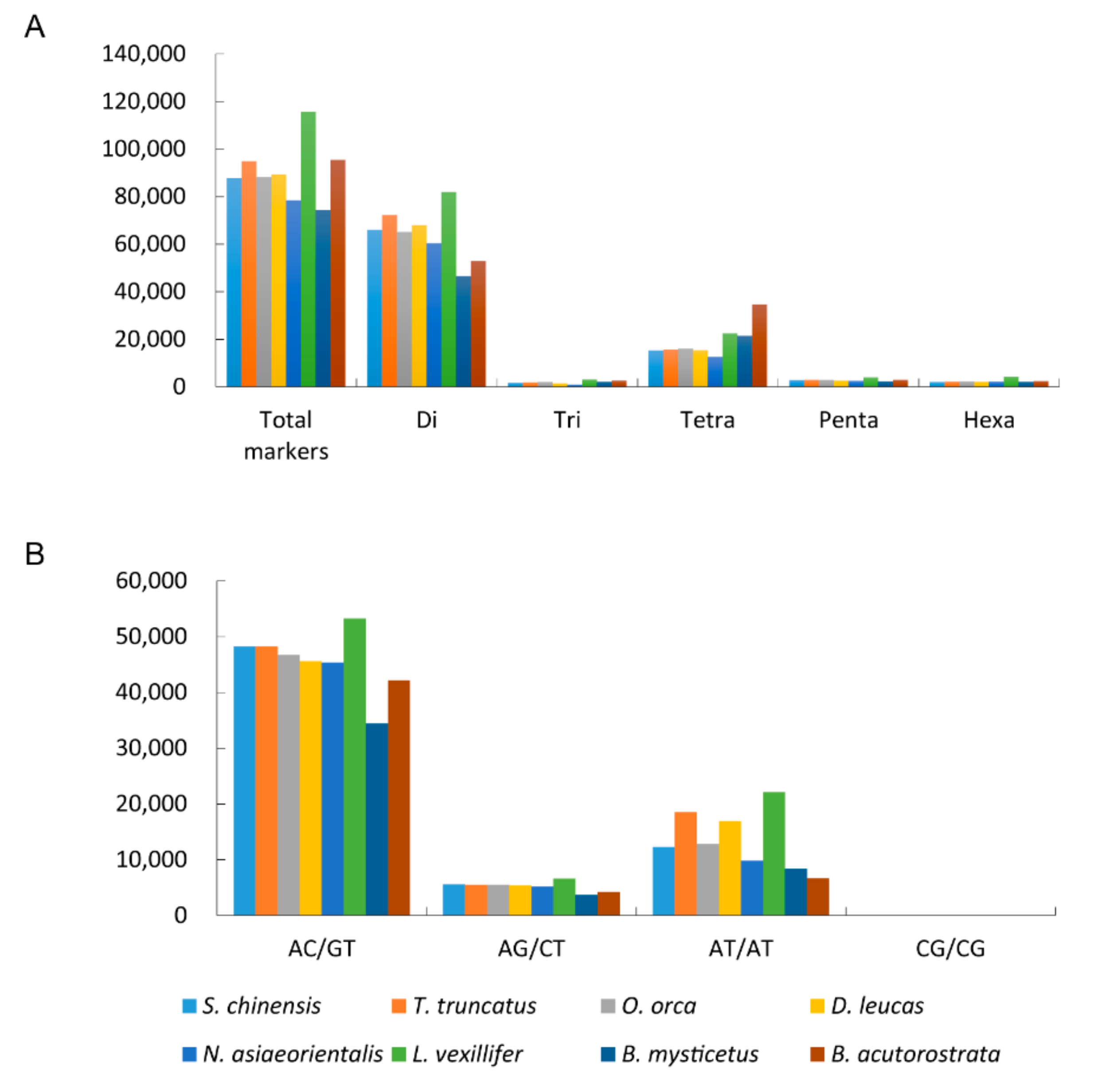
3.2. The Microsatellites Characteristics Comparison among Cetacean Genomes

B. mysticetus, B. acutorostrata, O. orca, T. truncates, D. leucas, L. vexillifer, and N. asiaeorientalis genomes were applied for microsatellites development (Table S4). The results represented similar characteristics to S. chinensis in terms of microsatellite composition (Figure 1A). Odontoceti possessed more total dinucleotide microsatellites than Mysticeti (Figure 1A). Besides, the number of dinucleotide microsatellites with different motifs was also higher in Odontoceti than Mysticeti (Figure 1A). On the other hand, Mysticeti had more tetranucleotide microsatellites than Odontoceti (Figure 1A). The total number of microsatellites in cetaceans ranged from 70,000 to 120,000 (Figure 1A, Table S1). AC/GT and AT/AT-rich repeats were the main types of dinucleotide microsatellites (Figure 1B, Table S5).

3.3. Microsatellites Distribution in the S. chinensis Genome

We investigated the distribution of microsatellites in the genome’s coding and non-coding regions. Most microsatellites were located in non-coding regions (87,347; 99.5%), and only about 0.5% (410) were located in coding regions, which was expected, as coding regions represent only a very small fraction of the genome. Based on the former reports, microsatellites were not evenly distributed throughout the whole genome [46,47,48,49]. Numerous microsatellites were distributed in non-coding regions and rarely in coding regions of eukaryotic species [50], e.g., about 7~10% in higher plants [51], and 9~15% in vertebrates [36,52]. In the genome of a swamp eel, about 1% of genes contain microsatellites in the coding regions of the genome [44]. In our study, only 0.8% of the genes contained microsatellites, a lower percentage than in vertebrates such as primates and rabbits [36,52], but one that is close to that of the swamp eel [44].
Next, GO function terms of the genes containing microsatellites in the coding regions were analyzed based on the genome annotation information [29]. Microsatellites were distributed in 195 genes, mainly enriched in 53 GO terms. Methylation processes such as histone acetyltransferase activity (GO: 0004402), histone acetylation (GO: 0016573), histone modification (GO: 0016570), histone methyltransferase complex (GO: 0035097), histone methyltransferase activity (H3-K4 specific) (GO: 0042800), and histone-lysine N-methyltransferase activity (GO: 0018024) were enriched (Table 1). Others were associated with metabolic and biosynthetic process, such as the regulation of the RNA metabolic process (GO: 0051252), the regulation of the nitrogen compound metabolic process (GO: 0051171), the regulation of the primary metabolic process (GO: 0080090), the regulation of the cellular metabolic process (GO: 0031323), the RNA metabolic process (GO: 0016070), etc (Table 1). The GO term annotation results suggest that the microsatellites were located in functional gene regions and may have impacts on gene function, such as the metabolic and methylation process. DNA methylation is readily detected in mammals, which is critical for species’ normal development and genome stability. Repetitive sequences have been regarded as ‘epigenetic elements’ that can regulate gene expression [53,54]. In plants and mammals, a large proportion of methylated cytosines are found in repeat elements [55,56]. The methylation of repeat elements is thought to have been compiled persistently during the evolution process and plays an important role in the epigenetic regulation of genes [57]. Most studies have focused on transposable elements, such as Alu elements (Alu), and long interspersed element-1 (LINE-1) [58,59]. The relationship between methylation and simple repeats still needs to be further studied. Thus, the analysis of genes containing microsatellites and their functions could probably be used to reveal potential mechanisms and characteristics of gene expression and regulation in S. chinensis.

3.4. Validation of Microsatellites in S. chinensis

Several accurate and widely used techniques can be applied to identify cetacean species, such as morphology, fingerprinting, and DNA barcoding [60,61]. However, it remains challenging to investigate the genetic diversity of cetaceans, due to the difficulty of obtaining accurate samples, especially for species threatened with extinction. S. chinensis is currently listed as Vulnerable by IUCN. It is also classified as a first-class protected animal by the Chinese National Key Protected Wild Aquatic Animals List. Live animal experiments are illegal and harmful to them. Cetacean stranding provides a unique opportunity to gain biological and ecological knowledge of cetaceans [62]. Previous studies have obtained microsatellites loci from stranded S. chinensis and identified them via PCR amplification [18,24]. We aligned the previous identified microsatellite markers to our genome. Of these total 46 markers, all could be mapped to the genome sequence. Then, we checked the positions and motif types between the published markers and the markers we isolated from the genome. Overall, 39 published markers were found in our markers that had the same location on the genome and the same motif types (+/−), and 7 published markers (SGATA47, SGATA35, SGATA28, SGATA25, SGATA18, SGATA13, SCA30) were not found in our markers at the corresponding position of the genome (Table 2). However, we extracted the sequences of the above seven marker-locatable genomic regions and developed the microsatellites using looser parameters: flank length of SSRs = 20 bp, and length of SSRs ≥ 8 bp in the genome. The results showed that the motif and the repeat numbers of these six microsatellites (※) were the same as those reported, except for SGATA18 (#) (Table 2). As a result, 45 of 46 markers could be confirmed at the genomic scale, with an overall verification rate of about 97.8%, indicating that the method of microsatellites isolation from the whole-genome sequence was comprehensive and accurate.

3.5. Polymorphic Microsatellites Detection Based on Male and Female Markers in S. chinensis

We reported the potential polymorphic microsatellites between male and female individuals. These markers may play an important role in the gender evolution or may be associated with sex chromosomes in mammals [63]. These microsatellites may also provide candidate markers for assessing population structure (male to female ratio) and the social behavior of S. chinensis. A total of 86,432 microsatellites were developed based on the male S. chinensis genome [30] (Table S6). Among these markers, 65,571 (75.87%) were dinucleotides microsatellites, followed by 14,677 (16.98%) tetranucleotides (Table S7). There were 15,678 candidate polymorphic microsatellites from the female and male markers, implying the presence of different PCR lengths and repeat numbers in both genomes (Table S8). Among them, 14,377 (91.71%) polymorphic microsatellites were dinucleotides, followed by 1063 (6.77%) tetranucleotides, 108 (0.69%) trinucleotides, 68 (0.43%) pentanucleotides, and 62 (0.40%) hexanucleotides (Table S9). The same trend of microsatellite composition was found in female and male S. chinensis. Dinucleotides and tetranucleotides were the predominant motif types among all the microsatellites isolated from the cetacean genome sequences (Figure 1A). These markers are of great value for conducting population surveys of S. chinensis in the field.

3.6. Specific Markers of Male S. chinensis

Until now, no cetacean genome has been found to have intact individual Y chromosome (a single and complete Y chromosome) assembled sequences. Only the bottlenose dolphin has the candidate Y chromosome-associated scaffolds. The bottlenose dolphin is a close relative of the S. chinensis [64]. Therefore, to obtain Y chromosome-specific microsatellites in the genome of male S. chinensis, we aligned unanchored scaffold sequences other than autosomal and X chromosome sequences in male S. chinensis with candidate Y chromosome-associated scaffold sequences of bottlenose dolphins. We obtained candidate contigs on the Y chromosome of S. chinensis genome based on the alignment results. This is the first time that the Y chromosome region sequence of this protected species has been identified. Finally, 1349 microsatellites were isolated from the candidate contigs, which can be applied as candidate markers for male dolphins (Table S10). These markers will have promising applications in the sex identification of S. chinensis after testing on additional samples. Since cetaceans have highly conserved genomes, these male-specific markers will also be important in the sex ratio investigation in S. chinensis and other cetaceans.

4. Conclusions

We explored and identified useful microsatellite markers in S. chinensis from a genome-wide scale and designed primers for these markers. A comparison with the published marker sequences revealed that the markers developed in this study had high reliability. Microsatellites in the S. chinensis genome are mainly distributed in the non-coding regions, and only approximately 0.5% are distributed in coding regions. Their associated genes presented potential methylation impacts in dolphins. Dinucleotide repeat motif type microsatellites are the most abundant, and this motif type is dominated by the AC-rich type. Cetacean species share the same microsatellite composition characteristics, which possibly reveals the conserved nature of the cetacean genomes. The study also yielded polymorphic microsatellite data for male and female S. chinensis, which provided a valuable database for the investigation and demography of dolphin populations. Moreover, the male-specific marker mining provided useful candidate markers for the sex identification of S. chinensis, as well as other cetacean species. Given that many marine mammals are species at risk of extinction, there is an urgent need to develop diverse conservation tools. The extensive and reliable genome-wide microsatellite information we identified in this study is expected to expand to other cetacean species and will play an important role in their population genetics studies and conservation.

Supplementary Materials

The following supporting information can be downloaded at: https://www.mdpi.com/article/10.3390/jmse10060834/s1, Table S1: The composition and classification of microsatellites in cetaceans; Table S2: The microsatellites in female Indo-Pacific humpback dolphin; Table S3: The composition and classification of dinucleotide microsatellites in female Indo-Pacific humpback dolphin; Table S4: The microsatellites in cetacean genomes; Table S5: The composition and classification of dinucleotide microsatellites in cetaceans; Table S6: The microsatellites in male Indo-Pacific humpback dolphin; Table S7: The composition and classification of microsatellites in male Indo-Pacific humpback dolphin; Table S8: The polymorphic microsatellites between female and male Indo-Pacific humpback dolphin; Table S9: The composition and classification of polymorphic microsatellites between male and female Indo-Pacific humpback dolphin; Table S10: Specific markers in male Indo-Pacific humpback dolphin.

Author Contributions

Y.M. and W.L. (Wenhua Liu) designed the entire research; X.Y. and W.L. (Wei Liu) prepared the samples; Y.M. analyzed the data; Y.M. wrote the manuscript with significant contributions from X.Y., W.L. (Wei Liu), J.W. and W.L. (Wenhua Liu). All authors have read and agreed to the published version of the manuscript.

Funding

This research was supported by the National Natural Science Foundation of China (NSFC, 41776174), Key Special Project for Introduced Talents Team of Southern Marine Science and Engineering Guangdong Laboratory (Guangzhou) (grant number GML2019ZD0606), 2019 Guangxi One Thousand Young and Middle-Aged College and University Backbone Teachers Cultivation Pro-gram, the Ministry of Agriculture and Rural Affairs of the People’s Republic of China (Chinese White Dolphin Conservation Action, 171821301354051004 and 171721301354051016), and CNOOC.

Institutional Review Board Statement

As we only used the published genome sequences data for the analysis, the study did not require ethical approval. We choose to exclude this statement.

Informed Consent Statement

We choose to exclude this statement because the study did not involve humans.

Data Availability Statement

We used the published data for the analysis in this study. The data resources were explained and described in the manuscript. The results and maker sequences created by this study have all been attached in the supplementary materials. We choose to exclude this statement.

Acknowledgments

We thank Edmond Sanganyado for polishing the manuscript writing.

Conflicts of Interest

The authors declare that they have no conflict of interest.

References

  1. Powell, W.; Machray, G.C.; Provan, J. Polymorphism revealed by simple sequence repeats. Trends Plant Sci. 1996, 1, 215–222. [Google Scholar] [CrossRef]
  2. Li, Y.C.; Korol, A.B.; Fahima, T.; Beiles, A.; Nevo, E. Microsatellites: Genomic distribution, putative functions and mutational mechanisms: A review. Mol. Ecol. 2002, 11, 2453–2465. [Google Scholar] [CrossRef] [PubMed]
  3. Abdul-Muneer, P.M. Application of microsatellite markers in conservation genetics and fisheries management: Recent advances in population structure analysis and conservation strategies. Genet. Res. Int. 2014, 2014, 691759. [Google Scholar] [CrossRef] [PubMed] [Green Version]
  4. Vieira, M.L.C.; Santini, L.; Diniz, A.L.; Munhoz, C.d.F. Microsatellite markers: What they mean and why they are so useful. Genet. Mol. Biol. 2016, 39, 312–328. [Google Scholar] [CrossRef] [PubMed] [Green Version]
  5. Li, S.-F.; Wang, B.-X.; Guo, Y.-J.; Deng, C.-L.; Gao, W.-J. Genome-wide characterization of microsatellites and genetic diversity assessment of spinach in the Chinese germplasm collection. Breed. Sci. 2018, 68, 455–464. [Google Scholar] [CrossRef] [PubMed] [Green Version]
  6. Pandey, G.; Misra, G.; Kumari, K.; Gupta, S.; Parida, S.K.; Chattopadhyay, D.; Prasad, M. Genome-wide development and use of microsatellite markers for large-scale genotyping applications in foxtail millet [Setaria italica (L.)]. DNA Res. Int. J. Rapid Publ. Rep. Genes Genomes 2013, 20, 197–207. [Google Scholar] [CrossRef]
  7. Khajuria, Y.P.; Saxena, M.S.; Gaur, R.; Chattopadhyay, D.; Jain, M.; Parida, S.K.; Bhatia, S. Development and Integration of Genome-Wide Polymorphic Microsatellite Markers onto a Reference Linkage Map for Constructing a High-Density Genetic Map of Chickpea. PLoS ONE 2015, 10, e0125583. [Google Scholar] [CrossRef] [Green Version]
  8. Abdelkrim, J.; Robertson, B.C.; Stanton, J.-A.L.; Gemmell, N.J. Fast, cost-effective development of species-specific microsatellite markers by genomic sequencing. BioTechniques 2009, 46, 185–192. [Google Scholar] [CrossRef]
  9. Sharma, P.C.; Grover, A.; Kahl, G. Mining microsatellites in eukaryotic genomes. Trends Biotechnol. 2007, 25, 490–498. [Google Scholar] [CrossRef]
  10. Taheri, S.; Lee Abdullah, T.; Yusop, M.R.; Hanafi, M.M.; Sahebi, M.; Azizi, P.; Shamshiri, R.R. Mining and Development of Novel SSR Markers Using Next Generation Sequencing (NGS) Data in Plants. Molecules 2018, 23, 399. [Google Scholar] [CrossRef] [Green Version]
  11. Wang, Q.; Fang, L.; Chen, J.; Hu, Y.; Si, Z.; Wang, S.; Chang, L.; Guo, W.; Zhang, T. Genome-Wide Mining, Characterization and Development of Microsatellite Markers in Gossypium Species. Sci. Rep. 2015, 5, 10638. [Google Scholar] [CrossRef] [PubMed] [Green Version]
  12. Shukla, N.; Kuntal, H.; Shanker, A.; Sharma, S.N. Mining and analysis of simple sequence repeats in the chloroplast genomes of genus Vigna. Biotechnol. Res. Innov. 2018, 2, 9–18. [Google Scholar] [CrossRef]
  13. Dettori, M.T.; Micali, S.; Giovinazzi, J.; Scalabrin, S.; Verde, I.; Cipriani, G. Mining microsatellites in the peach genome: Development of new long-core SSR markers for genetic analyses in five Prunus species. SpringerPlus 2015, 4, 337. [Google Scholar] [CrossRef] [PubMed] [Green Version]
  14. Wang, D.; Liao, X.; Cheng, L.; Yu, X.; Tong, J. Development of novel EST-SSR markers in common carp by data mining from public EST sequences. Aquaculture 2007, 271, 558–574. [Google Scholar] [CrossRef]
  15. Srivastava, S.; Avvaru, A.K.; Sowpati, D.T.; Mishra, R.K. Patterns of microsatellite distribution across eukaryotic genomes. BMC Genom. 2019, 20, 153. [Google Scholar] [CrossRef] [Green Version]
  16. Valsecchi, E.; Amos, W. Microsatellite markers for the study of cetacean populations. Mol. Ecol. 1996, 5, 151–156. [Google Scholar] [CrossRef]
  17. Shinohara, M.L.; Domingoroura, X.; Takenaka, O. Microsatellites in the bottlenose dolphin Tursiops truncatus. Mol. Ecol. 1997, 6, 695–696. [Google Scholar] [CrossRef]
  18. Chen, L.; Yang, G. A set of polymorphic dinucleotide and tetranucleotide microsatellite markers for the Indo-Pacific humpback dolphin (Sousa chinensis) and cross-amplification in other cetacean species. Conserv. Genet. 2008, 10, 697–700. [Google Scholar] [CrossRef]
  19. Nater, A.; Kopps, A.M.; Krutzen, M. New polymorphic tetranucleotide microsatellites improve scoring accuracy in the bottlenose dolphin Tursiops aduncus. Mol. Ecol. Resour. 2009, 9, 531–534. [Google Scholar] [CrossRef]
  20. Chen, I.; Nishida, S.; Yang, W.C.; Isobe, T.; Tajima, Y.; Hoelzel, A.R. Genetic diversity of bottlenose dolphin (Tursiops sp.) populations in the western North Pacific and the conservation implications. Mar. Biol. 2017, 164, 202. [Google Scholar] [CrossRef] [Green Version]
  21. Perez-Alvarez, M.J.; Olavarria, C.; Moraga, R.; Baker, C.S.; Hamner, R.M.; Poulin, E. Microsatellite markers reveal strong genetic structure in the endemic Chilean dolphin. PLoS ONE 2015, 10, e0123956. [Google Scholar] [CrossRef] [Green Version]
  22. Sumiyama, D.; Kitamura, S.; Terasawa, F.; Hori, Y.; Murata, K.; Kulski, J.K.; Inoko, H. Paternity determination of captive bottlenose dolphins (Tursiops truncatus) using microsatellite DNA analysis. J. Vet. Med. Sci. 2008, 70, 711–713. [Google Scholar] [CrossRef] [PubMed] [Green Version]
  23. Gui, D.; Jia, K.; Xia, J.; Yang, L.; Chen, J.; Wu, Y.; Yi, M. De novo assembly of the Indo-Pacific humpback dolphin leucocyte transcriptome to identify putative genes involved in the aquatic adaptation and immune response. PLoS ONE 2013, 8, e72417. [Google Scholar] [CrossRef] [PubMed] [Green Version]
  24. Dai, Y.; Chantra, R.; Kittiwattanawong, K.; Zhao, L.; Sakornwimon, W.; Aierken, R.; Wu, F.; Wang, X. Genetic structure of the endangered Irrawaddy dolphin (Orcaella brevirostris) in the Gulf of Thailand. Genet. Mol. Biol. 2021, 44, e20200365. [Google Scholar] [CrossRef]
  25. Chen, B.Y.; Zheng, D.M.; Wang, L.; Xu, X.R.; Yang, G. The northernmost distribution of Indo-Pacific humpback dolphin (Sousa chinensis) in the world: Evidence from preliminary survey in Ningde, China. Pak. J. Zool. 2012, 44, 1209–1214. [Google Scholar]
  26. Hung, S.K.; Würsig, B.; Wang, J.; Sims, M.P.; Piwetz, M.S. Monitoring of Marine Mammals in Hong Kong Waters (2011–12); Agriculture, Fisheries and Conservation Department of Hong Kong SAR Government: Hong Kong, China, 2012.
  27. Xu, X.; Song, J.; Zhang, Z.; Li, P.; Yang, G.; Zhou, K. The world’s second largest population of humpback dolphins in the waters of Zhanjiang deserves the highest conservation priority. Sci. Rep. 2015, 5, 8147. [Google Scholar] [CrossRef] [PubMed] [Green Version]
  28. Wang, J.; Yang, Y.; Yang, F.; Li, Y.; Li, L.; Lin, D.; He, T.; Liang, B.; Zhang, T.; Lin, Y. A framework for the assessment of the spatial and temporal patterns of threatened coastal delphinids. Sci. Rep. 2016, 6, 19883. [Google Scholar] [CrossRef] [PubMed] [Green Version]
  29. Ming, Y.; Jian, J.; Yu, X.; Wang, J.; Liu, W. The genome resources for conservation of Indo-Pacific humpback dolphin, Sousa chinensis. Sci. Data 2019, 6, 68. [Google Scholar] [CrossRef]
  30. Zhang, P.; Zhao, Y.; Li, C.; Lin, M.; Dong, L.; Zhang, R.; Liu, M.; Li, K.; Zhang, H.; Liu, X.; et al. An Indo-Pacific Humpback Dolphin Genome Reveals Insights into Chromosome Evolution and the Demography of a Vulnerable Species. iScience 2020, 23, 101640. [Google Scholar] [CrossRef]
  31. Thiel, T.; Michalek, W.; Varshney, R.K.; Graner, A. Exploiting EST databases for the development and characterization of gene-derived SSR-markers in barley (Hordeum vulgare L.). Theor. Appl. Genet. 2003, 106, 411–422. [Google Scholar] [CrossRef]
  32. Untergasser, A.; Cutcutache, I.; Koressaar, T.; Ye, J.; Faircloth, B.C.; Remm, M.; Rozen, S.G. Primer3—New capabilities and interfaces. Nucleic Acids Res. 2012, 40, e115. [Google Scholar] [CrossRef] [PubMed] [Green Version]
  33. Kent, W.J. BLAT—The BLAST-like alignment tool. Genome Res. 2002, 12, 656–664. [Google Scholar] [PubMed] [Green Version]
  34. Bourret, V.; Macé, M.; Bonhomme, M.; Crouau-Roy, B. Microsatellites in cetaceans: An overview. Open Mar. Biol. J. 2008, 2, 38–42. [Google Scholar] [CrossRef]
  35. Beckmann, J.S.; Weber, J.L. Survey of human and rat microsatellites. Genomics 1992, 12, 627–631. [Google Scholar] [CrossRef]
  36. Jurka, J.; Pethiyagoda, C. Simple repetitive DNA sequences from primates: Compilation and analysis. J. Mol. Evol. 1995, 40, 120–126. [Google Scholar] [CrossRef] [PubMed]
  37. Moran, C. Microsatellite repeats in pig (Sus domestica) and chicken (Gallus domesticus) genomes. J. Hered. 1993, 84, 274–280. [Google Scholar] [CrossRef]
  38. Nadir, E.; Margalit, H.; Gallily, T.; Ben-Sasson, S.A. Microsatellite spreading in the human genome: Evolutionary mechanisms and structural implications. Proc. Natl. Acad. Sci. USA 1996, 93, 6470–6475. [Google Scholar] [CrossRef] [Green Version]
  39. Manee, M.M.; Algarni, A.T.; Alharbi, S.N.; Al-Shomrani, B.M.; Ibrahim, M.A.; Binghadir, S.A.; Al-Fageeh, M.B. Genome-wide characterization and analysis of microsatellite sequences in camelid species. Mammal Res. 2020, 65, 359–373. [Google Scholar] [CrossRef] [Green Version]
  40. Song, X.; Yang, T.; Zhang, X.; Yuan, Y.; Yan, X.; Wei, Y.; Zhang, J.; Zhou, C. Comparison of the Microsatellite Distribution Patterns in the Genomes of Euarchontoglires at the Taxonomic Level. Front. Genet. 2021, 12, 227. [Google Scholar] [CrossRef]
  41. Getino-Mamet, L.N.; Valdivia-Carrillo, T.; Gomez Daglio, L.; Garcia-De Leon, F.J. Isolation and characterization of 14 tetranucleotide microsatellite loci for the cannonball jellyfish (Stomolophus sp.) by next generation sequencing. Mol. Biol. Rep. 2017, 44, 257–260. [Google Scholar] [CrossRef]
  42. Landinez-Garcia, R.M.; Marquez, E.J. Development and characterization of 24 polymorphic microsatellite loci for the freshwater fish Ichthyoelephas longirostris (Characiformes: Prochilodontidae). PeerJ 2016, 4, e2419. [Google Scholar] [CrossRef] [Green Version]
  43. Guo, W.; Guo, C.; Wang, Y.; Hu, W.; Mei, J. Population structure and genetic diversity in yellow catfish (Pelteobagrus fulvidraco) assessed with microsatellites. J. Genet. 2019, 98, 1–4. [Google Scholar] [CrossRef]
  44. Li, Z.; Chen, F.; Huang, C.; Zheng, W.; Yu, C.; Cheng, H.; Zhou, R. Genome-wide mapping and characterization of microsatellites in the swamp eel genome. Sci. Rep. 2017, 7, 3157. [Google Scholar] [CrossRef] [PubMed]
  45. Lei, Y.; Zhou, Y.; Price, M.; Song, Z. Genome-wide characterization of microsatellite DNA in fishes: Survey and analysis of their abundance and frequency in genome-specific regions. BMC Genom. 2021, 22, 421. [Google Scholar] [CrossRef]
  46. Toth, G.; Gaspari, Z.; Jurka, J. Microsatellites in different eukaryotic genomes: Survey and analysis. Genome Res. 2000, 10, 967–981. [Google Scholar] [CrossRef] [PubMed] [Green Version]
  47. Ellegren, H. Microsatellites: Simple sequences with complex evolution. Nat. Rev. Genet. 2004, 5, 435–445. [Google Scholar] [CrossRef]
  48. Rajendrakumar, P.; Biswal, A.K.; Balachandran, S.M.; Srinivasarao, K.; Sundaram, R.M. Simple sequence repeats in organellar genomes of rice: Frequency and distribution in genic and intergenic regions. Bioinformatics 2007, 23, 1–4. [Google Scholar] [CrossRef] [Green Version]
  49. Li, Y.-C.; Korol, A.B.; Fahima, T.; Nevo, E. Microsatellites within genes: Structure, function, and evolution. Mol. Biol. Evol. 2004, 21, 991–1007. [Google Scholar] [CrossRef]
  50. Metzgar, D.; Bytof, J.; Wills, C. Selection against frameshift mutations limits microsatellite expansion in coding DNA. Genome Res. 2000, 10, 72–80. [Google Scholar]
  51. Wang, Z.; Weber, J.L.; Zhong, G.; Tanksley, S. Survey of plant short tandem DNA repeats. Theor. Appl. Genet. 1994, 88, 1–6. [Google Scholar] [CrossRef]
  52. Van Lith, H.; Van Zutphen, L. Characterization of rabbit DNA micros extracted from the EMBL nucleotide sequence database. Anim. Genet. 1996, 27, 387–395. [Google Scholar] [CrossRef] [PubMed]
  53. Pardue, M.L.; Gall, J.G. Chromosomal localization of mouse satellite DNA. Science 1970, 168, 1356–1358. [Google Scholar] [CrossRef] [PubMed]
  54. McClintock, B. Chromosome organization and genic expression. In Cold Spring Harbor Symposia on Quantitative Biology; Cold Spring Harbor Laboratory Press: Long Island, NY, USA, 1951; pp. 13–47. [Google Scholar]
  55. Suzuki, M.M.; Bird, A. DNA methylation landscapes: Provocative insights from epigenomics. Nat. Rev. Genet. 2008, 9, 465–476. [Google Scholar] [CrossRef] [PubMed]
  56. Law, J.A.; Jacobsen, S.E. Establishing, maintaining and modifying DNA methylation patterns in plants and animals. Nat. Rev. Genet. 2010, 11, 204–220. [Google Scholar] [CrossRef]
  57. Teixeira, F.K.; Colot, V. Repeat elements and the Arabidopsis DNA methylation landscape. Heredity 2010, 105, 14–23. [Google Scholar] [CrossRef] [PubMed]
  58. Zheng, Y.; Joyce, B.T.; Liu, L.; Zhang, Z.; Kibbe, W.A.; Zhang, W.; Hou, L. Prediction of genome-wide DNA methylation in repetitive elements. Nucleic Acids Res. 2017, 45, 8697–8711. [Google Scholar] [CrossRef] [Green Version]
  59. Bollati, V.; Galimberti, D.; Pergoli, L.; Dalla Valle, E.; Barretta, F.; Cortini, F.; Scarpini, E.; Bertazzi, P.A.; Baccarelli, A. DNA methylation in repetitive elements and Alzheimer disease. Brain Behav. Immun. 2011, 25, 1078–1083. [Google Scholar] [CrossRef] [Green Version]
  60. Kriangwanich, W.; Buddhachat, K.; Poommouang, A.; Chomdej, S.; Thitaram, C.; Kaewmong, P.; Kittiwattanawong, K.; Nganvongpanit, K. Feasibility of melting fingerprint obtained from ISSR-HRM curves for marine mammal species identification. PeerJ 2021, 9, e11689. [Google Scholar] [CrossRef]
  61. Silva, V.S.; Skueresky, N.; Lopes, F.; Koch, T.K.; Ott, P.H.; Siciliano, S.; Barreto, A.S.; Secchi, E.R.; de Meirelles, A.C.O.; Carvalho, V.L. Integrating morphology and DNA barcoding to assess cetacean diversity in Brazil. Mammal Res. 2021, 66, 349–369. [Google Scholar] [CrossRef]
  62. Zhu, J.; Yu, X.; Zhang, Q.; Li, Y.; Tan, S.; Li, D.; Yang, Z.; Wang, J. Cetaceans and microplastics: First report of microplastic ingestion by a coastal delphinid, Sousa chinensis. Sci. Total Environ. 2019, 659, 649–654. [Google Scholar] [CrossRef]
  63. Ziemniczak, K.; Traldi, J.B.; Nogaroto, V.; De Almeida, M.C.; Artoni, R.F.; Moreirafilho, O.; Vicari, M.R. In situ Localization of (GATA)n and (TTAGGG)n Repeated DNAs and W Sex Chromosome Differentiation in Parodontidae (Actinopterygii: Characiformes). Cytogenet. Genome Res. 2015, 144, 325–332. [Google Scholar] [CrossRef] [PubMed]
  64. Ming, Y.; Jian, J.; Yu, F.; Yu, X.; Wang, J.; Liu, W. Molecular footprints of inshore aquatic adaptation in Indo-Pacific humpback dolphin (Sousa chinensis). Genomics 2018, 111, 1034–1042. [Google Scholar] [CrossRef] [PubMed]
Figure 1. Statistics of microsatellites with different repeat types in eight cetacean genomes (Sousa chinensis, Orcinus orca, Balaenoptera acutorostrata, Balaena mysticetus, Tursiops truncatus, Delphinapterus leucas, Lipotes vexillifer, and Neophocaena asiaeorientalis). (A) Statistics of all types of microsatellite repeats: Di (Dinucleotides), Tri (Trinucleotides), Tetra (Tetranucleotides), Penta (Pentanucleotides), and Hexa (Hexanucleotides); (B) Statistics of dinucleotides motif mircosatellites in cetacean species. Dinucleotides motifs contain four types: AT, CG, AC/GT and AG/CT (AT = TA, CG = GC, AC = CA = GT = TG, AG = GA = CT = TC).
Jmse 10 00834 g001
Table 1. GO function terms of the genes containing microsatellites in the genomic coding regions.
GO IDGO TermGO ClassCorrected p-Value
GO:0010468regulation of gene expressionBP1.76 × 10−19
GO:0051252regulation of RNA metabolic processBP1.73 × 10−17
GO:2000112regulation of cellular macromolecule biosynthetic processBP2.08 × 10−17
GO:0006355regulation of transcription, DNA-templatedBP2.41 × 10−17
GO:0006351transcription, DNA-templatedBP2.41 × 10−17
GO:0051171regulation of nitrogen compound metabolic processBP1.77 × 10−16
GO:0080090regulation of primary metabolic processBP2.23 × 10−16
GO:0031323regulation of cellular metabolic processBP4.73 × 10−16
GO:0016070RNA metabolic processBP5.11 × 10−14
GO:0090304nucleic acid metabolic processBP2.73 × 10−11
GO:0005634nucleusCC8.34 × 10−9
GO:0006725cellular aromatic compound metabolic processBP2.00 × 10−8
GO:1901360organic cyclic compound metabolic processBP3.74 × 10−8
GO:0003676nucleic acid bindingMF9.51 × 10−8
GO:0010467gene expressionBP1.57 × 10−7
GO:0005488bindingMF5.25 × 10−7
GO:0003677DNA bindingMF8.27 × 10−7
GO:0044451nucleoplasm partCC8.87 × 10−6
GO:0031981nuclear lumenCC1.07 × 10−5
GO:0034645cellular macromolecule biosynthetic processBP1.11 × 10−5
GO:0043227membrane-bounded organelleCC1.93 × 10−5
GO:0043565sequence-specific DNA bindingMF2.44 × 10−5
GO:0044271cellular nitrogen compound biosynthetic processBP2.59 × 10−5
GO:0050789regulation of biological processBP3.32 × 10−5
GO:0004402histone acetyltransferase activityMF4.15 × 10−5
GO:0043231intracellular membrane-bounded organelleCC6.23 × 10−5
GO:0050794regulation of cellular processBP6.46 × 10−5
GO:0140110transcription regulator activityMF1.21 × 10−4
GO:0044428nuclear partCC1.74 × 10−4
GO:0065007biological regulationBP2.80 × 10−4
GO:0016573histone acetylationBP2.80 × 10−4
GO:0006325chromatin organizationBP3.29 × 10−4
GO:0016570histone modificationBP3.43 × 10−4
GO:0035097histone methyltransferase complexCC5.69 × 10−4
GO:0009058biosynthetic processBP6.57 × 10−4
GO:0018205peptidyl-lysine modificationBP7.91 × 10−4
GO:0034641cellular nitrogen compound metabolic processBP8.80 × 10−4
GO:1901363heterocyclic compound bindingMF1.27 × 10−3
GO:0097159organic cyclic compound bindingMF1.30 × 10−3
GO:0003712transcription coregulator activityMF1.43 × 10−3
GO:0044260cellular macromolecule metabolic processBP1.49 × 10−3
GO:0042800histone methyltransferase activity (H3-K4 specific)MF4.83 × 10−3
GO:0016286small conductance calcium-activated potassium channel activityMF4.83 × 10−3
GO:0043170macromolecule metabolic processBP7.22 × 10−3
GO:0005515protein bindingMF9.68 × 10−3
GO:0016592mediator complexCC0.015
GO:1990234transferase complexCC0.019
GO:0003700DNA-binding transcription factor activityMF0.023
GO:0018024histone-lysine N-methyltransferase activityMF0.023
GO:0003713transcription coactivator activityMF0.024
GO:0005516calmodulin bindingMF0.029
GO:0071339MLL1 complexCC0.029
Note: Biological Process, BP; Cellular Component, CC; Molecular Function, MF.
Table 2. Identification of microsatellites using published markers.
Published MarkerRepeat TypeSSR ID in S. chinensisRepeat TypeNote
SGATA42(GATA)11scaffold428.1_57256(GATA)11+
SCA6(CA)17scaffold113.1_88883(CA)19+
SGATA47(TATC)5/(TAGA)5
SGATA45(AGAT)8scaffold2.1_32807(ATCT)9
SGATA35(TCTA)4/(GATA)4
SGATA33(ATAG)7scaffold470.1_58268(TCTA)8
SGATA30(AGAT)7scaffold162.1_47075(TATC)7
SGATA28(AT)4/(AT)4
SGATA25(GATA)5/(AGAT)5
SGATA21(TAGA)10scaffold35.1_72245(ATAG)10+
SGATA18(GATA)3GAT(GATA)3AATA(GATA)2//#
SGATA13(TCTA)4/(TCTA)4
SCA66(CA)11scaffold85.1_89945(GT)11
SCA64(CA)23scaffold77.1_66768(CA)22+
SCA58(AC)12scaffold18.1_1347(GT)12
SCA56(CA)15scaffold284.1_42084(CA)13+
SCA55(AC)14scaffold249.1_12650(AC)14+
SCA54(CA)20scaffold199.1_4149(AC)18+
SCA48(CA)16scaffold319.1_1085(TG)16
SCA39(AC)20scaffold196.1_39358(AC)24+
SCA37(AC)23scaffold57.1_92416(GT)23
SCA33(AC)22scaffold34.1_17501(AC)17+
SCA30(CA)9/(TG)9
SCA27(AC)21scaffold70.1_71178(TG)22
SCA22(CT)7TTCT(CA)36scaffold54.1_109484(GT)28GAGAAA(GA)7
SCA17(AC)16scaffold66.1_63598(AC)19+
SCA12(AC)13scaffold20.1_86731(AC)13+
SCA9(CA)23scaffold329.1_58395(AC)37
Sch5878(CAAC)12scaffold108.1_36155(TTGG)11
Sch6660(AAGG)13scaffold10.1_97587(CCTT)13
Sch443(CCAT)12scaffold10.1_97555(GGAT)12
Sch10207(CATC)12scaffold240.1_14114(GATG)12
Sch843(AAAT)11scaffold25.1_94818(TTTA)11
Sch7424(ATGG)13scaffold89.1_41509(CCAT)13
Sch7357(ATGG)11scaffold20.1_86885(ATGG)11+
Sch193(AGAGA)12scaffold26.1_104752(TCTCT)11
Sch8186(CCAT)11scaffold15.1_6749(CCAT)11+
Sch123(CCTAAC)7scaffold66.1_64097(CCTAAC)7+
Sch4657(TTCC)11scaffold304.1_101153(AGGA)11
Sch5373(GATG)11scaffold91.1_67861(TCCA)11
Sch9144(ATCT)14scaffold11.1_40258(TAGA)15
Sch2513(CATC)13scaffold165.1_13454(CATC)13+
Sch5685(AGGA)12scaffold9.1_107855(AGGA)13+
Sch974(GTTTT)13scaffold32.1_92270(AAAAC)10
Sch5094(TCTA)11scaffold28.1_42911(GATA)11
Sch8947(CTAT)12scaffold11.1_40359(CTAT)12+
Note: The table comprises published microsatellite IDs, repeat types [18,24], the corresponding microsatellite IDs, and repeat types extracted from the S. chinensis genome. +/− denotes forward and reverse mapping in the genome and present in the extracting markers; ※ denotes the published markers could be mapped to the genome but not included in our results, after which these sequences were checked and found to be shorter SSRs (these markers can be isolated from the genome if looser parameters are set when scanning the genome sequence with MISA (v1.0) software); # denotes the markers could be mapped to the genome, but not included in our isolated microsatellites, even when changing the search parameters.
Publisher’s Note: MDPI stays neutral with regard to jurisdictional claims in published maps and institutional affiliations.

