Article

How Do Gene Expression Patterns Change in Response to Osmotic Stresses in Kuruma Shrimp (Marsupenaeus japonicus)?

School of Marine Science and Engineering, Qingdao Agricultural University, Qingdao 266109, China
*
Author to whom correspondence should be addressed.
These authors contributed equally to this work.
J. Mar. Sci. Eng. 2022, 10(12), 1870; https://doi.org/10.3390/jmse10121870
Submission received: 14 October 2022 / Revised: 15 November 2022 / Accepted: 22 November 2022 / Published: 2 December 2022
(This article belongs to the Special Issue Evolution and Adaptation in Marine Invertebrates)

Abstract

:
Euryhaline crustaceans cope with external salinity changes by mechanisms of osmoregulation. In the current study, we first cloned and confirmed the ORF sequences of the ion-transportation-related genes Na+/K+-ATPase α subunit (NKAα), cytoplasmic carbonic anhydrase (CAc), and V-type H+-ATPase G subunit (VHA-G), and water channels of aquaporins (AQP3, AQP4, and AQP11) from kuruma shrimp (Marsupenaeus japonicus). Further tissue expression patterns showed a higher expression of MjAQP4, MjCAc, MjNKAα, and MjVHA-G in the gills, as well as a higher expression of MjAQP3 and MjAQP11 in the intestine and muscle, respectively. Then, qPCR analysis was used to assess the mRNA expression levels of those osmoregulatory genes in both post-larvae and adult shrimp when they were exposed to acute salinity stress or salinity acclimation. The results revealed significantly decreased expression levels of MjAQP3, MjAQP11, MjNKAα, and MjCAc, and higher expression levels of MjAQP4 and MjVHA-G when the post-larvae shrimp were directly subjected to 10‰ or 50‰ salinity. Moreover, similar expression patterns were also observed in the post-larvae shrimp during the accommodation to 10‰ or 50‰ salinity. As to the adult shrimp, significantly higher expression levels of those genes were observed in the gills after exposure to 10‰ salinity, whereas only the expression levels of MjAQP3, MjAQP11, and MjNKAα were up-regulated in the gills at 40‰ salinity. In contrast, the expression of MjVHA-G was significantly decreased at 40‰ salinity. Finally, during the acclimation to 10‰ salinity, the expression levels of MjAQP3, MjAQP11, and MjNKAα were also significantly elevated, while the expression of MjCAc was significantly decreased in the gills. In addition, the expression levels of MjAQP3, MjAQP4, MjCAc, and MjVHA-G were significantly decreased in the gills during the acclimation to 55‰ salinity. The findings of the study suggest that the examined genes are critical for the adaptation of aquatic crustaceans to changing environmental salinity. Our study lays as the foundation for further research on osmoregulation mechanisms in M. japonicus.

1. Introduction

Salinity is one of the major environmental factors that exert crucial effects on aquatic organisms. Variation in salinity can impose osmotic stress, alter osmolyte concentrations, and affect the presence, physiology, abundance, and distribution of aquatic animals [1]. Osmoregulation is believed to be the primary physiological function that aquatic animals use to maintain the ionic and water balance between the body fluid and the environment [2,3]. The widespread distribution of crustaceans in marine, brackish, and freshwater environments demonstrates an extraordinary capacity to adapt to variable salinities, which benefits from their powerful osmotic regulation abilities [4].
Researchers have discovered the physiological and morphological characteristics of crustaceans that facilitate their adaptation to a wide range of salinities [5,6]. The gill is considered the primary organ for osmoregulation and the epithelium of the gill contains specialized cells responsible for regulating osmotic pressure, controlling gas exchange, and maintaining an acid–base balance [7]. Moreover, crustaceans can respond to environmental salinity changes through the expression of osmoregulatory genes involved in active ion transport and exchange, the tolerance of osmotic stress, water channel maintenance, and cell volume control [8]. Ion transport and exchange are mostly attributed to the activity of the Na+/K+-ATPase (NKA), V-type H+-ATPase (VHA), and carbonic anhydrase (CA) in the gills of crustaceans [2,3,9]. NKA has been the most extensively examined gene in osmoregulatory studies of euryhaline animals because it is a critical enzyme responsible for establishing electrochemical gradients across the cell membrane in the gills [10]. VHA actively participates in osmoregulatory ion uptake across the gill epithelium of freshwater-tolerant crustaceans and is also responsible for acid–base balance and nitrogen excretion [2,9,11,12], whereas CA rapidly converts carbon dioxide to bicarbonate and carbonate, which affects the acid–base balance, ion transport, and gas exchange at both the tissue and cellular levels [13,14]. Apart from these, aquaporins (AQPs) are small transmembrane channel proteins that mainly facilitate the transport of water and small solutes through cellular membranes [15,16]. AQPs are classified into classical aquaporins, aquaglyceroporins, and unorthodox aquaporins [17,18]. Moreover, the roles of AQPs in osmoregulation have been indicated by their transcriptional regulation upon salinity stress in crustaceans in the last decade [19,20,21,22,23,24]. Recently, the AQP family from Pacific white shrimp (Litopenaeus vannamei) has been characterized and their expression patterns in response to salinity changes have been clarified [25,26].
The kuruma shrimp (Marsupenaeus japonicus) is one of the major shrimp species with high economic value that is mainly cultivated in China, accounting for more than 5% of the total shrimp production [27]. Nevertheless, due to the influence of estuaries, seasonal ocean currents, and other factors, changes in salinity are inevitable during aquaculture, which results in large economic losses in the industry. On the other hand, the distribution of salinity in aquatic waters in China is not uniform. There are large areas of inland saline waters and coastal high-salinity waters in China [28,29,30], wherein most of them have been in an abandoned state for a long time [31]. As M. japonicus is a species capable of tolerating a wide range of salinity [32], it can be developed as a promising aquaculture species in these waters to expand the space for aquaculture and increase the incomes of farmers. Thus, studies of the salinity adaptation mechanism of M. japonicus could provide new strategies for developing and optimizing successful shrimp farming in variable salinity conditions.
Up to now, few studies have addressed how the osmoregulatory genes respond to salinity challenges in M. japonicus. Therefore, the current study aimed to elucidate the potential roles of ion-transportation-related genes (NKA, CA, and VHA) and water channel genes (AQPs) with respect to variable salinity. These genes were first identified and characterized from the transcriptome of M. japonicus. Then, real-time quantitative PCR (qPCR) analysis was performed to clarify their tissue distributions and expression patterns in response to acute salinity challenges or salinity acclimation at two developmental stages. The investigation of their expression profiles can provide insights into not only the osmoregulation mechanisms of crustaceans but also into the cultivation of M. japonicus in the inland saline waters and coastal high-salinity waters of China.

2. Materials and Methods

2.1. Experimental Animals

The post-larvae shrimp (P5) were obtained from a farm in Weifang city, Shandong province, China. Shrimp were cultured in PVC buckets (30 L) containing 20 L of aerated natural seawater (salinity 30‰, pH 8.0, and 25 ± 0.5 °C) with a 30% daily exchange rate and fed Artemia four times a day (8:00, 13:00, 18:00, and 23:00). The following salinity stress experiments of post-larvae shrimp were also conducted at this farm.
Adult shrimp (average weight: 10.0 ± 0.5 g; body length: 11.2 ± 0.4 cm) were reared in open ponds in Qingdao city, Shandong province, China. The temperature of the ponds was 18 ± 1 °C and the salinity was 25‰. Then, the shrimp were transferred to the School of Marine Science and Engineering, Qingdao Agricultural University. They were acclimated in PVC buckets (300 L) and fed three times per day (9:00, 15:00, and 21:00; 5% of shrimp body weight per feeding) with a commercial pelleted feed (CP Group Feed, Qingdao, China). The temperature and salinity of seawater were re-adjusted to that of the shrimp’s original habitat in ponds. The feces and uneaten feed were removed daily with a siphon tube. Dissolved oxygen was kept above 6.0 mg/L.
All handling of this study was performed in strict accordance with the guidelines of the Animal Experimentation Ethics Committee of Qingdao Agricultural University, which also approved the protocol.

2.2. Effects of Acute Salinity Changes on Post-Larvae Shrimp

Artificial high-salinity (35‰, 40‰, 45‰, 50‰, and 55‰) seawater was prepared by adding sea salt to natural seawater (30‰), while low-salinity (25‰, 20‰, 15‰, 10‰, and 5‰) seawater was made by the addition of pre-aerated freshwater to natural seawater. A water quality detector (YSI incorporated, USA) was used to measure and monitor the salinity of seawater until it reached the target salinity. The pH and water temperature of artificial seawater were consistent with those of natural seawater. The post-larvae shrimp were directly placed from seawater of 30‰ salinity to seawater of high-salinity with ascending gradient or low-salinity with descending gradient. Beakers containing 500 mL of seawater with 10 shrimp each were prepared in triplicate for each salinity concentration. The number of surviving individuals in each group was counted and the dead ones were removed at 12 h and 24 h (hours) after the salinity challenges.
Based on the survival rates of shrimp from the above experiment, 10‰ and 50‰ salinity concentrations were set as the treatment salinity. Forty-five post-larvae shrimp (fifteen shrimp/bucket × three buckets) reared at 30‰ salinity were flash-frozen in liquid nitrogen and set as the control group (0 h) for this experiment. Then, shrimp were separately transferred from seawater of 30‰ salinity to 10‰ or 50‰ salinity and each treatment was replicated thrice (40 shrimp per replicate). Forty-five shrimp per treatment (fifteen shrimp/replicate × three replicates) were randomly selected, fixed by liquid nitrogen at 6 h and 12 h after salinity challenge, and stored at −80 °C.

2.3. Salinity Acclimation Experiment of Post-Larvae Shrimp

Forty-five post-larvae shrimp (fifteen shrimp/bucket × three buckets) from seawater of 30‰ salinity were first sampled and set as the control group. Then, 1200 shrimp were randomized equally into 6 PVC buckets (140 L) with the consistent culture conditions as described above. They were divided into high-salinity and low-salinity acclimation groups with each containing three replicates (200 individuals per replicate). The salinity of each group was changed daily by the addition of high-salinity artificial seawater or freshwater during the water exchange process at 07:00 am. The salinity of the high-salinity acclimation experimental group was increased daily by 5‰ until it reached 50‰ salinity and was maintained at 50‰ salinity for 7 days. The low-salinity treatment group was acclimated from 30‰ to 10‰ through daily 5‰ decrements, following a continuous acclimation at 10‰ salinity for 7 days. Forty-five shrimp per treatment (fifteen shrimp/replicate × three replicates) were sampled at 07:00 pm of the day that the salinity reached the target salinity. Then, another sampling was performed after the 7 days of acclimation.

2.4. Acute Salinity Exposure Experiment of Adult Shrimp

Nine shrimp at 25‰ salinity (three shrimp/bucket × three buckets) were randomly selected as the control group (0 h), and the tissues of gill, hepatopancreas, intestine, and muscle were dissected, quickly frozen in liquid nitrogen, and stored at −80 °C. Based on a previous report from Boyd and Fast (1992), the salinity concentrations of 10‰ and 40‰ were chosen for the acute salinity exposure. Adult shrimp were directly transferred from seawater of 30‰ salinity to 10‰ or 40‰ salinity and each treatment had three replicates (20 shrimp per replicate). Nine shrimp per treatment (three shrimp/replicate × 3 replicates) were chosen to fix the tissues as described above at 6 h and 12 h after salinity stress.

2.5. Salinity Acclimation Experiment of Adult Shrimp

Nine shrimp at 25‰ salinity (three shrimp/bucket × three buckets) were set as the control group and sampled as described in Section 2.4. Then, 180 shrimp in 6 buckets were divided into high-salinity and low-salinity acclimation groups with each containing 3 replicates (30 individuals per replicate). The two treatment groups were separately acclimated to 55‰ salinity or 10‰ salinity through daily 5‰ increments or decrements by using methods similar to those described in Section 2.3. Then, the sampling interval was set at 15‰ salinity and 9 shrimp per treatment (3 shrimp/replicate × 3 replicates) were sampled 12 h after reaching the salinity concentrations of 10‰, 40‰, and 55‰. Except for the sampled shrimp, few shrimp died during the acclimation period. In addition, no apparent changes in behavior, activity, and food intake were observed for the shrimp exposed to different salinities.

2.6. RNA Isolation and cDNA Synthesis

Total RNA of five post-larvae shrimp per replicate was extracted together using a Quick-RNA Microprep kit (Jianshi Bio, Beijing, China), and the total RNA of sampled tissues from adult shrimp was individually isolated by TRIzol Reagent (Vazyme, Nanjing, China). Thus, three RNA samples were obtained for each replicate. The RNA integrity was detected by 1% agarose gel electrophoresis. The purity and concentration of RNA were measured by NanoDrop 2000 spectrophotometer (Thermo Fisher Scientific, Waltham, MA, USA). Then, the first-strand cDNA was generated from an RNA sample of 1 μg with PrimeScriptTM RT Reagent Kit with gDNA Eraser (Vazyme, Nanjing, China).

2.7. Identification and Characterization of Osmoregulatory Genes

The mRNA sequences of AQP3, AQP4, AQP11, CAc, NKAα, and VHA-G from L. vannamei were used as query sequences to conduct online BLASTN (http://www.ncbi.nlm.nih.gov/BLAST/, accessed on 11 May 2021) against the TSA database of M. japonicus accessed on 11 May 2021. Transcripts homologous to the above genes were identified and specific primers were designed to amplify and validate the open reading frames (ORFs) of the transcripts (Table 1). Multiple sequence alignment of the obtained genes was conducted using the MUSCLE method in MEGA X software and visualized through Jalview software [33,34]. Phylogenetic analysis of these genes was performed by the neighbor-joining (NJ) method with 1000 bootstrap values in MEGA X software. Then, the phylogenetic trees were visualized using Interactive Tree of Life (iTOL) version 6 [35]. Transmembrane regions of NKAα protein were predicted with TMHMM 2.0 (http://www.cbs.dtu.dk/services/TMHMM, accessed on 11 May 2021).

2.8. Expression Analysis of Osmoregulatory Genes

Specific primers of target genes and reference gene (elongation factor 1 alpha, EF1α) were designed for the tissue distribution of the target genes and their expression patterns under salinity stresses (Table 2). The qPCR reaction mixture contained 5.0 μL of 2 × ChamQ Universal SYBR qPCR Master Mix (Vazyme, Nanjing, China), 2.0 μL of cDNA (5 ng/μL), 0.2 μL each of 10 μM forward and reverse primers, and 2.6 μL of RNase-free water. The entire reaction was carried out in 96-well PCR plates by a CFX96 Touch Real-Time PCR Detection System (Bio-Rad, Hercules, CA, USA). The qPCR amplification process consisted of pre-incubation at 95 °C (30 s) followed by 40 cycles of 95 °C (10 s) and 60 °C (30 s). Standard melt curve analysis was performed to confirm the amplification of individual qPCR products. The accumulation of fluorescence signals from the SYBR Green dye was recorded in the 60 °C (30 s) phase during each cycle. A negative control (no-template reaction) was included throughout. Each sample was analyzed in triplicates. The relative gene expression levels were calculated by the comparative Ct method using the formula 2−ΔΔCt (Livak and Schmittgen, 2001). Statistical analysis of the qPCR results was evaluated by one-way analysis of variance (ANOVA) or Two-way ANOVA-ordinary, followed by Tukey’s test embedded in Graphpad Prism 8.0 (GraphPad Software, San Diego, CA, USA), wherein p < 0.05 denoted a statistically significant difference.

3. Results

3.1. Molecular Characterization of Osmoregulatory Genes

Three transcripts from the M. japonicus TSA database were identified to encode three types of AQPs proteins. The ORF sequences were further verified by PCR amplification and Sanger sequencing. The mRNA sequences were submitted to GenBank with the accession numbers OP823702, OP823703, and OP823704. The ORF of MjAQP3, MjAQP4, and MjAQP11 consisted of 978, 786, and 780 nucleotides, encoding 325, 261, and 259 amino acids, respectively (Figure S1). Multiple sequence alignment of MjAQPs with AQPs of other animals showed that MjAQP3 and MjAQP4 had conserved NPA motifs, while MjAQP11 had non-canonical cysteine-proline-tyrosine (CPY) and asparagine-proline-valine (NPV) motifs (Figures S1 and S2). Phylogenetic analysis showed that they were closely clustered with their homologous proteins from L. vannamei and separately belonged to the groups of aquaglyceroporins, the classical aquaporins, and the unorthodox aquaporins (Figure 1A). Therefore, the MjAQPs were named MjAQP3, MjAQP4, and MjAQP11 based on their homologous genes in L. vannamei.
Transcripts separately encoding the NKA α subunit (NKAα), cytoplasmic CA (CAc), and VHA G subunit (VHA-G) were also confirmed, and the ORF and deduced amino acids sequences are shown in Figure S2. The mRNA sequences were submitted to GenBank with the accession numbers OP823705, OP823706, and OP823707. The ORF of MjNKAα, MjCAc, and MjVHA-G consisted of 3039, 813, and 357 nucleotides, encoding 1012, 270, and 118 amino acids, respectively (Figure S3). The deduced MjCAc contained an alpha carbonic anhydrase signature, a proton acceptor site, and three zinc-binding histidine residues (Figures S3A and S4). Phylogenetic analysis revealed that the CA proteins were classified into three sub-groups and the deduced MjCAc was clustered with the CAc proteins from decapod crustaceans (Figure 1B). In addition, the TMHMM prediction indicated that the MjNKAα contained eight trans-membrane helices and the trans-membrane domains were highly conserved among decapod NKAα proteins (Figures S3B and S5). Based on the phylogenetic analysis, the decapod NKAα proteins were grouped into two branches (Pleocyemata and Dendrobranchiata) and were distinct from the insect NKAα proteins. MjNKAα belonged to the Dendrobranchiata sub-group and was clustered with NKAα proteins from penaeid shrimp (Figure 1C). Moreover, multiple sequence alignments showed that MjVHA-G shared high similarities with the decapod VHA-G proteins and MjVHA-G was clustered with the VHA-G proteins from penaeid shrimp in the phylogenetic tree (Figure 1D and Figure S6)

3.2. Tissue Expression Patterns of Genes in Adult Shrimp

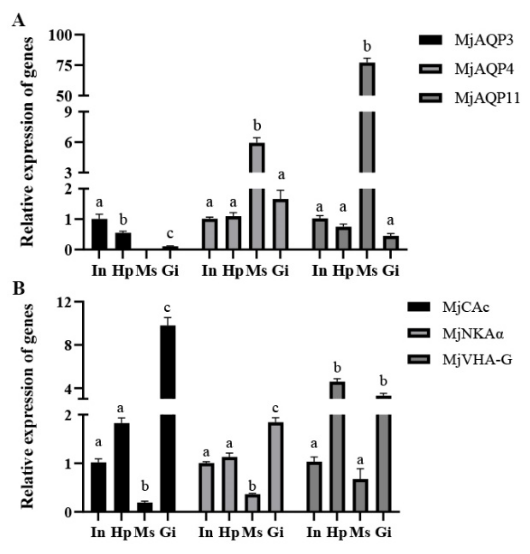
Tissue expression analysis revealed the differential expression patterns of the MjAQPs genes across the osmoregulatory tissues. MjAQP3 was expressed at the highest level in the intestine, while it was not detected in the gills (Figure 2A). In addition, the MjAQP3 gene was the most highly expressed aquaporin gene in the intestine (Figure S7). Although the highest expression levels of MjAQP4 and MjAQP11 were both observed in the muscle, the MjAQP11 gene was much more highly expressed than MjAQP4 in the muscle (Figure 2A and Figure S7). On the contrary, the expression level of MjAQP4 was higher than those of other MjAQPs in the gill (Figure S7).
The tissue distribution patterns of the ion-transportation-related genes were relatively uniform among the osmoregulatory tissues. (Figure 2B). The MjCAc expression profile was similar to that of the MjNKAα gene with the highest levels in the gills and the lowest levels in the muscle, while both genes showed intermediate expression in the hepatopancreas and muscle. As to the expression of MjVHA-G, it was highly expressed in the gills and hepatopancreas and lowly expressed in the muscle and intestine.

3.3. Effects of Acute Salinity Stress on Post-Larvae Shrimp

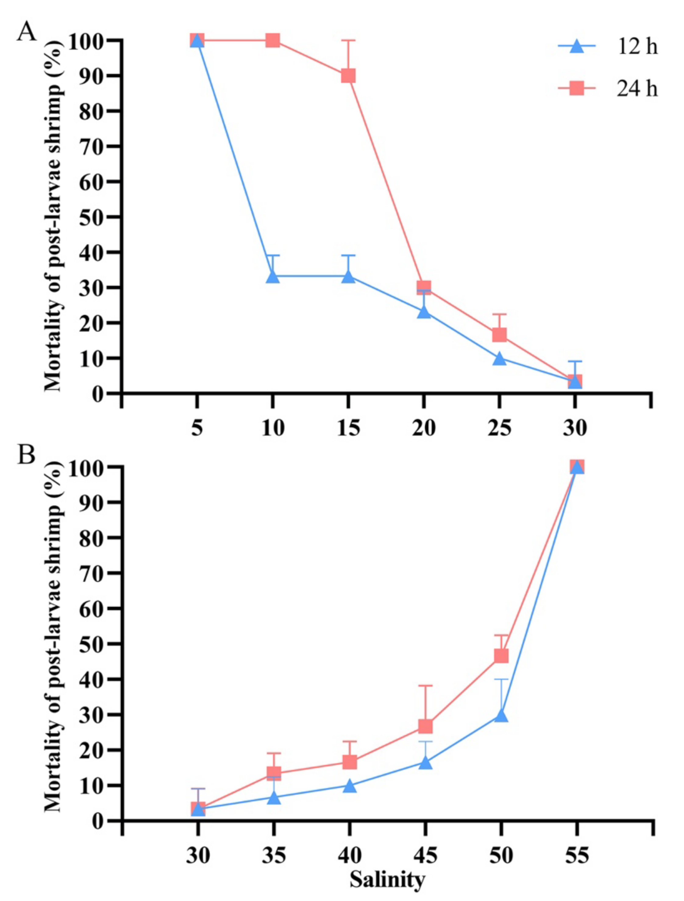
It was noted that with the declining salinity, the mortality of post-larvae shrimp increased over time (Figure 3A). More than half of the post-larvae shrimp could survive for 12 h when directly exposed to 10‰ salinity, while all died under 5‰ salinity after exposure for 12 h. As the salinity stresses continued for 24 h, the mortality sharply elevated at 10‰ and 15‰ salinity. Similar trends were observed for mortality with the rising salinity (Figure 3B). In addition, the majority of the post-larvae shrimp could survive for 12 h when directly exposed to 50‰ salinity, while all died under 55‰ salinity after exposure for 12 h. Moreover, the mortality rate of the shrimp at 50‰ salinity increased slightly during the following 12 h, showing a good tolerance to high-salinity stress. Based on the performance of post-larvae shrimp at different salinity concentrations, exposure to 10‰ and 50‰ salinity for 12 h were chosen for the following acute salinity stresses.
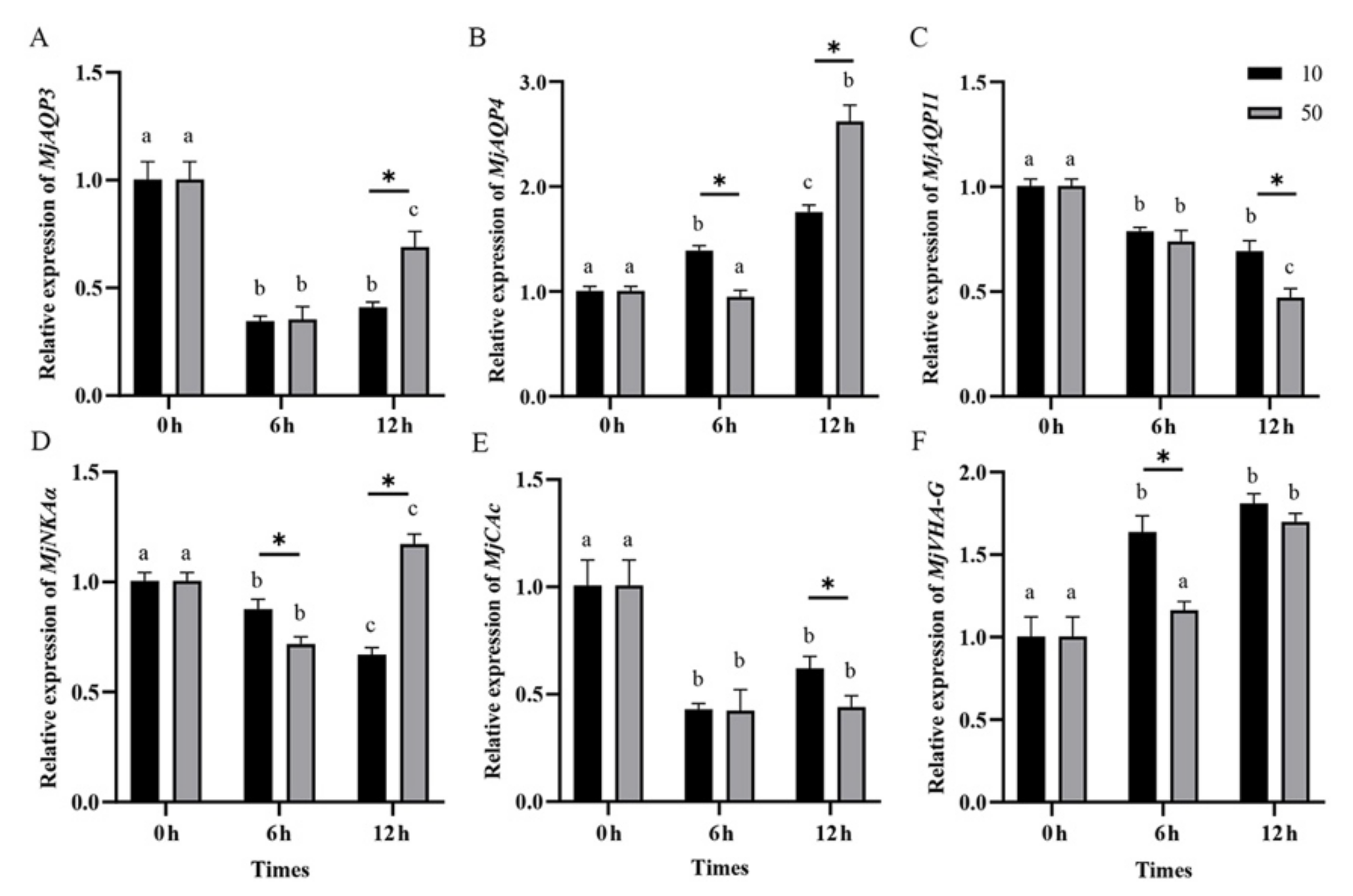
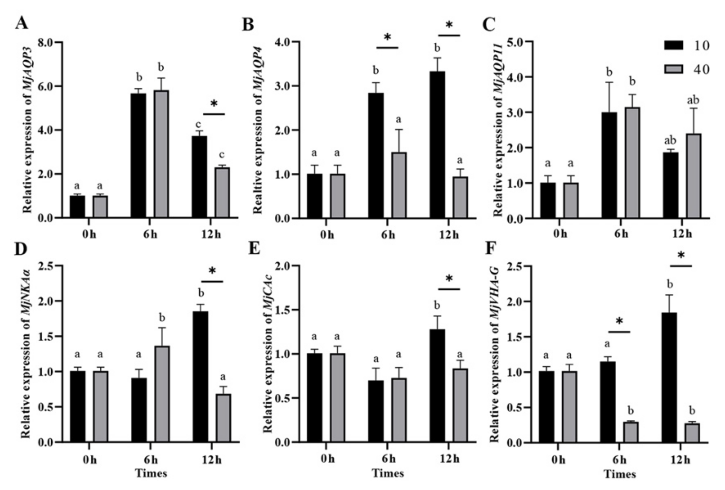
The expression level of MjAQP3 was significantly decreased at 6 h and then remained unchanged from 6 h to 12 h under 10‰ salinity (Figure 4A). In addition, the expression of MjAQP3 was also significantly down-regulated at 6 h, up-regulated from 6 h to 12 h, and still significantly lower than the initial level at the salinity of 50‰. Nevertheless, the expression level of MjAQP3 at 50‰ salinity was significantly higher than that at 10‰ salinity after 12 h of exposure (Figure 4A). There was a progressive increase in the expression levels of MjAQP4 from 0 h to 12 h at 10‰ salinity (Figure 4B). The expression levels of MjAQP4 were not significantly changed at 6 h, and then significantly increased from 6 h to 12 h at 50‰ salinity (Figure 4B). Moreover, there was a significant decline in the expression levels of MjAQP11 from 0 h to 12 h at both 10‰ and 50‰ salinity (Figure 4C).
A progressive decline in the expression level of MjNKAα was found from 0 h to 12 h at 10‰ salinity, whereas the expression of MjNKAα was significantly down-regulated at 6 h, then up-regulated from 6 h to 12 h, and was significantly higher than the initial level at the salinity of 50‰ (Figure 4D). The expression levels of MjCAc were significantly decreased at 6 h and then remained unchanged from 6 h to 12 h at both 10‰ and 50‰ salinity concentrations (Figure 4E). Additionally, a significantly elevated expression of MjVHA-G was observed at 6 h and the expression did not change significantly from 6 h to 12 h at 10‰ salinity. The expression of MjVHA-G was not significantly changed at 6 h, and then significantly increased from 6 h to 12 h at 50‰ salinity (Figure 4F).

3.4. Gene Expression in Post-Larvae Shrimp during the Salinity Acclimation

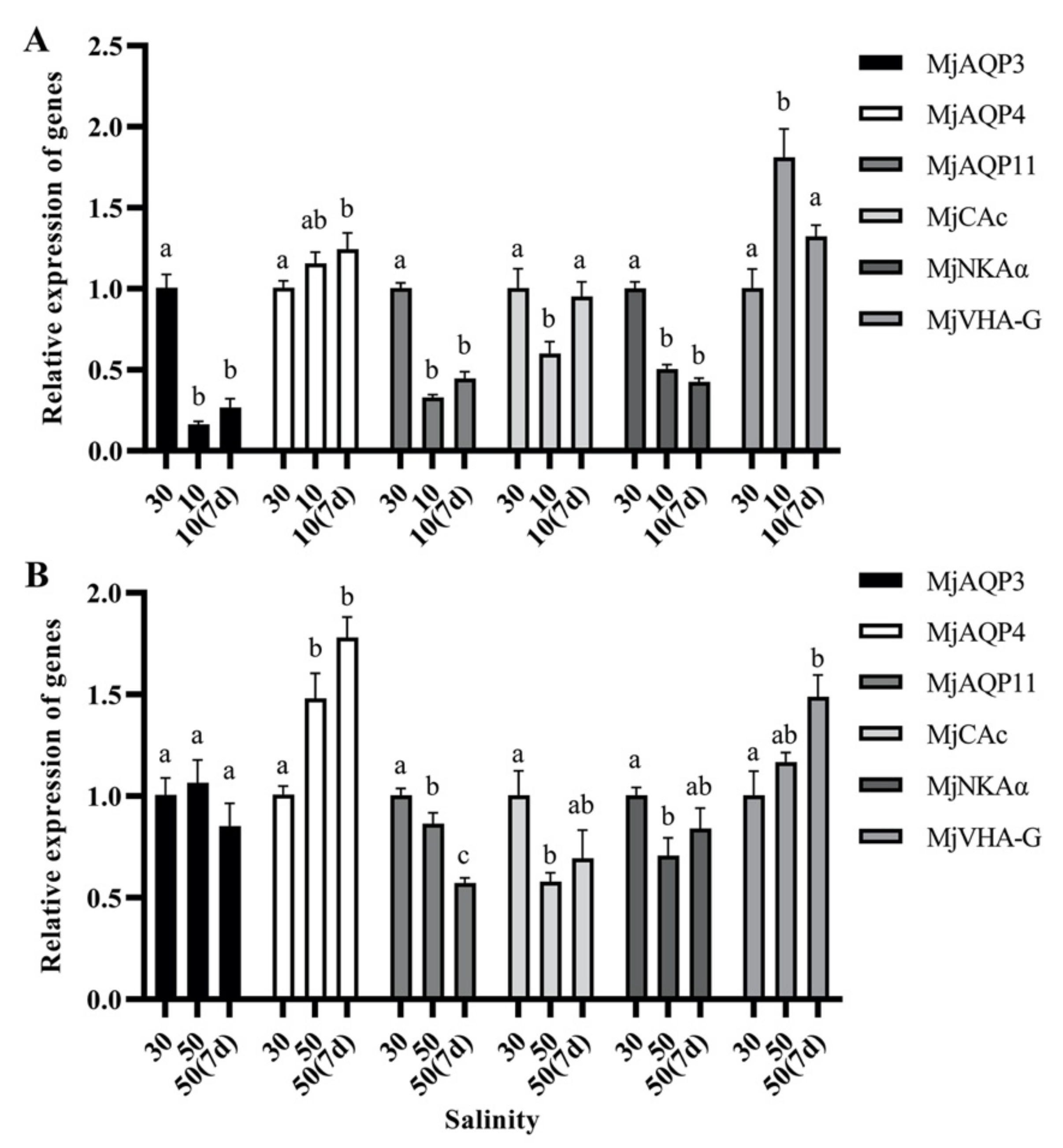
Four types of expression patterns of the genes were found during the acclimation to 10‰ salinity (Figure 5A). The expression levels of MjAQP3, MjAQP11, and MjNKAα were significantly decreased when the salinity declined to 10‰ salinity and remained constant in the following acclimation period. Additionally, the expression of MjAQP4 was slightly but not significantly elevated upon reaching 10‰ salinity, whereas its level was significantly higher than the initial value at the end of the acclimation period. Moreover, although the changing trend in the expression level of MjCAc was opposite to that noted for MjVHA-G when they acclimated to 10‰ salinity, both expression levels returned to the initial values after 7 d of long-term acclimation at 10‰ salinity.
These genes responded diversely to the high-salinity acclimation experiment (Figure 5B). There was no significant change in the expression of MjAQP3 during the whole acclimation period. The expression of MjAQP4 increased significantly and was maintained at a higher level, while the expression level of MjAQP11 was significantly decreased at 50‰ salinity, followed by a further reduction after 7 d of acclimation. Moreover, the expressions of MjCAc and MjNKAα first significantly declined at 50‰ salinity, and then returned to the initial level during further acclimation. MjVHA-G expression was slightly but not significantly elevated at 50‰ salinity, whereas its level was significantly increased during the following acclimation.

3.5. Gene Expression in Response to Acute Salinity Stresses in the Gills

There was a significant increase in the expression of MjAQP3 and MjAQP11 after 6 h of acute low-salinity and high-salinity stress (Figure 6A,C). Then, MjAQP3 expression declined and was still higher than the initial level, while MjAQP11 expression decreased to the initial value from 6 h to 12 h. Moreover, MjAQP4’s expression level at 40‰ salinity did not vary significantly up to 12 h, while its expression level at 10‰ salinity increased significantly at 6 h and then remained high from 6 h to 12 h (Figure 6B).
The expression of MjNKAα at 40‰ salinity showed a trend of rising first and then falling to the initial level, while it increased significantly from 6 h to 12 h at 10‰ salinity (Figure 6D). In addition, the MjCAc expression level at 40‰ did not vary significantly up to 12 h. Nevertheless, its expression level at 10‰ increased significantly from 6 h to 12 h and was significantly higher than that at 10‰ after 12 h of salinity exposure (Figure 6E). MjVHA-G exhibited the opposite response to low-salinity and high-salinity stress. The expression of MjVHA-G at 40‰ salinity decreased significantly at 6 h and then remained low from 6 h to 12 h, while it increased significantly from 6 h to 12 h at 10‰ salinity (Figure 6F).

3.6. Gene Expression in Response to Salinity Acclimation in the Gills

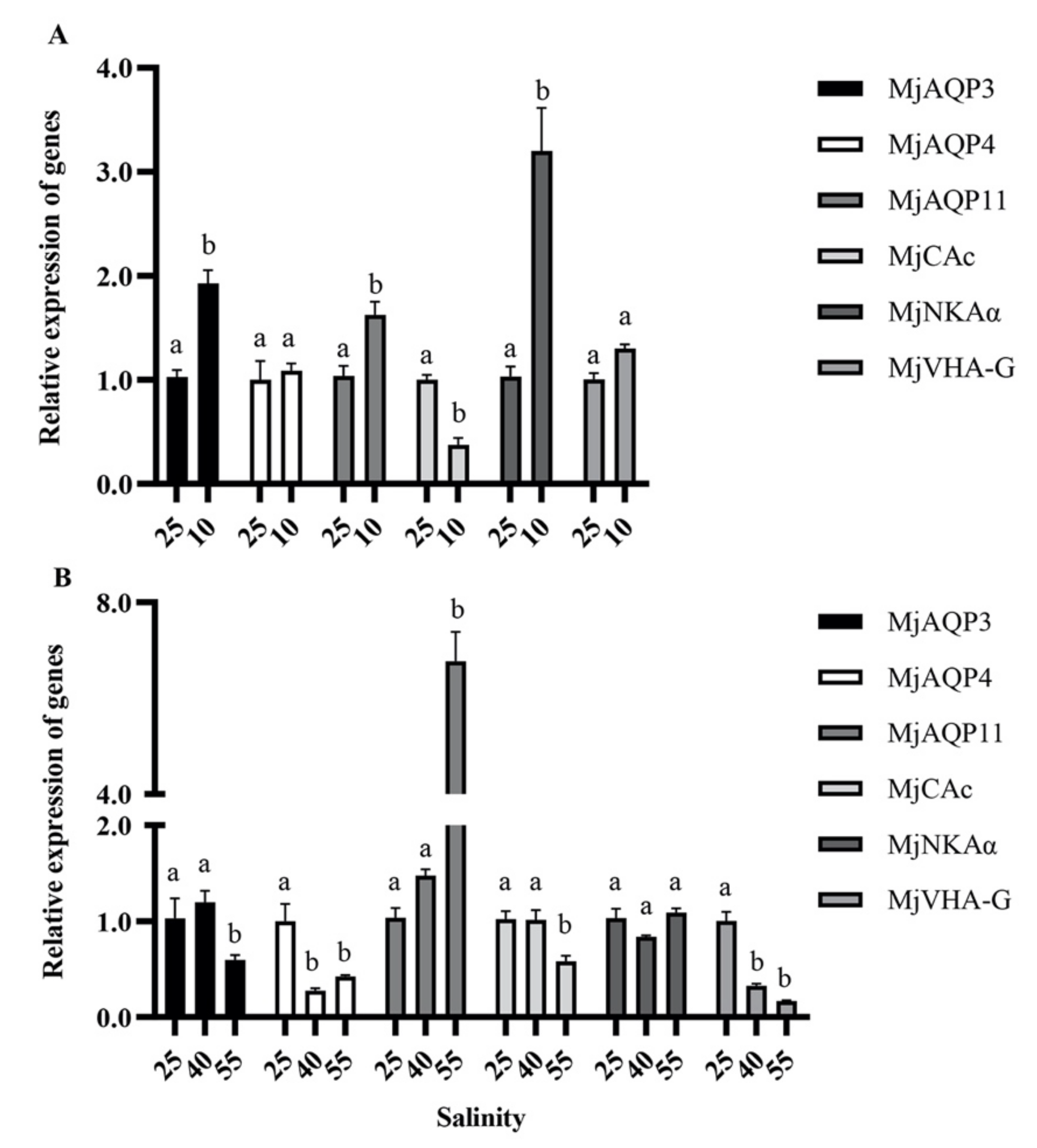
There was a significant increase in the expression of MjAQP3, MjAQP11, and MjNKAα concomitant with a significant decrease in the expression of MjCAc in the gills, whereas the expression of MjAQP4 and MjVHA-G remained unchanged when adult shrimp were acclimated from 25‰ to 10‰ salinity (Figure 7A). During the high-salinity acclimation period, the expression of MjAQP4 and MjVHA-G significantly declined at 40‰ salinity and was maintained at low levels at up to 55‰ salinity (Figure 7B). The expression levels of MjAQP3 and MjCAc were not significantly changed at 40‰ salinity, while they were significantly decreased at 55‰ salinity. In contrast, the expression of MjAQP11 was significantly elevated when it acclimated from 40‰ salinity to 55‰. Aside from the latter, there was no significant difference in the expression level of MjNKAα across the whole high-salinity acclimation period (Figure 7B).

4. Discussion

4.1. Tolerance of M. japonicus to Salinity Challenges

It has been reported that the kuruma shrimp can tolerate a wide range of salinities from 10‰ and 40‰ [32]. In this study, the post-larvae shrimp could not survive under the acute stress at 5‰ salinity for 12 h, suggesting that 5‰ salinity is lethal for M. japonicus. Recently, juvenile kuruma shrimp were reported to survive at 8‰ salinity for 96 h [36]. These results further confirm that the lower limit of salinity for M. japonicus to survive is ~10‰ salinity. Minor differences may result from the differences in the developmental stages of shrimp. Nevertheless, the intriguing findings are that M. japonicus could tolerate salinity up to 50‰ for the post-larvae shrimp and 55‰ for the adult shrimp during the acclimation process. In previous studies, salinities of 40‰ or 42‰ were commonly used as the salinity of acclimation in M. japonicus [37,38,39,40]. The current study is the first to report that the kuruma shrimp can be acclimated to 55‰ salinity. As the salinity of the high-salinity waters in northern China is more than 40‰ [30], M. japonicus has significant potential for aquaculture in these areas. The post-larvae shrimp could be first acclimated to the target salinity and then cultivated in outdoor ponds with high-salinity seawaters. Further studies are needed to comprehensively evaluate the growth, survival rate, and disease resistance of M. japonicus during long-term cultivation in high-salinity seawaters.

4.2. Expression of MjAQPs in Response to Salinity Challenges

Depending on the tertiary structure and selective permeability, vertebrate AQPs are classified into three categories: the classical aquaporins (AQP0, 1, 2, 4, 5, 6, and 8) for water transportation, the aquaglyceroporins (AQP3, 7, 9, and 10) potentially transporting water, and other small solutes, and the unorthodox aquaporins (AQP11 and 12) with an undetermined function [17,18]. However, although the three sub-families of AQPs have also been identified in crustaceans, the members of each sub-family differ significantly from those in vertebrates. A previous study has reported the identification and characterization of unique AQPs for each sub-type of L. vannamei [26]. In this study, three MjAQPs were found through an online BLAST search against AQPs from L. vannamei, which separately belong to the groups of aquaglyceroporins, classical aquaporins, and unorthodox aquaporins.
The MjAQP3 mRNA level in the post-larvae shrimp at 30‰ salinity was higher than that under acute low-salinity stress or long-term acclimation to low salinity. Similarly, the expression level of an aquaglyceroporin, CmGLP1, was decreased in the green crab (Carcinus maenas) under low-salinity exposure in comparison to crabs at normal seawater salinity [41]. Moreover, the expression of MjAQP3 in the post-larvae shrimp at 30‰ salinity was greater than that under acute high-salinity stress. In addition, the expression of AQP3 was also down-regulated in the intestine under acute high-salinity challenges in L. vannamei [26]. Besides water, the aquaglyceroporins also transport glycerol, urea, and other small solutes [16,42]. Moreover, MjAQP3 is primarily expressed in the intestine and hepatopancreas. The MjAQP3 gene may be involved in not only water transportation but also glycerol transportation for nutrient absorption along the intestine and hepatopancreas. The decreased expression of MjAQP3 implies weak functions regarding the digestion and absorption of nutrition under salinity stress. Aside from MjAQP3, the expression levels of MjAQP11 were also decreased in the post-larvae shrimp when exposed to low-salinity or high-salinity stress. Similar expression patterns were found for AQP11 in the muscle of L. vannamei [26]. Although the unorthodox AQPs have been reported to transport water and glycerol in the oocytes of medaka and zebrafish [43,44], there is still a lack of understanding regarding how unorthodox aquaporins regulate osmotic pressure in crustaceans. Nevertheless, these results imply the response of unorthodox aquaporins to the salinity changes in the shrimp. In contrast to the expression profiles of MjAQP3 and MjAQP11, the expression of MjAQP4 was up-regulated in the post-larvae shrimp under acute and long-term salinity challenges. The expression of a classical aquaporin, CsAQP1, is increased in larval blue crabs (Callinectes sapidus) after being exposed to low-salinity stress for 96 h [45]. In addition, AQP4 expression is elevated in the hepatopancreas of L. vannamei under acute high-salinity stress [46]. As the unique classical AQP in the shrimp, MjAQP4 should be primarily for transporting water and regulating cell volume, and thus has a critical role in the salinity adaption of the post-larvae shrimp.
In the post-larvae shrimp, we could only detect changes in the global expression levels of MjAQPs. However, a tissue distribution analysis implied that the MjAQPs have distinct tissue specificity, suggesting their different functions in different tissues. A higher expression of MjAQP3 and MjAQP11 was found in the gills under salinity stresses. Elevated expression levels of AQP3 have been reported in the gills of teleost fish in a high-salinity environment [43,47,48,49,50,51]. However, the results contradict their expression trends in the post-larvae shrimp. These inconsistent expression profiles may be related to the lower expression levels of MjAQP3 and MjAQP11 in the gills. Thus, the increased expression levels in the gills could not counteract the declined levels in other tissues. Therefore, it is necessary to further analyze the MjAQPs’ expression in one tissue to elucidate the roles of MjAQPs in adaption to salinity stress.
As the primary organ for osmoregulation, the gills sense salinity changes in their surrounding environment and adapt through the modulation of gene expression. The expression of MjAQP4 was not significantly changed in the gills under acute high-salinity stress, while it was significantly increased in the gills under acute low-salinity stress. Moreover, the expression of MjAQP3 and MjAQP11 was elevated in both treatments. As the most prominently expressed AQP gene in the gills, MjAQP4 is likely to play a dominant role in water transport. Accordingly, the MjAQP3 and MjAQP11 genes may have an assisting role in water transport. The results suggest that the global expression of MjAQPs may be orchestrated by the extent of the salinity shift. In this study, the kuruma shrimp show better tolerance to high-salinity stress. Thus, we conclude that low-salinity stress may present greater osmotic stress for the gills. Therefore, the expression of MjAQPs quickly increased under acute low-salinity stress, whereas only the up-regulation of MjAQP3 and MjAQP11 is required under acute high-salinity stress. The gills face the challenges of water influx in hypoosmotic conditions and the loss of water under hyperosmotic conditions [20,52,53]. Osmotic pressure from salinity changes drives water flow across the plasma membrane through AQPs, while the AQP channel determines the speed but not the direction of water flow [54]. The increased expression of AQPs is needed to excrete the excess water of the cells or to gain more water from the ambient environment and maintain the balance of intracellular volume and osmotic pressure. The expression of AQP is also higher at 0‰ and 10‰ salinity treatments than at 5‰ salinity in the gills of the prawn Macrobrachium australiense [23].
Challenges to the gills during the salinity acclimation process are different from those under acute salinity stress. Thus, the MjAQPs showed differential patterns in the gills in response to the salinity acclimation challenge. The salinity decreased by 5‰ daily during the low-salinity acclimation period and such minor salinity perturbations may cause reduced osmotic stress on the gills. This situation is similar to that for the gills under acute high salinity. Thus, the expression of MjAQP4 was not significantly changed, and the expression of MjAQP3 and MjAQP11 was elevated in the gills during the acclimation to 10‰ salinity. Moreover, the continuous high-salinity stress possibly imposes pressure on excessive water loss. To limit the rate at which water equilibrated across tissues during high-salinity acclimation, MjAQP4 expression significantly decreased at 40‰ and 55‰ salinity, leading to reductions in water transport in the gills.

4.3. Expression of Ion-Transportation-Related Genes in Response to Salinity Challenges

NKA is thought to be the main force driving the trans-epithelial movement of monovalent ions across the gill epithelia in crustaceans [7]. It consists of a single α-subunit, a glycosylated β-subunit, and a γ-subunit, of which the α-subunit contains an ATP-binding site, a phosphorylation site, and the amino acids essential for the binding of sodium and potassium ions [55]. Thus, NAKα has been shown to play an important role in osmoregulation in aquatic organisms under salinity challenges. In this study, the ORF sequence of NKAα was cloned from M. japonicus and a further phylogenetic analysis showed that MjNKA belonged to the shrimp NKAα sub-group. Previous studies have reported the enhanced expression of NKAα in the gills of euryhaline crustaceans subjected to osmotic stress [56,57,58,59,60]. The expression of MjNKAα increased significantly in the gills of the shrimp exposed to 10‰ or that acclimated to 10‰ salinity. The higher expression of MjNKAα may function to maintain the osmolality, composition, and volume of the hemolymph by actively transporting and exchanging Na+ and other critical cations through the gills. Nevertheless, its expression did not significantly change in the gills under high-salinity conditions. Significant changes in NKAα expression were not detected in the gills of swimming crabs (Portunus trituberculatus) under the low- and high-salinity conditions [22]. In addition, the NKA activity was unchanged in the gills of blue crabs (Callinectes danae) acclimated to 15‰ salinity and giant freshwater prawns (Macrobrachium rosenbergii) that acclimated from high-salinity to freshwater conditions [61,62]. These results suggest that the expression of NKAα might relate to the variable salinity tolerance among species. Beyond that, NKA activity has been shown to correlate with the developmental stages of M. japonicas [63], which may be the reason for the down-regulation of MjNKAα in the post-larvae shrimp in low-salinity conditions. Another reason may be that the expression of MjNKAα decreased in the other tissues of post-larvae shrimp. Recently, the expression of MjNKAα was found to be down-regulated in the hepatopancreas of juvenile shrimp after 24 h of low-salinity stress [36].
CA catalyzes the reversible hydration reaction of carbon dioxide (CO2) into protons (H+) and bicarbonate (HCO3) [64]. There are three types of CA in arthropods, including cytoplasmic CA (CAc), glycosyl-phosphatidylinositol-linked CA (CAg), and beta CA (CAb) [65]. The CAc isoform exists in the cytoplasm and provides counter ions (H+ and HCO3) for the exchange of Na+/H+ and Cl/HCO3 across the plasma membrane; thus, it is directly involved in osmotic and ionic regulation [66]. In this study, the ORF sequence of one CA was cloned from M. japonicus and further phylogenetic analysis confirmed that the MjCA was clustered into the CAc sub-group. The expression of CAc typically increases rapidly to supply the counter ions used in NaCl uptake under low-salinity challenges in the gills of euryhaline crustaceans [14,67,68,69,70,71]. The expression of MjCAc in the gills also increased significantly after a 12 h exposure to low salinity. However, beyond that, the expression levels of MjCAc were significantly decreased in the post-larvae shrimp following acute low-salinity stress. Moreover, decreased MjCAc expression was also observed in the gills of shrimp after the acclimation to low salinity. CA has been reported to show a lower expression level in P. trituberculatus after exposure to a low salinity of 11 ppt [72]. The reduction in the expression of CA mRNA might be due to the extremely low-salinity conditions. In this study, 10‰ salinity nearly reaches the limit of salinity for the survival of M. japonicus. The activity of CA and expression of CA mRNA in the gills increased with the decreased salinity when the C. maenas subjects were transferred from 32 ppt salinity to 25, 20, and 15 ppt salinity, while both reduced when the salinity continued to drop to 10 ppt [67]. These results suggest that extreme hypoosmotic stress may impede the induction of CA mRNA. Furthermore, the expression of MjCA was also significantly decreased in the post-larvae shrimp or the gills when exposed to high-salinity conditions. The expression of CA was down-regulated in the gills of P. trituberculatus after their long-term adaption to high salinity [22]. In hyperosmotic conditions, organisms must prevent high concentrations of external ions from penetrating the body. Thus, the down-regulation of MjCAc may involve the active secretion of NaCl to compensate for the passive NaCl influx from the external environment.
VHA is a multiple-subunit protein containing two domains: V0 and V1. The V1 domain consists of eight different subunits (A–H) responsible for ATP hydrolysis, and the V0 domain is composed of six subunits and is involved in the translocation of protons across the membrane [73]. Thus, VHA can participate in ion regulation by actively transporting protons into the medium, which in turn allows Na+ to flow in through Na channels and Na+/H+ exchangers [74,75]. A previous transcriptome analysis showed that the VHA-G subunit had the highest expression levels and was differentially expressed under salinity stress in the gills of L. vannamei [1]. Based on this result, we cloned the VHA-G subunit of M. japonicus to explore the function of MjVHA-G in response to salinity stress. The expression of MjVHA-G in the gills increased under low-salinity stress and was down-regulated following high-salinity stress. Similar expression of patterns of VHA subunit mRNA have been reported in crustaceans [4,76,77,78]. However, the expression of MjVHA-G in the post-larvae shrimp increased significantly at high-salinity conditions, which was not consistent with those in the gills. As it also shows high expression in the hepatopancreas, this inconformity may be due to its expression in other tissues.
Taken together, the expression levels of MjNKAα, MjCAc, and MjVHA-G were all promoted in the gills under acute low-salinity conditions. MjCA catalyzes the hydration of CO2 to provide H+ and HCO3, and then MjVHA complements MjNAK by powering sodium and chloride ions’ uptake by the excretion of H+ and HCO3 in lowly ionic environments. In addition, MjCAc and MjVHA-G are down-regulated in response to high salinity to reduce the intake of sodium and chloride ions by the epithelial cells in the gills.

5. Conclusions

It is essential for crustaceans to maintain ionic balance and water homeostasis to regulate osmoregulation in a salinity-varying environment. This study mainly investigated the expression patterns of key osmoregulatory genes involved in ion and water transportation in M. japonicus at different salinities. The results reveal remarkable differences in the patterns of gene expression depending on the intensity, exposure time, and treatment conditions of salinity stress. Overall, MjAQPs show varied tissue distribution patterns and are operative to different extents in different tissues and osmotic conditions. On the contrary, MjNKAα, MjCAc, and MjVHA-G have some consistent tissue distribution patterns and tend to act in tight cooperation to allow for effective osmoregulation.

Supplementary Materials

The following are available online at https://www.mdpi.com/article/10.3390/jmse10121870/s1, Figure S1: The ORFs and deduced amino acid sequences of (A) MjAQP3, (B) MjAQP4, and (C) MjAQP11. The asterisk stands for the stop codon. The NPA motifs are underlined with single line, Figure S2: Multiple alignment of deduced amino acid sequences of MjAQPs against selected AQPs sequences. Dark gray shading indicates residues conserved in >50% of the sequences, light gray shading indicates residues with similar properties. The NPA motifs are underlined with single line, Figure S3: The ORFs and deduced amino acid sequences of (A) MjNKAα, (B) MjCAc, and (C) MjVHA-G. The asterisk stands for the stop codon. The alpha-carbonic anhydrases signature is underlined. The zinc-binding histidine residues of MjCAc are in black box and the proton acceptor site of MjCAc is in black circle. The eight transmembrane domains of MjNKAα are underlined with single line, Figure S4: Multiple alignment of deduced amino acid sequences of MjCAc against selected CAc sequences. The conserved zinc-binding histidine residues are in red box and the most common proton acceptor sites are in blue box. Dark gray shading indicates residues conserved in >50% of the sequences, light gray shading indicates residues with similar properties, Figure S5: Multiple alignment of deduced amino acid sequences of MjNKAα against selected NKAα sequences. The eight transmembrane domains of NKAα are in red box. Dark gray shading indicates residues conserved in >50% of the sequences, light gray shading indicates residues with similar properties, Figure S6: Multiple alignment of deduced amino acid sequences of MjVHA-G against selected VHA-G sequences. Dark gray shading indicates residues conserved in >50% of the sequences, light gray shading indicates residues with similar properties, Figure S7: The relative expression levels of MjAQPs in different tissues of M. japonicus. Abbreviations: In, intestine; Hp, hepatopancreas; Ms, muscle; Gi, gill. The relative expression levels of MjAQP3 in the intestine, hepatopancreas, and gill, and the relative expression level of MjAQP4 in the muscle are separately used as calibrator. The data are presented as mean + SD (n = 3). Means labeled with different letters are significantly different (p < 0.05, one-way ANOVA, Tukey).

Author Contributions

Conceptualization, Y.C. and Y.L.; methodology, Z.Z.; software, Z.W.; validation, Y.L., F.L. and Y.C.; formal analysis, Z.Z. and L.Z.; investigation, R.Z. and X.W.; resources, Z.D. and J.S.; data curation, R.W.; writing—original draft preparation, Z.Z.; writing—review and editing, Y.C.; visualization, P.H.; supervision, Y.C.; project administration, Y.C.; funding acquisition, Y.C. and Y.L. All authors have read and agreed to the published version of the manuscript.

Funding

This work was supported by the Key Research and Development Plan in Shandong Province (2021LZGC027); the National Science Foundation of China (31802269; 31902407); the Shrimp and Crab Innovation Team of Shandong Agriculture Research System (SDAIT-15-011); the Natural Science Foundation of Shandong Province (ZR2019BC013); the High-level Talents Research Fund of Qingdao Agricultural University (663/1119054); and the “First Class Fishery Discipline” program in Shandong Province.

Institutional Review Board Statement

Not applicable.

Informed Consent Statement

Not applicable.

Data Availability Statement

The mRNA sequences in this study were submitted to GenBank with the accession numbers OP823702-OP823707.

Conflicts of Interest

The authors declare no conflict of interest.

References

  1. Li, Y.; Chen, Y.; Cui, Y.; Shen, M.; Wang, R.; Wang, Z. Transcriptome Analysis of Pacific White Shrimp (Litopenaeus vannamei) Under Prolonged High-Salinity Stress. J. Ocean Univ. China 2022, 21, 430–444. [Google Scholar] [CrossRef]
  2. McNamara, J.C.; Faria, S.C. Evolution of Osmoregulatory Patterns and Gill Ion Transport Mechanisms in the Decapod Crustacea: A Review. J. Comp. Physiol. B 2012, 182, 997–1014. [Google Scholar] [CrossRef]
  3. Rahi, M.L.; Moshtaghi, A.; Mather, P.B.; Hurwood, D.A. Osmoregulation in Decapod Crustaceans: Physiological and Genomic Perspectives. Hydrobiologia 2018, 825, 177–188. [Google Scholar] [CrossRef]
  4. Rahi, M.L.; Amin, S.; Mather, P.B.; Hurwood, D.A. Candidate Genes That Have Facilitated Freshwater Adaptation by Palaemonid Prawns in the Genus Macrobrachium: Identification and Expression Validation in a Model Species (M. Koombooloomba). PeerJ 2017, 5, e2977. [Google Scholar] [CrossRef] [Green Version]
  5. McNamara, J.C.; Freire, C.A.; Torres, A.H., Jr.; Faria, S.C. The Conquest of Fresh Water by the Palaemonid Shrimps: An Evolutionary History Scripted in the Osmoregulatory Epithelia of the Gills and Antennal Glands. Biol. J. Linn. Soc. 2015, 114, 673–688. [Google Scholar] [CrossRef] [Green Version]
  6. Thabet, R.; Ayadi, H.; Koken, M.; Leignel, V. Homeostatic Responses of Crustaceans to Salinity Changes. Hydrobiologia 2017, 799, 1–20. [Google Scholar] [CrossRef]
  7. Lucu, Č.; Towle, D.W. Na++K+-ATPase in Gills of Aquatic Crustacea. Comp. Biochem. Physiol. Part A Mol. Integr. Physiol. 2003, 135, 195–214. [Google Scholar] [CrossRef]
  8. Boudour-Boucheker, N.; Boulo, V.; Charmantier-Daures, M.; Anger, K.; Charmantier, G.; Lorin-Nebel, C. Osmoregulation in Larvae and Juveniles of Two Recently Separated Macrobrachium Species: Expression Patterns of Ion Transporter Genes. Comp. Biochem. Physiol. Part A Mol. Integr. Physiol. 2016, 195, 39–45. [Google Scholar] [CrossRef] [PubMed] [Green Version]
  9. Tsai, J.-R.; Lin, H.-C. V-Type H+-ATPase and Na+, K+-ATPase in the Gills of 13 Euryhaline Crabs during Salinity Acclimation. J. Exp. Biol. 2007, 210, 620–627. [Google Scholar] [CrossRef] [PubMed] [Green Version]
  10. Havird, J.C.; Henry, R.P.; Wilson, A.E. Altered Expression of Na+/K+–ATPase and Other Osmoregulatory Genes in the Gills of Euryhaline Animals in Response to Salinity Transfer: A Meta-Analysis of 59 Quantitative PCR Studies over 10 years. Comp. Biochem. Physiol. Part D Genom. Proteom. 2013, 8, 131–140. [Google Scholar] [CrossRef]
  11. Leone, F.A.; Lucena, M.N.; Garçon, D.P.; Pinto, M.R.; McNamara, J.C. Gill Ion Transport ATPases and Ammonia Excretion in Aquatic Crustaceans. In Acid-Base Balance and Nitrogen Excretion in Invertebrates; Springer: Cham, Switzerland, 2017; pp. 61–107. [Google Scholar]
  12. Weihrauch, D.; Ziegler, A.; Siebers, D.; Towle, D.W. Molecular Characterization of V-Type H(+)-ATPase (B-Subunit) in Gills of Euryhaline Crabs and Its Physiological Role in Osmoregulatory Ion Uptake. J. Exp. Biol. 2001, 204, 25–37. [Google Scholar] [CrossRef]
  13. Burnett, L.E.; Dunn, T.N.; Infantino, R.L. The Function of Carbonic Anhydrase in Crustacean Gills. In Proceedings of the Transport Processes, Iono- and Osmoregulation; Gilles, R., Gilles-Baillien, M., Eds.; Springer: Berlin/Heidelberg, Germany, 1985; pp. 159–168. [Google Scholar]
  14. Ge, Q.; Li, J.; Wang, J.; Li, Z.; Li, J. Characterization, Functional Analysis, and Expression Levels of Three Carbonic Anhydrases in Response to PH and Saline–Alkaline Stresses in the Ridgetail White Prawn Exopalaemon carinicauda. Cell Stress Chaperones 2019, 24, 503–515. [Google Scholar] [CrossRef] [PubMed]
  15. Abascal, F.; Irisarri, I.; Zardoya, R. Diversity and Evolution of Membrane Intrinsic Proteins. Biochim. Biophys. Acta BBA Gen. Subj. 2014, 1840, 1468–1481. [Google Scholar] [CrossRef] [PubMed] [Green Version]
  16. Finn, R.N.; Cerdà, J. Evolution and Functional Diversity of Aquaporins. Biol. Bull. 2015, 229, 6–23. [Google Scholar] [CrossRef] [PubMed]
  17. Benga, G. On the Definition, Nomenclature and Classification of Water Channel Proteins (Aquaporins and Relatives). Mol. Asp. Med. 2012, 33, 514–517. [Google Scholar] [CrossRef] [PubMed]
  18. Ishibashi, K.; Morishita, Y.; Tanaka, Y. The Evolutionary Aspects of Aquaporin Family. In Aquaporins; Advances in Experimental Medicine and Biology; Springer: Dordrecht, The Netherlands, 2017; Volume 969, pp. 35–50. [Google Scholar] [CrossRef]
  19. Boyle, R.T.; Oliveira, L.F.; Bianchini, A.; Souza, M.M. The Effects of Copper on Na+/K--ATPase and Aquaporin Expression in Two Euryhaline Invertebrates. Bull. Environ. Contam. Toxicol. 2013, 90, 387–390. [Google Scholar] [CrossRef] [PubMed]
  20. Foguesatto, K.; Boyle, R.T.; Rovani, M.T.; Freire, C.A.; Souza, M.M. Aquaporin in Different Moult Stages of a Freshwater Decapod Crustacean: Expression and Participation in Muscle Hydration Control. Comp. Biochem. Physiol. Part A Mol. Integr. Physiol. 2017, 208, 61–69. [Google Scholar] [CrossRef] [PubMed]
  21. Lind, U.; Järvå, M.; Alm Rosenblad, M.; Pingitore, P.; Karlsson, E.; Wrange, A.-L.; Kamdal, E.; Sundell, K.; André, C.; Jonsson, P.R. Analysis of Aquaporins from the Euryhaline Barnacle Balanus improvisus Reveals Differential Expression in Response to Changes in Salinity. PLoS ONE 2017, 12, e0181192. [Google Scholar] [CrossRef] [Green Version]
  22. Lv, J.; Liu, P.; Wang, Y.; Gao, B.; Chen, P.; Li, J. Transcriptome Analysis of Portunus trituberculatus in Response to Salinity Stress Provides Insights into the Molecular Basis of Osmoregulation. PLoS ONE 2013, 8, e82155. [Google Scholar] [CrossRef] [PubMed]
  23. Moshtaghi, A.; Rahi, M.L.; Mather, P.B.; Hurwood, D.A. An Investigation of Gene Expression Patterns That Contribute to Osmoregulation in Macrobrachium australiense: Assessment of Adaptive Responses to Different Osmotic Niches. Gene Rep. 2018, 13, 76–83. [Google Scholar] [CrossRef]
  24. Stavang, J.A.; Chauvigné, F.; Kongshaug, H.; Cerdà, J.; Nilsen, F.; Finn, R.N. Phylogenomic and Functional Analyses of Salmon Lice Aquaporins Uncover the Molecular Diversity of the Superfamily in Arthropoda. BMC Genom. 2015, 16, 618. [Google Scholar] [CrossRef] [PubMed] [Green Version]
  25. Wang, C.; Zhang, Z.; Deng, Z.; Sun, J.; Zhao, R.; Cui, Y.; Wang, R.; Li, Y.; Wang, Z. Classical Aquaporins from Pacific White Shrimp (Litopenaeus vannamei): Molecular Characterization and Expression Analysis in Hypersalinity. Aquac. Rep. 2022, 23, 101016. [Google Scholar] [CrossRef]
  26. Wang, Z.; Chen, Y.; Wang, C.; Zhao, N.; Zhang, Z.; Deng, Z.; Cui, Y.; Wang, R.; Li, Y. Aquaporins in Pacific White Shrimp (Litopenaeus vannamei): Molecular Characterization, Expression Patterns, and Transcriptome Analysis in Response to Salinity Stress. Front. Mar. Sci. 2022, 9, 817868. [Google Scholar] [CrossRef]
  27. Zhong, S.; Mao, Y.; Wang, J.; Liu, M.; Zhang, M.; Su, Y. Transcriptome Analysis of Kuruma Shrimp (MarsuPenaeus japonicus) Hepatopancreas in Response to White Spot Syndrome Virus (WSSV) under Experimental Infection. Fish Shellfish Immunol. 2017, 70, 710–719. [Google Scholar] [CrossRef] [PubMed]
  28. Chen, X.; Song, L.; Li, W.; Wu, J. Acute Toxicity of Salinity to Sinogastromyzon szechuanensis. J. Phys. Conf. Ser. 2020, 1629, 012025. [Google Scholar] [CrossRef]
  29. He, W.; Yu, T.; Cai, Y. Analysis of Control and Use of Alkaline Soil. J. Anshan Univ. Sci. Technol. 2010, 29, 158–160. [Google Scholar]
  30. Li, C.; Li, N.; Dong, T.; Fu, Q.; Cui, Y.; Li, Y. Analysis of Differential Gene Expression in Litopenaeus vannamei under High Salinity Stress. Aquac. Rep. 2020, 18, 100423. [Google Scholar] [CrossRef]
  31. Lv, X.; Xu, H.; Li, L.; Zhao, Y. Agricultural Sustainable Utilization and Evaluation of Saline-Alkali Land. Soils 2012, 44, 203–207. [Google Scholar]
  32. Boyd, C.E.; Fast, A.W. Pond Monitoring and Management: Marine Shrimp Culture Principles an Practices; Elsevier Science Publishing Comp. Inc.: New York, NY, USA, 1992. [Google Scholar]
  33. Kumar, S.; Stecher, G.; Li, M.; Knyaz, C.; Tamura, K. MEGA X: Molecular Evolutionary Genetics Analysis across Computing Platforms. Mol. Biol. Evol. 2018, 35, 1547–1549. [Google Scholar] [CrossRef]
  34. Waterhouse, A.M.; Procter, J.B.; Martin, D.M.; Clamp, M.; Barton, G.J. Jalview Version 2—A Multiple Sequence Alignment Editor and Analysis Workbench. Bioinformatics 2009, 25, 1189–1191. [Google Scholar] [CrossRef] [Green Version]
  35. Letunic, I.; Bork, P. Interactive Tree of Life (ITOL) v5: An Online Tool for Phylogenetic Tree Display and Annotation. Nucleic Acids Res. 2021, 49, W293–W296. [Google Scholar] [CrossRef]
  36. Chen, T.; Li, Z.; Liu, J.; Liang, C.; Yuan, L. Transcriptome of Hepatopancreas in Kuruma Shrimp MarsuPenaeus japonicus under Low-Salinity Stress. J. Oceanol. Limnol. 2022, 40, 745–765. [Google Scholar] [CrossRef]
  37. Cheng, S.-Y.; Lee, W.-C.; Chen, J.-C. Increase of Uricogenesis in the Kuruma Shrimp MarsuPenaeus japonicus Reared under Hyper-Osmotic Conditions. Comp. Biochem. Physiol. Part B Biochem. Mol. Biol. 2004, 138, 245–253. [Google Scholar] [CrossRef] [PubMed]
  38. Koyama, H.; Kuniyoshi, H.; Piyapattanakorn, S.; Watabe, S. Changes in Metabolite Concentrations in the Abdominal Muscle of the Kuruma Shrimp MarsuPenaeus japonicus in Response to Different Salinities. Fish. Sci. 2021, 87, 383–401. [Google Scholar] [CrossRef]
  39. Koyama, H.; Mizusawa, N.; Hoashi, M.; Tan, E.; Yasumoto, K.; Jimbo, M.; Ikeda, D.; Yokoyama, T.; Asakawa, S.; Piyapattanakorn, S. Changes in Free Amino Acid Concentrations and Associated Gene Expression Profiles in the Abdominal Muscle of Kuruma Shrimp (MarsuPenaeus japonicus) Acclimated at Different Salinities. J. Exp. Biol. 2018, 221, jeb168997. [Google Scholar] [CrossRef] [PubMed] [Green Version]
  40. Lee, W.-C.; Chen, J.-C. Hemolymph Ammonia, Urea and Uric Acid Levels and Nitrogenous Excretion of MarsuPenaeus japonicus at Different Salinity Levels. J. Exp. Mar. Biol. Ecol. 2003, 288, 39–49. [Google Scholar] [CrossRef]
  41. Nash, M.T.; Quijada-Rodriguez, A.R.; Allen, G.J.P.; Wilson, J.M.; Weihrauch, D. Characterization of 3 Different Types of Aquaporins in Carcinus maenas and Their Potential Role in Osmoregulation. Comp. Biochem. Physiol. Part A Mol. Integr. Physiol. 2022, 272, 111281. [Google Scholar] [CrossRef]
  42. Ishibashi, K.; Tanaka, Y.; Morishita, Y. Chapter One - Perspectives on the Evolution of Aquaporin Superfamily. In Vitamins and Hormones; Elsevier Inc.: Amsterdam, The Netherlands, 2020; Volume 112, pp. 1–27. [Google Scholar] [CrossRef]
  43. Kim, Y.K.; Lee, S.Y.; Kim, B.S.; Kim, D.S.; Nam, Y.K. Isolation and MRNA Expression Analysis of Aquaporin Isoforms in Marine Medaka Oryzias dancena, a Euryhaline Teleost. Comp. Biochem. Physiol. Part A Mol. Integr. Physiol. 2014, 171, 1–8. [Google Scholar] [CrossRef]
  44. Tingaud-Sequeira, A.; Calusinska, M.; Finn, R.N.; Chauvigné, F.; Lozano, J.; Cerdà, J. The Zebrafish Genome Encodes the Largest Vertebrate Repertoire of Functional Aquaporins with Dual Paralogy and Substrate Specificities Similar to Mammals. BMC Evol. Biol. 2010, 10, 38. [Google Scholar] [CrossRef] [Green Version]
  45. Chung, J.S.; Maurer, L.; Bratcher, M.; Pitula, J.S.; Ogburn, M.B. Cloning of Aquaporin-1 of the Blue Crab, Callinectes sapidus: Its Expression during the Larval Development in Hyposalinity. Aquat. Biosyst. 2012, 8, 21. [Google Scholar] [CrossRef]
  46. Gao, Y.; Hu, C.; Ren, C.; Qian, J.; He, X.; Jiang, X.; Huang, W. Molecular Cloning of Aquaporin-4 (AQP4) Gene in the Pacific White Shrimp (Litopenaeus vannamei) and the Effect of Salinity Stress on Its Expression in Hepatopancreas. Mar. Sci. 2017, 41, 61–70. [Google Scholar]
  47. Breves, J.P.; Inokuchi, M.; Yamaguchi, Y.; Seale, A.P.; Hunt, B.L.; Watanabe, S.; Lerner, D.T.; Kaneko, T.; Grau, E.G. Hormonal Regulation of Aquaporin 3: Opposing Actions of Prolactin and Cortisol in Tilapia Gill. J. Endocrinol. 2016, 230, 325–337. [Google Scholar] [CrossRef] [Green Version]
  48. Cutler, C.P.; Phillips, C.; Hazon, N.; Cramb, G. Cortisol Regulates Eel (Anguilla anguilla) Aquaporin 3 (AQP3) MRNA Expression Levels in Gill. Gen. Comp. Endocrinol. 2007, 152, 310–313. [Google Scholar] [CrossRef] [PubMed]
  49. Ellis, L.V.; Bollinger, R.J.; Weber, H.M.; Madsen, S.S.; Tipsmark, C.K. Differential Expression and Localization of Branchial AQP1 and AQP3 in Japanese Medaka (Oryzias latipes). Cells 2019, 8, 422. [Google Scholar] [CrossRef] [Green Version]
  50. Giffard-Mena, I.; Boulo, V.; Aujoulat, F.; Fowden, H.; Castille, R.; Charmantier, G.; Cramb, G. Aquaporin Molecular Characterization in the Sea-Bass (Dicentrarchus labrax): The Effect of Salinity on AQP1 and AQP3 Expression. Comp. Biochem. Physiol. Part A Mol. Integr. Physiol. 2007, 148, 430–444. [Google Scholar] [CrossRef] [PubMed]
  51. Tipsmark, C.K.; Sørensen, K.J.; Madsen, S. Aquaporin Expression Dynamics in Osmoregulatory Tissues of Atlantic Salmon during Smoltification and Seawater Acclimation. J. Exp. Biol. 2010, 213, 368–379. [Google Scholar] [CrossRef] [Green Version]
  52. Moshtaghi, A.; Rahi, M.L.; Mather, P.B.; Hurwood, D.A. Understanding the Genomic Basis of Adaptive Response to Variable Osmotic Niches in Freshwater Prawns: A Comparative Intraspecific RNA-Seq Analysis of Macrobrachium australiense. J. Hered. 2017, 108, 544–552. [Google Scholar] [CrossRef] [Green Version]
  53. Towle, D.W.; Henry, R.P.; Terwilliger, N.B. Microarray-Detected Changes in Gene Expression in Gills of Green Crabs (Carcinus maenas) upon Dilution of Environmental Salinity. Comp. Biochem. Physiol. Part D Genom. Proteom. 2011, 6, 115–125. [Google Scholar] [CrossRef] [PubMed]
  54. Wang, S.C.; Parpura, V.; Wang, Y.-F. Astroglial Regulation of Magnocellular Neuroendocrine Cell Activities in the Supraoptic Nucleus. Neurochem. Res. 2021, 46, 2586–2600. [Google Scholar] [CrossRef] [PubMed]
  55. Geering, K. Functional Roles of Na, K-ATPase Subunits. Curr. Opin. Nephrol. Hypertens. 2008, 17, 526–532. [Google Scholar] [CrossRef]
  56. Li, J.; Ma, P.; Liu, P.; Chen, P.; Li, J. The Roles of Na+/K+-ATPase α-Subunit Gene from the Ridgetail White Prawn Exopalaemon carinicauda in Response to Salinity Stresses. Fish Shellfish Immunol. 2015, 42, 264–271. [Google Scholar] [CrossRef] [PubMed]
  57. Lovett, D.L.; Verzi, M.P.; Burgents, J.E.; Tanner, C.A.; Glomski, K.; Lee, J.J.; Towle, D.W. Expression Profiles of Na+, K+-ATPase during Acute and Chronic Hypo-Osmotic Stress in the Blue Crab Callinectes sapidus. Biol. Bull. 2006, 211, 58–65. [Google Scholar] [CrossRef] [PubMed]
  58. Mendonça, N.; Masui, D.; McNamara, J.; Leone, F.; Furriel, R. Long-Term Exposure of the Freshwater Shrimp Macrobrachium olfersii to Elevated Salinity: Effects on Gill (Na+, K+)-ATPase α-Subunit Expression and K+-Phosphatase Activity. Comp. Biochem. Physiol. Part A Mol. Integr. Physiol. 2007, 146, 534–543. [Google Scholar] [CrossRef] [PubMed]
  59. Paulsen, R.; Weihrauch, D.; Lignot, J.; Towle, D. ATPase in Gills of the Blue Crab Callinectes sapidus: CDNA Sequencing and Salinit-Related Expression of Alpha Subunit MRNA and Protein; American Society of Zoologists: Lawrence, KS, USA, 2000; Volume 40, pp. 1166–1167. [Google Scholar]
  60. Sun, H.; Zhang, L.; Ren, C.; Chen, C.; Fan, S.; Xia, J.J.; Lin, H.; Hu, C. The Expression of Na, K-ATPase in Litopenaeus vannamei under Salinity Stress. Mar. Biol. Res. 2011, 7, 623–628. [Google Scholar] [CrossRef]
  61. Masui, D.C.; Mantelatto, F.L.; McNamara, J.C.; Furriel, R.P.; Leone, F.A. Na+, K+-ATPase Activity in Gill Microsomes from the Blue Crab, Callinectes danae, Acclimated to Low Salinity: Novel Perspectives on Ammonia Excretion. Comp. Biochem. Physiol. Part A Mol. Integr. Physiol. 2009, 153, 141–148. [Google Scholar] [CrossRef]
  62. Wilder, M.N.; Atmomarsono, M.; Hien, T.T.T.; Phu, T.Q.; Yang, W.-J. Characterization of Na/K-ATPase in Macrobrachium rosenbergii and the Effects of Changing Salinity on Enzymatic Activity. Comp. Biochem. Physiol. Part A Mol. Integr. Physiol. 2000, 125, 377–388. [Google Scholar] [CrossRef]
  63. Bouaricha, N.; Thuet, P.; Charmantier, G.; Charmantier-Daures, M.; Trilles, J.-P. Na+-K+ ATPase and Carbonic Anhydrase Activities in Larvae, Postlarvae and Adults of the Shrimp Penaeus japonicus (Decapoda, Penaeidea). Comp. Biochem. Physiol. A Physiol. 1991, 100, 433–437. [Google Scholar] [CrossRef]
  64. Gilmour, K.M. New Insights into the Many Functions of Carbonic Anhydrase in Fish Gills. Respir. Physiol. Neurobiol. 2012, 184, 223–230. [Google Scholar] [CrossRef]
  65. Syrjänen, L.; Tolvanen, M.; Hilvo, M.; Olatubosun, A.; Innocenti, A.; Scozzafava, A.; Leppiniemi, J.; Niederhauser, B.; Hytönen, V.P.; Gorr, T.A. Characterization of the First Beta-Class Carbonic Anhydrase from an Arthropod (Drosophila melanogaster) and Phylogenetic Analysis of Beta-Class Carbonic Anhydrases in Invertebrates. BMC Biochem. 2010, 11, 28. [Google Scholar] [CrossRef] [Green Version]
  66. Henry, R.P.; Garrelts, E.E.; McCarty, M.M.; Towle, D.W. Differential Induction of Branchial Carbonic Anhydrase and Na+/K+ ATPase Activity in the Euryhaline Crab, Carcinus maenas, in Response to Low Salinity Exposure. J. Exp. Zool. 2002, 292, 595–603. [Google Scholar] [CrossRef]
  67. Henry, R.P.; Thomason, K.L.; Towle, D.W. Quantitative Changes in Branchial Carbonic Anhydrase Activity and Expression in the Euryhaline Green Crab, Carcinus maenas, in Response to Low Salinity Exposure. J. Exp. Zool. Part A Comp. Exp. Biol. 2006, 305, 842–850. [Google Scholar] [CrossRef]
  68. Liu, M.; Liu, S.; Hu, Y.; Pan, L. Cloning and Expression Analysis of Two Carbonic Anhydrase Genes in White Shrimp Litopenaeus vannamei, Induced by PH and Salinity Stresses. Aquaculture 2015, 448, 391–400. [Google Scholar] [CrossRef]
  69. Pongsomboon, S.; Udomlertpreecha, S.; Amparyup, P.; Wuthisuthimethavee, S.; Tassanakajon, A. Gene Expression and Activity of Carbonic Anhydrase in Salinity Stressed Penaeus monodon. Comp. Biochem. Physiol. Part A Mol. Integr. Physiol. 2009, 152, 225–233. [Google Scholar] [CrossRef]
  70. Serrano, L.; Halanych, K.M.; Henry, R.P. Salinity-Stimulated Changes in Expression and Activity of Two Carbonic Anhydrase Isoforms in the Blue Crab Callinectes sapidus. J. Exp. Biol. 2007, 210, 2320–2332. [Google Scholar] [CrossRef] [Green Version]
  71. Serrano, L.; Henry, R.P. Differential Expression and Induction of Two Carbonic Anhydrase Isoforms in the Gills of the Euryhaline Green Crab, Carcinus maenas, in Response to Low Salinity. Comp. Biochem. Physiol. Part D Genom. Proteom. 2008, 3, 186–193. [Google Scholar] [CrossRef] [PubMed]
  72. Gao, B.; Sun, D.; Lv, J.; Ren, X.; Liu, P.; Li, J. Transcriptomic Analysis Provides Insight into the Mechanism of Salinity Adjustment in Swimming Crab Portunus trituberculatus. Genes Genom. 2019, 41, 961–971. [Google Scholar] [CrossRef]
  73. Toei, M.; Saum, R.; Forgac, M. Regulation and Isoform Function of the V-ATPases. Biochemistry 2010, 49, 4715–4723. [Google Scholar] [CrossRef] [PubMed] [Green Version]
  74. Beyenbach, K.W.; Wieczorek, H. The V-Type H+ ATPase: Molecular Structure and Function, Physiological Roles and Regulation. J. Exp. Biol. 2006, 209, 577–589. [Google Scholar] [CrossRef] [PubMed] [Green Version]
  75. Nishi, T.; Forgac, M. The Vacuolar (H+)-ATPases—Nature’s Most Versatile Proton Pumps. Nat. Rev. Mol. Cell Biol. 2002, 3, 94–103. [Google Scholar] [CrossRef]
  76. Qi, T.; Liu, J.; Zhao, P.; Ge, B.; Liu, Q.; Jiang, S.; Wang, Z.; Zhang, H.; Tang, B.; Ding, G. A Novel Modulation of Physiological Regulation in Cultured Chinese Mitten Crab (Eriocheir japonica sinensis) in Response to Consistent Salinity Changes. Gene 2020, 756, 144914. [Google Scholar] [CrossRef]
  77. Wang, L.; Wang, W.-N.; Liu, Y.; Cai, D.-X.; Li, J.-Z.; Wang, A.-L. Two Types of ATPases from the Pacific White Shrimp, Litopenaeus vannamei in Response to Environmental Stress. Mol. Biol. Rep. 2012, 39, 6427–6438. [Google Scholar] [CrossRef] [PubMed]
  78. Yang, Z.; Zhou, J.; Wei, B.; Cheng, Y.; Zhang, L.; Zhen, X. Comparative Transcriptome Analysis Reveals Osmotic-Regulated Genes in the Gill of Chinese Mitten Crab (Eriocheir sinensis). PLoS ONE 2019, 14, e0210469. [Google Scholar] [CrossRef] [PubMed]
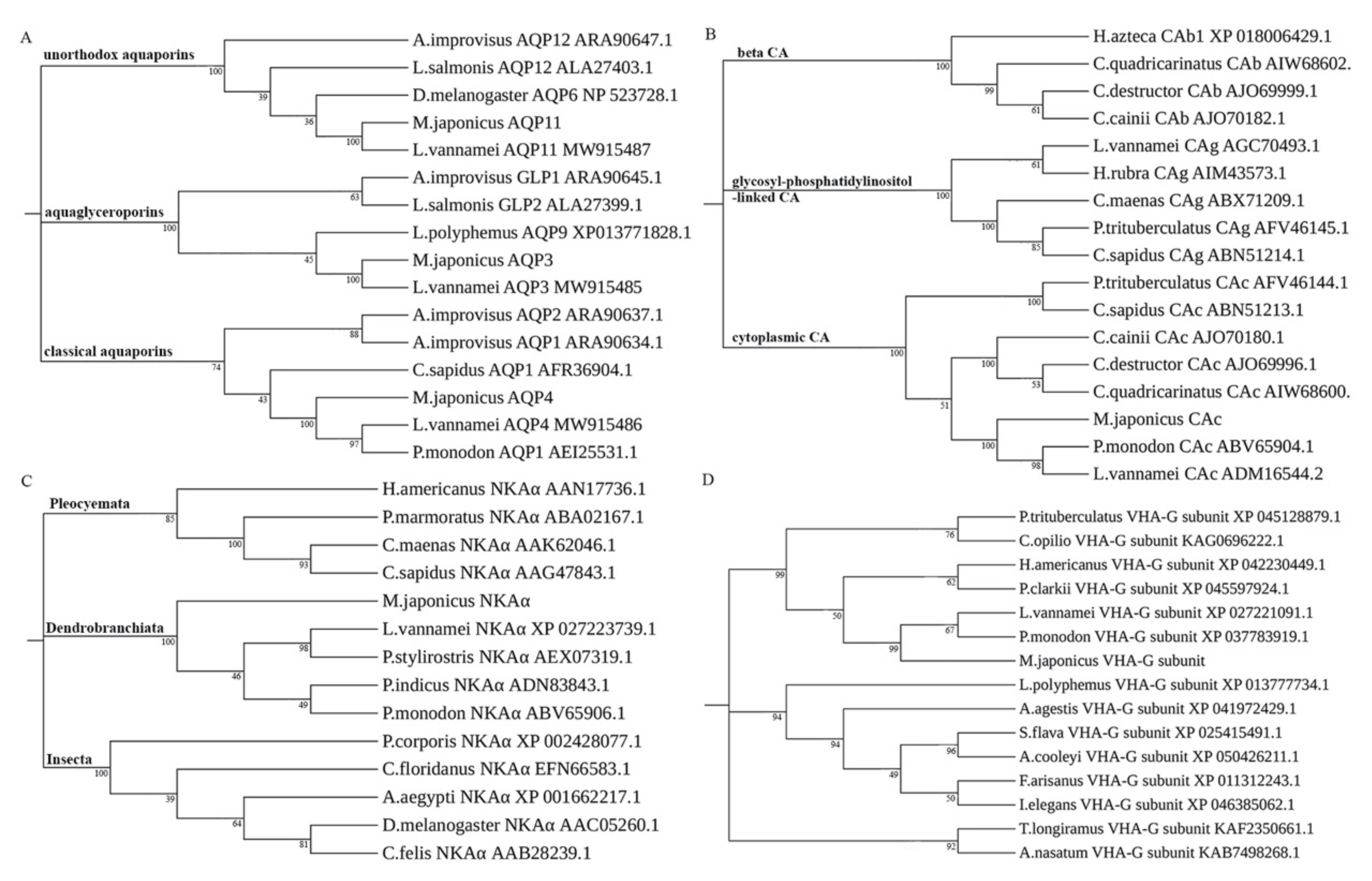
Figure 1. Phylogenetic analysis of (A) MjAQPs, (B) MjCAc, (C) MjNKAα, and (D) MjVHA-G based on NJ method. Phylogenetic trees based on the amino acid sequences of the AQPs, CA, NKAα, and VHA-G in various species were constructed by using MUSCLE alignment and the NJ method through MEGA X software. The node value indicates the percentage of 1000 bootstrap replicates.
Jmse 10 01870 g001
Figure 2. The relative expression levels of (A) MjAQPs and (B) MjCAc, MjNKAα, and MjVHA-G in different tissues of M. japonicus. Abbreviations: In, intestine; Hp, hepatopancreas; Ms, muscle; Gi, gill. The relative expression level of each gene in the intestine is separately used as calibrator. The data are presented as mean + SD (n = 3). Means labeled with different letters are significantly different (p < 0.05, one-way ANOVA, Tukey).
Jmse 10 01870 g002
Figure 3. Cumulative mortality rates of post-larvae shrimp under acute (A) low-salinity and (B) high-salinity stress. Each bar represents the mean value from three repetitions with SD.
Jmse 10 01870 g003
Figure 4. The relative expression levels of (A) MjAQP3, (B) MjAQP4, (C) MjAQP11, (D) MjNKAα, (E) MjCAc, and (F) MjVHA-G in the post-larvae shrimp at 10‰ and 50‰ salinity stresses. The relative expression level of each gene at 0 h is separately used as calibrator. The data are presented as mean + SD (n = 3). Lowercase letters indicate significant differences (p < 0.05) with the same salinity at different exposure times. Asterisks above black bars denote significant differences (p < 0.05) among different salinities at the same treatment time.
Jmse 10 01870 g004
Figure 5. The relative expression levels of genes during the low-salinity (A) and high-salinity (B) acclimation periods in the post-larvae shrimp. The relative expression level of each gene at 30‰ salinity is separately used as calibrator. The data are presented as mean + SD (n = 3). Lowercase letters indicate significant differences (p < 0.05) with each gene.
Jmse 10 01870 g005
Figure 6. The relative expression levels of (A) MjAQP3, (B) MjAQP4, (C) MjAQP11, (D) MjNKAα, (E) MjCAc, and (F) MjVHA-G in the gills at 10‰ and 40‰ salinity stresses. The relative expression level of each gene at 0 h is separately used as calibrator. The data are presented as mean + SD (n = 3). Lowercase letters indicate significant differences (p < 0.05) with the same salinity at different exposure times. Asterisks above black bars denote significant differences (p < 0.05) among different salinities at the same treatment time.
Jmse 10 01870 g006
Figure 7. The relative expression levels of genes during the low-salinity (A) and high-salinity (B) acclimation periods in the gills. The relative expression level of each gene at 25‰ salinity is separately used as calibrator. The data are presented as mean + SD (n = 3). Lowercase letters indicate significant differences (p < 0.05) with each gene.
Jmse 10 01870 g007
Table 1. Specific primers used for gene cloning.
Gene NamePrimer TypeSequence (5′-3′)
MjAQP3ForwardAGTGTACAGGAACAAAGGCTTAC
ReverseACTACTCCTCCTTAGTCGCTCTC
MjAQP4ForwardGAAGAGCAAGAAACCACCATC
ReverseCCGAGCCAGTAGATCTTTCAT
MjAQP11ForwardCATCTCCGCAACCATGTCGAT
ReverseTGTGACACTACCGAAAATCCTC
MjNKAαForwardGCCTAGTGGCTTGTACATAAGTG
ReverseTGAGCTCTAGTAATTCTTGCGC
MjCAcForwardGACAGTTAAACATGGTTGGCTG
ReverseGAGTGTCACTTATTTCTCTGGTCA
MjVHA-GForwardACAAGACACACACAGCAGACG
ReverseCAGTTCTACACACTTATCATGGAGT
Table 2. Specific primers used for qPCR analysis.
Gene NamePrimer TypeSequence (5′-3′)Melting TemperatureEfficiency
MjAQP3ForwardACCTTGGGTGTGCTCGTATCT86.0 °C97.23%
ReverseTGGGCAATAACATACACGGG
MjAQP4ForwardTGGCTGCTGCTCTTATCTACTCC81.0 °C95.36%
ReverseCGTTGTTGGTTCGCTTATTGTA
MjAQP11ForwardCGACCTCCAAGTGCCTGTAG85.0 °C96.53%
ReverseAAGCACGGGATTGAAGTAACC
MjNKAαForwardACCCGCCGTAACTCTATTGTC82.5 °C96.97%
ReverseTGCCTGGGGTGTAGGAAAG
MjCAcForwardACATTCATGGAAGGCTCAGGT84.0 °C95.67%
ReverseAGTAGCAGCGACCATTGACG
MjVHA-GForwardTGAATAAGCAGGTTGCCCAC81.5 °C96.78%
ReverseCCTTCTTAGCGTTGGTGTGC
EF1αForwardGGAACTGGAGGCAGGACC85.0 °C98.98%
ReverseAGCCACCGTTTGCTTCAT
Publisher’s Note: MDPI stays neutral with regard to jurisdictional claims in published maps and institutional affiliations.