Article

The m6A Methylation Profile Identified That OsHMT9.1 Deregulates Chromium Toxicity in Rice (Oryza sativa L.) Through Negative Regulatory Functions

1
Key Laboratory of Germplasm Enhancement, Physiology and Ecology of Food Crops in Cold Region, Ministry of Education, Northeast Agricultural University, Harbin 150030, China
2
Suihua Branch of Heilongjiang Academy of Agricultural Science, Suihua 152052, China
3
Institute of Crop Cultivation and Tillage, Heilongjiang Academy of Agricultural Sciences, Harbin 150030, China
*
Authors to whom correspondence should be addressed.
These authors contributed equally to this work.
Agriculture 2025, 15(5), 519; https://doi.org/10.3390/agriculture15050519
Submission received: 18 January 2025 / Revised: 16 February 2025 / Accepted: 24 February 2025 / Published: 27 February 2025
(This article belongs to the Special Issue Genetic Research and Breeding to Improve Stress Resistance in Rice)

Abstract

:
Chromium (Cr) is a toxic heavy metal that affects the food chain and poses a severe threat to food safety. Nonetheless, the N6-methyladenosine (m6A) transcriptomic regulation mechanisms of Cr tolerance genes in rice are not well understood. This study found that rice roots exhibit competitive and synergistic interactions with trace elements under Cr stress. Through a comprehensive transcriptome analysis of m6A methylation profiles under Cr stress, differentially methylated genes (DMGs) closely related to the plasma membrane, oxidoreductase activity, and protein phosphorylation were identified. A significant number of differentially expressed genes (DEGs) associated with heavy metal transporter domains, metalloproteases, metal ion transporters, and other cation transporters were strongly induced by Cr. Additionally, OsHMT9.1 exhibited extensive hypomethylation and up-regulation in Cr-exposed roots and was confirmed to be a regulatory factor for Cr tolerance. Enhanced plant resistance to Cr in oshmt9.1 was accompanied by increased levels of P, K, S, and Ca and decreased levels of Mn and Cu. These results suggest that knocking out OsHMT9.1 can promote Cr detoxification in rice by modulating the balance between Cr and other trace elements. These findings provide new insights into the molecular regulation and stress response of rice under Cr stress through transcriptome m6A methylation patterns.

1. Introduction

Heavy metal (HM) contamination has emerged as a significant issue in sustainable agriculture due to its detrimental impacts on crop growth, soil quality, food safety, and market viability [1]. Among various HMs, chromium (Cr) is particularly hazardous and lacks any essential role in plant metabolism. It ranks as the second most prevalent metal contaminant found in soil, groundwater, and sediments, posing considerable environmental risks [2]. Cr exists in different chemical states within the soil, with hexavalent Cr [Cr(VI)] being more toxic than its trivalent counterpart. In plants, physiological functions such as photosynthesis, hydration, and nutrient uptake can be adversely affected by Cr stress [3]. Previous research on the impact of Cr exposure on the roots of developing rice seedlings indicated that the induction of oxidative stress is a primary biological mechanism responsible for Cr toxicity in plants [4]. Although extensive investigations have been conducted on plant responses to heavy metal exposure, the effects of excess Cr(VI) on plant systems remain inadequately explored, and our understanding of the plant’s response to Cr(VI) stress at the gene expression level is limited. Moreover, plants may activate defense-related genes when subjected to excessive heavy metal exposure [5]. Consequently, conducting a comprehensive study on the molecular mechanisms governing the rice response to Cr(VI) stress is crucial for elucidating how higher plants endure heavy metal stress.
N6-methyladenosine (m6A) RNA methylation is crucial for development across various species, including yeasts and plants [6]. However, the understanding of m6A RNA methylation in monocots remains limited. In the context of plants, many investigations into the mechanisms and functions of m6A methylation have primarily progressed in Arabidopsis, demonstrating that these methylation events influence plant responses to growth, development, biotic stress, and abiotic stressors [7,8,9,10]. Rice, a vital food source in China, serves as a significant model organism for monocots. One study was the first to indicate that the RNA N6-methyladenosine methyltransferase subunit, OsFIP, plays a role in the early degeneration of microspores in rice [11]. Additionally, recent research has shown that m6A methylation is involved in the virulence processes of the rice blast fungus Pyricularia oryzae, Rice black-streaked dwarf virus, and the interactions between plant viruses and rice [12,13]. Furthermore, contemporary studies have provided an extensive reference map of gene activity through multi-omics analysis approaches, indicating that the m6A signaling pathway is essential in the carcinogenic processes associated with heavy metals such as cadmium (Cd) in soybeans [14] and barley [15], as well as lead (Pb) and cadmium in soybeans [16]. In rice, the significant uptake and accumulation of Cr can induce severe phytotoxic effects, including decreased seed germination traits, reduced seed vigor and plant biomass, inhibited development, impaired photosynthesis, diminished water retention, increased electrolyte leakage, and unbalanced micronutrient uptake [17]. Although studies on m6A-mediated heavy metal responses have been conducted in rice [18], the relationship between m6A modification and Cr tolerance in rice remains unexplored.
Numerous gene families responsible for metal transport, including HMA (heavy metal ATPase), ABC (ATP binding cassette) superfamily, CDF (cation diffusion facilitator), NRAMP (natural resistance-associated macrophage protein), and ZIP (ZRT, IRT-like protein), have been identified as playing essential roles in the movement of various metals from the roots to the shoots [3,19]. Despite their vital contributions to metal uptake, transport, sequestration, and tolerance, our understanding of the involvement of these transporter families with Cr in plants remains limited. In rice plants, the presence of Cr (VI) has been shown to induce the production of reactive oxygen species (ROS) and Ca2+, subsequently activating NADPH oxidase and calcium-dependent protein kinase, which are crucial for downstream signaling pathways [20]. Increasing evidence highlights the reciprocal interaction between calcium and ROS signaling systems, which has significant implications for optimizing cellular signaling networks. Additionally, various transcription factors associated with Cr signaling pathways have been identified, including WRKY and AP2/ERF TF genes, supporting their involvement in defense mechanisms against metal stress. Similarly, many genes related to phosphate kinases have been recognized in response to Cr (VI) exposure, further indicating their potential role in regulating multiple signaling cascades during Cr stress. Previous research on gene expression in rice plants subjected to Cr stress demonstrated the inactivation of pathways related to gibberellic acid, alongside the activation of ethylene, abscisic acid, and jasmonate-mediated signaling cascades, offering new insights into the functions of diverse hormones during Cr stress [20]. A separate investigation indicated that the transcriptome analysis of rice plants subjected to Cr (VI) exposure revealed a unique gene expression pattern. Specifically, genes associated with membrane transport, signal transduction, xenobiotic management, amino acid metabolism, and secondary metabolite biosynthesis were found to be up-regulated, while those linked to cellular growth and energy metabolism were down-regulated. Cr (VI) is capable of activating numerous genes associated with reactive oxygen species (ROS), calcium signaling, MAPKs, and CDPK-like kinases, all of which play crucial roles in perception and signaling pathways [21]. Similarly, distinct regulatory miRNAs were identified in tobacco plants under Cr (VI) stress conditions [22]. Conversely, studies focusing on the metabolomics and proteomics of rice plants after Cr exposure revealed that the response proteins were involved in various cellular processes, including cell wall formation, electron transport, primary metabolic activities, energy generation, detoxification, and hormone accumulation, which contributed to the defensive mechanisms against oxidative stress in rice plants [23,24]. These findings further underscore the significance of omics approaches in uncovering the essential components involved in Cr signal perception and transduction. Nevertheless, integrating multi-omics with gene knockout experiments is essential to elucidate the functions of distinct genes and other critical signaling components in Cr signaling, thereby offering novel perspectives for developing Cr-tolerant crop varieties.
To address this inquiry, transcriptome-wide m6A sequencing on rice roots exposed to Cr conditions were conducted. Our investigation focused on the ion response of roots to Cr stress by integrating analyses of m6A methylation related to Cr-responsive genes and alterations in transcription. A strong positive correlation between m6A methylation and the expression levels of genes responsive to Cr were discovered, which includes both upstream signaling and transcriptional regulatory genes. Furthermore, we identified that the heavy metal transporter domain gene OsHMT9.1 (Os09g0272000) underwent methylation and was up-regulated in response to Cr stress, facilitating the coordination and competition of ions in plants following the reception of the Cr signal, as it was expressed in various tissues.

2. Materials and Methods

2.1. Plant Material and Growth Conditions

The germination of rice seeds (Oryza sativa L. cv. Longdao-18) took place in a growth chamber maintained at 30 °C under dark conditions. After sterilization with hydrogen peroxide, the Longdao-18 seeds were placed in a Petri dish lined with moist filter paper and incubated at 30 °C for three days in the absence of light. Subsequently, seeds exhibiting uniform sprout lengths were transferred to a 96-well PCR plate, which was then positioned in a germination box (Figure 1A). For plant nourishment, Hoagland solution was utilized as the nutrient medium, comprising the following components: 506 mg of KNO3, 80 mg of NH4NO3, 136 mg of KH2PO4, 241 mg of MgSO4, 36.7 mg of FeNaEDTA, 0.83 mg of KI, 6.2 mg of H3BO3, 22.3 mg of MnSO4, 8.6 mg of ZnSO4, 0.25 mg of Na2MoO4, 0.025 mg of CuSO4, 0.025 mg of CoCl2, and 945 mg of Ca(NO3)2. The plants were cultivated in a growth chamber at 27 °C, under cool white fluorescent light with a light intensity of 100 μmol·m−2·s−1, and maintained under long-day conditions (16 h of light/8 h of darkness). The rice seedlings reached the three-leaf stage, at which point a concentration of 200 μM K2CrO4 (Sigma-Aldrich, St. Louis, MO, USA) was applied to induce Cr ion stress for a duration of six days. Following this treatment, selected rice plants underwent phenotypic, RNA-Seq, and MeRIP-Seq analyses to investigate their responses to the Cr stress conditions.

2.2. Phenotypic and Ion Concentration Measurements

Following treatment, the roots and leaves of rice from both the control and treatment groups were collected and thoroughly rinsed five times with distilled water to eliminate any residual nutrient solution and potassium dichromate present on the tissue surface. Ion concentrations were assessed according to methods detailed in previous studies [15]. A 0.2 g dry sample was weighed into a PTFE digestion tank, and 5 mL of nitric acid was added for overnight soaking. The mixture was then placed in a constant temperature drying oven at 80 °C for two hours, maintained at 120 °C for two hours, and subsequently raised to 160 °C for four hours. After this, it was allowed to cool naturally to room temperature in the oven, after which it was reheated to concentrate the acid until nearly dry. The resulting digest was transferred into a 25 mL volumetric flask, with the tank and lid washed three times using a small amount of 1% nitric acid solution. The washings were combined into the volumetric flask, which was then filled to the mark with 1% nitric acid and mixed thoroughly for future analysis. Once the sample digestion is completed, use an Inductively Coupled Plasma Optical Emission Spectrometer (ICP-OES, Santa Clara, CA, USA) for analysis, measuring the wavelength corresponding to each element’s characteristic spectral line. The concentrations of Cr, Zn, Mn, and Fe were quantified using an ICP-MS instrument (7500CE, Agilent, Santa Clara, CA, USA). In summary, the signal intensity of the spectral line for each measured element correlates directly with its concentration, allowing for accurate quantitative analysis. Each trait was analyzed using three independent biological replicates for each sample, along with three technical replicates for every biological replicate.

2.3. Semi-Thin Section of Rice Root Tip

After Cr treatment, the main root tips (about 0.8 cm) of rice seedlings were fixed in 50% FAA fixative at 4 °C for 24 h and then transferred to 75% ethanol at 4 °C overnight. After that, they were immersed in 85% and 95% anhydrous ethanol for 1 h. The material was transferred to a pre-osmotic solution (95% ethanol mixed with an equal volume of base liquid technovit 7100) and placed overnight at 4 °C. After condensation and hardening in the silica gel mold, the root tip materials were transferred to an oven at 37 °C for three days. A tungsten steel knife is used for slicing, and the slice thickness is set to 5 μm.

2.4. m6A Immunoprecipitation (IP) and cDNA Library Construction

TRIzol (Vazyme, Nanjing, China) was utilized for the isolation of total RNA following the provided guidelines. Following this, RNA samples were incubated with an m6A-specific antibody (Synaptic Systems, Goettingen, Germany) in IP buffer (which contains 100 mM Tris-HCl, 300 mM NaCl, and 0.5% [v/v] IGEPAL CA-630, Sigma, Burbank, CA, USA) along with protein A-conjugated beads for a duration of 3 h at 4 °C. To facilitate the elution of compounds that had bound nonspecifically, a specialized IP elution buffer was used, consisting of 10 mL 0.05 M DTT, 300 mM NaCl, 50 mM Tris-HCl (pH = 7.5), 0.1 mM EDTA, 0.05% SDS, and an inhibitor. Subsequently, samples of immunoprecipitated RNA (IP group) and input RNA (Input group, which also contributed to the RNA-Seq analysis) from three separate biological replicates for each treatment group were applied for the production of cDNA libraries using TruePrep RNA Library Prep reverse transcriptase (Vazyme, Nanjing, China). Additional details can be found in a previously published protocol [14].

2.5. High-Throughput MeRIP-Seq and RNA-Seq

For the high-throughput sequencing of cDNA libraries, the Illumina NovaSeq 6000 platform (LC-Bio Technology Co., Ltd., Hangzhou, China) was employed. Subsequently, the initial filtration of raw m6A-seq and RNA-seq data was performed using FastQC (https://www.bioinformatics.babraham.ac.uk/projects/fastqc/) (accessed on 10 January 2022) with default settings to eliminate reads containing undetermined bases or exhibiting low quality. The resulting datasets were then aligned to the rice reference genome IRGSP-1 (https://plants.ensembl.org/Oryza_sativa/Info/Index) (accessed on 18 January 2022) using HISAT2. To identify differentially expressed genes (DEGs) and differentially methylated genes (DMGs), a differential expression analysis was conducted by comparing various treatment groups with strict criteria: |log2 (fold change)| > 1 and p-value < 0.05 [25]. The subsequent results were evaluated using fragments per kilobase of exon per million fragments mapped (FPKM) values. Annotation and enrichment analyses for the Kyoto Encyclopedia of Genes and Genomes [26] and Gene Ontology [27] were performed on DEGs and/or genes located within differentially methylated peak (DMP) regions.

2.6. Vector Construction and Transgenic Rice Production

To establish CRISPR/Cas9 knockout lines, target sites were detected using CRISPR-P 2.0. The resulting plasmids were subsequently introduced into the Agrobacterium tumefaciens strain EHA105. The transformation of IRAT109 with A. tumefaciens was performed following the procedures outlined in our previous study [28]. After the transformation, the targeted genomic regions in the transgenic plants underwent PCR-based sequencing with the primers specified in Table S1 for verification. To assess the role of the DEG in plant tolerance to Cr, transgenic plants associated with all candidate genes were treated with K2CrO4, followed by an evaluation of the phenotype and Cr concentration in the plants.

2.7. Expression Pattern Analysis of OsHMT9.1

To investigate the expression levels of OsHMT9.1 in response to Cr induction, total RNA was isolated from rice roots at various time intervals and subjected to different concentrations of Cr using TRIzol reagent (Invitrogen, Waltham, MA, USA). The expression of OsHMT9.1 was measured through qRT-PCR, utilizing the Actin1 gene as a reference [29]. Primers were designed with Primer Premier v. 5.0 (PREMIER Biosoft International, Palo Alto, CA, USA), and they are detailed in Table S1. qRT-PCR analysis was conducted using the Roche Light Cycler 2.10 (Basel, Switzerland) alongside a 2 × SYBR Green I PCR master mix. For examining the tissue-specific expression of OsHMT9.1, the promoter region (approximately 1.5 kb) upstream of OsHMT9.1 was amplified with specific primers and subsequently ligated into the pMDC162 vector to create a GUS expression vector, which was then transformed into the rice variety Longdao 18. The tissues from OsHMT9.1::GUS plants were employed for GUS histochemical staining.

2.8. Subcellular Localization of OsHMT9.1

To determine the subcellular localization of OsHMT9.1, the full-length CDS of OsHMT9.1 was constructed into CaMV35S::GFP. The CaMV35S::OsHMT9.1-GFP vector was transformed into Agrobacterium tumefaciens EHA105 with nuclear and plasma membrane markers, respectively, and co-transformed into N.benthamiana leaves. After 24 h of dark induction at 28 °C, the fluorescence signal was observed by a confocal laser scanning microscope (FV3000, Olympus, Tokyo, Japan).

3. Results

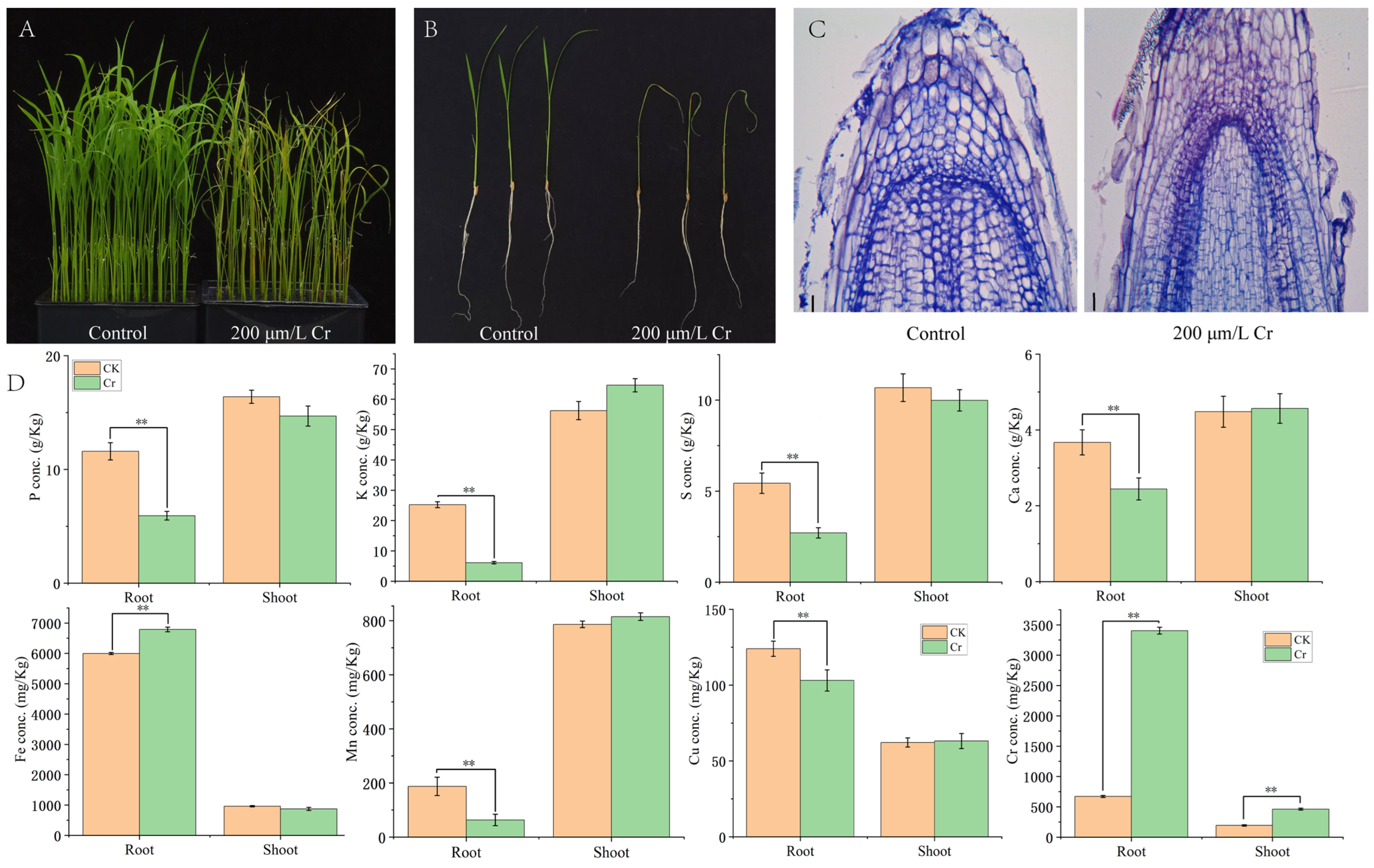
3.1. Phenotypic and Ionic Responses of Rice Seedlings to Cr Stress

After a treatment period of 6 days with 200 μM potassium dichromate, the growth of rice seedlings’ roots, stems, and leaves exhibited a significant reduction compared to the control group (Figure 1A,B). Additionally, the cells at the root tips suffered damage due to Cr stress, suggesting that this damage adversely affected the growth of both aerial and subterranean parts of the plant (Figure 1C). Regarding ion responses, the Cr levels following treatment were markedly elevated in contrast to the control group, with the concentration of Cr found in the roots being significantly greater than that present in the shoots (Figure 1D). Conversely, the levels of phosphorus, potassium, calcium, sulfur, manganese, and copper within the roots experienced a notable decline. Furthermore, as the copper content in the roots diminished, the iron concentration within the same tissue increased. These findings indicate that the reduction in root growth caused by Cr stress is intricately linked to the elevated Cr levels in rice, positioning Cr as a primary competitive agent in the absorption and inhibition of trace elements.

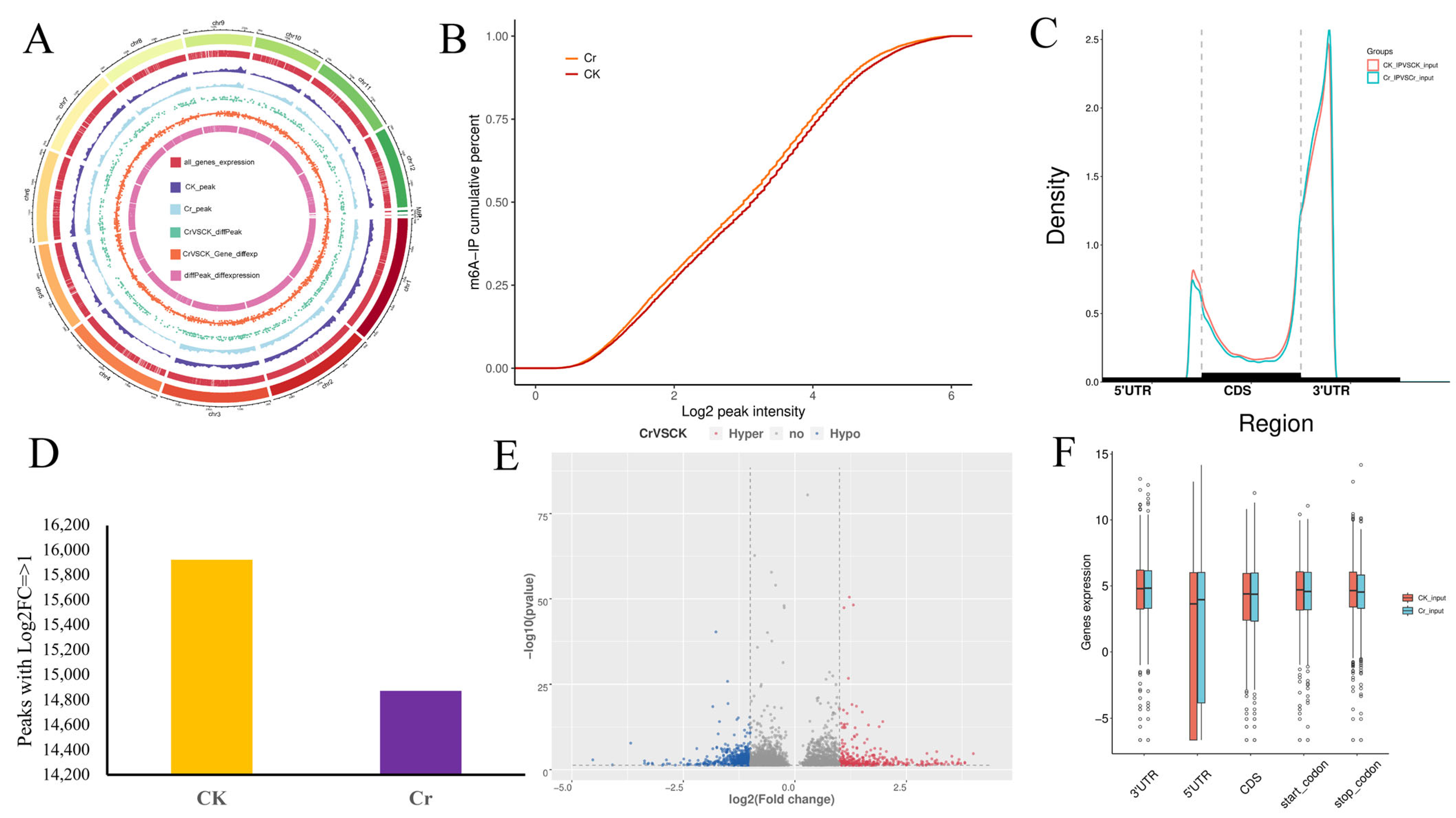
3.2. The Overview of Transcriptome-Wide m6A Methylation in the Roots of Rice Under Cr Stress

Alterations in the methylation patterns of m6A RNA can influence how crops respond to metal ions. To investigate the impact of Cr stress on the transcriptional activity in rice roots, a comprehensive transcriptome analysis focusing on m6A methylation were conducted. Following quality control measures for the sequences obtained from each sample, we achieved high-quality sequence data with an alignment rate exceeding 90% across three biological replicates. A total of approximately 474 Mb of clean reads was generated through high-throughput MeRIP-seq. The Q 30% values varied between 95.12% and 95.67%. The average GC content was measured at 48.81% for the immunoprecipitated (IP) group and 55.21% for the input group (Table S2). The distribution of m6A peaks and full-length transcripts across the rice genome was relatively uniform, with a notable enrichment of m6A peaks at the telomeric regions of each chromosome compared to other genomic areas (Figure 2A). In the control conditions, peak calls were nearly uniformly spread between transcription start sites (TSS) and transcription end sites (TES); conversely, under Cr exposure, a greater number of peak calls were identified in the TSS region as opposed to the TES region (Figure S1). Additionally, we observed significant enrichment of m6A peaks in proximity to the termination codon and within the 3′UTR regions of the genes (Figure 2B). When comparing the control to Cr conditions, the levels of m6A methylation across the genome increased under Cr stress (Figure 2C). 23,218 and 21,094 peaks in the CK and Cr samples were identified, respectively; among these, 21,761 and 19,908 peaks corresponded to 15,924 and 14,874 genes at a high confidence level (log2|fold change| ≥ 1, p-value < 0.05) (Figure 2D, and Tables S3 and S4). A total of 927 differentially m6A-modified genes were found in the comparison between Cr and CK conditions (Figure 2E, Table S5). Genes exhibiting m6A modifications in the regions of 3′UTR, TSS, and TES displayed elevated expression levels compared to those with modifications in the coding sequence (CDS) under Cr stress (Figure 2F).

3.3. Differentially m6A-Modified Peaks and Genes in Response to Cr

Analyses for GO and KEGG enrichment were conducted on the differentially modified m6A genes derived from the comparison between Cr and CK (Figure S2). Among the top 10 GO terms identified for 106 hypermethylated and 169 hypomethylated m6A modified genes in the Cd versus CK comparison were the galactolipid biosynthetic process, digalactosyldiacylglycerol synthase activity, UDP-galactosyltransferase activity, and aldehyde dehydrogenase (NAD+) activity, along with GTP 3′,8′-cyclase activity, tetrahydrofolate metabolic process, pantothenate biosynthetic process, nucleus, and the molybdopterin synthase complex (Figure S2, Table S6). Additionally, prominent KEGG pathways enriched included limonene degradation, biosynthesis of cofactors, sulfur relay system, plant hormone signal transduction, glycerolipid metabolism, cyanoamino acid metabolism, and ABC transporters (Figure S2, Table S7). Collectively, these findings indicate that differential m6A RNA modifications in response to Cd stress significantly impact genes related to sugar metabolism, plant hormone signaling, lipid metabolism, amino acid metabolism, and ABC transport mechanisms.

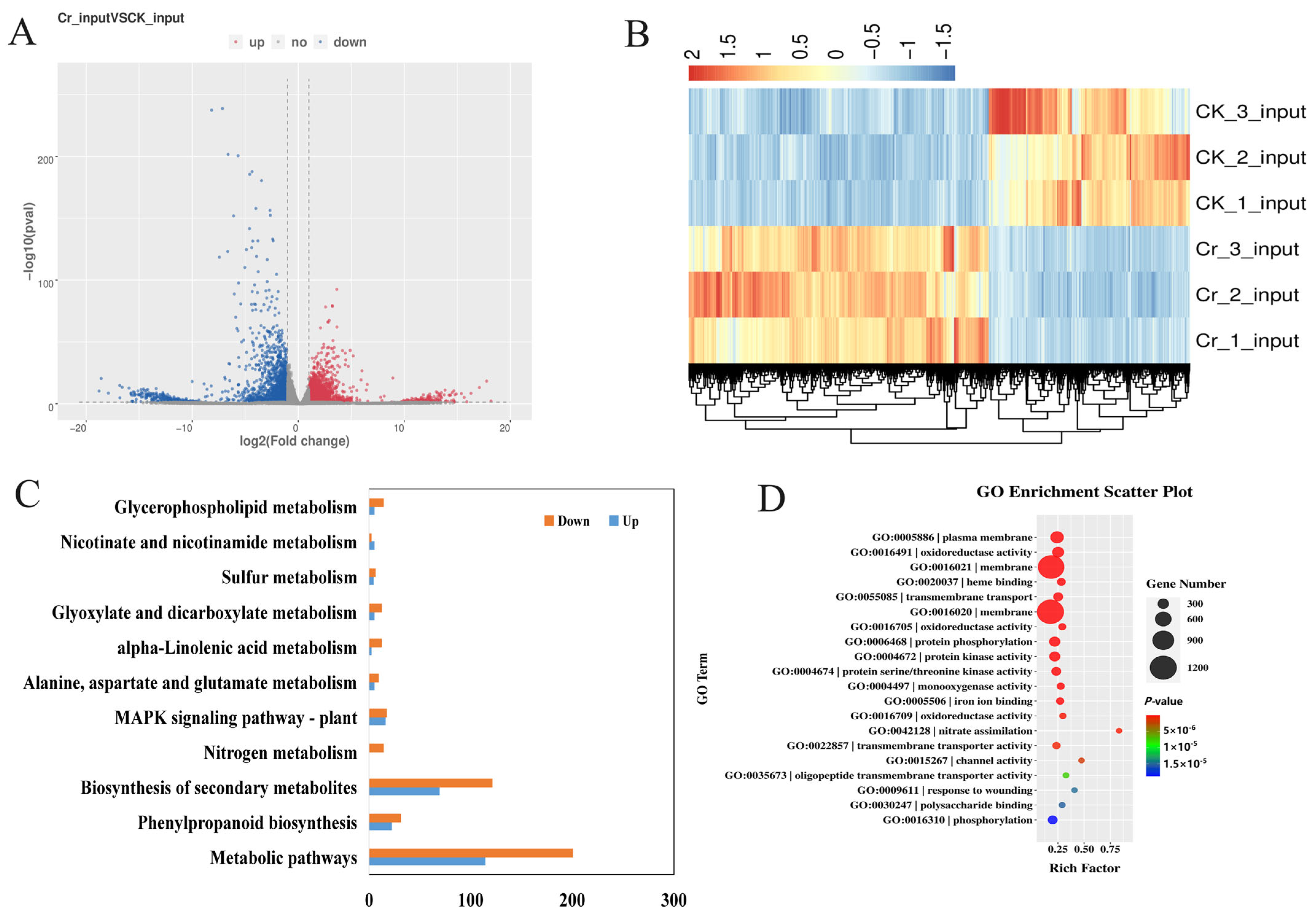
3.4. Differentially Expressed Genes in Rice Roots Under Cr Stress

A total of 5387 DEGs were discovered when comparing Cr and CK (Table S8), with 3301 genes showing up-regulation and 2085 genes exhibiting down-regulation due to Cd stress (Figure 3A). The heatmap of the DEGs illustrated the consistency across three biological replicates and highlighted the significant differences between the Cd-treated and control conditions (Figure 3B). KEGG analysis indicated that all notable DEGs were enriched in 252 metabolic or biological pathways. Notably, the pathways of “phenylpropanoid biosynthesis”, “biosynthesis of secondary metabolites”, “mapk signaling pathway-plant”, and “metabolic pathway” prominently featured both up-regulated and down-regulated DEGs (Figure 3C). Gene ontology (GO) analysis revealed that these DEGs were significantly enriched in categories such as “plasma membrane”, “oxidoreductase activity”, “protein phosphorylation”, and “iron ion binding” (Figure 3D). These findings suggested that the expression levels of genes in rice roots responding to Cr were likely influenced by m6A modifications induced by Cr stress.

3.5. Differentially m6A-Modified DEGs in Rice Roots Under Cr Stress

We subsequently pinpointed genes that exhibited changes in both transcript abundance and m6A methylation as a result of Cr stress. By analyzing the DMPs alongside the DEGs identified in the Cr vs. CK comparison, 150 genes with both differential expression and m6A methylation changes were discovered. Among these, 25 genes were classified as m6A hypermethylated and up-regulated (Hyper-up), 42 as hypermethylated and down-regulated (Hyper-down), 51 as hypomethylated and up-regulated (Hypo-up), and 32 as hypomethylated and down-regulated (Hypo-down) (Figure 4A, Table S9). This indicates that roughly 30% of the identified DEGs experienced hypermethylation under Cr stress. Additionally, a KEGG analysis of these Cr-responsive genes plotted in the four-quadrant diagram revealed enrichment in 49 pathways, which included pyruvate metabolism, phenylpropanoid biosynthesis, and glutathione metabolism (Figure 4B, Table S10). Following Cr exposure, the counts of genes from the Hyper-up and Hypo-down groups associated with “pyruvate metabolism”, “metabolic pathways”, “glutathione metabolism”, and “phenylpropanoid biosynthesis” were 3, 17, 3, and 4, respectively. Importantly, all genes linked to “fatty acid degradation” and “limonene degradation” were found in the Hyper-up category, while “taurine and hypotaurine metabolism” appeared in the Hypo-down category. Simultaneously, GO analysis demonstrated significant enrichment of these genes in processes such as the “oxidation-reduction process”, “glutathione catabolic process”, and “membrane” (Figure 4C, Table S11). Furthermore, the majority of m6A peaks were closely associated with a core motif sequence of 5′-UGUAAAU-3′ (Figure 4D). These findings, therefore, affirm that the DEGs triggered by Cr, which are involved in the antioxidant system and signal transduction within rice roots, showed a pronounced response to m6A RNA modification.

3.6. DEGs and DMPs Play Roles of Cr Stress Response in Rice Roots

To withstand abiotic stress, signaling molecules crucial for downstream signaling pathways are particularly significant in plants. These include calcium, reactive oxygen species (ROS), hormones, protein kinases, and transcription factors [1]. In our study, a total of 21 DEGs and DMPs were identified, which comprised 6 transcription factors, 4 kinases, 7 oxidases, 2 enzymes related to glutathione, and 1 enzyme associated with hormone synthesis (Table S12). Many of these are implicated in responses to low temperatures, drought, salinity, abscisic acid (ABA), and reactive oxygen species, among others [15,30,31,32,33]. Additionally, the majority of genes undergo modifications primarily through m6A methylation within non-coding regions, resembling the methylation changes observed in barley under cadmium stress conditions [15]. Furthermore, to investigate transcriptional alterations in metal ion transporters particularly linked to Cr transport, 54 candidate DEGs related to metal transport were identified (Table S13). Most of these were characterized by functions associated with the heavy metal transport protein domain (HMT), heavy metal ATPase (HMA), metal ion transporters, cadmium-related transporters, and other cation transporters. Among the 21 OsHMT genes, 7 exhibited down-regulated expression, while 14 were up-regulated, with two (Os09g0272000, OsHIPP16) displaying m6A methylation modifications. OsHIPP16 is noted for its high activity in cadmium transport and accumulation, significantly aiding in the detoxification of cadmium by modulating its accumulation in rice [34]. In addition, OsHIPP24 plays a role in the transport of trace metal elements throughout vascular tissues [35]. This shows that a large number of OsHMT genes are induced to express under Cr stress, while a few genes are methylated. This suggests that OsHMT genes play a crucial role in rice’s physiological reaction to Cr stress through their transcriptional activity. In contrast, both OsHMA genes showed methylation. OsHMA9 is activated by elevated levels of copper, zinc, and cadmium ions, with primary expression occurring in vascular bundles and anthers [36]. On the other hand, OsHMA1 is categorized within the Zn/Co/Cd/Pb subgroup; however, its role in the migration or detoxification processes of zinc and cadmium in plants remains ambiguous [37].
A total of three out of the 17 identified cation transporter genes exhibited up-regulation when exposed to Cr stress. The gene OsMTP8.1 functions specifically as a manganese transporter, capable of sequestering Mn2+ into vacuoles, which is crucial for the manganese tolerance of rice shoots [38]. The transporter OZT1 is known to enhance the tolerance of rice against Zn2+ and Cd2+, playing a significant role in the movement of heavy metal ions such as zinc and cadmium, as well as maintaining ion homeostasis [39]. The transport of cadmium ions (Cd2+) via OsZIP6 is facilitated by pH conditions, with its transport functionality increasing in more acidic environments [40]. Serving as a metal-nicotinamide transporter, OsYSL2 is responsible for the phloem transport of iron and manganese, including their translocation to rice grains [41]. The iron-regulated transporters OsIRT1 and OsIRT2 uptake cadmium ions from the soil and deliver them to the shoots [42]. OsZIP5, functioning as a ZRT- and IRT-like protein, responds to cadmium by up-regulating its expression in rice roots [43]. Notably, none of the 17 cation transporter genes examined exhibited M6A methylation modifications. Additionally, one of the six identified Metallothionein-like proteins, OsMT1e, supports rice growth under cadmium stress by shielding organelles from cadmium toxicity [44]. Meanwhile, only OsMT2a showed methylation in its 3′UTR. Furthermore, five proteins associated with cadmium ion transport were identified, among which HsfA4 enhances the cadmium tolerance of both rice and wheat [45]. OsLCT1, a low-affinity cation transport protein in rice, regulates the transfer of cadmium through the phloem to the grains [46]. Lastly, OsNRAMP6, classified as a natural resistance-associated macrophage protein (NRAMP), fine-tunes the transport of iron and manganese through alternative splicing to create diverse protein variants [47], while OsNRAMP5 serves as the primary transporter for the absorption of external Mn2+, Cd2+, and Fe2+ within rice root cells [48].

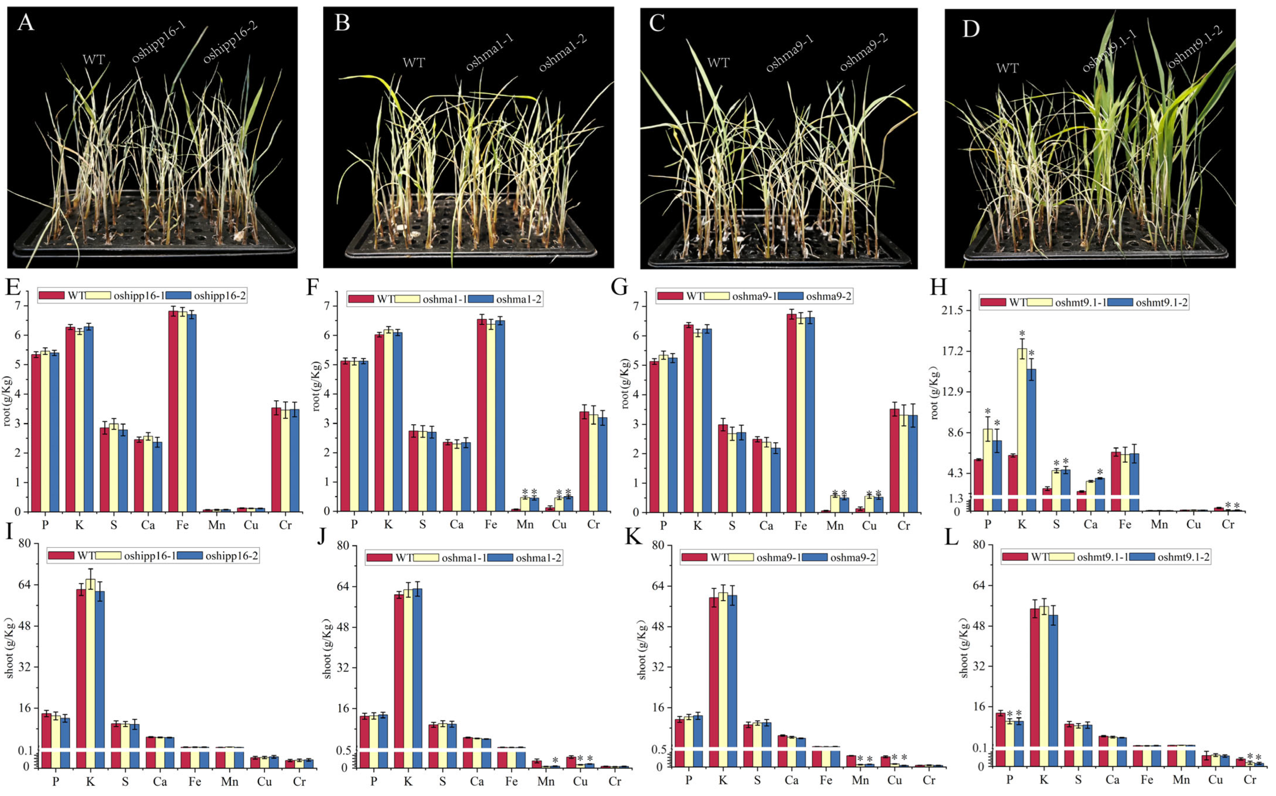
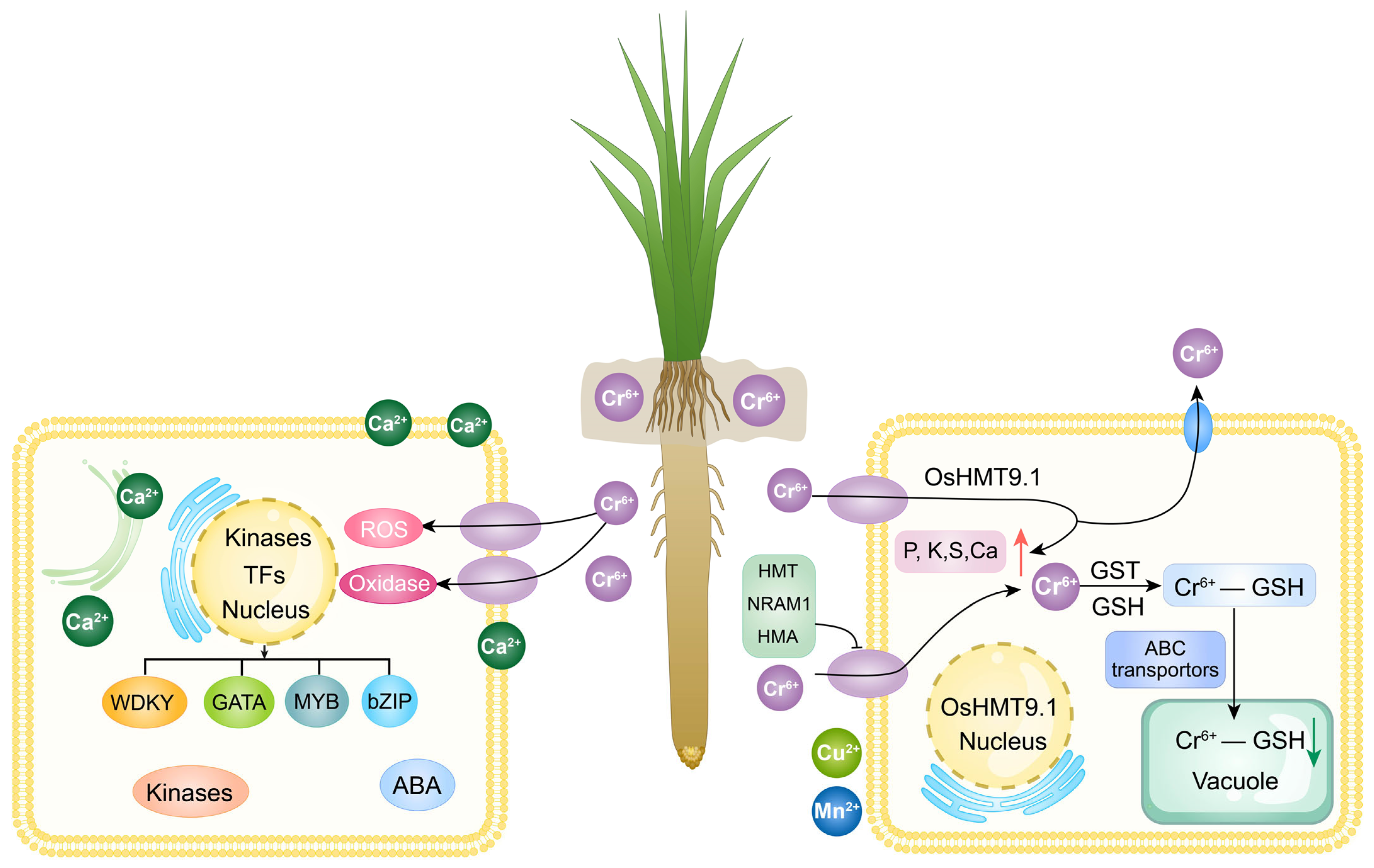
3.7. OsHMT9.1 Negatively Regulates Chromium Stress in Rice

Among the 54 DEGs associated with metal ions, methylation events were noted solely in OsHIPP16, OsHMA1, OsHMA9, and Os09g0272000. To investigate the role of these genes in Cr ion tolerance, we treated knockout transgenic plants expressing the four genes with 200 μM potassium dichromate for 6 days. All transgenic seedlings, with the exception of Os09g0272000, succumbed (Figure 5A–D). When compared to the control (Figure S3), following Cr stress, the concentrations of trace elements in the roots and shoots of the OsHIPP16 transgenic lines were comparable to those of the wild type (Figure 5E,I). In contrast, the roots of the transgenic lines of OsHMA1 and OsHMA9 showed an increased accumulation of Mn and Cu ions, while the leaves exhibited opposite trends. This indicates that these three genes do not serve a functional role in the absorption and transport of Cr ions (Figure 5F,G,I,J). Notably, during Cr stress, Os09g0272000 significantly enhanced the uptake of P, K, S, and Ca while inhibiting Cr absorption in the plant roots, with no impact on Cu and Mn uptake (Figure 5H,L). These results imply that OsHMA1 and OsHMA9 are up-regulated by Cr, which influences the uptake of Cu and Mn in plants. The increased tolerance associated with the reduced Cr levels in the roots and seedlings of the Os09g0272000 knockout may be linked to the enhanced absorption of P, K, S, and Ca. Furthermore, Os09g0272000 is identified as a protein involved in heavy metal transport and detoxification, designated as OsHMT9.1.

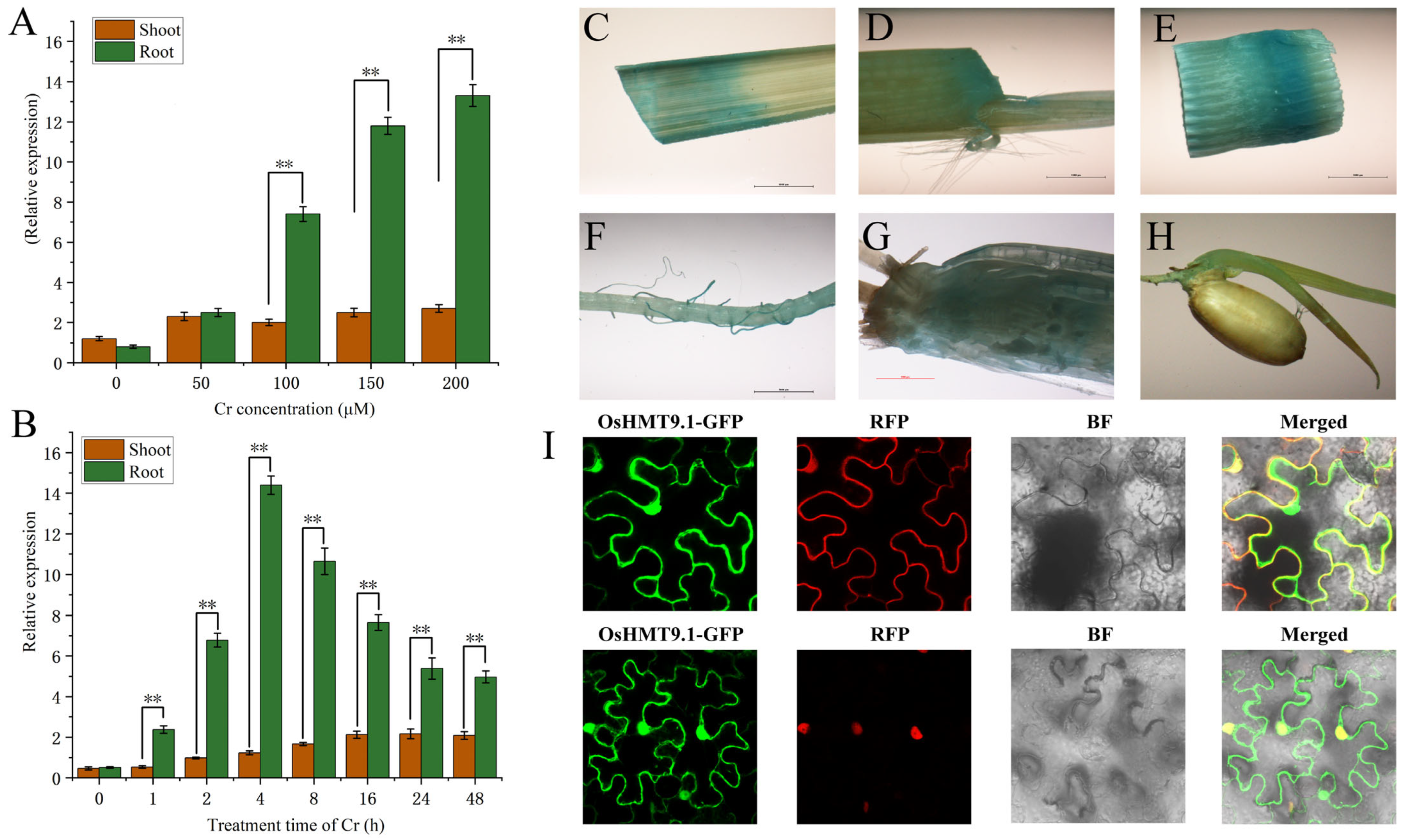
3.8. Expression Characteristics of OsHMT9.1

To investigate the transcriptional response of OsHMT9.1 to Cr, wild-type rice seedlings were subjected to various concentrations of Cr (0, 50, 100, 150, and 200 μM) for a duration of 12 h, resulting in a notable increase in OsHMT9.1 transcription levels. In roots exposed to 100–200 μM Cr, the transcript levels of OsHMT9.1 were found to be amplified by at least three-fold compared to the control group (Figure 6A). Additionally, OsHMT9.1 was markedly stimulated under different durations of Cr exposure (Figure 6B). To elucidate the transcriptional behavior of OsHMT9.1 across various tissues, transgenic lines expressing OsHMT9.1::GUS were developed. The pOsHMT9.1::GUS construct exhibited high levels of expression in leaves (Figure 6C) and leaf sheaths (Figure 6D) during the three-leaf developmental stage. Moreover, significant expression was observed in stem internodes (Figure 6E), root hairs (Figure 6F), and the root base (Figure 6G) of rice at the heading stage. GUS activity was also identified in germinated seeds (Figure 6H) at later developmental stages. To examine the subcellular localization of OsHMT9.1, a fusion expression vector of GFP and OsHMT9.1 was constructed and introduced into tobacco leaves. The fluorescence microscopy revealed that the fusion protein co-localized with markers for the nucleus and plasma membrane (Figure 6I).

4. Discussion

Cr stress poses a significant threat to crops due to the presence of heavy metals. In the soil, Cr can be found in various forms, notably Cr (III) “trivalent” and Cr (VI) “hexavalent”, with Cr (VI) being the most widespread and highly toxic to living organisms. Hexavalent Cr ions typically bond with oxygen, occurring in nature primarily as chromate (CrO42−) and dichromate (Cr2O72−), and they jeopardize crop growth and development through their toxic effects and by competing for ions [3]. Exposure to Cr stress induces metabolic disruptions in plant roots, hampers the uptake of water and essential nutrients, and interferes with the division and elongation of root cells [49]. This results in diminished root activity, inhibited root elongation, and a lower number of lateral roots, which consequently affects root dry matter accumulation and overall plant height [50]. Although a specific mechanism governing Cr absorption and transport in plants has not been identified, Cr can infiltrate plants and accumulate in various tissues by vying for absorption sites and transporters alongside essential elements that share similar structural features [3]. Hence, minimizing Cr uptake in rice is crucial for safeguarding food safety, which necessitates a thorough examination of the mechanisms by which Cr is absorbed and transported within the roots. In the present study, Cr stress was found to hinder the growth of rice roots, reduce plant height, and alter the physiological structure of root tip cells (Figure 1A–C). Following Cr exposure, the uptake of phosphorus (P), potassium (K), calcium (Ca), sulfur (S), manganese (Mn), and copper (Cu) decreased, while the uptake of iron (Fe) rose, and the concentration of Cr ions in roots was significantly greater than that in shoots (Figure 1D). This suggested that Cr disturbed the physiological equilibrium of roots, establishing a competitive interaction with P, K, Ca, S, Mn, and Cu, while fostering a synergistic relationship with Fe.
Cell wall receptors, ion channels, calcium, ROS, hormones, protein kinases, and transcription factors are crucial components in downstream signaling pathways. This approach effectively addresses both biological and numerous non-biological stress sources. The contributions of these signaling components have been extensively explored in response to various biotic and abiotic stressors. Nevertheless, their involvement in heavy metal-related stressors, particularly Cr, remains largely acknowledged. With the advent of multi-omics methodologies, the understanding of Cr signal transduction mechanisms—originating from the cell wall and extending to the plasma membrane and cytoplasm—has gained attention, especially regarding transcriptional, translational, and metabolic reprogramming in diverse plant systems following Cr exposure. For instance, in rice, exposure to Cr (VI) promotes the generation of ROS and Ca2+, subsequently triggering NADPH oxidase activation and calcium-dependent protein kinase, which are pivotal for downstream signaling pathways [20]. Following Cr treatment, our analyses revealed 3301 up-regulated differentially expressed genes (DEGs) and 2085 down-regulated DEGs within rice root tissue. A majority of the total 5386 DEGs were found to be enriched in pathways related to phenylpropanoid biosynthesis, biosynthesis of secondary metabolites, plant MAPK signaling pathway, and metabolic pathways. It implied that specific transporters identified may serve as potential Cr transporters regulating Cr levels within roots. Notably, the expression of OsMTP1 decreased (−1.28) after Cr exposure, indicating its significant role in transporting heavy metal ions like Zn and Cd while maintaining homeostasis [39]. Similarly to OsMTP1, OsMT1a also demonstrates a reduction in expression levels. As a metal-binding protein, OsMT1a plays a crucial role in protecting organelles from cadmium toxicity and regulates rice growth during cadmium exposure [51]. Furthermore, Cr stress induces an up-regulation (2.02) of OsHIPP24, which facilitates the transport of trace metal elements through vascular tissues and is essential for rice growth [33]. Additionally, 19 genes associated with heavy metal transporter or detoxification protein domains and 6 genes resembling metallothioneins exhibited varying degrees of up-regulation or down-regulation, significantly contributing to the uptake and transport of metal ions in rice [34,35,51]. However, the regulatory mechanisms underlying these Cr-responsive genes in the context of rice m6A methylationomics remain poorly understood.
The dynamic changes in the m6A methylation pattern are regulated by a network of writer, eraser, and reader proteins [16]. Heavy metal stress often influences crop methylation events through various signaling pathways, including MAPK, WRKY, and MYB members in response to cadmium stress in barley [15], Ca2+ signal transduction, and ROS in soybean under cadmium stress [14], as well as arginine, proline, and glycerolipid metabolism in rice subjected to cadmium stress [18]. In this study, following Cr stress, the m6A peak exhibited the highest enrichment at the telomeres of each chromosome (Figure 2B), revealing significant differences in m6A density between Cr-treated and normal conditions (Figure 2C). Among all DEGs, only 150 transcripts were identified as DMGs (Figure 4A, Table S9). Under Cr induction, upstream DEGs associated with the antioxidant system and signal transduction demonstrated a strong response to m6A RNA modification, which aligns with previous findings regarding m6A RNA modification in plants under various metal ion stresses [15,16]. The m6A RNA modification of genes in rice exposed to different metal ion stresses often involves several common KEGG pathways. Under cadmium stress, the KEGG annotations of DEGs in rice were predominantly related to tryptophan metabolism, pyruvate metabolism, plant hormone signal transduction, and arginine and proline metabolism. These pathways were also the four most frequently annotated DEG KEGG pathways in this study, suggesting that the methylation events in rice under different metal stresses may share commonalities across several KEGG pathways.
The regulation of metal stress tolerance in plants involves multiple genes [52,53,54]. This study identified a total of 54 DEGs associated with metal ion transport in response to Cr stress. Among these, 15 DEGs have been linked to the regulation of tolerance against various metal ion stresses, including copper ion stress (OsHIPP24) [35], cadmium ion stress (OsHIPP16, OsIRT2, OsMT1e, OsHsfA4a, OsLCT1) [34,42,44,55], manganese ion stress (OsMTP8.1) [38], and multi-metal ion stress (OsHMA9, OsHMA1, OsMTP1/OZT1, OsZIP6, OsYSL2, OsIRT1, OsZIP9, OsZIP2, OsNRAMP5) [36,40,41,42,48,55]. This suggests that genes responsive to Cr stress may also be activated by various other metal ions. Notably, the significant responsiveness of these genes to Cr stress was a key finding of this research. Further investigations revealed that only four genes related to metal ion transport exhibited methylation. To determine the functional involvement of these genes in the regulation of Cr stress, transgenic knockout seedlings were generated. Analyses of phenotypes and trace elements indicated that OsHMT9.1 plays a role in maintaining the physiological balance of rice seedlings by negatively influencing Cr tolerance. As a heavy metal transport protein, OsHMT9.1 is primarily expressed in root tissues and its immediate environment throughout its lifecycle, and it can be up-regulated in response to either the concentration or duration of Cr stress (Figure 6). This behavior is comparable to that of other heavy metal transport genes, such as OsHIPP16 [34], OsMHA2 [56], and OsHIPP42 [57]. The current analysis demonstrated significant hypomethylation and increased expression of OsHMT9.1 in roots exposed to Cr. Additionally, histochemical staining of the GUS reporter gene validated that the transcriptional activity of rice OsHMT9.1 was enhanced in response to Cr stress, with strong GUS signals observed in roots, leaves, and seeds. This pattern aligns with recent findings related to OsHIPP16 [34], OsHIPP42, and OsHIPP56 [57]. Such a multi-tissue expression profile may elucidate the mechanism through which OsHMT9.1 facilitates the transport of ions from the roots to various organs in response to Cr exposure. Nonetheless, further investigations are necessary for confirmation. In conclusion, we developed a methylation transcriptome map of rice roots under Cr ion stress (Figure 7) and comprehensively examined the regulatory processes initiated by Cr ions.
Cr interferes with the absorption of essential elements in rice through competitive absorption, thereby hindering the normal metabolism of roots and leaves. Analysis of m6A methylation in rice roots under Cr stress shows that m6A peaks are most enriched on telomeres, and there is a significant difference in m6A density between Cr treatment and normal conditions. A total of 927 differentially methylated m6A genes were identified, and their expression levels in the 3′UTR, TSS and TES regions are higher than those in the CDS region. Compared with CK, 5387 DEGs were found in Cr treatment. These DEGs are mainly enriched in metabolic or biological pathways such as phenylpropanoid biosynthesis and plant MAPK signaling pathway. OsHMT9.1 is one of four methylated genes among the 54 metal ion-related DEGs. Under Cr stress, it enhances the absorption of P, K, S, and Ca, inhibits Cr absorption, and improves rice tolerance to Cr. Its transcription level significantly increases under different concentrations and durations of Cr treatment, and strong GUS signals are detected in multiple tissues, highlighting its key role in rice’s response to Cr stress.

5. Conclusions

Cr interferes with the absorption of essential elements in rice through competitive absorption, thereby hindering the normal metabolism of roots and leaves. Analysis of m6A methylation in rice roots under Cr stress shows that m6A peaks are most enriched on telomeres, and there is a significant difference in m6A density between Cr treatment and normal conditions. A total of 927 differentially methylated m6A genes were identified, and their expression levels in the 3′UTR, TSS and TES regions are higher than those in the CDS region. Compared with CK, 5387 DEGs were found in Cr treatment. These DEGs are mainly enriched in metabolic or biological pathways such as phenylpropanoid biosynthesis and plant MAPK signaling pathway. OsHMT9.1 is one of four methylated genes among the 54 metal ion-related DEGs. Under Cr stress, it enhances the absorption of P, K, S, and Ca, inhibits Cr absorption, and improves rice tolerance to Cr. Its transcription level significantly increases under different concentrations and durations of Cr treatment, and strong GUS signals are detected in multiple tissues, highlighting its key role in rice’s response to Cr stress. In conclusion, these results highlight essential genes and pathways involved in the response of rice roots to heavy metal stress and warrant further exploration as potential candidates for enhancing heavy metal tolerance in rice through genetic improvement.

Supplementary Materials

The following supporting information can be downloaded at: https://www.mdpi.com/article/10.3390/agriculture15050519/s1, Figure S1. Based on the IGV software, the m6A methylation modification on the target gene was visualized. The upper region shows the reads distribution of each sample (the same sample includes IP and input libraries); Figure S2. GO and KEGG enrichment analyses were performed on the differentially m6A-modified genes from comparison of the Cr vs. CK; Figure S3. Under control conditions, the ion content distribution of roots and leaves of the four genes and their wild-type plants. The data are the means of three biological replicates. * p-value < 0.05, Student’s t-test. Table S1. Primer sequences used in this study; Table S2. Statistical summary of the MeRIP sequencing data; Table S3. Differential expression peaks by the comparison of CK_IP vs. CK_Input; Table S4. Differential expression peaks by the comparison of Cr_IP vs. Cr_Input; Table S5. Differential m6A modified peaks (DMPs) for Cr vs. CK; Table S6. Differentially m6A-modified genes in the Cr vs. CK comparison were annotated with GO terms; Table S7. Differentially m6A-modified genes in the Cr vs. CK comparison were annotated with KEGG terms; Table S8. Differential expression gene by the comparison of Cr_Input vs. Cr_Input; Table S9. 150 genes whose transcripts were both differentially expressed and differentially m6A methylated; Table S10. KEGG enrichment analysis of differentially m6A methylated and differential genes; Table S11. GO enrichment analysis of differentially m6A methylated and differential genes; Table S12. DEGs and DPs play roles of Cr stress response in rice roots; Table S13. The regulation patterns of m6A methylation and gene expression associated with metal ion transport in the roots of rice under Cr stress.

Author Contributions

Data curation, J.W., H.L. and L.L.; H.Z., W.X., Y.H., X.K., S.W., J.L. and C.L. helped with the investigation and formal analysis. Writing—original draft preparation, Y.H., X.K., J.L., C.L., L.Y., Z.W., S.X. and D.Z.; writing—review and editing, Z.W., D.Z. and L.Y. All authors have read and agreed to the published version of the manuscript.

Funding

This work was supported by the National Natural Science Foundation of China (Grant numbers 32372140 and U23A20193), the Heilongjiang Province Key R&D Program (2022ZX02B03), and the Natural Science Foundation of Heilongjiang Province, China (LH2022C021), and the Breeding and industrialization of high-quality multi-resistant aromatic rice varieties (SHFY2022-02).

Institutional Review Board Statement

Not applicable.

Data Availability Statement

All raw sequencing data have been deposited at the NCBI Sequence Read Archive (https://www.ncbi.nlm.nih.gov/sra (accessed on 18 May 2024); PRJNA1119091).

Conflicts of Interest

The authors declare that they have no competing interests.

References

  1. Ali, S.; Mir, R.A.; Tyagi, A.; Manzar, N.; Kashyap, A.S.; Mushtaq, M.; Raina, A.; Park, S.; Sharma, S.; Mir, Z.A.; et al. Chromium Toxicity in Plants: Signaling, Mitigation, and Future Perspectives. Plants 2023, 12, 1502. [Google Scholar] [CrossRef] [PubMed]
  2. Rahman, Z.; Singh, V.P. The relative impact of toxic heavy metals (THMs) (arsenic (As), cadmium (Cd), chromium (Cr)(VI), mercury (Hg), and lead (Pb)) on the total environment: An overview. Environ. Monit. Assess. 2019, 191, 419. [Google Scholar] [CrossRef] [PubMed]
  3. Shanker, A.K.; Cervantes, C.; Loza-Tavera, H.; Avudainayagam, S. Chromium toxicity in plants. Environ. Int. 2005, 31, 739–753. [Google Scholar] [CrossRef] [PubMed]
  4. Panda, S.K. Chromium-mediated oxidative stress and ultrastructural changes in root cells of developing rice seedlings. J. Plant Physiol. 2007, 164, 1419–1428. [Google Scholar] [CrossRef]
  5. Sudo, E.; Itouga, M.; Yoshida-Hatanaka, K.; Ono, Y.; Sakakibara, H. Gene expression and sensitivity in response to copper stress in rice leaves. J. Exp. Bot. 2008, 59, 3465–3474. [Google Scholar] [CrossRef]
  6. Wei, L.H.; Song, P.; Wang, Y.; Lu, Z.; Tang, Q.; Yu, Q.; Xiao, Y.; Zhang, X.; Duan, H.C.; Jia, G. The m(6)A Reader ECT2 Controls Trichome Morphology by Affecting mRNA Stability in Arabidopsis. Plant Cell 2018, 30, 968–985. [Google Scholar] [CrossRef]
  7. Arribas-Hernández, L.; Bressendorff, S.; Hansen, M.H.; Poulsen, C.; Erdmann, S.; Brodersen, P. An m(6)A-YTH Module Controls Developmental Timing and Morphogenesis in Arabidopsis. Plant Cell 2018, 30, 952–967. [Google Scholar] [CrossRef]
  8. Duan, H.C.; Wei, L.H.; Zhang, C.; Wang, Y.; Chen, L.; Lu, Z.; Chen, P.R.; He, C.; Jia, G. ALKBH10B Is an RNA N(6)-Methyladenosine Demethylase Affecting Arabidopsis Floral Transition. Plant Cell 2017, 29, 2995–3011. [Google Scholar] [CrossRef] [PubMed]
  9. Shen, L.; Liang, Z.; Gu, X.; Chen, Y.; Teo, Z.W.; Hou, X.; Cai, W.M.; Dedon, P.C.; Liu, L.; Yu, H. N(6)-Methyladenosine RNA Modification Regulates Shoot Stem Cell Fate in Arabidopsis. Dev. Cell 2016, 38, 186–200. [Google Scholar] [CrossRef]
  10. Yue, H.; Nie, X.; Yan, Z.; Weining, S. N6-methyladenosine regulatory machinery in plants: Composition, function and evolution. Plant Biotechnol. J. 2019, 17, 1194–1208. [Google Scholar] [CrossRef] [PubMed]
  11. Zhang, F.; Zhang, Y.C.; Liao, J.Y.; Yu, Y.; Zhou, Y.F.; Feng, Y.Z.; Yang, Y.W.; Lei, M.Q.; Bai, M.; Wu, H.; et al. The subunit of RNA N6-methyladenosine methyltransferase OsFIP regulates early degeneration of microspores in rice. PLoS Genet. 2019, 15, e1008120. [Google Scholar] [CrossRef] [PubMed]
  12. Shi, Y.; Wang, H.; Wang, J.; Liu, X.; Lin, F.; Lu, J. N6-methyladenosine RNA methylation is involved in virulence of the rice blast fungus Pyricularia oryzae (syn. Magnaporthe oryzae). FEMS Microbiol. Lett. 2019, 366, fny286. [Google Scholar]
  13. Tian, S.; Wu, N.; Zhang, L.; Wang, X. RNA N(6) -methyladenosine modification suppresses replication of rice black streaked dwarf virus and is associated with virus persistence in its insect vector. Mol. Plant Pathol. 2021, 22, 1070–1081. [Google Scholar] [CrossRef] [PubMed]
  14. Han, X.; Wang, J.; Zhang, Y.; Kong, Y.; Dong, H.; Feng, X.; Li, T.; Zhou, C.; Yu, J.; Xin, D.; et al. Changes in the m6A RNA methylome accompany the promotion of soybean root growth by rhizobia under cadmium stress. J. Hazard. Mater. 2023, 441, 129843. [Google Scholar] [CrossRef] [PubMed]
  15. Su, T.; Fu, L.; Kuang, L.; Chen, D.; Zhang, G.; Shen, Q.; Wu, D. Transcriptome-wide m6A methylation profile reveals regulatory networks in roots of barley under cadmium stress. J. Hazard. Mater. 2022, 423, 127140. [Google Scholar] [CrossRef] [PubMed]
  16. Ma, C.; Ma, S.; Yu, Y.; Feng, H.; Wang, Y.; Liu, C.; He, S.; Yang, M.; Chen, Q.; Xin, D.; et al. Transcriptome-wide m(6)A methylation profiling identifies GmAMT1;1 as a promoter of lead and cadmium tolerance in soybean nodules. J. Hazard. Mater. 2024, 465, 133263. [Google Scholar] [CrossRef]
  17. Basit, F.; Chen, M.; Ahmed, T.; Shahid, M.; Noman, M.; Liu, J.; An, J.; Hashem, A.; Fahad Al-Arjani, A.B.; Alqarawi, A.A.; et al. Seed Priming with Brassinosteroids Alleviates Chromium Stress in Rice Cultivars via Improving ROS Metabolism and Antioxidant Defense Response at Biochemical and Molecular Levels. Antioxidants 2021, 10, 1089. [Google Scholar] [CrossRef]
  18. Cheng, Q.; Wang, P.; Wu, G.; Wang, Y.; Tan, J.; Li, C.; Zhang, X.; Liu, S.; Huang, S.; Huang, T.; et al. Coordination of m(6)A mRNA methylation and gene transcriptome in rice response to cadmium stress. Rice 2021, 14, 62. [Google Scholar] [CrossRef]
  19. Shahid, M.; Shamshad, S.; Rafiq, M.; Khalid, S.; Bibi, I.; Niazi, N.K.; Dumat, C.; Rashid, M.I. Chromium speciation, bioavailability, uptake, toxicity and detoxification in soil-plant system: A review. Chemosphere 2017, 178, 513–533. [Google Scholar] [CrossRef]
  20. Trinh, N.N.; Huang, T.L.; Chi, W.C.; Fu, S.F.; Chen, C.C.; Huang, H.J. Chromium stress response effect on signal transduction and expression of signaling genes in rice. Physiol. Plant. 2014, 150, 205–224. [Google Scholar] [CrossRef] [PubMed]
  21. Huang, T.L.; Huang, L.Y.; Fu, S.F.; Trinh, N.N.; Huang, H.J. Genomic profiling of rice roots with short- and long-term chromium stress. Plant Mol. Biol. 2014, 86, 157–170. [Google Scholar] [CrossRef]
  22. Bukhari, S.A.; Shang, S.; Zhang, M.; Zheng, W.; Zhang, G.; Wang, T.Z.; Shamsi, I.H.; Wu, F. Genome-wide identification of chromium stress-responsive micro RNAs and their target genes in tobacco (Nicotiana tabacum) roots. Environ. Toxicol. Chem. 2015, 34, 2573–2582. [Google Scholar] [CrossRef] [PubMed]
  23. Dubey, S.; Misra, P.; Dwivedi, S.; Chatterjee, S.; Bag, S.K.; Mantri, S.; Asif, M.H.; Rai, A.; Kumar, S.; Shri, M.; et al. Transcriptomic and metabolomic shifts in rice roots in response to Cr (VI) stress. BMC Genom. 2010, 11, 648. [Google Scholar] [CrossRef]
  24. Zeng, F.; Qiu, B.; Wu, X.; Niu, S.; Wu, F.; Zhang, G. Glutathione-mediated alleviation of chromium toxicity in rice plants. Biol. Trace Elem. Res. 2012, 148, 255–263. [Google Scholar] [CrossRef] [PubMed]
  25. Love, M.I.; Huber, W.; Anders, S. Moderated estimation of fold change and dispersion for RNA-seq data with DESeq2. Genome Biol. 2014, 15, 550. [Google Scholar] [CrossRef]
  26. Kanehisa, M.; Goto, S. KEGG: Kyoto encyclopedia of genes and genomes. Nucleic Acids Res. 2000, 28, 27–30. [Google Scholar] [CrossRef]
  27. Ashburner, M.; Ball, C.A.; Blake, J.A.; Botstein, D.; Butler, H.; Cherry, J.M.; Davis, A.P.; Dolinski, K.; Dwight, S.S.; Eppig, J.T.; et al. Gene ontology: Tool for the unification of biology. The Gene Ontology Consortium. Nat. Genet. 2000, 25, 25–29. [Google Scholar] [CrossRef] [PubMed]
  28. Yang, L.; Lei, L.; Wang, J.; Zheng, H.; Xin, W.; Liu, H.; Zou, D. qCTB7 positively regulates cold tolerance at booting stage in rice. Theor. Appl. Genet. 2023, 136, 135. [Google Scholar] [CrossRef]
  29. Siahpoosh, M.R.; Sanchez, D.H.; Schlereth, A.; Scofield, G.N.; Furbank, R.T.; van Dongen, J.T.; Kopka, J. Modification of OsSUT1 gene expression modulates the salt response of rice Oryza sativa cv. Taipei 309. Plant Sci. 2012, 182, 101–111. [Google Scholar] [CrossRef] [PubMed]
  30. Feng, L.; Gao, Z.; Xiao, G.; Huang, R.; Zhang, H. Leucine-Rich Repeat Receptor-Like Kinase FON1 Regulates Drought Stress and Seed Germination by Activating the Expression of ABA-Responsive Genes in Rice. Plant Mol. Biol. Report. 2014, 32, 1158–1168. [Google Scholar] [CrossRef]
  31. Islam, M.A.; Du, H.; Ning, J.; Ye, H.; Xiong, L. Characterization of Glossy1-homologous genes in rice involved in leaf wax accumulation and drought resistance. Plant Mol. Biol. 2009, 70, 443–456. [Google Scholar] [CrossRef] [PubMed]
  32. Wu, T.M.; Lin, W.R.; Kao, C.H.; Hong, C.Y. Gene knockout of glutathione reductase 3 results in increased sensitivity to salt stress in rice. Plant Mol. Biol. 2015, 87, 555–564. [Google Scholar] [CrossRef] [PubMed]
  33. Zhang, C.J.; Zhao, B.C.; Ge, W.N.; Zhang, Y.F.; Song, Y.; Sun, D.Y.; Guo, Y. An apoplastic h-type thioredoxin is involved in the stress response through regulation of the apoplastic reactive oxygen species in rice. Plant Physiol. 2011, 157, 1884–1899. [Google Scholar] [CrossRef] [PubMed]
  34. Cao, H.W.; Zhao, Y.N.; Liu, X.S.; Rono, J.K.; Yang, Z.M. A metal chaperone OsHIPP16 detoxifies cadmium by repressing its accumulation in rice crops. Environ. Pollut. 2022, 311, 120058. [Google Scholar] [CrossRef] [PubMed]
  35. Chen, G.; Xiong, S. OsHIPP24 is a Copper Metallochaperone Which Affects Rice Growth. J. Plant Biol. 2021, 64, 145–153. [Google Scholar] [CrossRef]
  36. Lee, S.; Kim, Y.Y.; Lee, Y.; An, G. Rice P1B-type heavy-metal ATPase, OsHMA9, is a metal efflux protein. Plant Physiol. 2007, 145, 831–842. [Google Scholar] [CrossRef] [PubMed]
  37. Takahashi, R.; Bashir, K.; Ishimaru, Y.; Nishizawa, N.K.; Nakanishi, H. The role of heavy-metal ATPases, HMAs, in zinc and cadmium transport in rice. Plant Signal. Behav. 2012, 7, 1605–1607. [Google Scholar] [CrossRef]
  38. Chen, Z.; Fujii, Y.; Yamaji, N.; Masuda, S.; Takemoto, Y.; Kamiya, T.; Yusuyin, Y.; Iwasaki, K.; Kato, S.; Maeshima, M.; et al. Mn tolerance in rice is mediated by MTP8.1, a member of the cation diffusion facilitator family. J. Exp. Bot. 2013, 64, 4375–4387. [Google Scholar] [CrossRef] [PubMed]
  39. Lan, H.X.; Wang, Z.F.; Wang, Q.H.; Wang, M.M.; Bao, Y.M.; Huang, J.; Zhang, H.S. Characterization of a vacuolar zinc transporter OZT1 in rice (Oryza sativa L.). Mol. Biol. Rep. 2013, 40, 1201–1210. [Google Scholar] [CrossRef] [PubMed]
  40. Kavitha, P.G.; Kuruvilla, S.; Mathew, M.K. Functional characterization of a transition metal ion transporter, OsZIP6 from rice (Oryza sativa L.). Plant Physiol. Biochem. 2015, 97, 165–174. [Google Scholar]
  41. Zhang, H.; Li, Y.; Pu, M.; Xu, P.; Liang, G.; Yu, D. Oryza sativa POSITIVE REGULATOR OF IRON DEFICIENCY RESPONSE 2 (OsPRI2) and OsPRI3 are involved in the maintenance of Fe homeostasis. Plant Cell Environ. 2020, 43, 261–274. [Google Scholar] [CrossRef] [PubMed]
  42. Nakanishi, H.; Ogawa, I.; Ishimaru, Y.; Mori, S.; Nishizawa, N.K. Iron deficiency enhances cadmium uptake and translocation mediated by the Fe2+ transporters OsIRT1 and OsIRT2 in rice. Soil Sci. Plant Nutr. 2010, 52, 464–469. [Google Scholar] [CrossRef]
  43. Tan, L.; Qu, M.; Zhu, Y.; Peng, C.; Wang, J.; Gao, D.; Chen, C. ZINC TRANSPORTER5 and ZINC TRANSPORTER9 Function Synergistically in Zinc/Cadmium Uptake. Plant Physiol. 2020, 183, 1235–1249. [Google Scholar] [CrossRef]
  44. Rono, J.K.; Le Wang, L.; Wu, X.C.; Cao, H.W.; Zhao, Y.N.; Khan, I.U.; Yang, Z.M. Identification of a new function of metallothionein-like gene OsMT1e for cadmium detoxification and potential phytoremediation. Chemosphere 2021, 265, 129136. [Google Scholar] [CrossRef]
  45. Mittal, D.; Chakrabarti, S.; Sarkar, A.; Singh, A.; Grover, A. Heat shock factor gene family in rice: Genomic organization and transcript expression profiling in response to high temperature, low temperature and oxidative stresses. Plant Physiol. Biochem. 2009, 47, 785–795. [Google Scholar] [CrossRef]
  46. Uraguchi, S.; Kamiya, T.; Sakamoto, T.; Kasai, K.; Sato, Y.; Nagamura, Y.; Yoshida, A.; Kyozuka, J.; Ishikawa, S.; Fujiwara, T. Low-affinity cation transporter (OsLCT1) regulates cadmium transport into rice grains. Proc. Natl. Acad. Sci. USA 2011, 108, 20959–20964. [Google Scholar] [CrossRef]
  47. Peris-Peris, C.; Serra-Cardona, A.; Sánchez-Sanuy, F.; Campo, S.; Ariño, J.; San Segundo, B. Two NRAMP6 Isoforms Function as Iron and Manganese Transporters and Contribute to Disease Resistance in Rice. Mol. Plant-Microbe Interact. 2017, 30, 385–398. [Google Scholar] [CrossRef]
  48. Ishimaru, Y.; Bashir, K.; Nakanishi, H.; Nishizawa, N.K. OsNRAMP5, a major player for constitutive iron and manganese uptake in rice. Plant Signal. Behav. 2012, 7, 763–766. [Google Scholar] [CrossRef]
  49. Barceló, J.; Poschenrieder, C.; Gunsé, B. Water Relations of Chromium VI Treated Bush Bean Plants (Phaseolus vulgaris L. cv. Contender) under both Normal and Water Stress Conditions. J. Exp. Bot. 1986, 37, 178–187. [Google Scholar] [CrossRef]
  50. Panda, S.K.; Patra, H.K. Effect of salicylic acid potentiates cadmium-induced oxidative damage in Oryza sativa L. leaves. Acta Physiol. Plant. 2007, 29, 567–575. [Google Scholar] [CrossRef]
  51. Yang, D.; Li, S.; Xiao, Y.; Lu, L.; Zheng, Z.; Tang, D.; Cui, H. Transcriptome analysis of rice response to blast fungus identified core genes involved in immunity. Plant Cell Environ. 2021, 44, 3103–3121. [Google Scholar] [CrossRef] [PubMed]
  52. Abe, T.; Ito, M.; Takahashi, R.; Honma, T.; Kuramata, M.; Ishikawa, S. QTL Pyramiding and Its Use in Breeding for Increasing the Phytoextraction Efficiency of Soil Cd via High-Cd-Accumulating Rice. Plants 2022, 11, 2178. [Google Scholar] [CrossRef]
  53. Ali, M.K.; Sun, Z.H.; Yang, X.M.; Pu, X.Y.; Duan, C.L.; Li, X.; Wang, L.X.; Yang, J.Z.; Zeng, Y.W. NILs of Cold Tolerant Japonica Cultivar Exhibited New QTLs for Mineral Elements in Rice. Front. Genet. 2021, 12, 789645. [Google Scholar] [CrossRef]
  54. Pan, X.; Li, Y.; Liu, W.; Liu, S.; Min, J.; Xiong, H.; Dong, Z.; Duan, Y.; Yu, Y.; Li, X. QTL mapping and candidate gene analysis of cadmium accumulation in polished rice by genome-wide association study. Sci. Rep. 2020, 10, 11791. [Google Scholar] [CrossRef]
  55. Tian, S.; Liang, S.; Qiao, K.; Wang, F.; Zhang, Y.; Chai, T. Co-expression of multiple heavy metal transporters changes the translocation, accumulation, and potential oxidative stress of Cd and Zn in rice (Oryza sativa). J. Hazard. Mater. 2019, 380, 120853. [Google Scholar] [CrossRef]
  56. Yamaji, N.; Xia, J.; Mitani-Ueno, N.; Yokosho, K.; Feng Ma, J. Preferential delivery of zinc to developing tissues in rice is mediated by P-type heavy metal ATPase OsHMA2. Plant Physiol. 2013, 162, 927–939. [Google Scholar] [CrossRef] [PubMed]
  57. Khan, I.U.; Rono, J.K.; Liu, X.S.; Feng, S.J.; Yang, Z.M. Functional characterization of a new metallochaperone for reducing cadmium concentration in rice crop. J. Clean. Prod. 2020, 272, 123152. [Google Scholar] [CrossRef]
Figure 1. Phenotype and ion content analysis of rice seedlings under chromium (Cr) stress. (A) Three-leaf period seedlings of Longdao 18 under control condition and Cr stress conditions; (B) Morphological comparison of roots and seedlings of rice seedlings under control and Cr stress conditions; (C) Semi-thin sections of rice root tips under control and Cr stress conditions, Bar = 100 μm; (D) Analysis of element content in rice roots and stems under control and Cr stress conditions. The data in (D) are the means of three biological replicates. ** p-value < 0.001, Student’s t-test.
Agriculture 15 00519 g001
Figure 2. Overview of m6A methylome and transcriptome in rice roots under chromium (Cr) and control (CK) conditions. (A) Circos plot of m6A methylation group in rice root genome under Cr stress; (B) Cumulative distribution of Log2 peak intensity of m6A modification under Cr and CK conditions; (C) Distribution of m6A peak density in the 5′UTR, CDS, and 3′UTR regions of transcripts; (D) Histogram of the number of m6a peaks identified in the CK and Cr groups based on the criteria log2|fold change| ≥ 1 and p-value < 0.05; (E) The distribution of differentially m6A-modified genes under Cr and CK; (F) The gene structure was divided into five groups (3′UTR, start codon, CDS, stop codon and 5′UTR) according to the annotation results of m6A peak. input group: No antibody was added as a control to reflect the group of basal RNA abundance.
Agriculture 15 00519 g002
Figure 3. Analysis of differentially expressed genes (DEGs) in rice roots under chromium (Cr) and CK conditions. (A) Volcanic maps of up-regulated (red) and down-regulated (blue) genes between Cr and CK; (B) A heat map of the correlation of gene expression between the three biological replicates under Cr and CK treatment; (C) Top 11 KEGG enrichment of DEGs; (D) GO enrichment of DEGs.
Agriculture 15 00519 g003
Figure 4. Integration analysis of differentially modified m6A methylation and expression genes in rice roots after chromium (Cr) treatment. (A) The four-quadrant diagram between m6A methylation and gene expression. |Log2(FC)| > 1, p-value < 0.05 was used as the standard to evaluate the difference between m6A and gene expression. Hyper-up, hypermethylation, and up-regulated genes; Hyper-down, hypermethylation and down-regulated genes; Hypo-up, hypomethylation, and up-regulated genes; Hypo-down, hypomethylation and down-regulated genes; (B) The top 10 KEGG pathways of m6A hypermethylated (hyper-up, blue) and hypomethylated (hypo-down, orange) DEGs; (C) GO enrichment analysis of m6A hyper-up and hypo-down DEGs; (D) The sequence motif with the most m6A peaks.
Agriculture 15 00519 g004
Figure 5. Functional characterization of transgenic plants under chromium (Cr) stress. (AD) The growth of oshipp16, oshma1, oshma9, oshma9.1 and their wild type plants after 200 μm treatment for 6 days; (EH) The ion content of roots of oshipp16, oshma1, oshma9, oshma9.1, and their wild-type plants after 200 μm treatment for 6 days; (IL) The ion content of shoots of oshipp16, oshma1, oshma9, oshma9.1, and their wild-type plants after 200 μm treatment for 6 days. The data in (EL) are the means of three biological replicates. * p-value < 0.05, Student’s t-test.
Agriculture 15 00519 g005
Figure 6. Analysis of expression characteristics of oshma9.1. RFP, red fluorescent protein. (A) The expression levels of oshma9.1 in roots and shoots of rice treated with different concentrations of Cr for 6 h; (B) The expression level of oshma9.1 in rice roots and shoots under different treatment time; (CH) Histochemical staining of different organs of OsHMT9.1::GUS transgenic plants; (I) Transient expression of OsHMT9.1-GFP and nuclear and plasma membrane biomarker fusion proteins in tobacco epidermal cells. The data in (A,B) are the means of three biological replicates. ** p-value < 0.01, Student’s t-test.
Agriculture 15 00519 g006
Figure 7. Regulatory mechanisms of rice root response to chromium ions. HMT, Heavy metal transport; HMA, Heavy metal-associated; GST, Glutathione S-transferase; GSH, Glutathione. Red arrow indicate elevated P, K, Ca, and S contents in plant roots after knockdown of OsHMT9.1. Green arrow indicate decreased GSH in roots due to Cr stress.
Agriculture 15 00519 g007
Disclaimer/Publisher’s Note: The statements, opinions and data contained in all publications are solely those of the individual author(s) and contributor(s) and not of MDPI and/or the editor(s). MDPI and/or the editor(s) disclaim responsibility for any injury to people or property resulting from any ideas, methods, instructions or products referred to in the content.

