Article

Changes in Proteolytic System Activity Due to Varroa destructor Infestation in Apis mellifera Workers

by
Magdalena Kunat-Budzyńska
1,*,
Patrycja Staniszewska
2,
Krzysztof Olszewski
3 and
Aneta Strachecka
2,*
1
Department of Immunobiology, Institute of Biological Sciences, Faculty of Biology and Biotechnology, Maria Curie-Skłodowska University, Akademicka 19, 20-033 Lublin, Poland
2
Department of Invertebrate Ecophysiology and Experimental Biology, University of Life Sciences in Lublin, 20-950 Lublin, Poland
3
Subdepartment of Apidology, Institute of Biological Basis of Animal Production, Faculty of Animal Sciences and Bioeconomy, University of Life Sciences in Lublin, 20-950 Lublin, Poland
*
Authors to whom correspondence should be addressed.
Agriculture 2025, 15(18), 1942; https://doi.org/10.3390/agriculture15181942
Submission received: 14 August 2025 / Revised: 10 September 2025 / Accepted: 11 September 2025 / Published: 14 September 2025
(This article belongs to the Special Issue The Impact of Environmental Factors and Pesticides on Bee Behavior)

Abstract

The proteolytic system plays a crucial role in maintaining the homeostasis and defence against pathogens. Its proper functioning depends on the balance between the activities of proteases and their inhibitors. The disturbing of this balance, caused, for example, by Varroa destructor, brings about physiological/metabolic changes leading to premature aging. Therefore, our study aimed to investigate the effect of V. destructor on the activities of acidic, neutral, and alkaline proteases and their inhibitors in bee hemolymph and fat body segments (from tergite 3, tergite 5 and sternite). The parasite caused a decrease in the protease and protease inhibitor activities, accelerating the aging process. In healthy worker bees, proteolytic activity in the fat body segments increased with age, peaking at 21–28 days, and subsequently declined in 35-day-old workers. Additionally, it was observed that tergite 5 was the segment characterized by the highest activity of the proteolytic system, which indicates that it can be used as a biomarker of aging and immunity. Studying the proteolytic system is important because it allows for a more detailed understanding of immunity mechanisms, aging processes, and responses to infection, which may contribute to the development of preparations promoting apian health.

Graphical Abstract

1. Introduction

The honeybee (Apis mellifera) is a pollinator that plays a crucial ecological and economical role. Its lifespan depends on the number of environmental stressors, such as climate change, pesticides, and pathogens such as Varroa destructor, Nosema ceranae, and Paenibacillus larvae. Bees, like other insects, have a complex immune system composed of many interconnected mechanisms that help them respond to environmental stressors and pathogens.
The first line of defence is composed of anatomical and physiological barriers, which include the tough cuticle, tracheal tubes, and peritrophic membranes. Once these barriers are broken down, cellular and humoral immune responses are initiated. The cellular response involves hemolymph cells, known as hemocytes, whose role is to participate in nodulation, encapsulation, and phagocytosis. The humoral response, on the other hand, begins with the activation of two closely interconnected systems: the proteolytic and antioxidant one. The proteolytic system of Apis mellifera consists of proteases and their inhibitors, which are synthesized in the fat body and transported through the hemolymph. These enzymes are activated in the host after pathogen entry to degrade pathogen proteins. This degradation produces reactive oxygen species (ROS), which are then removed by antioxidant enzymes [1,2,3,4,5]. Proteases are involved in various processes, such as DNA repair, food digestion, receptor and zymogen activation, regulation of the mitochondrial function, stress response, intercellular signaling and many others [6]. Thus, they are responsible for maintaining body homeostasis and regulating the aging process. Among the proteases present in A. mellifera, we can distinguish aspartic, serine, thiolic and metallic proteases [2,7,8,9]. The largest group of bee proteases are alkaline proteases, which include serine proteases [10]. Their main functions are participation in hemolymph coagulation, immune protein production, melanization, and wound healing. Furthermore, they are involved in the activation of the prophenoloxidase cascade [7,9,11,12]. Importantly, genes encoding serine proteases have been shown to play a significant role in the embryonic development of bees [7]. Aspartic and cysteine proteases are primarily activated at an acidic pH. Their main functions include regulating cell proliferation and death (programmed cell death). They are also released during viral and bacterial infections [13]. Neutral proteases include, for example, metalloproteases, which are involved in the body’s defence processes during antimicrobial hemocyte or protein reactions and also participate in digestive processes, in the biosynthesis of peptide hormones and neurotransmitters, and in melanization, as well as influencing the development of the reproductive system and the proper development of larvae and pupae [14].
Bees also produce protease inhibitors, including serpins, which inhibit the activities of serine proteases. Serpins, also called suicide molecules, are a superfamily of proteins that act by permanently binding to proteolytic enzymes and blocking their functioning [15]. These molecules inhibit excessive melanization and production of immune proteins and help control the excessive activation of the immune system [7,8].
One of the diseases causing an accelerated aging of bees and the collapse of bee colonies is varroasis, caused by V. destructor. Characteristic symptoms of varroasis include reduced protein and sugar content, weight loss, and immunosuppression [16,17,18]. It is known that V. destructor feeds on apian fat bodies and hemolymph. While feeding, V. destructor secretes proteolytic enzymes that degrade the host proteins [19,20]. As a result, the parasite gains easy access to essential nutrients from the bee’s body [21,22,23]. The enzymes produced by V. destructor also include chitinases, esterases, and phosphatases [16,21]. Chitinase enables V. destructor to penetrate the bee cuticle [21]. Esterases, in turn, neutralize various chemicals, including pesticides and acaricides, and enable the deactivation of the host inhibitors. This may represent an example of a strategy for protecting the parasite from the host defences. At the same time, these enzymes, contained in the saliva and secretions of V. destructor constitute the first line of defence that is analogous to the anatomical and physiological barriers of the bee [16]. The consequence of hemolymph volume being reduced in bees as a result of parasitism caused by V. destructor, is a reduction in the concentration of hemocytes (by up to 50%), a decrease in the expression of immunity genes, secondary infections with pathogenic microorganisms, or even dysfunctions of other tissues and organs, e.g., the hypopharyngeal gland [24] the mandibular glands and smaller vessel glands [25], antennal sense organs [26], flight muscles and the midgut [27].
The available literature still lacks detailed information on the effects of V. destructor on aging and immunity in bees. Therefore, further research is necessary. Moreover, there is no information about the influence of V. destructor on the functioning of the proteolytic system in bees in two tissues that are key for immunity, i.e., the fat body and the hemolymph. Our analyses significantly contribute to our understanding of these biochemical processes that accompany V. destructor infestation. We assumed that V. destructor negatively affects the activity of the bee proteolytic system and formulated the following hypotheses: (1) workers infested with V. destructor exhibit lower activities of acidic, neutral, and alkaline proteases and their inhibitors compared to healthy workers in the fat body segments, i.e., tergites 3 and 5, and the sternite (H1), and (2) the activity of the proteolytic system in healthy worker bees increases with their age/aging, while in those infested with V. destructor it decreases (H2). Therefore, the aim of our work was to investigate the effect of V. destructor on the proteolytic system activity in the fat body of the sternite and tergite 3 and 5, as well as in the hemolymph of the workers, and how these activities change with age.

2. Materials and Methods

2.1. Collecting Honeybees for Experiments

For the experiments, eight healthy bee colonies were selected, maintained in Dadant hives at the apiary of the University of Life Sciences in Lublin (51°22′ N, 22°63′ E). The collection of one-day-old bees for the experiments was described in our earlier work by Kunat-Budzyńska et al. [28,29]. After obtaining one-day-old bees, 30 were collected for analysis to determine the proteolytic system activity, and 6000 were marked with a special marker (POSCA PC-3M marker, Uni Mitsubishi Pencil, Shinagawa, Tokyo, Japan). The marked workers were then placed in six mini-hives with small frames (210 mm × 170 mm), which were divided into two groups: a control group (three healthy bee colonies) and a test group (three bee colonies infested with V. destructor).
In the control group, V. destructor was effectively controlled in July and October (during the brooding/beekeeping season preceding the experiment) by temporarily isolating the queen (a biotechnical method devised by Olszewski [30] to prevent brood presence in the colonies). This prevented the reproduction of V. destructor and, consequently, its spreading in the control group. After achieving a broodless state, the colonies were treated with oxalic acid vaporization, which further increased the effectiveness of the treatment against V. destructor.
For the experiments, 14-, 21-, 28-, and 35-day-old workers were collected from the control group (free from V. destructor—healthy workers; 3 colonies × 4 samplings × 10 bees). However, only 14- and 21-day-old workers with a visible presence of the V. destructor parasite on their bodies were collected from the test group (3 colonies × 2 samplings × 10 workers). Due to a high mortality in the test group, the 28- and 35-day variants could not be collected.
Ten worker bees of the respective ages were sampled from each colony in the control and test groups to obtain a representative number of workers. In total, 210 worker bees were sampled for analysis.

2.2. Fat Body and Hemolymph Collection

Hemolymph was collected from the abdomen of the living worker bees by inserting a glass capillary between the third and fourth tergites (20 µL, “end-to-end” type, without anticoagulant; Medlab Products, Raszyn, Poland) according to the method described by Łoś and Strachecka [31]. 5 µL of hemolymph was collected from each bee and placed in 25 µL of ice-cold 0.6% NaCl in an Eppendorf tube. The hemolymph solutions were then stored at −40 °C for further analysis. The abdomina of the bees were dissected along the pleural membrane with microsurgical scissors and gently opened using tweezers or a dissecting needle. Using thin needles, the tergite side of the abdomen was attached to a polystyrene insert. To avoid contaminating the fat body, the digestive tract and trachea were removed. Next, under a stereoscopic microscope, the fat body was collected from the selected segments, representing the most metabolically active ones, i.e., tergites 3 and 5 and the sternite, according to the methodology of Kunat-Budzyńska et al. [28], Wójcik et al. [32], and Strachecka et al. [33]. The collected tissue was placed in a 0.6% sodium chloride solution. The tissues from the fat body segments were manually homogenized and then centrifuged (3000× g, 4 °C, 1 min). The obtained supernatants were stored at −25 °C until further biochemical analyses [33].

2.3. Determination of Protein Concentration

Protein concentration in the hemolymph and fat body segments, i.e., tergites 3 and 5 and the sternite, was determined using the Lowry method, as modified by Schacterle and Pollack [34] and Łoś and Strachecka [31]. 10 μL of the hemolymph/fat body supernatant was supplemented with 10 μL of an alkaline copper solution, and the samples were incubated at 25 °C for 10 min. After incubation, 40 μL of Folin’s reagent (1:17) were added to the samples, and the samples were incubated again at 55 °C for 5 min. Protein concentration [mg/mL] was determined spectrophotometrically [Synergy HTX (S1LFA); Warsaw, Poland] at a wavelength of 650 nm.

2.4. Determination of Proteolytic Activity and Protease Inhibitor Activity

To determine proteolytic activity, 2 µL of substrate, i.e., hemoglobin (1% w/v), was added to 1 µL of hemolymph and fat body supernatant and incubated at 37 °C for 90 min in the appropriate buffer, depending on the type of proteases to be detected: acidic—100 mM glycine–HCl, pH 2.4; neutral—100 mM Tris–HCl, pH 7.0; alkaline—100 mM glycine–NaOH, pH 11.2. The enzymatic reaction was stopped by adding 8 µL of cold 5% trichloroacetic acid (TCA) to the samples. Undigested proteins were then precipitated and centrifuged for 1 min at 17,709× g. Absorbance was measured spectrophotometrically at 280 nm [2,35].
To determine the activities of acidic, neutral, and alkaline protease inhibitors, 1 µL of the enzyme pepsin (a marker for acidic proteases) or trypsin (a marker for neutral and alkaline proteases) at a concentration of 1 mg/mL was added to 1 µL of the hemolymph or fat body supernatant. The samples were incubated at 37 °C for 30 min. Then, 5 µL of hemoglobin (1%) dissolved in an appropriate buffer was added and incubation continued for 60 min. The enzymatic reaction was stopped by adding 12 µL of the trichloroacetic acid (TCA), and the samples were centrifuged at 17,709× g for 1 min. The absorbance of the resulting supernatants was measured spectrophotometrically at 280 nm [36].
The acidic, neutral, and alkaline proteases and their respective inhibitory activities were calculated per 1 mg of protein.

2.5. Statistical Analysis

The results were analyzed statistically using the Statistica software, version 13.3 (2017) for Windows, StatSoft Inc., Tulsa, OK, USA. Data distribution was checked using the Shapiro–Wilk test. The data were not normally distributed. The effects of the tissue/fat body location (the hemolymph and the fat body from tergite 3, tergite 5, and the sternite) in each age group (n  =  30 workers) on the activities of acidic, neutral and alkaline proteases and those of acidic, neutral and alkaline protease inhibitors were measured with the Kruskal–Wallis test. The same assessment was made to assess the effects of age (1, 14, 21, 28 and 35 days of age) on the activities of acidic, neutral and alkaline proteases and those of acidic, neutral and alkaline protease inhibitors for the particular tissues/fat body locations (the hemolymph and the fat body from tergite 3, tergite 5, and the sternite). For each tissue/fat body location, the activities of acidic, neutral and alkaline proteases and those of acidic, neutral and alkaline protease inhibitors were compared between the age groups with the Mann–Whitney U test.

3. Results

The tissue location (hemolymph vs. fat body from different segments—tergite 3, tergite 5, and the sternite) had a statistically significant effect on the activities of the acidic, neutral, and alkaline proteases, their inhibitors, and the concentrations of proteins in both the healthy and V. destructor-infested workers (Table 1).
The activities of acidic, neutral, and alkaline proteases, and those of their inhibitors, as well as the concentrations of proteins significantly differed statistically depending on the age of the workers (1, 14, 21, 28 and 35 days) (Table 2).
The highest protein concentrations were observed in tergite 5 and in the hemolymph. The protein levels increased with worker age, reaching a maximum concentration in the 21/28-day-old healthy workers, after which the protein levels decreased at 35 days of age. The V. destructor parasite leads to a decrease in protein levels, except in tergite 5, where the protein levels are comparable to or slightly higher than those in the healthy workers (Figure 1).
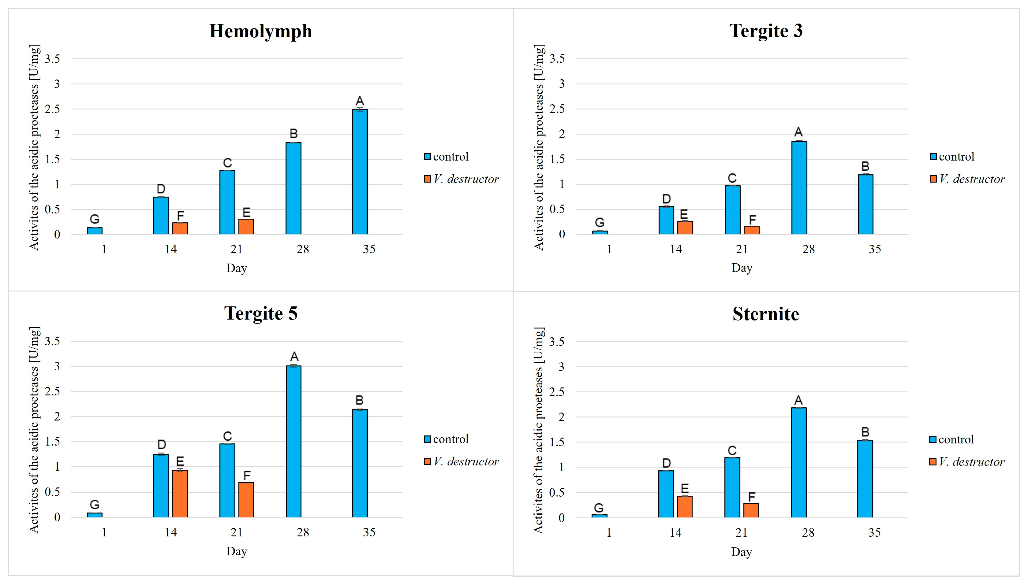
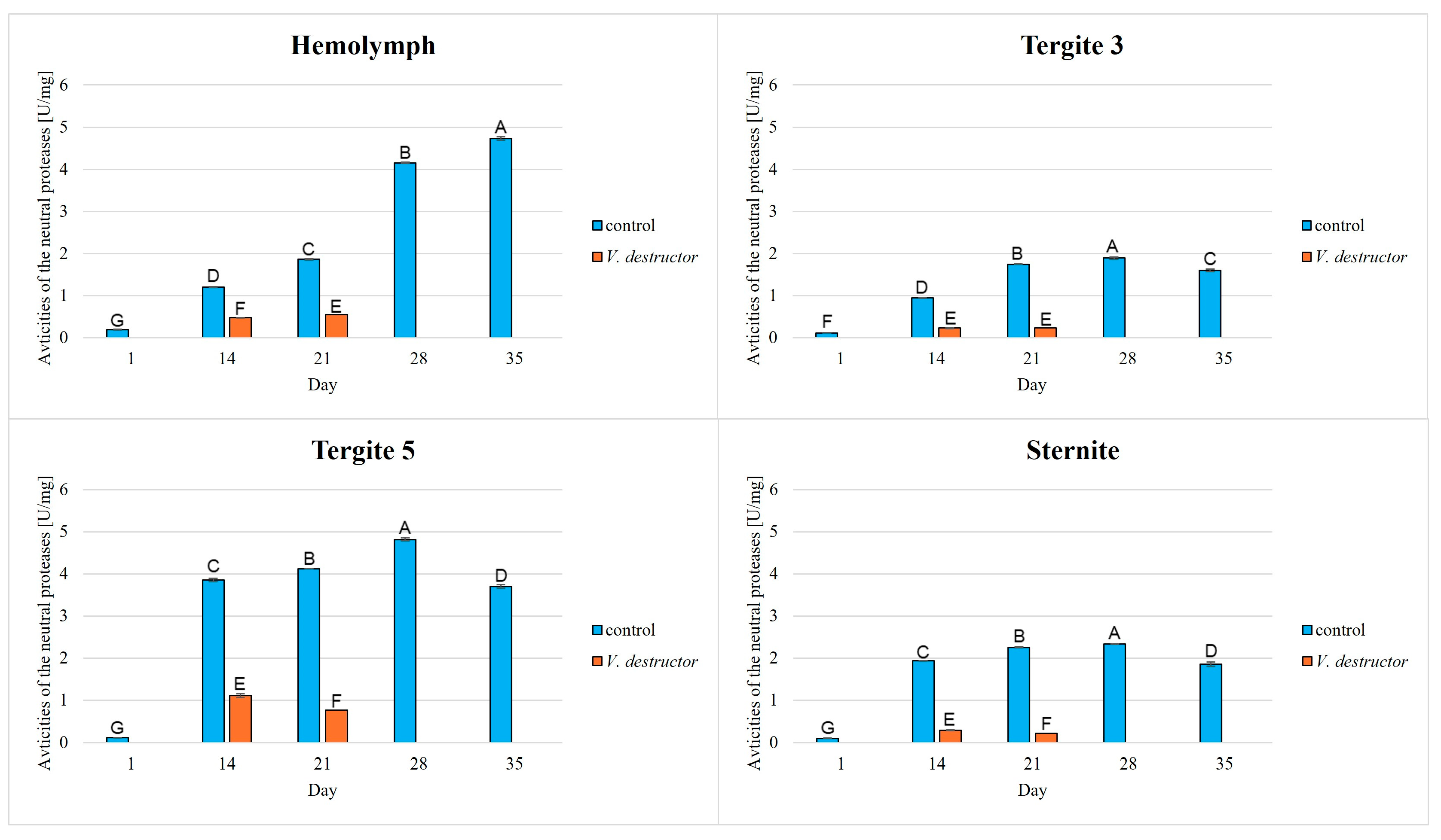
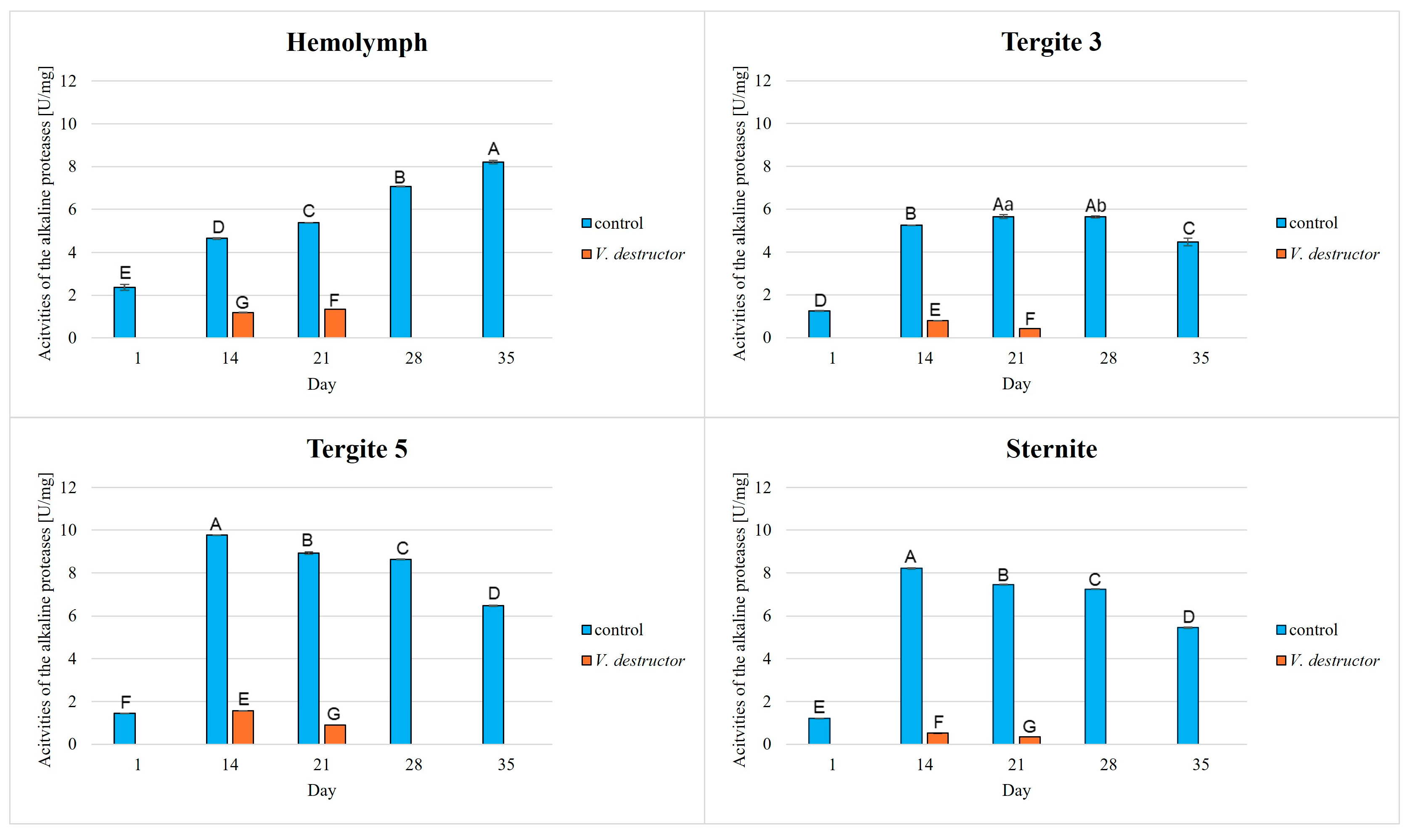
The highest activities were observed in the alkaline proteases. The activities of acidic and neutral proteases increased with age, reaching the highest levels in all the fat body segments of the 28-day-old healthy workers, and then decreased in those of the 35-day-old workers. In contrast, in the hemolymph, the activities of acidic and neutral proteases increased with age, reaching the highest values in the 35-day-old workers (Figure 2 and Figure 3). A different trend was observed in the case of the alkaline protease activities—the activities of these enzymes in the tergite 5 and sternite increased in the 14-day-old workers and then gradually decreased with age (Figure 4).
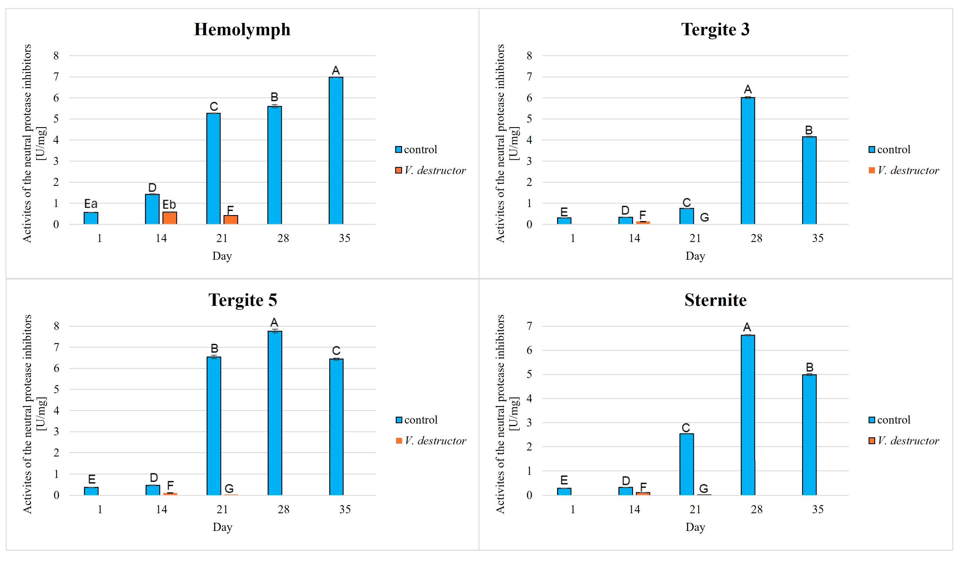
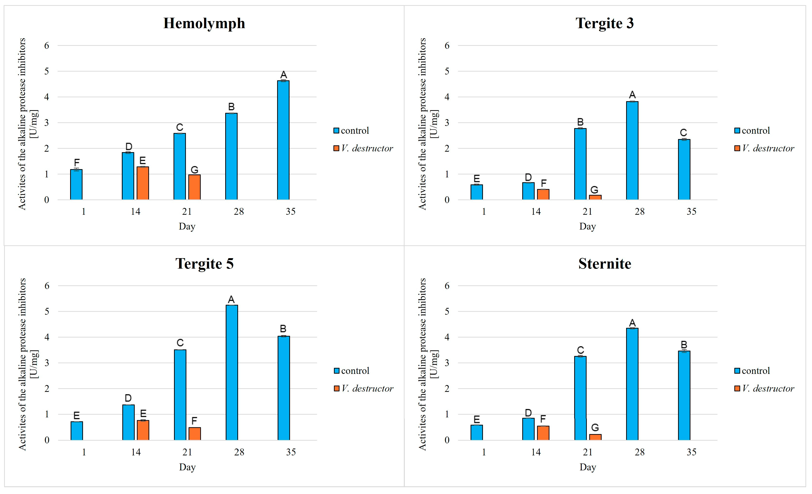
In the fat body segments (tergites 3 and 5, and the sternite), the activities of acidic, neutral, and alkaline protease inhibitors increased with age, reaching maximum values in the 28-day-old healthy workers and then decreasing in the 35-day-old workers (Figure 5, Figure 6 and Figure 7). Among the protease inhibitors tested, the acidic protease inhibitors exhibited the highest activity, while alkaline protease inhibitors displayed the lowest activity (Figure 5 and Figure 7).
Varroa destructor reduces the activities of acidic, neutral, and alkaline proteases and their inhibitors in the fat body segments and hemolymph (Figure 2, Figure 3, Figure 4, Figure 5, Figure 6 and Figure 7). The highest activities of proteases and their inhibitors were noted in the 14-day-old workers. In contrast, the lowest activities were observed in the 21-day-old workers infested with V. destructor. (Figure 2, Figure 3, Figure 4, Figure 5, Figure 6 and Figure 7).
Tergite 5 was characterized by the highest activities of proteases, while the highest activities of the protease inhibitors were found in the hemolymph (Figure 2, Figure 3, Figure 4, Figure 5, Figure 6 and Figure 7).

4. Discussion

The proteolytic system serves many functions in bees. Firstly, it is responsible for regulating numerous physiological processes, including protein digestion, and secondly, it is one of the bees’ primary defence barriers against viruses, parasites, fungi, and bacteria. Proteolytic enzymes, along with their inhibitors, can be found on the surface of the cuticle, constituting the first line of defence against pathogens. They are produced internally in the fat body and distributed through the hemolymph [2,37]. The activation of the protease cascade is responsible for rapid threat recognition and subsequent neutralization. Reduced protease activities are conducive to digestive disorders [38,39] and also lead to metabolic disorders, preventing zymogens from being activated. This results in the blockage of many processes occurring in bees. For example, proteins and hormones are not secreted properly, which weakens the immune system, making bees more susceptible to pathogens such as Nosema apis or Varroa destructor. Among the proteases, serine proteases play a key role in immunity, being responsible for initiating the proPO cascade, leading to melanization, sclerotization, and mounting effective defence against pathogens [11,40,41].
The activity of the proteolytic system is influenced by many environmental and anthropogenic factors, such as electromagnetic fields [42,43], the diet [10], pesticides [44], drugs [45], and the cell size in the comb [13,46], in addition to others. As mentioned earlier, the efficient functioning of proteases and their inhibitors is essential for the bee’s body to defend itself and fight pathogen infections. However, little attention has been paid to illustrating the effects of disease states on the activities of proteases and their inhibitors within the bee. Therefore, with this publication, we have filled this gap and demonstrated how V. destructor affects the activity of this crucial immune system. Furthermore, we focused on illustrating the effects wrought by Varroa in two tissues crucial for bee immunity and physiology: the hemolymph and the fat body. These tissues were selected because they serve as a nutrient source for the mites. To develop effective strategies for controlling this parasite, a thorough understanding of the mechanisms governing host–parasite interactions is essential. Additionally, we considered the fat body as a heterogeneous, segmented tissue [33] and presented the activities of acidic, neutral, and alkaline proteases and their inhibitors in tergites 3 and 5, and the sternite—the locations to which the mites most frequently attach [22].

4.1. Host (Honeybee)—Pathogen/Parasite Interaction

Varroa destructor, as one of the parasites causing losses in bee colonies, is an interesting model for research. In order to develop a strategy to combat this parasite, it is essential to understand its biology and physiology, including protease and protease inhibitor activities, which are crucial for the host defence.
Ramsey et al. [22] in 2019 showed that V. destructor feeds primarily on apian fat bodies, which allows it to maintain high reproductive rates. Piou et al. [20] provide new information on the proteome of V. destructor as it feeds on the hemolymph and fat bodies at various stages of bee development, i.e., larvae, pupae, and mature forms. V. destructor has two life stages: the first, the reproductive one, in which the parasite lays eggs in cells containing larvae or pupae, and the second, pertaining to dispersal, in which the parasite feeds on adult bees. Depending on the life stage, V. destructor requires different nutrients. During the dispersal phase, it feeds primarily on the fat bodies of adult bees, which provide them with energy for survival, while during the reproductive phase, it uses the larvae and pupae to extract the hemolymph, rich in proteins and amino acids essential for egg production. This allows its metabolism to adapt to current needs—from lipid burning to protein synthesis [47]. We add new information that the proteins used by V. destructor in the bee’s body come primarily from the fat body from tergite 3 and the sternite, as well as from the hemolymph (Figure 1). Moreover in these tissues/fat body locations, the protein concentrations in the workers from the infested colonies were very low. The protein concentrations in the fat body from tergite 5 in the workers with mites on their bodies were similar or even higher than in those in the control group. Piou et al. [20] showed, that the same proteins found in the bee hemolymph were identified in the gut of V. destructor mites. These proteins include apolipophorin, vitellogenin, hexamerin, and transferrin, which are involved in energy transport and storage. These proteins were observed to be consumed the most frequently and in large quantities by the parasite, regardless of the bee’s developmental stage. Furthermore, it was noted that the amount of protein in the mite gut is variable, which is due to the protein composition of the bee body changing throughout the apian life cycle. There was no correlation between the parasite’s proteome and the bee fat body. This may be due to the fact that the hemolymph is more readily available and contains a high proportion of nutrients as it flows around the fat body and transports these compounds away from it. It is from this bee tissue that the mites obtain a wide range of nutrients, as well as proteases and their inhibitors. We observed the highest activities of these compounds in the fat body of tergite 5 (see the control group; Figure 2, Figure 3, Figure 4, Figure 5, Figure 6 and Figure 7). The mites primarily penetrate the intersegmental membrane between tergites 3 and 4 and between the sternites; hence the activity of the proteolytic system in the fat body from these locations was drastically reduced compared to the control group. Moreover, Piou et al. [20] suggest that, the mite may encounter difficulties in directly obtaining nutrients from the fat body, which is connected with a significant energetic cost for them. The hemolymph is a more easily digestible tissue. However, as suggested by Tewarson and Jany [48] and Piou et al. [20], honeybee protease inhibitors may be used by the parasite to control protein degradation and physiology within its body. This indicates that the activity/efficiency of metabolic processes occurring in the mites is closely dependent on proteins, hormones, and enzymes derived from various bee tissues at different stages of development. Furthermore, as our research suggests, mites utilize the proteolytic system compounds from both the hemolymph and the fat body, regardless of its location, and in each of these tissues, we observed a significant reduction in these enzyme activities in the bodies of the infested bees.
The main source of the enzymes and enzyme inhibitors produced by the parasite is its salivary secretion, which contains substances such as cystatin-L2. The parasite injects saliva into the bee’s white-eyed pupa to digest its tissues and decrease the expression of 1483 genes responsible, among others, for ATP production and nutrient metabolism, which reduces the parasite’s energy consumption and facilitates its absorption of food to the detriment of the bee [49].
Proteolytic activity is assessed depending on the environmental pH and the enzyme’s origin, i.e., whether it comes from the host or the pathogen. In a study of host–parasite interactions, Frączek et al. [50] determined the activity of the proteolytic system in three pH ranges: 7.5—at this pH, bee enzymes exhibit high activity; 5—moderate activity in both the bees and V. destructor; and pH 3.5, where the parasite protease activity is the highest (Figure 8). They demonstrated that in all the pH ranges, V. destructor extracts inhibit the activities of bee proteases, primarily the serine proteases, resulting in a suppression of host defence mechanisms. V. destructor has been found to contain trypsin and its inhibitors, which aid the parasite in feeding on honeybees by preventing hemolymph clotting, which facilitates parasitism [50]. Strachecka et al. [19] showed that V. destructor which had no contact with acaricides (e.g., through the use of tau-fluvalinate) produced primarily aspartic and serine proteases, while the mites exposed to acaricides synthesized aspartic, serine, and thiol proteases, metalloproteases, as well as serine protease inhibitors. This is consistent with the results obtained in our study. Most likely, during the digestion of bee tissue by V. destructor, a reaction between the enzymes of these two organisms occurs, consequently inhibiting the activities of acidic, neutral, and alkaline proteases and their inhibitors in the host (Figure 2, Figure 3, Figure 4, Figure 5, Figure 6 and Figure 7). At this point, it is worth posing the following questions: what type of proteases and their inhibitors are these, and what is the precise mechanism by which they are activated and act within the bee? These questions will guide future research.
Nazzi et al. [51] demonstrated that in response to the presence of V. destructor and the accompanying infection with the deformed wing virus (DWV), the expression of genes encoding serine proteases increases in the bee body, which may indicate an active attempt on the part of the bee to defend itself against the pathogen. Furthermore, the research by Zou et al. [7] indicates that infestation with V. destructor and DWV can lead to a reduction in the activities of some serine proteases, suggesting a complex nature of the host–parasite interactions.
Similarly to the V. destructor parasite, other pathogens also cause changes in the activity of the proteolytic system, which can be observed as an increase or decrease in this activity. Malone and Gatehouse [52] demonstrated that N. apis reduces the activities of digestive enzymes, such as trypsin and chymotrypsin, compared to healthy bees. Furthermore, Doughuzlu et al. [53] demonstrated the presence of serine proteases with molecular masses in the range of 23–35 kDa and cathepsins in bees infected with N. ceranae. On the one hand, at low activities, these enzymes can act as immunostimulants and induce a host immune response. On the other hand, especially at high activities, they facilitate the penetration of host tissues by N. ceranae. The bacteria P. larvae infecting the larva produce proteases. Studies have shown that these are primarily metalloproteases, which play a significant role in pathogenesis. Therefore, they are considered the main factors responsible for the virulence of P. larvae. Importantly, protease production in P. larvae may be regulated by a quorum-sensing mechanism [54,55]. Monitoring changes in the bees’ proteolytic system in response to pathogens such as microsporidia, bacteria, and mites can be an effective way to assess bee colony health and infection levels. This will allow for an early detection of problems in the apiary and the implementation of protective measures to limit the spread of disease and improve the health of bee colonies.

4.2. The Relationship Between the Proteolytic System, Tissue Location and Aging Processes in the Workers

Maintaining proper homeostasis in the body involves various proteins. The synthesis and degradation of these proteins are controlled by proteases and their inhibitors. These enzymes work in balance to ensure proper cell function.
As the bees age, the efficiency of the proteolytic system declines. This leads to the accumulation of damaged proteins, decreased immunity, and higher susceptibility to various diseases. The available literature has reported that high levels of hemolymph proteins indicate that bees are more resistant to various pathogens, while a decrease in protein levels may indicate poor health. Low protein concentrations may be a consequence of an impaired functioning of proteases and their inhibitors, one of whose functions is the post-translational processing of proteins, including the enzymatic ones. We observe a phenomenon referred to as a vicious circle here: low activity of the proteolytic system—low concentration of functional proteins—even lower activity of the system [56,57]. This observation is consistent with our results, as we found that low levels of proteins, proteases, and their inhibitors are associated with V. destructor infestation in both the bee hemolymph and the fat body segments, i.e., tergites 3 and 5, and the sternite (Figure 1, Figure 2, Figure 3, Figure 4, Figure 5, Figure 6 and Figure 7). Thus, V. destructor is one of the factors that accelerate the aging process in bees.
It is known that the activity of the proteolytic system increases in the hemolymph of healthy bees with age/aging [10,43,45]. However, to our knowledge, the way these activities change in the bee fat body has not been presented in the literature. Strachecka et al. [2] focused on demonstrating differences in the activity of this system in the fat body between the bee castes, but only in 1-day-old bees. Bryś et al. [10] presented the activities of proteases and their inhibitors in various locations of the fat body of bees up to 14 days of age. In this publication, we expand on the existing knowledge about the functioning of the proteolytic system in both the hemolymph and the fat body of healthy summer workers up to 35 days of age. Furthermore, we showed how the activity of this system changes in varroa-infested workers up to 21 days of age. It is worth emphasizing that the bee samples were collected from apiary tests, not cage tests as in numerous publications. We demonstrated that protease activities increased in healthy workers until 21 or 28 days of age, after which a decline was observed in the 35-day-old individuals. A similar trend was observed in the bees infested with the V. destructor parasite—the highest protease activities were observed in the 14-day-old workers and the lowest in the 21-day-old workers (Figure 2, Figure 3, Figure 4, Figure 5, Figure 6 and Figure 7).
The apparent decline in the efficiency of the proteolytic system in 35-day-old healthy workers may indicate progressive degenerative changes associated with aging. Therefore, the values of its activity may be a biomarker of aging. As a result of these phenomena, bees experience accelerated aging that resembles the metabolic syndrome characteristic of vertebrates. In humans, the metabolic syndrome consists of various factors such as high blood sugar levels, hypertension, lipid metabolism disorders, and abdominal obesity. These factors ultimately lead to the development of diabetes and heart disease, which, if left untreated, accelerate aging. In insects, specifically dragonflies, a number of physiological changes resembling the metabolic syndrome have been observed. Dragonflies infected with gregarines (intestinal parasites) have been observed to exhibit high hemolymph sugar levels, impaired lipid metabolism, fat deposition in the thorax, and weakened flight muscles. These changes and infection result in impaired carbohydrate regulation and the activation of p38 MAP kinase, which is associated with stress and the immune response. These observations suggest that the metabolic syndrome occurs not only in vertebrates but can also be identified in insects. Based on the available research, we suggest that honeybees may be a valuable model for studying the mechanisms of the metabolic syndrome and the associated aging process [58,59].
We also observed that after emergence, the worker bees had the most active proteolytic system in their hemolymph. From day 7 until the end of their lives, these values were higher in the fat body, particularly elevated in tergite 5. This is consistent with our previous studies [28], which showed that the fat body in tergite 5 was characterized by the highest antioxidant activities. This may suggest that this location of the fat body is responsible for detoxification, neutralization, and proteolysis, and is the “bee liver” (analogous to the vertebrate liver). Comparing the activity of the proteolytic system in different locations of the fat body, we found the lowest values in tergite 3, and particularly depressed in the workers infested with V. destructor. The fat body of tergite 3 accumulates large amounts of energy compounds (glycogen, triacylglycerols, glucose) [33,60], which participate in the Krebs cycle and the respiratory chain [29]. This would explain why the mites choose this location, attaching themselves to the bee cuticle. The depletion of energy reserves and the destabilization of the proteolytic and antioxidant systems in the bee fat body progress with the aging process, which is clearly accelerated by V. destructor.

5. Conclusions

  • Our results showed that the activities of acidic, neutral, and alkaline proteases, and their inhibitors were highest in the fat body of tergite 5. Therefore, the fat body of tergite 5 not only plays a significant role in the antioxidant defence but also in other mechanisms, such as the activation of the proteolytic system. Understanding these various (other) mechanisms is crucial, particularly in the context of immunity and aging in bees. It is worthwhile to focus on analyzing the fat body from this segment in the future, as it may provide valuable information on bee colony health.
  • V. destructor reduces the activities of proteases and their inhibitors, which in turn causes an accelerated aging of the bees. It is important to monitor bee colonies for infestations with this parasite and to develop effective methods of combating this parasite, as it leads to the decline of bee colonies.
  • Serine proteases were the most active among these enzymes. Perhaps they could become a new indicator of immunity, which could help monitor the condition of bees and relatively quickly identify emerging stressors, such as pathogens or pesticides that threaten bee colonies.
  • Changes in the proteolytic system are one of the effects of aging. Therefore, it is important to develop strategies for the early detection of disturbances in the proteolytic system. It is also crucial to create new supplements and management methods for honey bee colonies that enhance immunity while simultaneously slowing the aging process.

Author Contributions

Conceptualization, A.S.; methodology, M.K.-B., K.O. and A.S.; software, M.K.-B. and A.S.; validation, A.S.; formal analysis, M.K.-B., P.S. and A.S.; resources, A.S. and K.O.; data curation, A.S.; writing—original draft preparation, M.K.-B.; writing—review and editing, M.K.-B., P.S., K.O. and A.S.; visualization, M.K.-B.; supervision, A.S.; project administration, M.K.-B. and A.S. All authors have read and agreed to the published version of the manuscript.

Funding

This study was funded by the University of Life Sciences in Lublin, No. LKESUBB.WLE.22.058. For the purpose of Open Access, the author has applied a CC-BY public copyright license to any author-accepted manuscript (AAM) version arising from this submission.

Institutional Review Board Statement

Not applicable.

Data Availability Statement

The original contributions presented in this study are included in the article. Further inquiries can be directed to the corresponding author.

Conflicts of Interest

The authors declare no conflicts of interest.

References

  1. Morfin, N.; Anguiano-Baez, R.; Guzman-Novoa, E. Honey bee (Apis mellifera) immunity. Vet. Clin. N. Am. Food Anim. Pract. 2021, 37, 521–533. [Google Scholar] [CrossRef]
  2. Strachecka, A.; Kuszewska, K.; Olszewski, K.; Skowronek, P.; Grzybek, M.; Grabowski, M.; Paleolog, J.; Woyciechowski, M. Activities of antioxidant and proteolytic systems and biomarkers in the fat body and hemolymph of young Apis mellifera females. Animals 2022, 12, 1121. [Google Scholar] [CrossRef]
  3. Larsen, A.; Reynaldi, F.J.; Guzmán-Novoa, E. Fundaments of the honey bee (Apis mellifera) immune system. Review. Rev. Mex. Cienc. Pecu. 2019, 10, 705–728. [Google Scholar] [CrossRef]
  4. DeGrandi-Hoffman, G.; Chen, Y. Nutrition, immunity and viral infections in honey bees. Curr. Opin. Insect Sci. 2015, 10, 170–176. [Google Scholar] [CrossRef]
  5. Skowronek, P.; Wójcik, Ł.; Strachecka, A. CBD supplementation has a positive effect on the activity of the proteolytic system and biochemical markers of honey bees (Apis mellifera) in the apiary. Animals 2022, 12, 2313. [Google Scholar] [CrossRef]
  6. Rai, M.; Curley, M.; Coleman, Z.; Demontis, F. Contribution of proteases to the hallmarks of aging and to age-related neurodegeneration. Aging Cell 2022, 21, e13603. [Google Scholar] [CrossRef]
  7. Zou, Z.; Lopez, D.L.; Kanost, M.R.; Evans, J.D.; Jiang, H. Comparative analysis of serine protease-related genes in the honey bee genome: Possible involvement in embryonic development and innate immunity. Insect Mol. Biol. 2006, 15, 603–614. [Google Scholar] [CrossRef] [PubMed]
  8. Bogaerts, A.; Baggerman, G.; Vierstraete, E.; Schoofs, L.; Verleyen, P. The hemolymph proteome of the honeybee: Gel-based or gel-free? Proteomics 2009, 9, 3201–3208. [Google Scholar] [CrossRef] [PubMed]
  9. Migdał, P.; Murawska, A.; Strachecka, A.; Bieńkowski, P.; Roman, A. Honey bee proteolytic system and behavior parameters under the influence of an electric field at 50 Hz and variable intensities for a long exposure time. Animals 2021, 11, 863. [Google Scholar] [CrossRef]
  10. Bryś, M.S.; Olszewski, K.; Strachecka, A. The relationship between pollen monodiets and the activities of proteolytic systems in the fat body and hemolymph of honeybee workers. PLoS ONE 2025, 20, e0326175. [Google Scholar] [CrossRef] [PubMed]
  11. Asgari, S.; Zhang, G.; Zareie, R.; Schmidt, O. A Serine proteinase homolog venom protein from an endoparasitoid wasp inhibits melanization of the host hemolymph. Insect Biochem. Mol. Biol. 2003, 33, 1017–1024. [Google Scholar] [CrossRef]
  12. Kim, B.Y.; Lee, K.S.; Wan, H.; Zou, F.M.; Choi, Y.S.; Yoon, H.J.; Kwon, H.W.; Je, Y.H.; Jin, B.R. Anti-elastolytic activity of a honeybee (Apis cerana) chymotrypsin inhibitor. Biochem. Biophys. Res. Commun. 2013, 430, 144–149. [Google Scholar] [CrossRef]
  13. Dziechciarz, P.; Strachecka, A.; Borsuk, G.; Olszewski, K. Workers of honey bee (Apis mellifera L.) reared in small-cell combs in apiary conditions show higher activity of the proteolytic system and lower protein concentrations on the cuticle surface than workers reared in standard-cell combs. Pol. J. Vet. Sci. 2025, 28, 111–122. [Google Scholar] [CrossRef] [PubMed]
  14. Bajda, M.; Łoś, A.; Schulz, M.; Kasperek, K. Metaloproteazy ssaków i owadów. Med. Weter 2016, 72, 408–412. (In Polish) [Google Scholar] [CrossRef]
  15. Liu, H.; Xu, J.; Wang, L.; Guo, P.; Tang, Z.; Sun, X.; Tang, X.; Wang, W.; Wang, L.; Cao, Y.; et al. Serpin-1a and Serpin-6 regulate the toll pathway immune homeostasis by synergistically inhibiting the Spätzle-processing enzyme CLIP2 in silkworm, Bombyx mori. PLoS Pathog. 2023, 19, e1011740. [Google Scholar] [CrossRef]
  16. Dmitryjuk, M.; Żółtowska, K.; Frączek, R.; Lipiński, Z. Esterases of Varroa destructor (Acari: Varroidae), parasitic mite of the honeybee. Exp. Appl. Acarol. 2014, 62, 499–510. [Google Scholar] [CrossRef] [PubMed]
  17. van Dooremalen, C.; Stam, E.; Gerritsen, L.; Cornelissen, B.; van der Steen, J.; van Langevelde, F.; Blacquière, T. Interactive effect of reduced pollen availability and Varroa destructor infestation limits growth and protein content of young honey bees. J. Insect Physiol. 2013, 59, 487–493. [Google Scholar] [CrossRef]
  18. Cournoyer, A.; Plamondon, L.; Bau-Gaudreault, L.; Deschamps, A.; Dubreuil, P.; Benoit-Biancamano, M.O. Effects of Varroa destructor on hemolymph sugars and secondary infections in honeybees (Apis mellifera). Appl. Sci. 2022, 12, 11630. [Google Scholar] [CrossRef]
  19. Strachecka, A.; Borsuk, G.; Olszewski, K.; Paleolog, J.; Lipiński, Z. Proteolysis on the body surface of pyrethroid-sensitive and resistant Varroa destructor. Acta Parasitol. 2013, 58, 64–69. [Google Scholar] [CrossRef] [PubMed]
  20. Piou, V.; Arafah, K.; Bocquet, M.; Bulet, P.; Vétillard, A. The proteomic content of Varroa destructor gut varies according to the developmental stage of its host. PLoS Pathog. 2024, 20, e1012802. [Google Scholar] [CrossRef]
  21. Colin, M.; Tchamitchian, M.; Bonmatin, J.M.; Di Pasquale, S. Presence of chitinase in adult Varroa destructor, an ectoparasitic mite of Apis mellifera. Exp. Appl. Acarol. 2001, 25, 947–955. [Google Scholar] [CrossRef]
  22. Ramsey, S.D.; Ochoa, R.; Bauchan, G.; Gulbronson, C.; Mowery, J.D.; Cohen, A.; Lim, D.; Joklik, J.; Cicero, J.M.; Ellis, J.D.; et al. Varroa destructor feeds primarily on honey bee fat body tissue and not hemolymph. Proc. Natl. Acad. Sci. USA 2019, 116, 1792–1801. [Google Scholar] [CrossRef]
  23. Garedew, A.; Schmolz, E.; Lamprecht, I. The energy and nutritional demand of the parasitic life of the mite Varroa destructor. Apidologie 2004, 35, 419–430. [Google Scholar] [CrossRef]
  24. Pinto, F.; Souza, G.; Sanches, M.; Serrão, J. Parasitic effects of Varroa destructor (Acari: Varroidae) on hypopharyngeal glands of africanized Apis mellifera (Hymenoptera: Apidae). Sociobiology 2011, 58, 769–778. [Google Scholar]
  25. Zakaria, M.E.; Abd El-Wahab, T.E. Scanning electron microscopic (SEM) of hypopharyngeal and mandibular glands of honey bee workers infested by Varroa mites (Varroa destructor). Egypt. J. Agric. Sci. 2004, 55, 375–384. [Google Scholar] [CrossRef]
  26. Abd El-Wahab, T.E.; Zakaria, M.E.; Nour, M.E. Influence of the infestation by Varroa mite Varroa destructor on some antennal sense organs of the worker and drone honey bees Apis mellifera L. J. Appl. Sci. Res. 2006, 2, 80–85. [Google Scholar]
  27. Power, K.; Martano, M.; Altamura, G.; Piscopo, N.; Maiolino, P. Histopathological features of symptomatic and asymptomatic honeybees naturally infected by deformed wing virus. Pathogens 2021, 10, 874. [Google Scholar] [CrossRef] [PubMed]
  28. Kunat-Budzyńska, M.; Staniszewska, P.; Olszewski, K.; Strachecka, A. Antioxidant activities in the hemolymph and fat body of physiologically and prematurely aging bees (Apis mellifera). Antioxidants 2025, 14, 373. [Google Scholar] [CrossRef]
  29. Kunat-Budzyńska, M.; Staniszewska, P.; Olszewski, K.; Cytryńska, M.; Strachecka, A. The efficiency of the krebs cycle and the respiratory chain in physiologically and prematurely aging bees (Apis mellifera). Int. J. Mol. Sci. 2025, 26, 7294. [Google Scholar] [CrossRef]
  30. Olszewski, K. Biotechniczne Zwalczanie Warrozy-Współczesne Możliwości „Archaicznych” Metod, 1st ed.; Bee & Honey Sp z o.o.: Klecza Dolna, Poland, 2025; pp. 1–114. [Google Scholar]
  31. Łoś, A.; Strachecka, A. Fast and cost-effective biochemical spectrophotometric analysis of solution of insect “blood” and body surface elution. Sensors 2018, 18, 1494. [Google Scholar] [CrossRef] [PubMed]
  32. Wójcik, Ł.; Chęć, M.; Skowronek, P.; Grabowski, M.; Persona, K.; Strachecka, A. Do the different life history strategies of ants and honeybees determine fat body morphology? Arthropod Struct. Dev. 2022, 69, 101186. [Google Scholar] [CrossRef]
  33. Strachecka, A.; Olszewski, K.; Kuszewska, K.; Chobotow, J.; Wójcik, Ł.; Paleolog, J.; Woyciechowski, M. Segmentation of the subcuticular fat body in Apis mellifera females with different reproductive potentials. Sci. Rep. 2021, 11, 13887. [Google Scholar] [CrossRef]
  34. Schacterle, G.R.; Pollack, R.L. A simplified method for the quantitative assay of small amounts of protein in biologic material. Anal. Biochem. 1973, 51, 654–655. [Google Scholar] [CrossRef]
  35. Anson, M.L. The estimation of pepsin, trypsin, papain, and cathepsin with hemoglobin. J. Gen. Physiol. 1938, 22, 79–89. [Google Scholar] [CrossRef] [PubMed]
  36. Lee, T.M.; Lin, Y.H. Trypsin inhibitor and trypsin-like protease activity in air- or submergence-grown rice (Oryza sativa L.) coleoptiles. Plant Sci. 1995, 106, 43–54. [Google Scholar] [CrossRef]
  37. Grzywnowicz, K.; Ciołek, A.; Tabor, A.; Jaszek, M. Profiles of the body-surface proteolytic system of honey bee queens, workers and drones: Ontogenetic and seasonal changes in proteases and their natural inhibitors. Apidologie 2009, 40, 4–19. [Google Scholar] [CrossRef]
  38. Haloi, K.; Kalita, M.K.; Devi, D. Regulation and characterization of amylase enzyme secretion in the digestive tract of Antheraea assamensis Helfer. Indian J. Entomol. 2023, 85, 556–562. [Google Scholar] [CrossRef]
  39. Sharifi, M.; Chitgar, G.; Ghadamyari, M.; Ajamhasani, M. Identification and characterization of midgut digestive proteases from the rosaceous branch borer, Osphranteria coerulescens redtenbacher (Coleoptera: Cerambycidae). Rom. J. Biochem. 2012, 49, 33–37. [Google Scholar]
  40. Murawska, A.; Migdał, P.; Roman, A. Effects of plant protection products on biochemical markers in honey bees. Agriculture 2021, 11, 648. [Google Scholar] [CrossRef]
  41. Wessler, I.; Gärtner, H.A.; Michel-Schmidt, R.; Brochhausen, C.; Schmitz, L.; Anspach, L.; Grünewald, B.; Kirkpatrick, C.J. Honeybees produce millimolar concentrations of non-neuronal acetylcholine for breeding: Possible adverse effects of neonicotinoids. PLoS ONE 2016, 11, e0156886. [Google Scholar] [CrossRef]
  42. Migdał, P.; Murawska, A.; Strachecka, A.; Bieńkowski, P.; Roman, A. Changes in the honeybee antioxidant system after 12 h of exposure to electromagnetic field frequency of 50 Hz and variable intensity. Insects 2020, 11, 713. [Google Scholar] [CrossRef]
  43. Migdał, P.; Murawska, A.; Bieńkowski, P.; Strachecka, A.; Roman, A. Effect of the electric field at 50 Hz and variable intensities on biochemical markers in the honey bee’s hemolymph. PLoS ONE 2021, 16, e0252858. [Google Scholar] [CrossRef]
  44. Paleolog, J.; Wilde, J.; Siuda, M.; Bąk, B.; Wójcik, Ł.; Strachecka, A. Imidacloprid markedly affects hemolymph proteolysis, biomarkers, DNA global methylation, and the cuticle proteolytic layer in western honeybees. Apidologie 2020, 51, 620–630. [Google Scholar] [CrossRef]
  45. Strachecka, A.; Olszewski, K.; Paleolog, J. Varroa treatment with bromfenvinphos markedly suppresses honeybee biochemical defence levels. Entomol. Exp. Appl. 2016, 160, 57–71. [Google Scholar] [CrossRef]
  46. Dziechciarz, P.; Strachecka, A.; Borsuk, G.; Olszewski, K. Effect of rearing in small-cell combs on activities of catalase and superoxide dismutase and total antioxidant capacity in the hemolymph of Apis mellifera workers. Antioxidants 2023, 12, 709. [Google Scholar] [CrossRef] [PubMed]
  47. Han, B.; Wu, J.; Wei, Q.; Liu, F.; Cui, L.; Rueppell, O.; Xu, S. Life-history stage determines the diet of ectoparasitic mites on their honey bee hosts. Nat. Commun. 2024, 15, 725. [Google Scholar] [CrossRef]
  48. Tewarson, N.C.; Jany, K.D. Determination of proteolytic activity in Varroa jacobsoni an ectoparasitic hemophagous mite of honey bees (Apis sp.). Apidologie 1982, 13, 383–389. [Google Scholar] [CrossRef]
  49. Zhou, H.; Duan, X.; Sun, C.; Huang, H.; Yang, M.; Huang, S.; Li, J. Salivary cystatin-L2-like of Varroa destructor causes lower metabolism activity and abnormal development in Apis Mellifera pupae. Animals 2023, 13, 3660. [Google Scholar] [CrossRef] [PubMed]
  50. Frączek, R.J.; Żółtowska, K.; Lipiński, Z.; Dmitryjuk, M. The mutual influence of proteins from Varroa destructor extracts and from honeybee haemolymph on their proteolytic activity-in vitro study. Acta Parasitol. 2013, 58, 317–323. [Google Scholar] [CrossRef]
  51. Nazzi, F.; Brown, S.P.; Annoscia, D.; Del Piccolo, F.; Di Prisco, G.; Varricchio, P.; Vedova, G.D.; Cattonaro, F.; Caprio, E.; Pennacchio, F. Synergistic parasite-pathogen interactions mediated by host immunity can drive the collapse of honeybee colonies. PLoS Pathog. 2012, 8, e1002735. [Google Scholar] [CrossRef]
  52. Malone, L.A.; Gatehouse, H.S. Effects of Nosema apis infection on honey bee (Apis Mellifera) digestive proteolytic enzyme activity. J. Invertebr. Pathol. 1998, 71, 169–174. [Google Scholar] [CrossRef]
  53. Doghuzlu, M.A.; Nabian, S.; Paghaleh, G.A.N.; Taheri, M.; Asadollahi, Z.; Akhzari, S. Zymography of proteases in honey bees (Apis mellifera) infected with Nosema ceranae. Vet. Glas. 2024, 78, 155–167. [Google Scholar] [CrossRef]
  54. Antúnez, K.; Anido, M.; Schlapp, G.; Evans, J.D.; Zunino, P. Characterization of secreted proteases of Paenibacillus larvae, potential virulence factors involved in honeybee larval infection. J. Invertebr. Pathol. 2009, 102, 129–132. [Google Scholar] [CrossRef]
  55. Pellegrini, M.C.; Zalazar, L.; Fuselli, S.R.; Ponce, A.G. Inhibitory action of essential oils against proteases activity of Paenibacillus larvae, the Etiological Agent of American Foulbrood Disease. Span. J. Agric. Res. 2017, 15, e0504. [Google Scholar] [CrossRef]
  56. Elfar, S.A.; Bahgat, I.M.; Shebl, M.A.; Lihoreau, M.; Tawfik, M.M. Intraspecific variability in proteomic profiles and biological activities of the honey bee hemolymph. Insects 2023, 14, 365. [Google Scholar] [CrossRef] [PubMed]
  57. Cabbri, R.; Ferlizza, E.; Nanetti, A.; Monari, E.; Andreani, G.; Galuppi, R.; Isani, G. Biomarkers of nutritional status in honeybee haemolymph: Effects of different biotechnical approaches for Varroa destructor treatment and wintering phase. Apidologie 2018, 49, 606–618. [Google Scholar] [CrossRef]
  58. Hashim, K.N.; Chin, K.Y.; Ahmad, F. The mechanism of honey in reversing metabolic syndrome. Molecules 2021, 26, 808. [Google Scholar] [CrossRef]
  59. Schilder, R.J.; Marden, J.H. Metabolic syndrome and obesity in an insect. Proc. Natl. Acad. Sci. USA 2006, 103, 18805–18809. [Google Scholar] [CrossRef]
  60. Bryś, M.S.; Staniec, B.; Strachecka, A. The effect of pollen monodiets on fat body morphology parameters and energy substrate levels in the fat body and hemolymph of Apis mellifera L. workers. Sci. Rep. 2024, 14, 15177. [Google Scholar] [CrossRef]
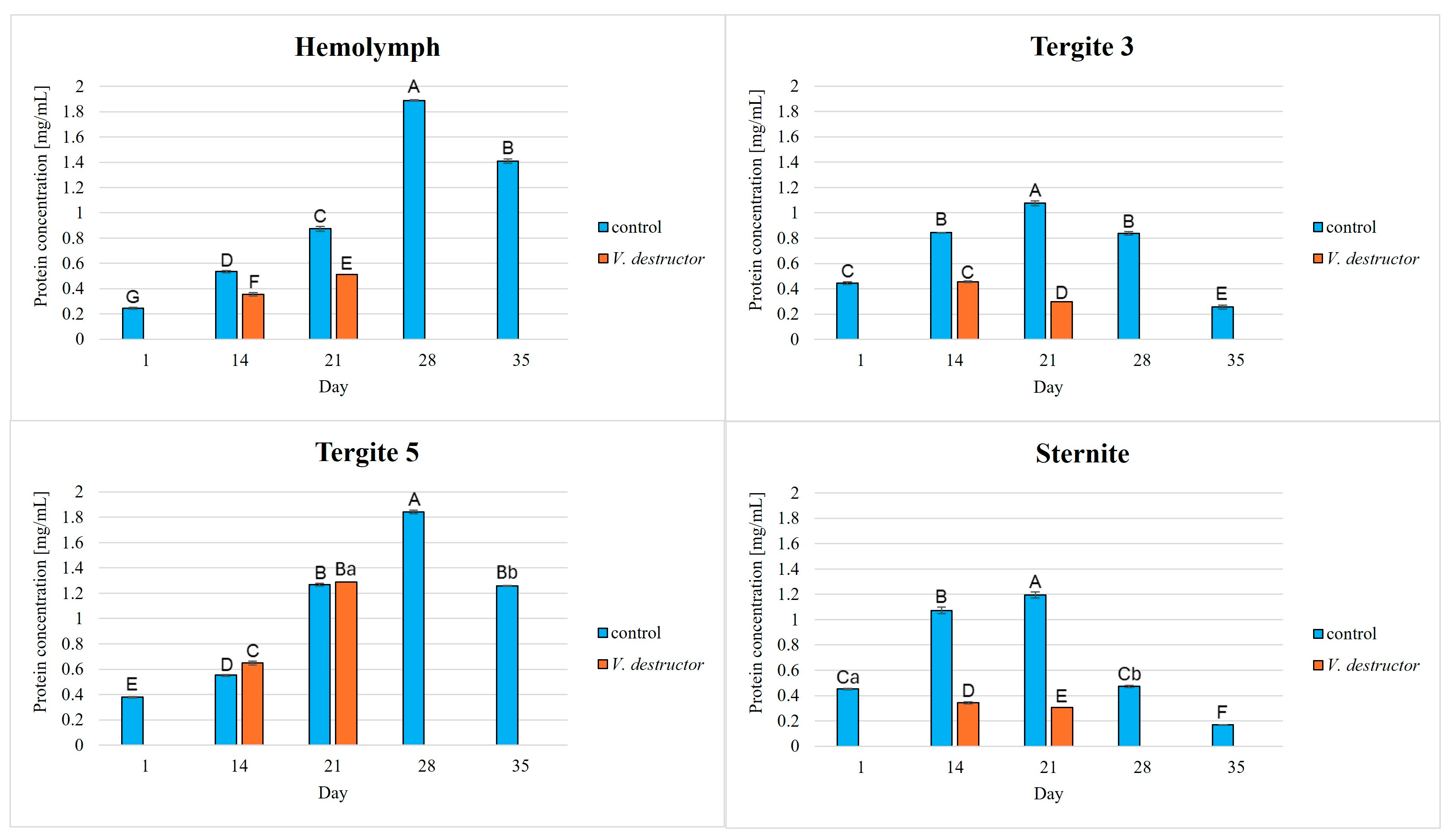
Figure 1. Protein concentrations in the hemolymph and the fat body from tergite 3, tergite 5, and the sternite of the healthy 1-, 14-, 21-, 28-, and 35-day-old workers and in the V. destructor-infested 14- and 21-day-old workers. A, B, C, D, E, F, G—capital letters indicate statistically significant differences between the groups at p ≤ 0.01. a, b—lowercase letters indicate statistically significant differences between the groups at p ≤ 0.05.
Agriculture 15 01942 g001
Figure 2. Activities of acidic proteases [U/mg protein] in the hemolymph and the fat body from tergite 3, tergite 5, and the sternite of the healthy 1-, 14-, 21-, 28-, and 35-day-old workers and in the V. destructor-infested 14- and 21-day-old workers. A, B, C, D, E, F, G—capital letters indicate statistically significant differences between the groups at p ≤ 0.01.
Agriculture 15 01942 g002
Figure 3. Activities of neutral proteases [U/mg protein] in the hemolymph and the fat body from tergite 3, tergite 5, and the sternite of the healthy 1-, 14-, 21-, 28-, and 35-day-old workers and in the V. destructor-infested 14- and 21-day-old workers. A, B, C, D, E, F, G—capital letters indicate statistically significant differences between the groups at p ≤ 0.01.
Agriculture 15 01942 g003
Figure 4. Activities of alkaline proteases [U/mg protein] in the hemolymph and the fat body from tergite 3, tergite 5, and the sternite of the healthy 1-, 14-, 21-, 28-, and 35-day-old workers and in the V. destructor-infested 14- and 21-day-old workers. A, B, C, D, E, F, G—capital letters indicate statistically significant differences between the groups at p ≤ 0.01. a, b—lowercase letters indicate statistically significant differences between the groups at p ≤ 0.05.
Agriculture 15 01942 g004
Figure 5. Activities of acidic protease inhibitors [U/mg protein] in the hemolymph and the fat body from tergite 3, tergite 5, and the sternite of the healthy 1-, 14-, 21-, 28-, and 35-day-old workers and in the V. destructor-infested 14- and 21-day-old workers. A, B, C, D, E, F, G—capital letters indicate statistically significant differences between the groups at p ≤ 0.01.
Agriculture 15 01942 g005
Figure 6. Activities of neutral protease inhibitors [U/mg protein] in the hemolymph and the fat body from tergite 3, tergite 5, and the sternite of the healthy 1-, 14-, 21-, 28-, and 35-day-old workers and in the V. destructor-infested 14- and 21-day-old workers. A, B, C, D, E, F, G—capital letters indicate statistically significant differences between the groups at p ≤ 0.01. a, b—lowercase letters indicate statistically significant differences between the groups at p ≤ 0.05.
Agriculture 15 01942 g006
Figure 7. Activities of alkaline protease inhibitors [U/mg protein] in the hemolymph and the fat body from tergite 3, tergite 5, and the sternite of the healthy 1-, 14-, 21-, 28-, and 35-day-old workers and in the V. destructor-infested 14- and 21-day-old workers. A, B, C, D, E, F, G—capital letters indicate statistically significant differences between the groups at p ≤ 0.01.
Agriculture 15 01942 g007
Figure 8. Dependence of the activities of proteolytic enzymes of bees and V. destructor on the pH value.
Agriculture 15 01942 g008
Table 1. The effect of tissue location: hemolymph, tergite 3, tergite 5, and the sternite in the particular age groups on the concentrations of proteins and the activities of acidic, neutral, and alkaline proteases and their inhibitors in both the healthy and V. destructor-infested workers.
Acidic
Proteases
Neutral
Proteases
Alkaline
Proteases
Protein
Concentration
HemolymphH = 175.49
p < 0.001
H = 175.19
p < 0.001
H = 138.93
p < 0.001
H = 181.96
p < 0.001
Tergite 3H = 168.18
p < 0.001
H = 146.89
p < 0.001
H = 62.45
p < 0.001
H = 78.56
p < 0.001
Tergite 5H = 168.18
p < 0.001
H = 131.76
p < 0.001
H = 53.56
p < 0.001
H = 187.20
p < 0.001
SterniteH = 168.17
p < 0.001
H = 129.95
p < 0.001
H = 22.83
p < 0.001
H = 76.66
p < 0.001
Acidic Protease InhibitorsNeutral Protease InhibitorsAlkaline Protease Inhibitors
HemolymphH = 141.16
p < 0.001
H = 135.04
p < 0.001
H = 138.01
p < 0.001
Tergite 3H = 124.71
p < 0.001
H = 132.38
p < 0.001
H = 98.88
p < 0.001
Tergite 5H = 125.14
p < 0.001
H = 101.75
p < 0.001
H = 141.32
p < 0.001
SterniteH = 131.60
p < 0.001
H = 131.62
p < 0.001
H = 131.43
p < 0.001
H—statistic from the Kruskal–Wallis test; p—probability value.
Table 2. The effect of age: 1, 14, 21, 28 and 35 days for the individual tissues (the hemolymph and fat body segments: tergites 3 and 5, and the sternite) on the concentrations of proteins and the activities of acidic, neutral, and alkaline proteases and those of their inhibitors in both the healthy and V. destructor-infested workers.
GroupAge (Days)Acidic
Proteases
Neutral
Proteases
Alkaline
Proteases
Protein
Concentration
control1H = 100.45
p < 0.001
H = 92.51
p < 0.001
H = 100.85
p < 0.001
H = 100.62
p < 0.001
control14H = 111.57
p < 0.001
H = 111.55
p < 0.001
H = 111.58
p < 0.001
H = 100.98
p < 0.001
V. destructor14H = 111.42
p < 0.001
H = 104.53
p < 0.001
H = 111.57
p < 0.001
H = 100.72
p < 0.001
control21H = 111.57
p < 0.001
H = 111.53
p < 0.001
H = 109.45
p < 0.001
H = 108.02
p < 0.001
V. destructor21H = 102.09
p < 0.001
H = 100.78
p < 0.001
H = 111.58
p < 0.001
H = 100.04
p < 0.001
control28H = 102.79
p < 0.001
H = 111.57
p < 0.001
H = 109.81
p < 0.001
H = 104.07
p < 0.001
control35H = 111.38
p < 0.001
H = 111.59
p < 0.001
H = 111.58
p < 0.001
H = 107.13
p < 0.001
GroupAge (Days)Acidic
protease inhibitors
Neutral
protease inhibitors
Alkaline
protease inhibitors
control1H = 103.75
p < 0.001
H = 102.10
p < 0.001
H = 100.59
p < 0.001
control14H = 111.53
p < 0.001
H = 100.45
p < 0.001
H = 111.57
p < 0.001
V. destructor14H = 111.57
p < 0.001
H = 85.47
p < 0.001
H = 110.60
p < 0.001
control21H = 110.55
p < 0.001
H = 111.57
p < 0.001
H = 110.316
p < 0.001
V. destructor21H = 111.57
p < 0.001
H = 99.37
p < 0.001
H = 108.79
p < 0.001
control28H = 111.57
p < 0.001
H = 111.56
p < 0.001
H = 111.57
p < 0.001
control35H = 111.57
p < 0.001
H = 111.56
p < 0.001
H = 111.58
p < 0.001
H—statistic from the Kruskal–Wallis test; p—probability value.
Disclaimer/Publisher’s Note: The statements, opinions and data contained in all publications are solely those of the individual author(s) and contributor(s) and not of MDPI and/or the editor(s). MDPI and/or the editor(s) disclaim responsibility for any injury to people or property resulting from any ideas, methods, instructions or products referred to in the content.