Article

Mycorrhizal Fungi Modulate the Development and Composition of Purslane (Portulaca oleracea L.) Bioactive Compounds

1
Department of Plant Physiology, Faculty of Biology, Sofia University “St. Kliment Ohridski”, 1164 Sofia, Bulgaria
2
Laboratory “Plant-Soil Interactions”, Institute of Plant Physiology and Genetics, Bulgarian Academy of Sciences, 1113 Sofia, Bulgaria
*
Author to whom correspondence should be addressed.
Agriculture 2025, 15(13), 1458; https://doi.org/10.3390/agriculture15131458
Submission received: 30 April 2025 / Revised: 3 July 2025 / Accepted: 5 July 2025 / Published: 7 July 2025
(This article belongs to the Special Issue Arbuscular Mycorrhiza in Cropping Systems)

Abstract

The present research focused on the physiological alterations and antioxidant potential of Portulaca oleracea L. due to mycorrhizal symbiosis with diverse strains. Purslane belongs to the plants that form a symbiosis with mycorrhizal fungi and show tolerance to various strains. Inoculation with Funneliformis mosseae gave better mycorrhizal colonization results and positively affected biomass accumulation and the concentration of reducing sugars. The total accumulation of plastid pigments was higher in symbiotic plants, although this effect was not specific to any particular strain. Mycorrhizal fungi increased the levels of carotenes in the shoots, while xanthophylls decreased, with the highest values observed in non-inoculated plants. Both strains influenced the ratio of betalains: Funneliformis mosseae promoted the accumulation of β-cyanins, while Claroideoglomus claroideum increased β-xanthines. The association with Funneliformis mosseae also affected antioxidant capacity, as indicated by the FRAP test, by altering the concentrations of secondary metabolites, particularly phenols and flavonoids. Targeted inoculation with specific strains boosts both non-enzymatic (including water-soluble and lipid-soluble metabolites) and enzymatic antioxidant activity; however, it was not dependent on the strain. These findings underscore the benefits of mycorrhizal associations in purslane cultivation, promoting sustainable ecological practices and enhancing its quality as a food product.

1. Introduction

Portulaca oleracea L. (common purslane) is an important member of the Portulacaceae family, which has been used since ancient times as a food and herb to treat many ailments. It exhibits a wide range of pharmacological effects, including antibacterial [1], antiulcerogenic [2], anti-inflammatory [3], and antioxidant [4].
The content of bioactive compounds is affected by different environmental factors, cultivation conditions, collection time, and extraction solvents [5]. The phytochemical profile of P. oleracea was determined by the isolated various bioactive compounds including flavonoids, alkaloids, fatty acids, terpenoids, polysaccharides, vitamins (such as vitamins A, B, C, and E) [6], sterols, proteins, and minerals (such as phosphorus, calcium, magnesium, and zinc) [6,7]. In the P. oleracea plant, different flavonoids are present, including kaempferol, myricetin, luteolin, apigenin, quercetin, genistein, isorhamnetin, campferol-3-O-glucoside, rutin, and oleracon C, D, E [7,8]. In addition to flavonoids, other important chemicals found in this plant are alkaloids, including dopa, dopamine, and noradrenalin [9]. Several phenolic acids such as caffeic acid, p-coumaric acid, ferulic acid, gallic acid, gentisic acid, benzoic acid, and anasonic acid have been reported in purslane [6]. The beneficial qualities of purslane are complemented by the fact that purslane is one of the main plant sources of omega-3 fatty acids, especially α-linolenic acid (up to 30%), and other essential fatty acids such as palmitoleic, palmitic, linoleic, oleic, stearic, eicosapentaenoic, and docosahexaenoic acid [5].
Another group of compounds contributing to purslane pharmacological potential are the unique pigments betalains (betacyanins and betaxanthins), which prove to be a promising alternative to supplement therapies in oxidative stress-, inflammation-, and dyslipidemia-related diseases such as stenosis of the arteries, atherosclerosis, hypertension, and cancer [10].
Purslane is a herbaceous succulent annual plant that occurs in warm climate regions and has a cosmopolitan distribution [9]. These properties are defined by the broad ecological plasticity of the plants expressed by unique features in the photosynthetic process [11] and high drought and salinity resistance [12]. P. oleracea has been identified as the first known plant with a C4 type of photosynthesis, which also uses a facultative CAM cycle (C4 + CAM) in response to drought or changes in photoperiod [13].
Significant evidence indicates that arbuscular mycorrhizal fungi (AMF) in optimal conditions stimulate plant nutrient and water uptake through a symbiotic relationship with roots, leading to increased growth, a reprogrammed plant primary and secondary metabolome, followed by higher plant product quality and stress resistance [14,15,16]. The major findings reported that medicinal and aromatic plants inoculation with AMF enhanced secondary metabolites directly by increasing nutrient and water uptake and also improving photosynthesis capacity or indirectly by stimulating secondary metabolite biosynthetic pathways through changes in phytohormonal concentrations and production of signaling molecules [17]. A limited number of authors reported the presence of an effective mycorrhizal association in purslane roots [18,19,20]. Sinegani and Yeganeh [20] indicated that the type of colonization in purslane was “Paris”. This type is rare in herbaceous plants and is characteristic of plants from semi-arid habitats [21].
P. oleracea L. has a high potential to be used as human and animal food and to be utilized as a pharmacological agent in medicine. While some plant responses to an AMF may be predictable based on existing research, overall plant reactions are generally complex and hard to hypothesize due to numerous interacting factors. Thus, the focus of the present study was to investigate the purslane physiological changes, bioactive compounds, and the antioxidant potential in general following diverse AMF strains inoculation.

2. Materials and Methods

2.1. Plant Material and Growing Conditions

Portulaca oleracea plants were grown from seeds (“Sortovi semena, Bulgaria EOOD”) for 120 days (from December to April) in laboratory conditions: photoperiod about 15 h; average temperature 21 °C (day/night); and average relative humidity 40%. Purslane was planted in 1 kg plastic pots (5 plants per pot) on soil/sand (3:1) substrate in four replicates per variant. The soil type was a leached cinnamon forest soil (Chromic Luvisols, FAO, Rome, Italy), 30–40 cm in depth, and had the following agrochemical characteristics: pH (H2O) = 6.1; 7.5 mgkg−1 soil total mobile nitrogen (N-NO3+ N-NH4+); 28 mg kg−1 soil P2O5; 119 mg kg−1 soil K2O. The mycorrhizal strains were Claroideoglomus claroideum (N. C. Schenk & G.S. Sm.) [22], (syn. Glomus claroideum N. C. Schenk & G. S. Sm.) and Funneliformis mosseae (Nicol. & Gerd.) [22], (syn. Glomus mosseae (Nicol. & Gerd.) Gerd & Trappe). The mycorrhizal strains were kindly provided by the AMF collection of Estación Experimental del Zaidín (CSIC Granada, Spain). Inoculation with the selected strains was performed by sowing the seeds on a thin layer of mycorrhizal inoculum (2 g kg−1 soil substrate) consisting of colonized roots and soil from 4-month-old oats previously grown on an autoclaved soil/sand (3:1) substrate [23], which is renewed by growing in a “trap-pot” before experimental series, allowing for the fungi to multiply in the soil. The inoculum was stored in a cool, dry place, avoiding extreme temperatures, direct sunlight, and freezing.
The following experimental variants were defined:
(1)
NM—Control non-mycorrhizal P. oleracea plants grown on soil substrate without added mycorrhizal inoculum;
(2)
AM1—P. oleracea plants grown on soil substrate with added mycorrhizal inoculum strain Claroideoglomus claroideum;
(3)
AM2—P. oleracea plants grown on soil substrate with mycorrhizal inoculum strain Funneliformis mosseae.

2.2. Quantification of Mycorrhizal Colonization

To visualize the mycorrhizal structures and prepare for microscopic observation, the roots were dried to a constant air-dry weight following 24 h incubation in 10% KOH to prepare for staining with 0.05% trypan blue [24]. Stained roots were stored in 80% lactic acid. The degree of mycorrhizal colonization was determined by counting mycorrhizal structures in a graphed Petri dish using binoculars (Nikon SMZ745, Tokyo, Japan) [25]. According to Trouvelot et al. [26], the stained root segments were placed on a slide in lactic acid and mycorrhizal parameters: frequency of mycorrhiza in the root system (F%), intensity of the mycorrhizal colonization in the root system (M%), intensity of the mycorrhizal colonization in root fragments (m%), arbuscule abundance in mycorrhizal parts of root fragments (a%), and arbuscule abundance in the root system (A%) were determined. Microscopic slides were prepared from each variant in triplicate. A Nikon eclipse Ts2 (100× and 200× total magnification) was used to count the mycorrhizal structures. Mycorrhizal structures were photographed at various magnifications with a DV-300 camera and LissView 6.1.4.1 program. The assessment of mycorrhizal symbiosis was performed according to the range of classes indicated by Trouvelot et al. [26].

2.3. Easily Extracted and Total Extracted Glomalin-Related Soil Proteins (EE-GRSP and TE-GRSP)

The quantity of the two fractions of the specific mycorrhizal protein glomalin was determined according to the methodology of Wright and Upadhyaya [27]. Soil samples were sieved, oven-dried (60 °C) to constant weight, and stored at 4 °C. To extract EE-GRSP, the soil samples were mixed with 20 mM sodium citrate (pH 7). Autoclaving for 30 min and centrifugation (10,000 rpm, 10 min) followed. For the extraction of TE-GRSP, the soil samples were mixed with 50 mM sodium citrate (pH 8). This was followed by autoclaving for 60 min and centrifugation (10,000 rpm, 10 min). The supernatants were stored at 4 °C and were used to determine the concentration of EE-GRSP andTE-GRSP in the soil. The concentration of the two glomalin fractions is expressed in mg glomalin−1 FW. Glomalin quantification (EE-GRSP and TE-GRSP) was carried out by the method of Bradford (1976) using protein dye reagent and bovine serum albumin (BSA, Sigma-Aldrich (Saint Louis, MO, USA)) as a standard [28].

2.4. Determination of Plant Growth and Physiological Development

The fresh weight (FW) of plants from the above-ground and root variants was measured. They were dried at room temperature to determine the dry weight (DW). An average of 10 plants (n ≥ 10) was analyzed. Mycorrhizal dependence (MD) expresses the degree of growth depending on the efficiency of arbuscular mycorrhizal colonization, and was expressed in % [29].
The amounts of reducing sugars in the shoots were determined according to Plummer [30] and Asare-Brown and Bullock [31] by a standard curve (mg mL−1) and calculated in mg glucose g−1 FW. Soluble protein content was determined by the method of Bradford using bovine serum albumin as a standard [28].
Contents of plastid pigments were determined in 80% acetone fresh plant extract measured by extinction at λ = 663.6, 646.6, 440.5 [32]. The analysis continued with xanthophylls and carotenes separation in the acetone extract, mixed with an equal amount of hexane. The polar carotenoids are calculated according to the equation of Porra et al. [32]. Non-polar carotenoids were determined following hexane fraction evaporation, and precipitation was dissolved in 80% acetone and 12.5% HCl. Absorption was determined at λ = 470 nm. Final results (µg mL−1) are presented in µg g−1 FW. The content of betalain pigments was determined in fresh plant material extracted with 15% ethanol (0.8 w/v) according to the method described by Zin et al. [33]. Spectrophotometric analysis for the quantification of β-cyanine and β-xanthine was measured at λ = 535 and λ = 480 nm, respectively. Appropriate dilutions were made for each type of measurement and the concentrations of the corresponding betalain compounds were calculated according to the equation as defined by Ravichandran et al. [34]. Final results are presented in mg g−1 FW.
The test for the presence of anthocyanins was applied according to the pH differential method with modifications [35,36] using two buffers with different pH (pH 1.0 and pH 4.5). The absorption was measured at 510 and 700 nm. The concentration of anthocyanins in the extract was expressed as cyanidin-3 glycoside (mg mL−1).

2.5. Evaluation of the Antioxidant (AO) Status

The total extract for determining the content of secondary metabolites (phenols and flavonoids) was prepared according to the methodology of Zhishen et al. [37] with modifications, extracted from dry plant material with 3 mL of 96% ethanol following ultrasonic bath. The supernatant was used for further analyses. The quantity of flavonoids was determined according to the method of Zhishen et al. [37]. The method is based on the formation of colored chelate complexes of the flavonoids with AlCl3 and interaction of the chelates with NaNO2 and NaOH. The absorbance of the mixture, pink in color, was measured at 510 nm. The flavonoid content was determined by a standard curve and is expressed as mg catechin−1 DW. Determination of total phenolic content was by the method of Singleton et al. [38]. Extinction was measured at 765 nm using a standard curve (E765 nm and gallic acid/GA, as standard) by which the amount of GA is determined in µg mL−1. The amount of total phenols is expressed as mgGA g−1 DW.
The FRAP (ferric-reducing antioxidant power) assay to determine AO activity is based on the reduction of a ferri-tripyridyl-S-triazine complex (Fe3+-TPTZ) to the ferro-form Fe2+-TPTZ (absorption maximum at 593 nm) [39]. The homogenate was prepared from fresh shoots parts extracted in 80% ethanol. Antioxidant activity is expressed as mmoles FRAPg−1 FW.
The spectrophotometric determination of water-soluble antioxidant (WS-AO) and lipid-soluble antioxidant capacity (LS-AO), expressed as equivalents of ascorbate and α-tocopherol based on the formation of a green-colored phosphomolybdenum complex were carried out according to the method of Prieto et al. [40]. The extract for the determination of LS-AO was prepared from dry plant material and C6H14 following mixture on a shaker for 1h, at room temperature, in the absence of light. The water-soluble AOs extracts were incubated at 95 °C for 90 min in the dark, while the lipid-soluble AO extracts were incubated at 37 °C for 90 min. The extinction was measured at 695 nm. The method was optimized and characterized with linearity, repeatability, and reproducibility range and molar absorption coefficients for the quantification of WS- and LS-AOs expressed as equivalents of ascorbate (mMAsc g−1 DW) and α-tocopherol (mMα-tocopherol g−1 DW) [40]. The absorption coefficients are: ascorbate = (3.4 ± 0.1) × 103 M−1 cm−1; α-tocopherol (4 ± 0.1) × 103 M−1 cm−1.
The enzyme extract was prepared from 0.5 g fresh plant material, homogenized with 0.1 M K2HPO4 buffer (pH = 7; 1 mM EDTA; 1%PVP). The homogenate was centrifuged at 12,000× g for 30 min and the supernatant was used as a crude enzyme extract. All steps in the preparation of the enzyme extract were carried out at 0–4 °C. The ascorbate peroxidase (APX) activity (E.C. 1.11.1.11) was determined following Nakano and Asada method [41]. The reaction mixture contained 50 mM phosphate buffer (pH 7.0), 0.1 M EDTA, 0.5 mM ascorbate, 0.1 mM H2O2, and 0.2 mL enzyme extract. The kinetics of ascorbate oxidation was monitored at 290 nm for 1 min. The specific APX activity (A) is expressed as μmol ascorbate.mg protein−1 min−1. The activity of the enzyme catalase (CAT) (E.C. 1.11.1.6) was determined by the rate of decrease in substrate absorbance (H2O2) correlated with its concentration measured at 240 nm for 1 min [42]. The reaction mixture consisted of 50 mM phosphate buffer (pH 7.0), 0.1 M EDTA, 15 mM H2O2, and 0.1 mL enzyme extract. The reaction was started by the addition of H2O2. The specific CAT activity is expressed as µmol H2O2 mg protein−1 min−1.
The determination of H2O2 was performed with modifications by extracted fresh plant material in 0.1% TCA [43]. The resulting pale yellow coloration was determined at λ = 390 nm. The concentration of H2O2 was determined according to a previously prepared standard curve (the curve covers 1 ÷ 100 nM H2O2). Results are recalculated for H2O2 μmol g−1 FW.

2.6. Statistics

Data were expressed as mean ± standard error, where n varies between 3 and 10, depending on the type of assay. Common letters within graphs indicate no significant differences as assessed after comparison of means by Fisher’s least significant difference (LSD) test at p ≤ 0.05 after ANOVA. The data had a normal distribution. A statistical software package (StatGraphics Plus, version 5.1. for Windows, The Plains, VA, USA) was used.

3. Results

3.1. Comparative Analysis of the Arbuscular Mycorrhizal Association in the Roots of Portulaca oleracea

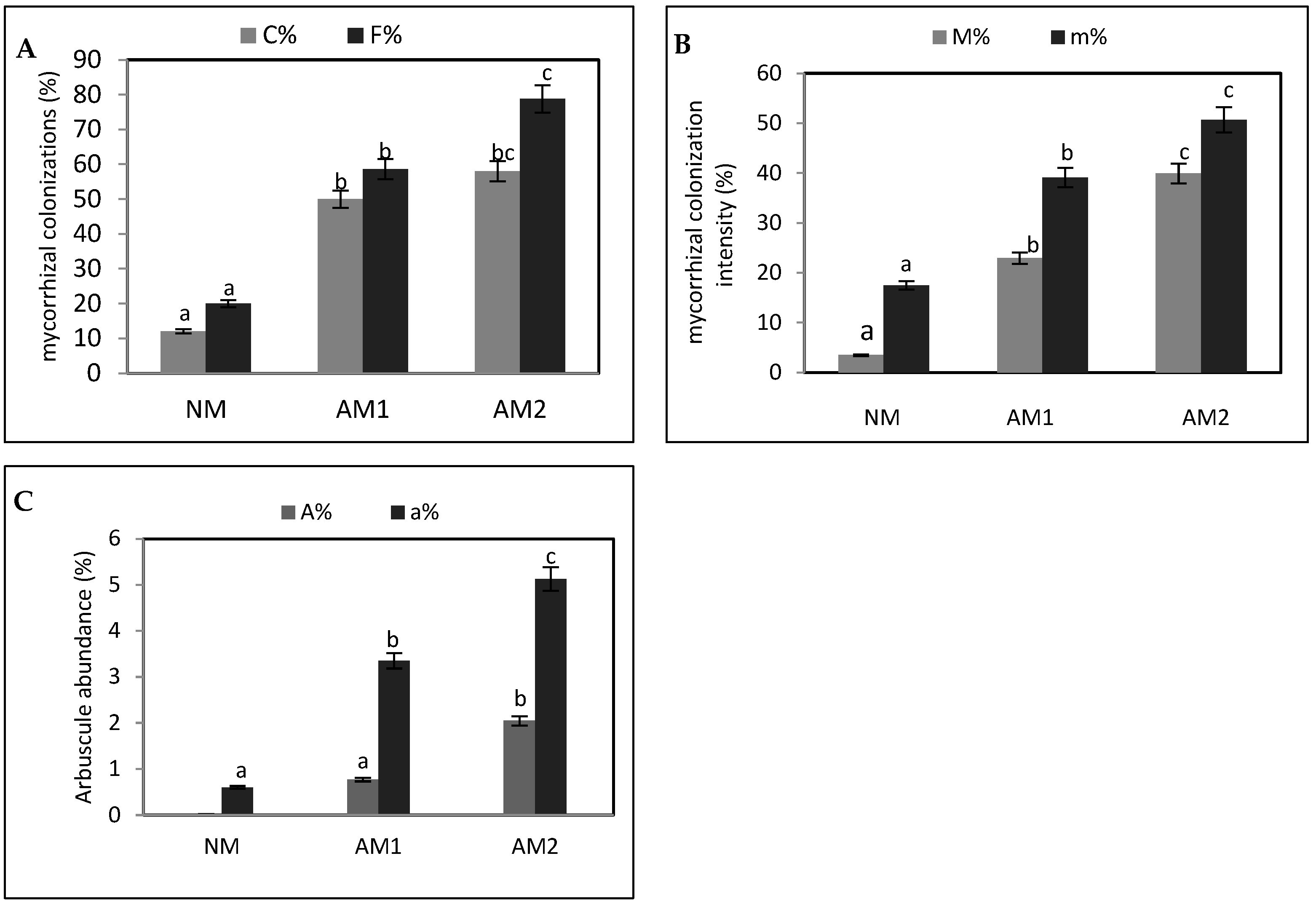
Qualitatively and quantitatively, the mycorrhizal association in the roots of P. oleracea was visualized by light-microscopic pictures (Figure 1). The mycorrhization of the roots, its frequency, intensity, and also arbuscule abundance of mycorrhizal colonization were determined (Figure 2). The microscopic pictures clearly showed the presence of the morphological structures of AMF in both studied strains (AM1—Claroideoglomus claroideum and AM2—Funneliformis mosseae), which is evidence of the presence of symbiosis between AMF and P. oleracea. Hyphae (intraradical hyphae and extraradical hyphae), arbuscules, and vesicles were found in the root microscopic preparations of the studied strains (Figure 1) at 10× and 20× magnifications. Quantitative analysis of mycorrhizal symbiosis (Figure 2) indicated that inoculation with F. mosseae (AM2) resulted in significantly higher values. This was evidenced by an increased degree and frequency of mycorrhizal colonization, as well as greater intensity and abundance of arbuscules, both in individual root fragments and throughout the entire root system of P. oleracea.
The presence of mycorrhizal colonization in the control variants is probably due to the natural diversity in the AMF soil, different from the species applied in the experiment (Figure 1 and Figure 2), since the soil/sand substrate used was not autoclaved.

3.2. Amount of the Specific Mycorrhizal Protein Glomalin (EE-GRSP and TE-GRSP) in the Rhizosphere of P. oleracea

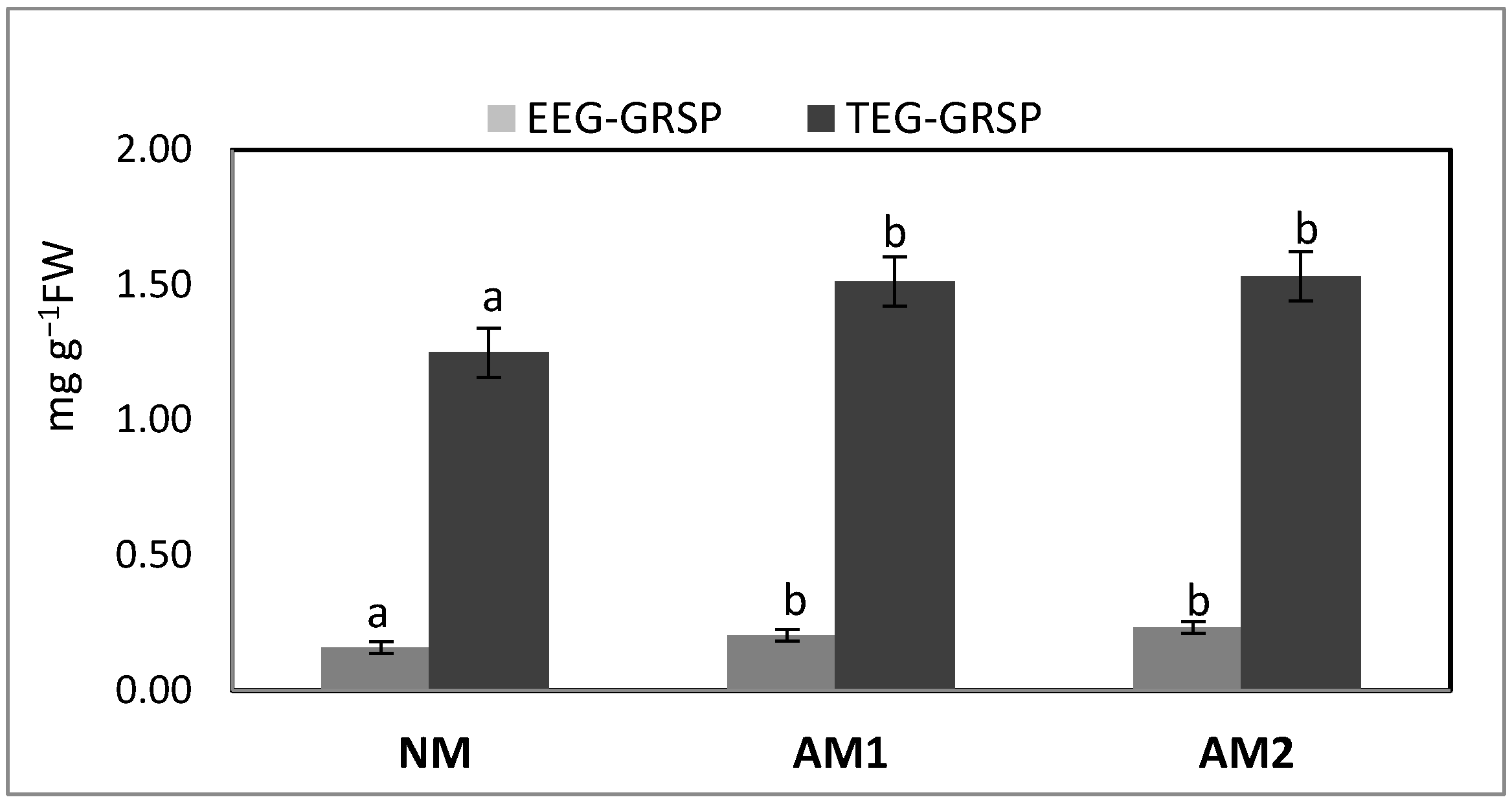
Significant differences were clearly identified in the first extracted fraction (EE-GRSP) and the total glomalin fraction (TE-GRSP) of soil samples from the inoculated plants when compared to the control group. However, there was no significant difference between the two strains (Figure 3).

3.3. Growth and Physiological Development of P. oleracea Plants Depended on Mycorrhizal Association Efficiency

According to the results, the biomass of both the fresh and dried plant shoots as well as the roots of the inoculated plants was increased. The fresh and dry weight of the shoots and roots were significantly higher in the variants that were inoculated with F. mosseae (Table 1). Mycorrhizal dependence (MD) indicates the extent of growth reliant on the efficiency of arbuscular mycorrhizal colonization.

3.4. Determining the Content of Total Proteins and Reducing Sugars in the Above-Ground Parts

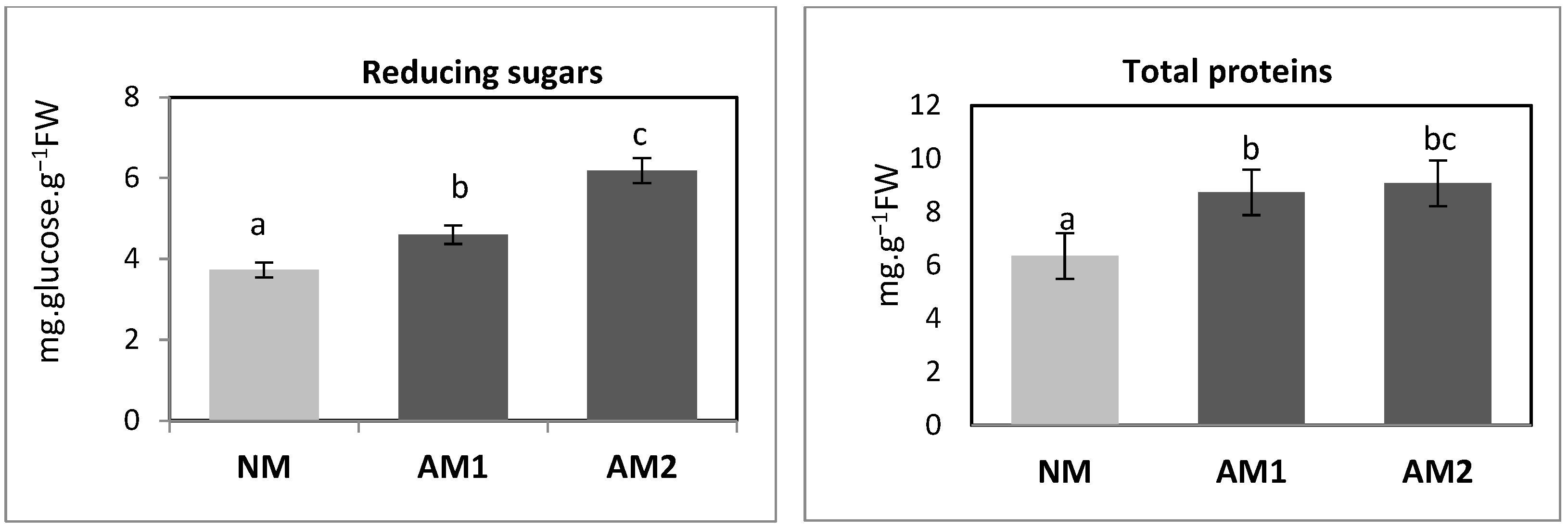
The analysis of primary metabolites, specifically total proteins and reducing sugars, indicated that plants in symbiotic relationships with arbuscular mycorrhizal fungi (AMF) exhibited higher values compared to non-inoculated control plants (refer to Figure 4). While differences in total protein content between the two strains were minimal, a notable increase in reducing sugars was specifically observed in the AM2 strain (Figure 4).

3.5. Content of Plastid Pigments: Chlorophylls and Carotenoids (Carotenes and Xanthophylls)

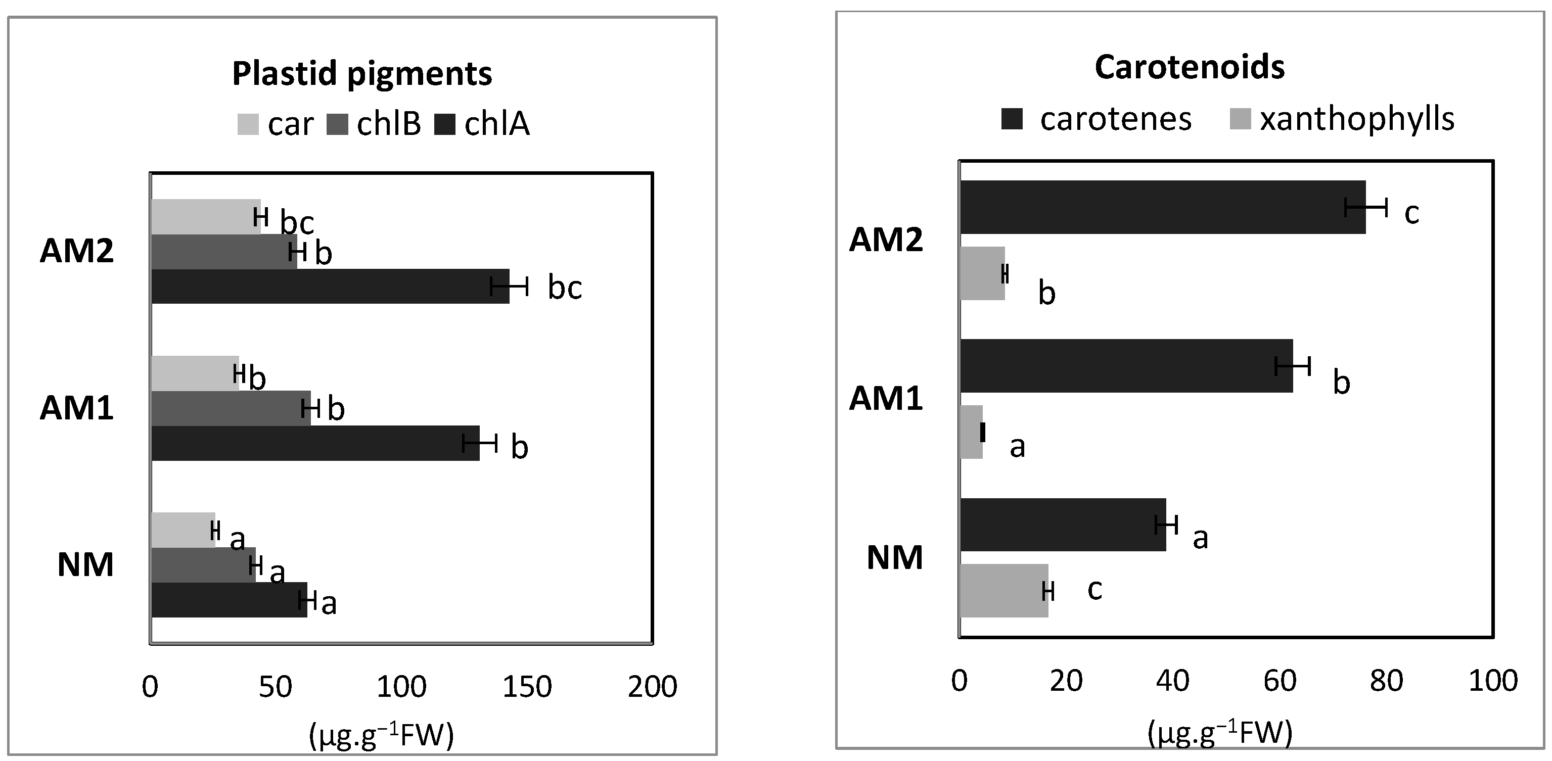
The results indicate that effective mycorrhizal colonization enhances the levels of plastid pigments, although no statistically significant differences were found between the strains (Figure 5).
The mycorrhizal fungi increased the total carotenoid content, particularly carotenes, while the opposite trend was noted for xanthophylls, which had the highest levels in non-inoculated plants (Figure 5).

3.6. Content of Anthocyanins and Betalains

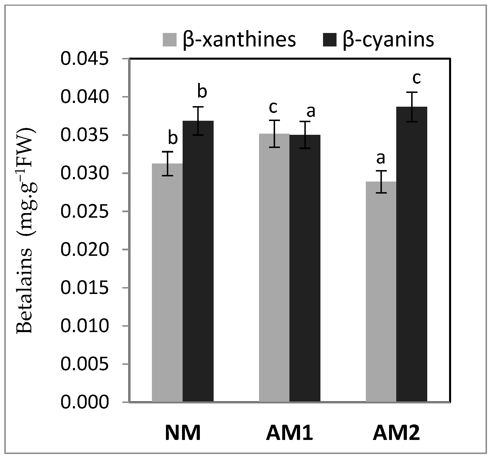
The three studied variants were tested for anthocyanin and betalain content. The analysis of anthocyanins did not show the presence of this type of pigment in contrast to the test for betalains (Figure 6). Both strains affect the ratio of betalains. F. mosseae stimulates the accumulation of β-cyanins, while C. claroideum raises β-xanthines (Figure 6).

3.7. Evaluation of P. oleracea L. Antioxidant Status Modified by the Mycorrhizal Strains

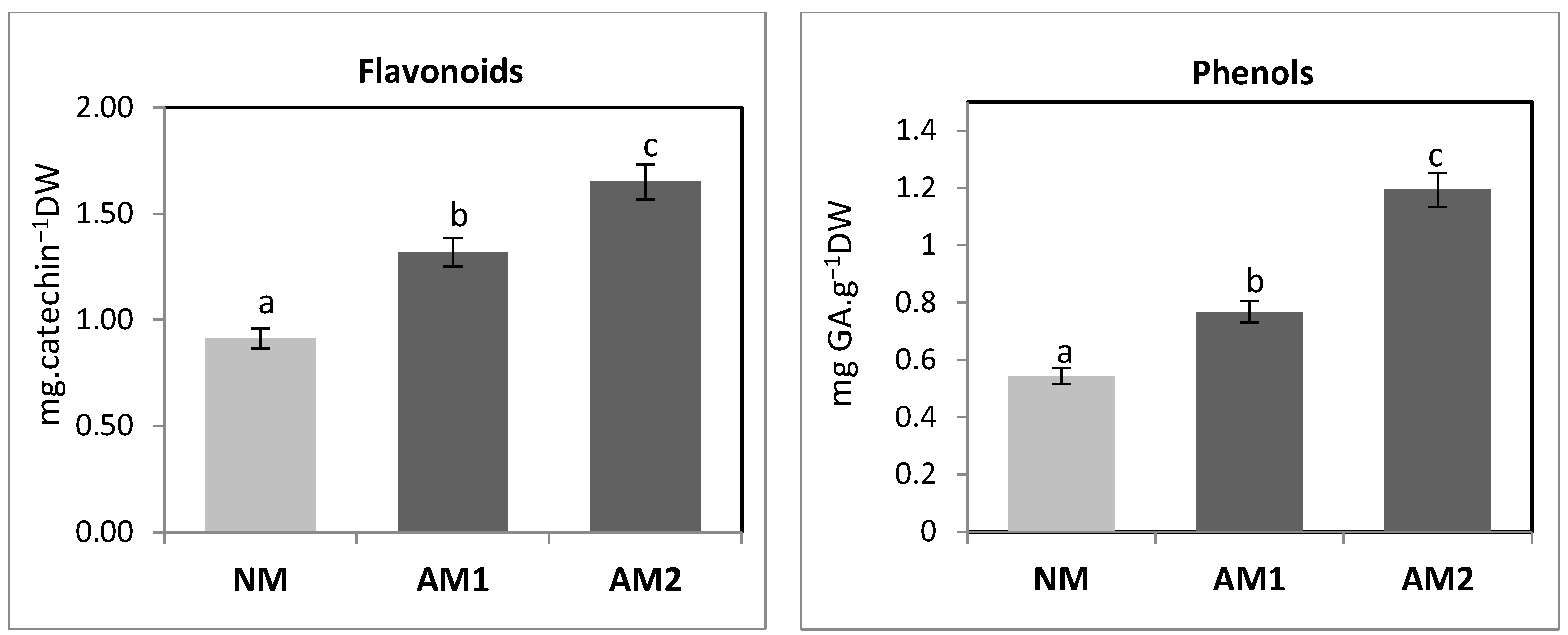
In the present research, it was found that the quantity of phenols in P. oleracea, expressed as gallic acid, increased following mycorrhizal symbiosis compared to the control (Figure 7). The most significant increase was reported in the F. mosseae variant.
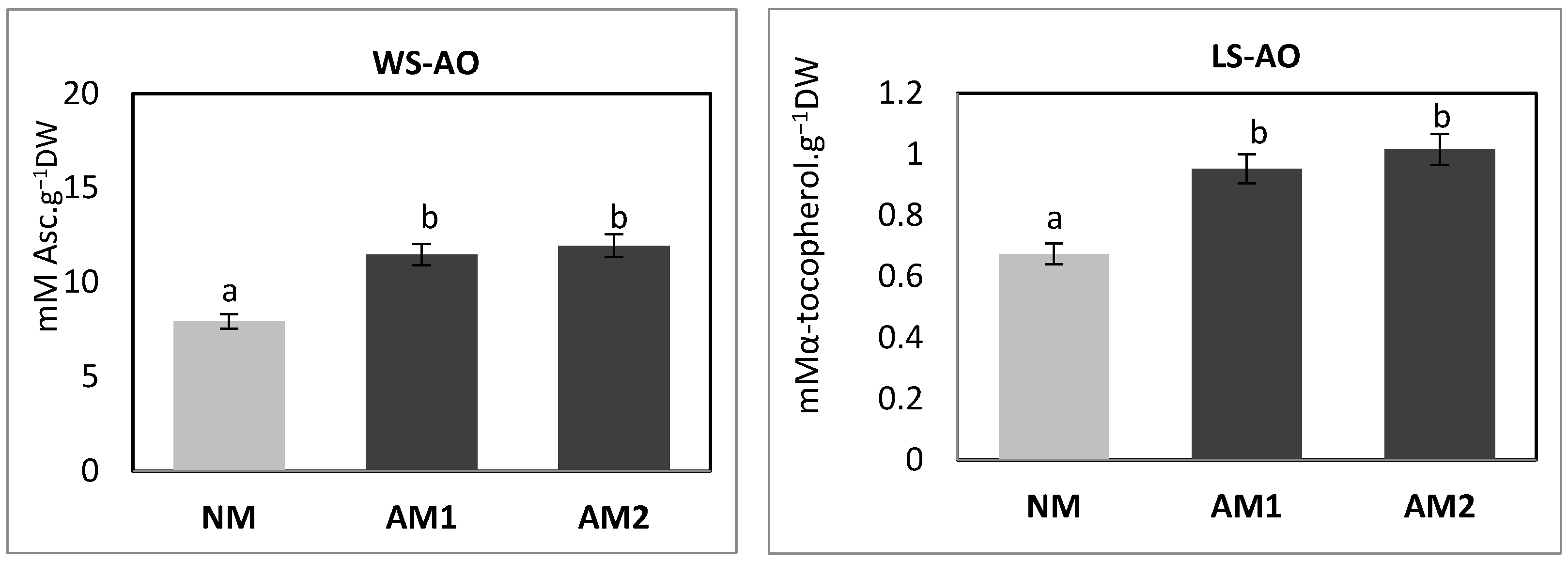
The study on flavonoid concentration indicated that catechin levels changed in a pattern similar to that of phenol content (Figure 7). This suggests that the values increased due to the influence of AMF, with the most significant increase observed in the F. mosseae variants. Additionally, the research found that the content of water-soluble antioxidants (WS-AO) increased following AMF inoculation. However, there was no significant difference between the two strains, as shown in Figure 8. Likewise, the levels of lipid-soluble antioxidants (LS-AO) exhibited a comparable pattern of variation between the inoculated and control plants, with no statistical difference noted between the two strains, C. claroideum and F. mosseae (Figure 8).
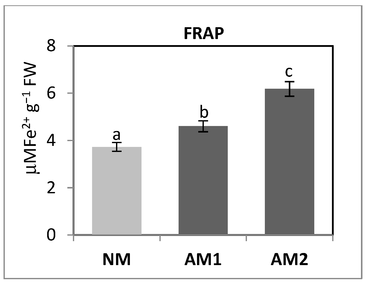
The results clearly demonstrate that AMF significantly reduce hydrogen peroxide (H2O2) content, as illustrated in Figure 9. Notably, the AM2 variant shows statistically higher levels of H2O2, which can be attributed to its more active metabolism and the accumulation of various cellular products. Furthermore, the reduced H2O2 levels in mycorrhizal plants correlate with enhanced CAT and APX activity, although there are no statistically significant differences observed between the strains (Figure 9). The antioxidant capacity was measured by the analysis of ferric-reducing antioxidant power (FRAP) and found that it increased due to mycorrhizal association (Figure 10). Furthermore, there was a statistically significant difference between the two strains, with AM2 showing greater benefits.

4. Discussion

Arbuscular mycorrhizal fungi (AMF) are universal microorganisms present in all types of soil and in association with a wide variety of plants from different taxonomic groups. They are microorganisms with proven qualities of biostimulators, applied to plants (roots and seeds) or in the soil, and activate physiological processes promoting better plant development, expressed in increased growth, greater resistance to stress, and increased product quality [17,44]. The mycorrhizal status of plants of the Portulacaceae family and more specifically of P. oleracea is poorly studied and data in the literature are limited and controversial [18].
The morphological structures of AMF are mainly divided into intraradical hyphae, extraradical hyphae, arbuscules, vesicles, accessory cells, and spores, which are produced inside the plant roots, while hyphae and spores can also be formed outside the roots, i.e., in the rhizosphere, and create an extensive surface area of membrane contact between plant root cells, AMF structures and soil resources [45].
A limited number of authors have reported the presence of effective mycorrhizal association in purslane roots [18,19,20]. Only in the publication of Sinegani and Yeganeh [20] is the type of colonization indicated, namely, “Paris”. This type is rare in herbaceous plants and is characteristic of plants of semi-arid habitats [21], which is probably evolutionarily related to the tolerance of purslane to drought. Brundrett et al. [46] found differences in mycorrhizal symbioses, dependent on the strain used, as different families showed variations in infection development patterns and resulting structures in root cells. The current research highlights that purslane forms a symbiosis with mycorrhizal fungi and demonstrates tolerance to various strains. Inoculation with F. mosseae resulted in more effective mycorrhizal colonization, indicated by higher degree and frequency, intensity, and abundance of arbuscules in both individual root fragments and the overall root system of P. oleracea L. The concentration of glomalin, a specific protein produced by mycorrhizal fungi, serves as an indicator of the fungal activity. Glomalins are a class of glycoproteins secreted by AMF hyphae with multiple functions mainly related to soil aggregation [27]. According to Burrows [47], levels of the immunoreactive protein glomalin varied seasonally, with higher levels in late summer than late spring, with no positive correlation between mycorrhizal infection potential and glomalin. Other authors suggest that glomalin secretion is a response to stress and may be linked to suboptimal mycelial growth [48]. The levels of glomalin in the soil—specifically the EE-GRSP and TE-GRSP fractions—were found to increase due to effective colonization of the roots of purslane. Contrary to previous research, this increase was not influenced by the type of fungal strain used. Some authors pointed out that different AMF species can have varying levels of glomalin production. For example, Claroideoglomus species may produce more glomalin compared to Glomus species [49]. The amount of glomalin in the soil is a complex issue influenced by fungal strains, the interactions of arbuscular mycorrhizal fungi with plants and their environment, and potentially other soil microorganisms. Additionally, successful mycorrhizal colonization had a positive impact on biomass accumulation in both the shoots and roots of purslane, particularly favoring the use of the fungal strain F. mosseae for inoculation. Inoculation with AMF significantly increased the concentration of macro- and micronutrients, which resulted in a significant improvement in biomass accumulation [50]. AMF improve the surface absorptive capacity of host roots through an extraradical mycelium, which is a physical extension of the plant’s root system, and thus the mycelium increases the surface area through which nutrients can be taken up. There is a lack of data in the literature to compare the effect of different strains on biomass accumulation in purslane. Hosseinzadeh et al. [51] treated purslane with a strain of Rhizophagusirregularis and reported an increase in biomass. Kanade and Bhosale [19] showed that when purslane is in symbiosis with mycorrhizal fungi, the interaction has an extremely favorable effect on the plants expressing through maximum growth and the largest number of leaves.
The biosynthesis of primary metabolites, such as total proteins and reducing sugars, is enhanced in P. oleracea plants that are grown in symbiotic conditions compared to non-inoculated plants. These results align with the findings related to purslane biomass, emphasizing that increased leaf mass leads to more efficient photosynthesis and higher accumulation of reducing sugars. The total protein content varied only slightly between the two strains, while the concentration of reducing sugars showed a positive influence from F. mosseae. AMF obtain sugars synthesized by the host plant through photosynthesis, storing and using them as carbon sources to support the growth of mycorrhizal structures [52]. Accumulated carbon-containing substances are difficult to assimilate and re-utilize by host plants, but they can stimulate carbohydrate concentration gradients between AMF and their plant partners that facilitate the continuous transport of carbohydrates from host plants to AMF, accelerating their metabolism and leading to synchronous development of both symbiotic partners [53].
AMF can increase the uptake of inorganic nutrients in almost all plants, leading to higher chlorophyll contents due to the effective trapping of nitrogen by chlorophyll molecules [54]. Effective mycorrhizal colonization increases the concentration of plastid pigments in P. oleracea, though this effect is not specific to certain strains of mycorrhizal fungi. These fungi stimulate the accumulation of carotenoids, particularly preferring carotenes, while the levels of xanthophylls tend to decrease. Notably, higher levels of β-carotene were observed in both chamber-grown and wild-grown purslane leaves compared to spinach leaves. Among the different parts of the plant, leaves contain the highest β-carotene content, followed by flowers and stems. In fact, the β-carotene content in leaves is twice that of stems and slightly higher than that in flowers [55].
The three studied purslane variants were tested for anthocyanins and betalains content. According to literature data, betalains and anthocyanins are not found in the same plant species and share mutual exclusion, with their precursors being tyrosine and phenylalanine, respectively, synthesized from arogenate [56]. Betalains (β-cyanins and β-xanthines) are water-soluble nitrogen-containing pigments with red-violet and yellow-orange colors [6]. The mechanism by which mycorrhizal symbiosis affects the content of betalains is not well-documented in the literature. This indicator can be considered indirectly as a general effect of AMF on the accumulation of secondary metabolites. Some authors report an increased synthesis of this type of pigments under the influence of stress factors [57], since structurally, all betacyanins and betaxanthins bearing residues of aromatic amino compounds are likely to stabilize radicals due to their aromatic nature [58]. According to D’Andrea et al. [59], in P. oleracea, protecting sensitive physiological processes such as photosynthesis from reactive oxygen species production under water deficit induces the biosynthesis of secondary metabolites such as flavonoids and betalains. The inoculation of P. oleracea with F. mosseae leads to a significant increase in the accumulation of β-cyanins. In contrast, the symbiotic relationship with C. claroideum results in elevated levels of β-xanthines.
The present study also evaluated the antioxidant status of symbiotically grown P. oleracea with diverse arbuscular mycorrhizal fungi strains. Plants have evolved a complex antioxidant system to protect cell membranes and organelles from the damaging effects of reactive oxygen species [60], which is composed of low molecular weight antioxidants (glutathione, ascorbate, and carotenoids) as well as enzymes (SOD, CAT, and APX) [12].
Mycorrhizal symbioses modulate the primary and secondary metabolism of host plants, stimulating the production of bioactive substances in roots and shoots [61]. Phenolic compounds are widely distributed in plants and have attracted much attention due to their antioxidant activity and their ability to scavenge free radicals. A number of phenolic acids such as caffeic, p-coumaric, ferulic, gallic, gentisic, benzoic, and anisic acids have been reported in purslane, and the database is constantly being updated [6]. The accumulation of phenolics in plants depends on the developmental stage of the symbiosis, also playing a key role in the cross-talk between plants and symbiotic fungi, as they affect spore germination and hyphal growth and stimulate root colonization [62]. Flavonoids are one of the main active ingredients of purslane. The main flavonoids present in this plant are kaempferol, myricetin, luteolin, apigenin, quercetin, genistein, and genistin [7]. Various studies have reported that levels of transcripts encoding the key enzyme of the shikimate pathway, phenylalanineammonia lyase, are increased as a result of mycorrhizal associations in a strain-dependent manner [63], as well as transcripts encoding chalcone synthase [64]. Following the successful establishment of mycorrhizal associations, there was a notable increase in the concentration of secondary metabolites, specifically phenols and flavonoids, in the shoots of purslane when compared to the control group. The most pronounced enhancement occurred as a result of inoculation with F. mosseae, which was positively correlated with a higher degree of mycorrhizal association development and an increase in biomass.
Purslane has been shown to be rich in vitamins such as vitamin A, B-complex vitamins (riboflavin, niacin, and pyridoxine), ascorbic acid, and α-tocopherol [5]. Physiological changes related to primary and secondary metabolism are due, on the one hand, to transient activation of host defense responses in colonized roots and the accumulation of antioxidant compounds [65], and, on the other hand, to the higher content of mineral nutrients [66,67]. Another reason for changes in the biochemical composition of active metabolites is that the primary metabolism of root cells, such as the plastid biosynthetic pathways and the Krebs cycle, is altered by arbuscular colonization, with an increase in amino acids, fatty acids, and apocarotenoids [68]. The concentration of non-enzymatic antioxidants (WS- and LS-AO)—specifically expressed, respectively, as ascorbate and α-tocopherol—were positively influenced in purslane by the association with mycorrhizae, while the specific strains did not have a significant impact.
A number of publications have reported an increase in AO status in above-ground plant parts in various AM-inoculated plants compared to non-mycorrhizal plants. Estrada et al. [69] found that AMF symbiosis induces AO enzymes, emphasizing that the degree of induction depends on the mycorrhizal strain used. In our previous studies on the influence of AMF and more specifically strain C. claroideum in symbiosis with Ocimumbasilicum L., an enhancement in the activity of AO enzymes was found [70]. The data in some articles related to the accumulation and disposal of reactive oxygen species and stimulation of the activity of AO enzymes under the influence of AMF, considered in the context of applied abiotic stress and its overcoming [44,71,72]. Also, the symbiotic nature of AMF associations suggests that the activity of antioxidant enzymes may be a consequence of a general plant defense response to mycorrhizal colonization.
Plant cells continuously synthesize hydrogen peroxide (H2O2) as a cellular metabolite, which serves as an important signaling molecule mediating various physiological and biochemical processes [73]. Mycorrhizal fungi have been shown to activate the antioxidant enzymes CAT and APX in P. oleracea. Notably, no statistically significant differences were detected between the two strains examined. Furthermore, the application of F. mosseae results in an enhancement of antioxidant enzyme activity, which subsequently leads to a reduction in hydrogen peroxide (H2O2) levels in the treated plants. In previous studies, it was found that treatment with AMF affects the physiological state of P. oleracea, increasing biomass, total antioxidant activity, and unsaturated fatty acid content in leaves [51]. The mycorrhizal association has been shown to enhance the antioxidant capacity, as indicated by the FRAP test, in P. oleracea. This effect is more pronounced in the variants associated with F. mosseae. This trend correlates with changes in the concentration of secondary metabolites, such as phenols and flavonoids. The mycorrhizal association boosts both non-enzymatic antioxidants (including WS- and LS-AO metabolites) and enzymatic antioxidant activity. Notably, this effect is independent of the specific strain applied. It is worth noting that the changes in the primary and secondary metabolites affected by AMF is context-dependent and could be influenced by the plant species, AMF species, environmental conditions, and the interaction of the three previously mentioned factors [14]. Mycorrhizal fungi exhibit different physiological traits that result in unique biochemical profiles. These variations arise mainly from their distinct strategies for acquiring nutrients and exchanging carbon with their plant hosts. For instance, some fungi have evolved the ability to efficiently uptake specific nutrients like phosphorus, which influences the types and quantities of biochemical compounds produced by both the fungi and the plants [74,75].According to Guo et al. [76], who consolidate the information about the specific plant mycorrhizal responses, host plants could specifically select fungal symbionts and result in unique AMF network topologies and community structures, because both the host plants and AMF could preferentially allocate resources to higher quality partners and consequently achieve optimal plant–AMF matches or plant–AMF specificity.
The limited data on the purslane microbiome underscores the need for research aimed at maximizing benefits through the selection of the appropriate strain. This research is essential to explore the diverse impacts on the plant’s primary and secondary metabolic pathways.

5. Conclusions

The current research highlights the potential of using non-conventional plants, such as purslane, as a food source. This is particularly important given the decreasing availability of traditional resources. Purslane is valued for its high nutritional content, which includes omega-3 fatty acids, bioactive compounds, and antioxidants. This study emphasizes the importance of cultivating purslane using sustainable ecological practices, particularly through the application of biostimulants, to enhance its quality as a food product. Our findings emphasize that arbuscular mycorrhizal fungi strains are influenced by the genotype of the host plant. These strains exhibit varying abilities to form effective mycorrhizal associations and can significantly affect the plant’s metabolism. When plants are grown in symbiosis with suitable AMF, they benefit in various ways. Inoculation with Funneliformis mosseae resulted in improved mycorrhizal colonization and positively influenced biomass accumulation and the concentration of reducing sugars. Symbiotic plants exhibited a higher total accumulation of plastid pigments; however, this effect was not specific to any particular strain. Mycorrhizal fungi significantly boosted the levels of carotenes in the shoots, while xanthophylls saw a remarkable decline, with the highest concentrations found in plants that were not inoculated. Furthermore, both fungal strains uniquely influenced the balance of betalains: Funneliformis mosseae elegantly enhanced the accumulation of β-cyanins, whereas Claroideoglomus claroideum skillfully elevated the levels of β-xanthines. The association purslane, Funneliformis mosseae, is distinguished significantly with increased antioxidant capacity, as evidenced by the FRAP test, by altering levels of secondary metabolites such as phenols and flavonoids. Targeted inoculation with specific strains unequivocally boosts both non-enzymatic and enzymatic antioxidant activity, and this enhancement is consistent regardless of the strain used.
It is vital to acknowledge the wild essence of this species, highlighting the urgent need to establish robust best practice guidelines. These guidelines should aim to refine management techniques, optimize yields, enhance nutritional values, and bolster stress tolerance. Embracing these principles will not only elevate our approach but also unlock the full potential of this remarkable species.

Author Contributions

Conceptualization, M.H. and K.V.; Data curation, M.H. and K.V.; Formal analysis, M.H. and K.V.; Investigation, M.H. and K.V.; Methodology, M.H. and M.G.; Resources, M.H., M.G. and K.V.; Software, M.H., M.G. and K.V.; Supervision, M.H.; Validation, M.H. and K.V.; Visualization, M.H. and K.V.; Writing and editing, M.H., M.G. and K.V. All authors have read and agreed to the published version of the manuscript.

Funding

The research study was funded through the institution’s regular annual budget and not through an external competitive grant award process.

Institutional Review Board Statement

Not applicable.

Data Availability Statement

All data, tables, and figures were obtained and produced by the authors of this work. The authors declare that the data supporting the findings of this study are available within the paper. Should any raw data files be needed in another format, they are available from the corresponding author upon reasonable request.

Conflicts of Interest

The authors declare no conflicts of interest.

Abbreviations

The following abbreviations are used in this manuscript:
AMFArbuscular mycorrhizal fungi
EE-GRSPEasilyextractedglomalin-related soil protein
TE-GRSPTotalextracted glomalin-related soil proteins
MDMycorrhizal dependence
CATCatalase
APXAscorbate peroxidase
WS-AOWater-soluble antioxidantcapacity
LS-AOLipid-soluble antioxidant capacity
FRAPFerric-reducing antioxidant power

References

  1. Zhang, X.J.; Ji, Y.B.; Qu, Z.Y.; Xia, J.C.; Wang, L. Experimental studies on antibiotic functions of Portulaca oleracea L. in vitro. Chin. J. Microbiol. 2002, 14, 277–280. [Google Scholar]
  2. Karimi, G.; Hosseinzadeh, H.; Ettehad, N. Evaluation of the gastric antiulcerogenic effects of Portulaca oleracea L. extracts in mice. Phytother. Res. 2004, 18, 484–487. [Google Scholar] [CrossRef] [PubMed]
  3. Chan, K.; Islam, M.W.; Kamil, M.; Radhakrishnan, R.; Zakaria, M.N.; Habibullah, M.; Attas, A. The analgesic and anti-inflammatory effects of Portulaca oleracea L. subsp Sativa (Haw.) Celak. J. Ethnopharmacol. 2000, 73, 445–451. [Google Scholar] [CrossRef]
  4. Chen, B.; Zhou, H.; Zhao, W.; Zhou, W.; Yuan, Q.; Yang, G. Effects of aqueous extract of Portulaca oleracea L. on oxidative stress and liver, spleen leptin, PARα and FAS mRNA expression in high-fat diet induced mice. Mol. Biol. Rep. 2012, 39, 7981–7988. [Google Scholar] [CrossRef]
  5. Petropoulos, S.; Karkanis, A.; Martins, N.; Ferreira, I. Phytochemical composition and bioactive compounds of common purslane (Portulaca oleracea L.) as affected by crop management practices. Trends Food Sci. Tech. 2016, 55, 1–10. [Google Scholar] [CrossRef]
  6. Kumar, A.; Sreedharan, S.; Kashyap, A.; Singh, P.; Ramchiary, N. A review on bioactive phytochemicals and ethnopharmacological potential of purslane (Portulaca oleracea L.). Heliyon 2022, 8, e08669. [Google Scholar] [CrossRef] [PubMed]
  7. Iranshahy, M.; Javadi, B.; Iranshahi, M.; Jahanbakhsh, S.; Mahyari, S.; Hassani, F.; Karimi, G. A review of traditional uses, phytochemistry and pharmacology of Portulaca oleracea L. J. Ethnopharmacol. 2017, 205, 158–172. [Google Scholar] [CrossRef]
  8. Yang, X.; Zhang, W.; Ying, X.; Stien, D. New flavonoids from Portulaca oleracea L. and their activities. Fitoterapia 2018, 127, 257–262. [Google Scholar] [CrossRef]
  9. Zhou, Y.-X.; Xin, H.-L.; Rahman, K.; Wang, S.-J.; Peng, C.; Zhang, H. Portulaca oleracea L: A Review of Phytochemistry and Pharmacological Effects. BioMed Res. Int. 2015, 1, 925631. [Google Scholar]
  10. Rahimi, P.; Abedimanesh, S.; Mesbah-Namin, S.; Ostadrahimi, A. Betalains, the nature-inspired pigments, in health and diseases. Crit. Rev. Food Sci. Nutr. 2019, 59, 2949–2978. [Google Scholar] [CrossRef]
  11. Moreno-Villena, J.; Zhou, H.; Gilman, S.; Tausta, L.; Cheung, M.; Edwards, J. Spatial resolution of an integrated C4+CAM photosynthetic metabolism. Sci. Adv. 2022, 8, eabn2349. [Google Scholar] [CrossRef] [PubMed]
  12. Yazici, I.; Türkan, I.; Sekmen, A.; Demiral, T. Salinity tolerance of purslane (Portulaca oleracea L.) is achieved by enhanced antioxidative system, lower level of lipid peroxidation and proline accumulation. Environ. Exp. Bot. 2007, 61, 49–57. [Google Scholar] [CrossRef]
  13. Koch, K.; Kennedy, R. Characteristics of crassulacean acid metabolism in the succulent C4 dicot, Portulaca oleracea L. Plant Physiol. 1980, 65, 193–197. [Google Scholar] [CrossRef]
  14. Dighton, J. Mycorrhizae. In Encyclopedia of Microbiology; Elsevier: Amsterdam, The Netherlands, 2009; pp. 153–162. [Google Scholar]
  15. Kaur, S.; Suseela, V. Unraveling arbuscular mycorrhiza-induced changes in plant primary and secondary metabolome. Metabolites 2020, 10, 335. [Google Scholar] [CrossRef]
  16. Rodrigues, M.; Baptistella, J.; Horz, D.; Bortolato, L.; Mazzafera, P. Organic plant biostimulants and fruit quality—A Review. Agronomy 2020, 10, 988. [Google Scholar] [CrossRef]
  17. Machiani, A.; Javanmard, A.; Machiani, R.; Sadeghpour, A. Arbuscular mycorrhizal fungi and changes in primary and secondary metabolites. Plants 2022, 11, 2183. [Google Scholar] [CrossRef]
  18. Kamble, V.; Pal, A. Assessment of three representative species of Portulacaceae with ambiguity about mycorhizal status. IOSR-JESTFT 2016, 10, 2319–2399. [Google Scholar]
  19. Kanade, A.; Bhosale, R. Effect of VA Mycorrhizae inoculation on vegetative growth in Portulaca oleracea L. (Purslane, Gholu). IJRAR 2019, 6, 710–712. [Google Scholar]
  20. Sinegani, A.; Yeganeh, M. The occurrence of arbuscular mycorrhizal fungi in soil and root of medicinal plants in Bu-Ali Sinagarden in Hamadan. Iran Biol. J. Microorg. 2017, 5, 43–59. [Google Scholar]
  21. McGee, P. Variation in propagule numbers of vesicular- arbuscular mycorrhizal fungi in a semi-arid soil. Mycol. Res. 1989, 92, 28–33. [Google Scholar] [CrossRef]
  22. Schüßler, A.; Walker, C. The Glomeromycota: A Species List with New Families and New Genera URL. Published in December 2010 in Libraries at The Royal Botanic Garden Edinburgh, The Royal Botanic Garden Kew, Botanische Staats Sammlung Munich, and Oregon State University Arthur Schüßler and Christopher Walker, Gloucester. 2010. Available online: http://www.amf-phylogeny.com (accessed on 10 April 2024).
  23. Jackson, N.; Franklin, R.; Miller, R. Effects of vesicular-arbuscular mycorrhizae on growth and phosphorus content of three agronomic crops. Soil Sci. Soc. Am. Proc. 1972, 36, 64–67. [Google Scholar] [CrossRef]
  24. Phillips, J.; Hayman, D. Improved procedures for clearing and staining parasitic and vesicular–arbuscular mycorrhizal fungi for rapid assessment of infection. Trans. Br. Mycol. Soc. 1970, 55, 158–161. [Google Scholar] [CrossRef]
  25. Giovannetti, M.; Mosse, B. An evaluation of techniques for measuring vesicular- arbuscular mycorrhizal infection in roots. New Phytopatol. 1980, 84, 489–500. [Google Scholar] [CrossRef]
  26. Trouvelot, A.; Kough, J.; Gianinazzi-Pearson, V. Mesure du taux de mycorhization VA d’un système radiculaire Recherche de methods d’estimation ayant une signification fonctionnelle. In Physiological and Genetical Aspects of Mycorrhizae; Gianinazzi-Pearson, V., Gianinazzi, S., Eds.; INRA Press: Paris, France, 1986; pp. 217–221. [Google Scholar]
  27. Wright, S.; Upadhyaya, A. Extraction of an abundant and unusual protein from soil and comparison with hyphal protein of arbuscular mycorrhizal fungi. Soil Sci. 1996, 161, 575–586. [Google Scholar] [CrossRef]
  28. Bradford, M. A rapid and sensitive method for the estimation of microgram quantities of protein utilizing the principle of protein–dye binding. Anal. Biochem. 1976, 72, 248–254. [Google Scholar] [CrossRef]
  29. Plenchette, C.; Fortm, J.; Furlan, V. Growthresponseofseveralplantspeciestomycorrhizaein a soil ofmoderate Pfertility.I.Mycorrhizal dependency under field conditions. Plant Soil 1983, 70, 199–209. [Google Scholar] [CrossRef]
  30. Plummer, D. An introduction to practical biochemistry. In Biochemical Education; McGraw Hill: New York, NY, USA, 1988; Volume 16, pp. 98–100. [Google Scholar]
  31. Asare-Brown, E.; Bullock, C. Simple practical investigation using invertase. Biochem. Educ. 1988, 16, 98–100. [Google Scholar] [CrossRef]
  32. Porra, R.; Thompson, W.; Kriedemann, P. Determination of accurate extinction coefficients and simultaneous equations for assaying chlorophylls a and b extracted with four different solvents: Verification of the concentration of chlorophyll standards by atomic absorption spectroscopy. Biochim. Biophys. Acta 1989, 975, 384–394. [Google Scholar] [CrossRef]
  33. Zin, M.; Márki, E.; Bánvölgyi, S. Conventional extraction of betalain compounds from beetroot peels with aqueous ethanol solvent. Acta Aliment. 2020, 49, 163–169. [Google Scholar] [CrossRef]
  34. Ravichandran, K.; Saw, N.; Mohdaly, A.; Gabr, A.; Kastell, A.; Riedel, H.; Cai, Z.; Knorr, D.; Smetanska, I. Impact of processing of red beet on betalain content and antioxidant activity. Food Res. Int. 2013, 50, 670–675. [Google Scholar] [CrossRef]
  35. Sellappan, S.; Akoh, C. Flavonoids and antioxidant capacity of Georgia-grown Vidalia onions. J. Agric. Food Chem. 2002, 50, 5338–5342. [Google Scholar] [CrossRef] [PubMed]
  36. Lee, J.; Durst, R.; Wrolstad, R. Flavonoids determination of total monomeric anthocyanin pigment content of fruit juices, beverages, natural colorants, and wines by the pH differential method: Collaborative study. JAOAC Int. 2005, 88, 1269–1278. [Google Scholar] [CrossRef]
  37. Zhishen, J.; Mengcheng, T.; Jianming, W. The determination of flavonoid contents in mulberry and their scavenging effects on superoxide radicals. Food Chem. 1999, 64, 555–559. [Google Scholar] [CrossRef]
  38. Singleton, V.; Orthofer, R.; Lamuela-Raventys, R. Analysis of total phenols and other oxidation substrates and antioxidants by means of Folin-Ciocalteu reagent. Method Enzymol. 1999, 299, 152–178. [Google Scholar]
  39. Benzie, I.; Szeto, Y. Total antioxidant capacity of teas by ferric reducing/antioxidant power assay. J. Agric. Food Chem. 1999, 47, 633–636. [Google Scholar] [CrossRef]
  40. Prieto, P.; Pineda, M.; Aguilar, M. Spectrophotometric quantitation of antioxidant capacity through the formation of a phosphomolybdenum complex: Specific application to the determination of vitamin E. Anal. Biochem. 1999, 269, 337–341. [Google Scholar] [CrossRef]
  41. Nakano, Y.; Asada, K. Purification of ascorbate peroxidase in spinach chloroplasts: Its inactivation in ascorbate-depleted medium and reactivation by monodehydroascorbate radical. Plant Cell Physiol. 1987, 28, 131–140. [Google Scholar]
  42. Aebi, H. Catalase. In Methods of Enzymatic Analysis, 3rd ed.; Bergmeyer, H.U., Ed.; Band, VCH, Verlagsgessellschaft mbH: Weinheim, Germany, 1986; pp. 273–286. [Google Scholar]
  43. Velikova, V.; Yordanov, I.; Edreva, A. Oxidative stress and some antioxidant systems in acid rain—Treated bean plants: Protective role of exogenous polyamines. Plant Sci. 2000, 151, 59–66. [Google Scholar] [CrossRef]
  44. Rouphael, Y.; Franken, P.; Schneider, C.; Schwarz, D.; Giovannetti, M.; Agnolucci, M.; De Pascale, S.; Bonini, P.; Colla, G. Arbuscular mycorrhizal fungi act as biostimulantsinhorticultural crops. Sci. Hortic. 2015, 196, 91–108. [Google Scholar] [CrossRef]
  45. Hodge, A.; Storer, K. Arbuscular mycorrhiza and nitrogen: Implications for individual plants through to ecosystems. Plant Soil 2015, 386, 1–9. [Google Scholar] [CrossRef]
  46. Brundrett, M.; Bougher, N.; Dell, B.; Grove, T.; Malajczuk, N. Working with Mycorrhizas in Forestry and Agriculture. In ACIAR Monograph Series, Pirie, Canberra; Australian Centre for International Agricultural Research: Canberra, Australia, 1996; Volume 374. [Google Scholar]
  47. Burrows, R. Glomalin production and infectivity of arbuscular-mycorrhizal fungi in response to grassland plant diversity. Am. J. Plant Sci. 2014, 5, 103–111. [Google Scholar] [CrossRef]
  48. Hammer, E.; Rillig, M. The influence of different stresses on glomalin levels in an arbuscular mycorrhizal fungus-salinity increases glomalin content. PLoS ONE 2011, 6, e28426. [Google Scholar] [CrossRef] [PubMed]
  49. Bedini, S.; Pellegrino, E.; Avio, L.; Pellegrini, S.; Bazzoffi, P.; Argese, E.; Giovannetti, M. Changes in soil aggregation and glomalin-related soil protein content as affected by the arbuscular mycorrhizal fungal species Glomus mosseae and Glomus intraradices. Soil Biol. Biochem. 2009, 41, 1491–1496. [Google Scholar] [CrossRef]
  50. Begum, N.; Qin, C.; Ahanger, M.A.; Raza, S.; Khan, M.I.; Ashraf, M.; Zhang, L. Role of arbuscular mycorrhizal fungi in plant growth regulation: Implications in abiotic stress tolerance. Front. Plant Sci. 2019, 10, 1–9. [Google Scholar] [CrossRef] [PubMed]
  51. Hosseinzadeh, M.; Ghalavand, A.; Mashhadi-Akbar-Boojar, M.; Modarres-Sanavy, S.; Mokhtassi-Bidgoli, A. Increased medicinal contents of purslane by nitrogen and arbuscular mycorrhiza under drought stress. Commun. Soil Sci. Plant 2020, 51, 118–135. [Google Scholar] [CrossRef]
  52. Luginbuehl, L.; Menard, G.; Kurup, S.; Erp, H.; Radhakrishnan, G.; Breakspear, A. Fatty acids in arbuscular mycorrhizal fungi are synthesized by the host plant. Science 2017, 356, 1175–1178. [Google Scholar] [CrossRef]
  53. Li, D.W.; Wang, D.M.; Yu, Z.Z. Advances on physiological and biochemistry effects of the symbiosis between AM fungi and plants. Acta Bot Boreali-Occident Sin. 2002, 22, 1255–1262. [Google Scholar]
  54. De Andrade, S.; Domingues, A.; Mazzafera, P. Photosynthesis is induced in rice plants that associate with arbuscular mycorrhizal fungi and are grown under arsenate and arsenite stress. Chemosphere 2015, 134, 141–149. [Google Scholar] [CrossRef]
  55. Uddin, M.K.; Juraimi, A.S.; Hossain, M.S.; Nahar, M.A.U.; Ali, M.E.; Rahman, M.M. Purslane Weed (Portulaca oleracea): A Prospective Plant Source of Nutrition, Omega-3 Fatty Acid, and Antioxidant Attributes. Sci. World J. 2014, 2014, 951019. [Google Scholar] [CrossRef]
  56. Tanaka, Y.; Sasaki, N.; Ohmiya, A. Biosynthesis of plant pigments: Anthocyanins, betalains and carotenoids. Plant J. 2008, 54, 733–749. [Google Scholar] [CrossRef]
  57. Sarker, U.; Oba, S. Salinity stress enhances color parameters, bioactive leaf pigments, vitamins, polyphenols, flavonoids and antioxidant activity in selected Amaranthus leafy vegetables. J. Sci. Food. Agric. 2018, 99, 2275–2284. [Google Scholar] [CrossRef]
  58. Florian, C.S.; Reinhold, C. Functional properties of anthocyanins and betalains in plants, food, and in human nutrition. Trends Food Sci. Technol. 2004, 15, 19–38. [Google Scholar]
  59. D’Andrea, R.M.; Andreo, C.S.; Lara, M.V.; Andrea, R.M.D.; Andreo, C.S.; Lara, M.V. Deciphering the mechanisms involved in Portulaca oleracea (C4) response to drought: Metabolic changes including crassulacean acid-like metabolism induction and reversal upon re-watering. Physiol. Plant 2014, 152, 414–430. [Google Scholar] [CrossRef]
  60. Mansoor, S.; Ali Wani, O.; Lone, J.K.; Manhas, S.; Kour, N.; Alam, P.; Ahmad, A.; Ahmad, P. Reactive oxygen species in plants: From source to sink. Antioxidants 2022, 11, 225. [Google Scholar] [CrossRef]
  61. Sbrana, C.; Avio, L.; Giovanetti, M. Beneficial mycorrhizal symbionts affecting the production of health-promoting phytochemicals. Electrophoresis 2014, 35, 1535–1546. [Google Scholar] [CrossRef]
  62. Abdel-Lateif, K.; Bogusz, D.; Hocher, V. The role of flavonoids in the establishment of plant roots endosymbioses with arbuscular mycorrhiza fungi, rhizobia and frankia bacteria. Plant Signal. Behav. 2012, 7, 636–641. [Google Scholar] [CrossRef]
  63. Blilou, I.; Ocampo, J.A.; Garcia, G.J. Induction of Ltp (lipid transfer protein) and Pal (phenylalanine ammonia-lyase) gene expression in rice roots colonized by the arbuscular mycorrhizal fungus Glomus mosseae. J. Exp. Bot. 2000, 51, 1969–1977. [Google Scholar] [CrossRef]
  64. Bonanomi, A.; Oetiker, J.H.; Guggenheim, R.; Boller, T.; Wiemken, A.; Vogeli-Lange, R. Arbuscular mycorrhiza in mini-mycorrhizotrons: First contact of Medicago truncatula roots with Glomus intraradices induces chalcone synthase. New Phytol. 2001, 150, 573–582. [Google Scholar] [CrossRef]
  65. Strack, D.; Fester, T. Isoprenoid metabolism and plastid reorganization in arbuscular mycorrhizal roots. New Phytol. 2006, 172, 22–34. [Google Scholar] [CrossRef]
  66. Larose, G.; Chenevert, R.; Moutoglis, P.; Gagne, S.; Piche, Y.; Vierheilig, H. Flavonoid levels in roots of Medicago sativa are modulated by the developmental stage of the symbiosis and the root colonizing arbuscular mycorrhizal fungus. J. Plant Physiol. 2002, 159, 1329–1339. [Google Scholar] [CrossRef]
  67. Schliemann, W.; Ammer, C.; Strack, D. Metabolic profiling of mycorrhizal roots of Medicago truncatula. Phytochemistry 2008, 69, 112–146. [Google Scholar] [CrossRef]
  68. Lohse, S.; Schliemann, W.; Ammer, C.; Kopk, J.; Strack, D.; Fester, T. Organisation and metabolism of plastids and mitochondria in arbuscular mycorrhizal roots of Medicago truncatula. Plant Physiol. 2005, 139, 329–340. [Google Scholar] [CrossRef]
  69. Estrada, B.; Aroca, R.; Barea, J.M.; Ruiz-Lozano, J.M. Native arbuscular mycorrhizal fungi isolated from a saline habitat improved maize antioxidant systems and plant tolerance to salinity. Plant Sci. 2013, 202, 42–51. [Google Scholar] [CrossRef]
  70. Hristozkova, M.; Gigova, L.; Geneva, M.; Stancheva, I.; Velikova, V.; Marinova, G. Influence of mycorrhizal fungi and microalgae dual inoculation on basil plants performance. Gesunde Pflanz. 2018, 70, 99–107. [Google Scholar] [CrossRef]
  71. Hristozkova, M.; Geneva, M.; Stancheva, I.; Iliev, I.; Azcon-Aguilar, C. Symbiotic association between golden berry (Physalis peruviana) and arbuscular mycorrhizal fungi in heavy metal-contaminated soil. J. Plant Prot. Res. 2017, 57, 173–184. [Google Scholar] [CrossRef]
  72. Malhi, G.S.; Kaur, M.; Kaushik, P.; Alyemeni, M.N.; Alsahli, A.A.; Ahmad, P. Arbuscular mycorrhiza in combating abiotic stresses in vegetables: An eco-friendly approach. Saudi J. Biol. Sci. 2021, 28, 1465–1476. [Google Scholar] [CrossRef]
  73. Niu, L.; Liao, W. Hydrogen peroxide signaling in plant development and abiotic responses: Crosstalk with nitric oxide and calcium. Front. Plant Sci. 2016, 7, 230. [Google Scholar] [CrossRef]
  74. Wahab, A.; Muhammad, M.; Munir, A.; Abdi, G.; Zaman, W.; Ayaz, A.; Khizar, C.; Reddy, S.P.P. Role of arbuscular mycorrhizal fungi in regulating growth, enhancing productivity, and potentially influencing ecosystems under abiotic and biotic stresses. Plants 2023, 12, 3102. [Google Scholar] [CrossRef]
  75. Sun, D.; Shang, X.; Cao, H.; Lee, S.-J.; Wang, L.; Gan, Y.; Feng, S. Physio-biochemical mechanisms of arbuscular mycorrhizal fungi enhancing plant resistance to abiotic stress. Agriculture 2024, 14, 2361. [Google Scholar] [CrossRef]
  76. Guo, X.; Wang, P.; Wang, X.; Li, Y.; Ji, B. Specific plant mycorrhizal responses are linked to mycorrhizal fungal species interactions. Front. Plant Sci. 2022, 13, 930069. [Google Scholar] [CrossRef]
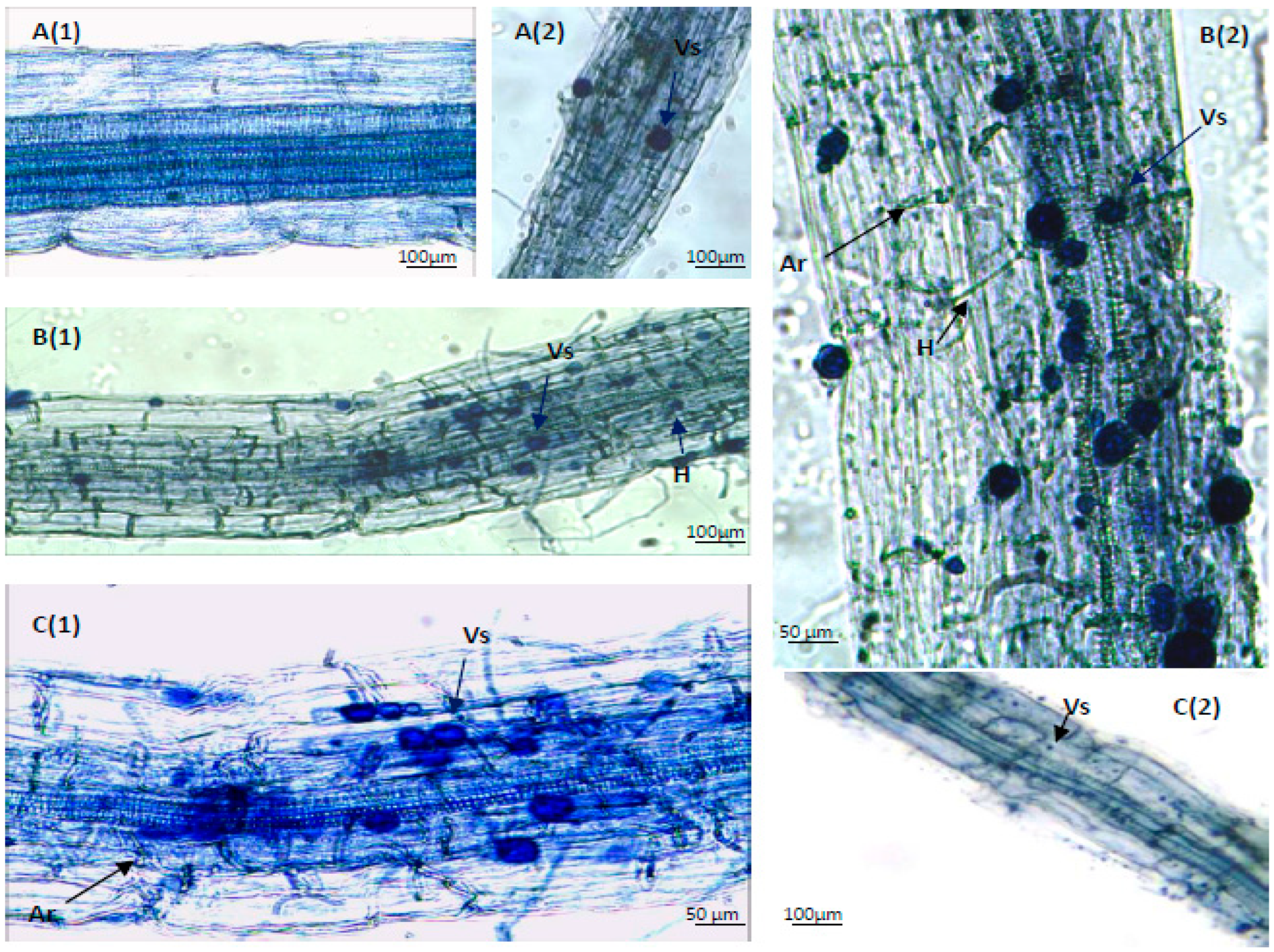
Figure 1. Visualization of the mycorrhizal colonization in the P. oleracea roots at 100× and 200× magnifications: (A1,A2) control; (B1,B2) AM1 strain C. claroideum; (C1,C2) AM2 strain F. mosseae. Vesicles (Vs), arbuscules (Ar), hyphae (H).
Agriculture 15 01458 g001
Figure 2. Assessment of mycorrhizal colonization in the roots of P. oleracea roots: (A) Degree (C%) and frequency (F%) of mycorrhiza in the root system. (B) Intensity of the mycorrhizal colonization in the root system (M%) and root fragments (m%). (C) Arbuscule abundance in mycorrhizal parts of root fragments (a%) and arbuscule abundance in the root system (A%). Variants: NM (control, non-mycorrhizal) and inoculated plants (AM1—C. claroideum; AM2—F. mosseae). Means ± SE are presented, n = 3; same letters within graph indicate no statistically significant differences as determined by Fisher’s LSD test (p ≤ 0.05) after ANOVA.
Agriculture 15 01458 g002
Figure 3. Glomalin concentration in the rhizosphere of P. oleracea: easily extracted fraction EE-GRSP and total glomalin fraction TE-GRSP. Variants: NM (non-mycorrhizal) and inoculated plants (AM1—C. claroideum; AM2—F. mosseae). Means ± SE are presented, n = 3; same letters within graph indicate no statistically significant differences as determined by Fisher’s LSD test (p ≤ 0.05) after ANOVA.
Agriculture 15 01458 g003
Figure 4. Content of reducing sugars and total proteins in the shoots of P. oleracea. Variants: NM (non-mycorrhizal) and inoculated plants (AM1—C. claroideum; AM2—F. mosseae). Means ± SE are presented, n = 3; same letters within graph indicate no statistically significant differences as determined by Fisher’s LSD test (p ≤ 0.05) after ANOVA.
Agriculture 15 01458 g004
Figure 5. Content of plastid pigments and carotenoids in P. oleracea shoots. Variants: NM (non-mycorrhizal) and inoculated plants (strains AM1—C. claroideum and AM2—F. mosseae). Means ± SE are presented, n = 3; same letters within graph indicate no statistically significant differences as determined by Fisher’s LSD test (p ≤ 0.05) after ANOVA.
Agriculture 15 01458 g005
Figure 6. Content of betalains in the aerial parts of P. oleracea. Variants: NM (non-mycorrhizal) and inoculated plants (strains AM1—C. claroideum and AM2—F. mosseae). Means ± SE are presented, n = 3; same letters within graph indicate no statistically significant differences as determined by Fisher’s LSD test (p ≤ 0.05) after ANOVA.
Agriculture 15 01458 g006
Figure 7. Content of flavonoids and phenols in the aerial parts of P. oleracea. Variants: NM (non-mycorrhizal) and inoculated plants (strains AM1—C. claroideum and AM2—F. mosseae). Means ± SE are presented, n = 3; identical letters within the graph indicate no statistically significant differences as determined by Fisher’s LSD test (p ≤ 0.05) after ANOVA.
Agriculture 15 01458 g007
Figure 8. Content of water- (WS-AO) and lipid- (LS-AO) soluble antioxidants in the aerial parts of P. oleracea. Variants: NM (non-mycorrhizal) and inoculated plants (strains AM1—C. claroideum and AM2—F. mosseae). Means ± SE are presented, n = 3; identical letters within the graph indicate no statistically significant differences as determined by Fisher’s LSD test (p ≤ 0.05) after ANOVA.
Agriculture 15 01458 g008
Figure 9. Activity of antioxidant enzymes (CAT and APX) and H2O2 content in P. oleracea shoots. Variants: NM (non-mycorrhizal) and inoculated plants (strains AM1—C. claroideum and AM1—F. mosseae). Means ± SE are presented, n = 3; identical letters within the graph indicate no statistically significant differences as determined by Fisher’s LSD test (p ≤ 0.05) after ANOVA.
Agriculture 15 01458 g009
Figure 10. Antioxidant potential (FRAP) of P. oleracea. Variants: NM (non-mycorrhizal) and inoculated plants (strains AM1—C. claroideum and AM1—F. mosseae). Means ± SE are presented, n = 3; identical letters within the graph indicate no statistically significant differences as determined by Fisher’s LSD test (p ≤ 0.05) after ANOVA.
Agriculture 15 01458 g010
Table 1. Fresh (FW) and dry (DW) weight of P. oleracea (shoots and roots) and relative mycorrhizal dependence (MD). Variants: NM (non-mycorrhizal) and inoculated plants (AM1—C. claroideum; AM2—F. mosseae).
VariantsShoot FW
(g−1 FW)
Shoot DW
(g−1 DW)
Root FW
(g−1 FW)
Root DW
(g−1 DW)
MD (%)
NM1.001 a*0.042 a0.050 a0.007 a-
AM11.749 b0.074 b0.070 b0.011 b53.593 a
AM21.894 c0.081 c0.098 c0.013 c57.381 b
* Means are presented, n = 3; identical letters within the table indicate no statistically significant differences as determined by Fisher’s LSD test (p ≤ 0.05) after ANOVA.
Disclaimer/Publisher’s Note: The statements, opinions and data contained in all publications are solely those of the individual author(s) and contributor(s) and not of MDPI and/or the editor(s). MDPI and/or the editor(s) disclaim responsibility for any injury to people or property resulting from any ideas, methods, instructions or products referred to in the content.