Article

The Suitability of Algae Solution in Pea Microgreens Cultivation under Different Light Intensities

by
Barbara Frąszczak
1,*,
Monika Kula-Maximenko
2,* and
Caihua Li
3
1
Department of Vegetable Crops, Poznań University of Life Sciences, ul. Dąbrowskiego 159, 60-594 Poznań, Poland
2
The Franciszek Górski Institute of Plant Physiology, Polish Academy of Sciences, ul. Niezapominajek 21, 30-239 Kraków, Poland
3
Economic Plant Research Institute, Jilin Academy of Agricultural Sciences, Changchun 130033, China
*
Authors to whom correspondence should be addressed.
Agriculture 2024, 14(10), 1665; https://doi.org/10.3390/agriculture14101665
Submission received: 31 July 2024 / Revised: 18 September 2024 / Accepted: 20 September 2024 / Published: 24 September 2024
(This article belongs to the Section Crop Production)

Abstract

:
Microgreens are young plants grown from vegetables, grain, or herb seeds in a controlled environment with artificial lighting. LED modules are the preferred option for indoor and vertical farming. Light intensity (LI) is crucial for plant growth and the synthesis of phytochemicals. The study aimed to assess whether growing microgreens under low light intensity but with the addition of algae would produce plants with similar parameters (biometric, active compound content) to those grown under higher light intensity. The experiment evaluated LED white light at two intensity levels: 115 µmol m−2 s−1 (low light, LL) and 230 µmol m−2 s−1 (high light, HL). Pea seeds were soaked in a 10% solution of Chlorella vulgaris algae or water before sowing, and the plants were watered or sprayed during growth with the same solutions. The results showed no positive effect of algae on plant biometric traits. However, plants treated with algae had a significantly higher chlorophyll and carotenoid content index. Light significantly influenced pea growth, with plants grown under high light (HL) showing greater weight, height, and plant area. Additionally, changes in the photosynthetic apparatus and light stress were observed in microgreens watered with water (AW and WW) under high light during the vegetative phase. Raman spectra also indicated changes in the chemical composition of microgreens’ leaves based on light intensity and treatment. Microgreens treated with algae solution during seed soaking and water during the vegetative phase produced much more carotenoids compared to other variants.

1. Introduction

Microgreens are young plants produced from vegetables, grain, or herb seeds. They have a stalk, cotyledons, and the actual first leaves that are sprouting. They are several centimeters long. Depending on the species, harvesting often occurs 7–21 days after germination [1,2]. There is an enormous variety of species that can be produced as microgreens. They differ in germination rate, flavor, and chemical composition. Cereals, legumes, oilseeds, and cruciferous plants—such as spinach, broccoli, pea, radish, sunflower, watercress, pumpkin, fenugreek, or chives—are some of the most commonly used species for microgreens [3,4]. They are a popular dish addition because of their delicate texture, distinctive color, and palatability. They go well with salads, sandwiches, soups, desserts, and drinks. They are used throughout the year, regardless of the season [5].
These nutrient-rich plants may provide health-promoting effects related to their ability to prevent the development of a wide range of chronic inflammatory diseases. Microgreens may be a promising new food source to satisfy consumer interest in healthy eating [2,6]. Microgreens are low in sugar and abundant in organic acids, carotenoids, and chlorophylls. They also show anticholinergic and antidiabetic properties. The consumption of microgreens can play a major role in preserving human health and preventing oxidative stress-related diseases [7].
Pea seedlings are one of the popular vegetables in oriental countries [8]. In addition to their striking appearance, pea seedlings provide fiber, vitamins, and the phytonutrients following: phenolic compounds, b-carotene, and chlorophyll. These natural antioxidants play an essential role in human health, as they eliminate free radicals and, consequently, can prevent the risk of cancer [7,8].
Because microgreens require a small area during production and their life cycle is very short, they are perfectly suited for cultivation under completely controlled conditions. Indoor farming is becoming a more and more common method for cultivating leafy vegetables because it gives growers the most control over the growing environment to maximize taste and morphology depending on market preferences and the ability to produce highly uniform crops year-round [9]. Cultivation under controlled conditions involves the use of artificial lighting. Light intensity (LI) is one of the most important factors in plant growth and development, including the synthesis of phytochemicals [10]. Artificial lighting conditions allow the use of varying light intensities to regulate plant growth and phytochemical accumulation, resulting in a high-quality product. The literature shows that light intensities of 100 to 300 µmol m2 s1 photosynthetic photon flux density (PPFD) are often used to produce microgreens [11]. Low light intensity is in the range of 100–140, medium intensity is in the range of 140–200, and high intensity is above 200 µmol m2 s1 [12]. Several studies have examined the connections between light intensity and different microgreens growth and yield parameters. In general, an increase in LI caused an increase in fresh and dry weight in Brassicaceae microgreens, and decreased hypocotyl length [13,14].
Low light intensities can potentially reduce the cost of electricity needed to grow microgreens in a plant factory. With the dimming option available for LED luminaires, energy consumption can be lowered by reducing the intensity of light delivered to the microgreens [11]. On the other hand, light intensity affects the rate of photosynthesis in plants and therefore plant growth [15], as well as the accumulation of various chemical compositions, including the production of secondary compounds [16]. Its reduction can affect the growth rate of plants as well as their quality, including the content of active compounds [13].
Biostimulants are products made from organic material, and they can be used in modest amounts to promote the growth and development of a variety of crops in both optimum as well as tough environments [17]. Microalgae are present in almost all terrestrial, aquatic, and sub-aerial surfaces, including all kinds of soil. They are also a component of phytoplankton [18,19]. The most common microalgae species are Chlorella sp., Arthrospira sp., Isochrysis sp., and Dunaliella sp. Among them, the two most widely grown and utilized microalgae species for commercial purposes are Arthrospira sp. and Chlorella sp. [20]. Due to the diverse properties of Chlorella vulgaris, it is the world’s most commercially cultivated algae species [21]. A lot of the beneficial effects of microalgae on soil and plants have been studied [22]. They can have a variety of useful functions in plant cultivation. Algae extracts are used for seed conditioning, as well as for soil or foliar application during the growth and flowering period. Additionally, they contribute to plant growth by producing bioactive substances such as phytohormones, and protect plants against phytopathogens and pests [23,24,25]. The application method impacts the response of the plant and the way of applying biostimulants depends primarily on the type of edible plant and its specific needs [26,27]. A study with lettuce seedlings showed that the response to Chlorella vulgaris extracts differed depending on the application methods. The foliar application had a major effect on the activity of enzymes involved in primary nitrogen metabolism, while the root watering application mainly influenced the activity of enzymes involved in primary carbon metabolism [28].
The pot experiment aimed to determine the effect of the application of algae on the biometrics, the efficiency of the photosynthetic apparatus, and the chemical composition of pea green microgreens cultivated under two different light intensities. The aim of the study was to investigate whether growing microgreens under low light intensity but with the addition of algae would produce plants with similar parameters (biometric, active compound content) to those grown under higher light intensity. Furthermore, the study aimed to establish the general validity of the use of algae in microgreens cultivation, based on biometric parameters and the analysis of active compounds using an FT-Raman spectrometer.

2. Materials and Methods

2.1. Plant Material and Growth Conditions

The experiment was conducted on green pea cultivation (Pisum sativum L. cv. Boogie) in a controlled environment. Pea seeds for the microgreens were obtained from a commercial source (“W. Legutko” Company, Jutrosin, Poland). The experiment was performed in the growth chamber. The temperature in the growth chamber was maintained at 23 °C until the seeds emerged. During the cultivation period, the day/night temperature was 21/17 ± 2 °C. The relative air humidity was maintained at 50–60%. The photoperiod was 16/8 h (day/night). The microgreens were grown under white light emitting diode (LED) panel units (Sowelo 70 72W, EKO-LED, Józefosław, Poland), with an emission wavelength of 400–700 nm, combining 88% red, 3.8% blue, and 8.2% green.
The microgreens were grown in special plastic tray vessels (13 × 9 × 3 cm, 0.35-litre capacity) filled with peat substrate (sphagnum peat). The substrate used was Hartmann’s universal substrate (Hartmannn, Poznań, Poland) pH 5.5–6.5 ± 0.2 (in H2O) with the addition of fertilizer PG Mix—NPK 14:16:18 + microelements at 1.0–1.3 kg m3 (±20%). The substrate was moistened before sowing the seeds. Twenty seeds in each container were sown. Then the top layer of the substrate in each container was covered with a thin layer of sand. Until the time of emergence, the containers were covered with transparent film to ensure higher humidity.

Light Intensity

Two levels of light intensity (LI) were used—high (HL) and low (LL). The photosynthetic photon flux density (PPFD) measured near the top of the plants was 230 (HL) and 115 (LL) µmol m2 s1 ±10 µmol m2 s1, respectively. The daily light dose was about 13.2 and 6.6 µmol m2, respectively, at a 16 h photoperiod.

2.2. Algae Cultivation Conditions

A 1000 mL Duran bottle was used as the culture vessel for microalgae growth (with 800 mL working volume and 32 mL algae suspension). Before using algae as a solution for pea microgreens, the algae were grown for 14 days at a photoperiod of 16/8 h (light/dark) at 25 °C. The culture light was white fluorescent with an intensity of 100 µmol photons m2 s1. Air was supplied to the cylinder using aeration pumps (SERA Air 550 R Plus, Trzmiel, Wrocław, Poland) to ensure the aeration and mixing of the algae culture.
The algae solution used for pea microgreens had an optical density (OD750 nm) of about 0.809 [a.u.] and the total chlorophyll content was 64.02 µg mL1.

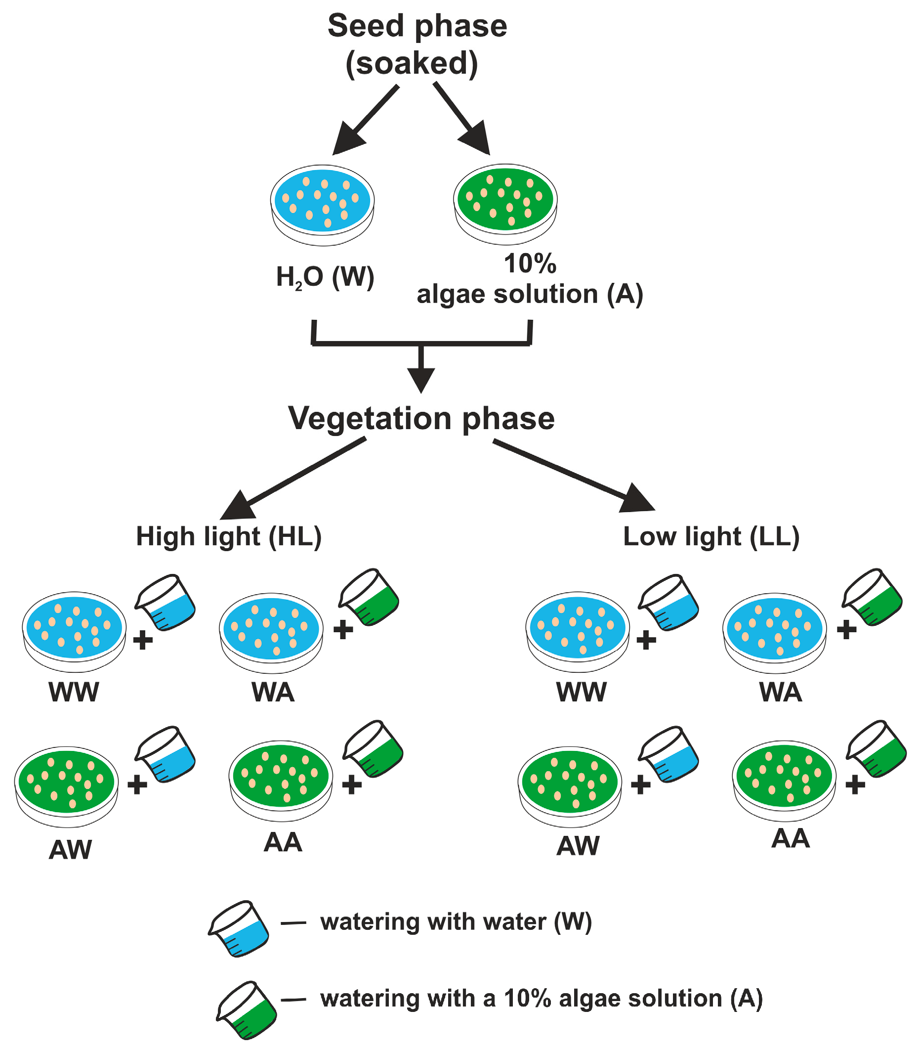
2.3. Treatment of Pea Microgreens

In the experiment, an algae solution (Chlorella vulgaris) at a concentration of 10% or water was used to soak seeds before sowing and to water or spray plants during growth (Figure 1). A 10% algal concentration was used based on previous studies (unpublished), in which the highest algal concentration (10%) had the best effect on pea microgreens biomass yield. The seeds were soaked in a solution of water with 10% algae concentration or only in pure water for 24 h at a temperature of 20 °C. The seeds were then sown into special plastic containers. Before sowing, the substrate in the containers was watered with 50 mL of an algae solution at a concentration of 10% or water, depending on the combination. During the growing season, the plants were sprayed with an algae solution at a concentration of 10% or water interchangeably with watering the substrate with water or algae solution, at a dose of 50 mL. Treatments were carried out every two days. During the 14 days of cultivation, plants were watered four times and spraying was performed three times. No plant protection products were used in the experiment, and no additional fertilization was applied.

2.4. Harvest, Biometrical and Chlorophyll Fluorescence Measurements

Since peas germinate hypogeically, the plants were harvested 14 days after emergence. The plants were harvested with scissors to cut at the lowest part of the stem. The fresh and dry weight were checked with an electronic weighing balance (parameters were measured using laboratory scales WTB200, R: 0.001 g; Radwag Poland). Dry mass (DM, mg) was measured by weighing dried microgreens. Plants were weighed after drying for 24 h at 100 °C. The dry matter ratio [%] was calculated with the following formula: DM = (Wdry/Wfr) × 100, where DM is the dry matter ratio [%], Wdry is the dry weight of the sample, and Wfr is the fresh weight of the sample. The length of plants as well as the length and width of leaves were measured using a measuring ruler in centimeters. These last parameters were used to calculate the values of the leaf shape index (the leaf length/width ratio). To calculate the area of the above-ground part of the plants was scanned and then the Skwer program was used to calculate the area. The plant area and dry mass [m2 kg−1 DM] were used to calculate the specific leaf area (SLA). All measurements were taken on six plants randomly selected from each replication. The chlorophyll content index (CCI) was measured using the OSI CCM-200 Plus leaf chlorophyll meter (ADC BioScientific Ltd., Hoddesdon, UK).
Chlorophyll fluorescence was measured using the OS1-FL modulated fluorometer (Optiscience, Hudson, NY, USA) half an hour after the termination of the period of exposure to light. All chlorophyll fluorescence parameters used in this study are described in Table 1.

2.5. FT-Raman Spectroscopy Measurements

The pea microgreens’ chemical composition was analyzed using FT-Raman spectrometer Nicolet NXR 9650, equipped with an Nd:YAG laser with a 1064 nm wavelength. The microgreens were lyophilized before measurement. FT-Raman spectra were measured with an aperture of 50 and a spectral resolution of 8 cm−1. The spectra were recorded at a laser power of 1.0 W, ranging from 400 to 3200 cm−1. For each spectrum, 64 scans were acquired. Measurements were made in 8 repetitions. For every spectrum, 9-point smoothing, vector normalization, and baseline correction were performed. Raman spectra were processed using the Omnic/Thermo Scientific and OriginPro 2020 software.

2.6. Experiment Design and Statistical Analysis

The study was conducted as a two-factor experiment. The first factor was light intensity (2 levels), and the second was the treatment of plants with an algae solution or water (4 variants) (Figure 1). The research was performed in four replicates for each combination (a single tray constituted one repetition). The experiment was conducted in two independent cycles. The results were the means of the two cycles. The data were analyzed with ANOVA. Differences between the means were estimated with the Newman–Keuls test for biometric parameters, and the Tukey’s HSD test for photosynthetic parameters at a significance level = 0.05. The data were analyzed statistically with the Statistica program (StatSoft, Kraków, Poland) and OriginPro 2020 software.
In the Raman spectra study, Principal Component Analysis (PCA) was used to distinguish between samples collected from the low light and high light. For this purpose, two Raman ranges were analyzed (400 cm−1–1800 cm−1 and 2700 cm−1– 3000 cm−1) and selected. The PCA analysis was performed using OriginPro 2020 software.

3. Results

3.1. Growth and Morphology of Pea Microgreens

The use of algae in microgreens cultivation did not significantly affect plant yields (Table 2). A more significant factor was light intensity (LI). Plants grown at high light intensity (HL) had significantly higher fresh weight compared to low light intensity (LL). Significant differences are particularly evident for combinations where plants were treated with algae only (AA). The dry mass content resulted in the lowest values for the combination with algae (AA) under both light treatments. The light intensity had no significant effect on dry mass content. Despite the lack of significant differences (except for the AA combination), higher plants were obtained for HL, individually for each algae/water combination. The largest differences in plant height between LL and HL were found for AA combinations. The lower leaf length was obtained for LL (although these differences were not always significant). Only for the AW combination were similar values obtained for both LI. The values for leaf width were similar. However, a significant reduction in leaf width was observed for this parameter in the algae-treated combinations. Also, leaf area as well as SLA were significantly lower for LL and algae-treated combinations. It is noteworthy that the greatest differences in SLA values between 115 and 230 µmol m−2 s−1 were obtained for the combinations treated with the algal solution during growth. A significantly higher CCI value was observed for plants treated with algae (AA) at both LI. In the combination where plants were treated with water only (WW) and in the combination where plants were watered (AW), a lower CCI value was found compared to plants watered with algae (WA and AA).

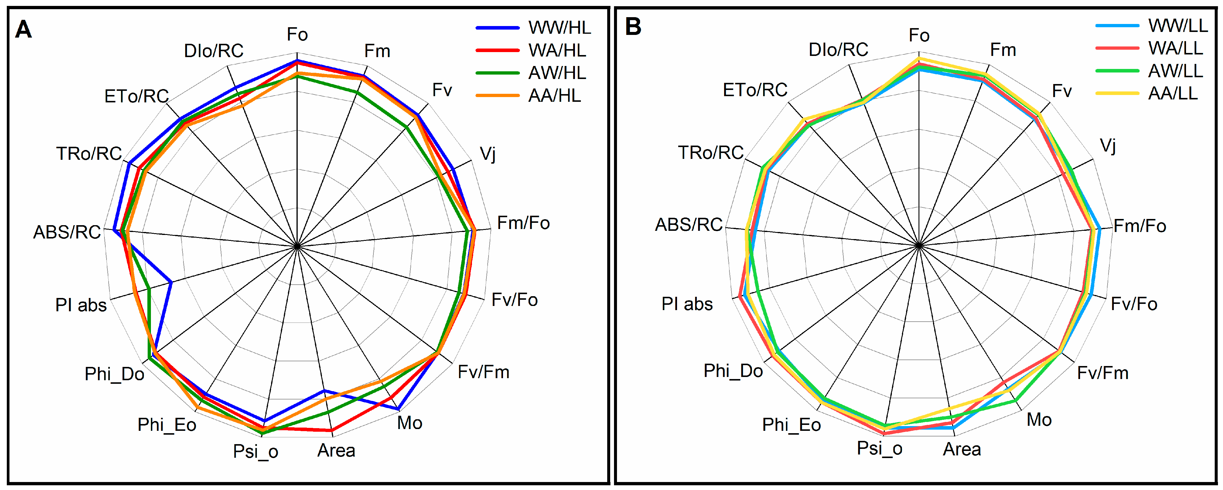
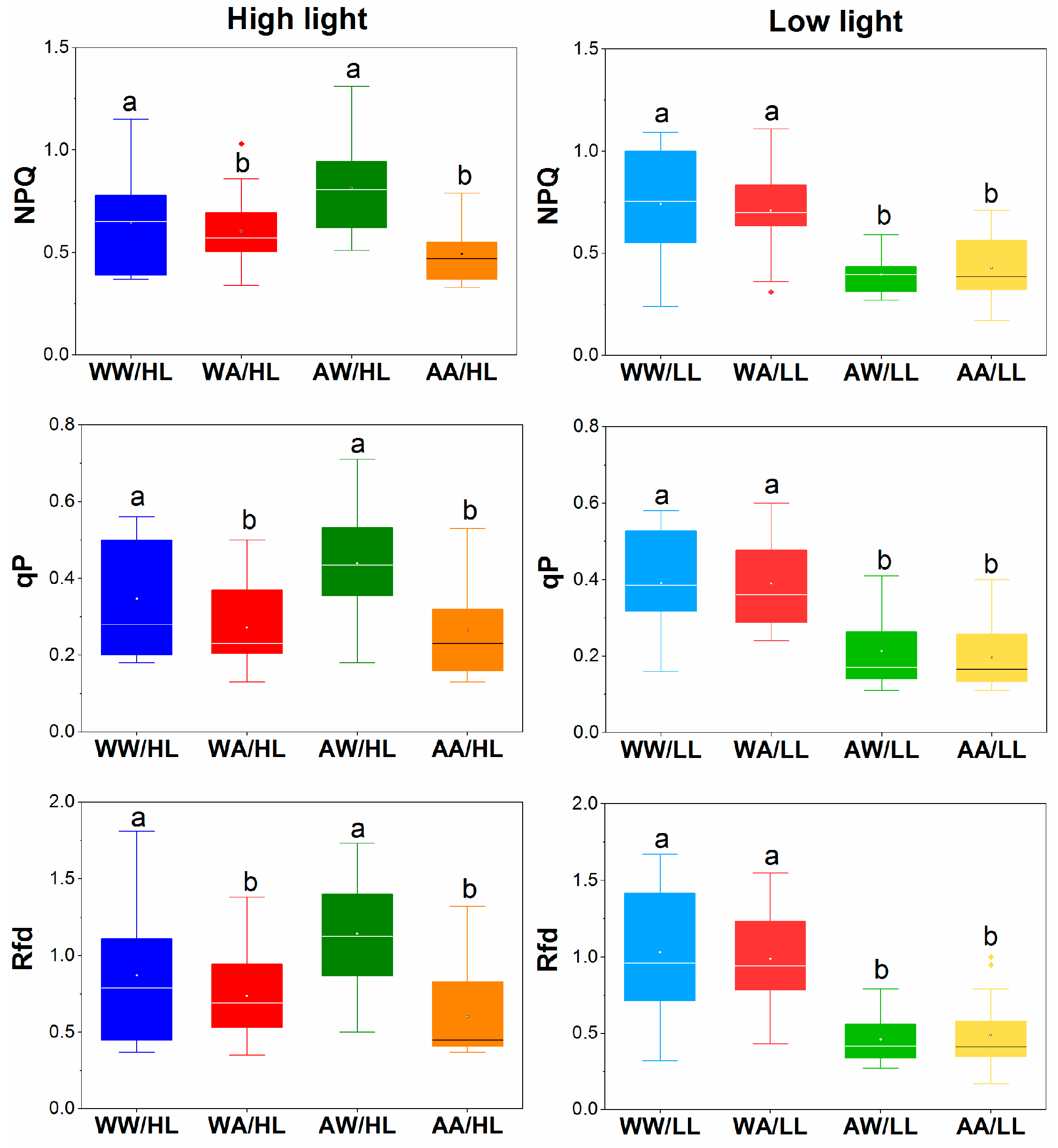
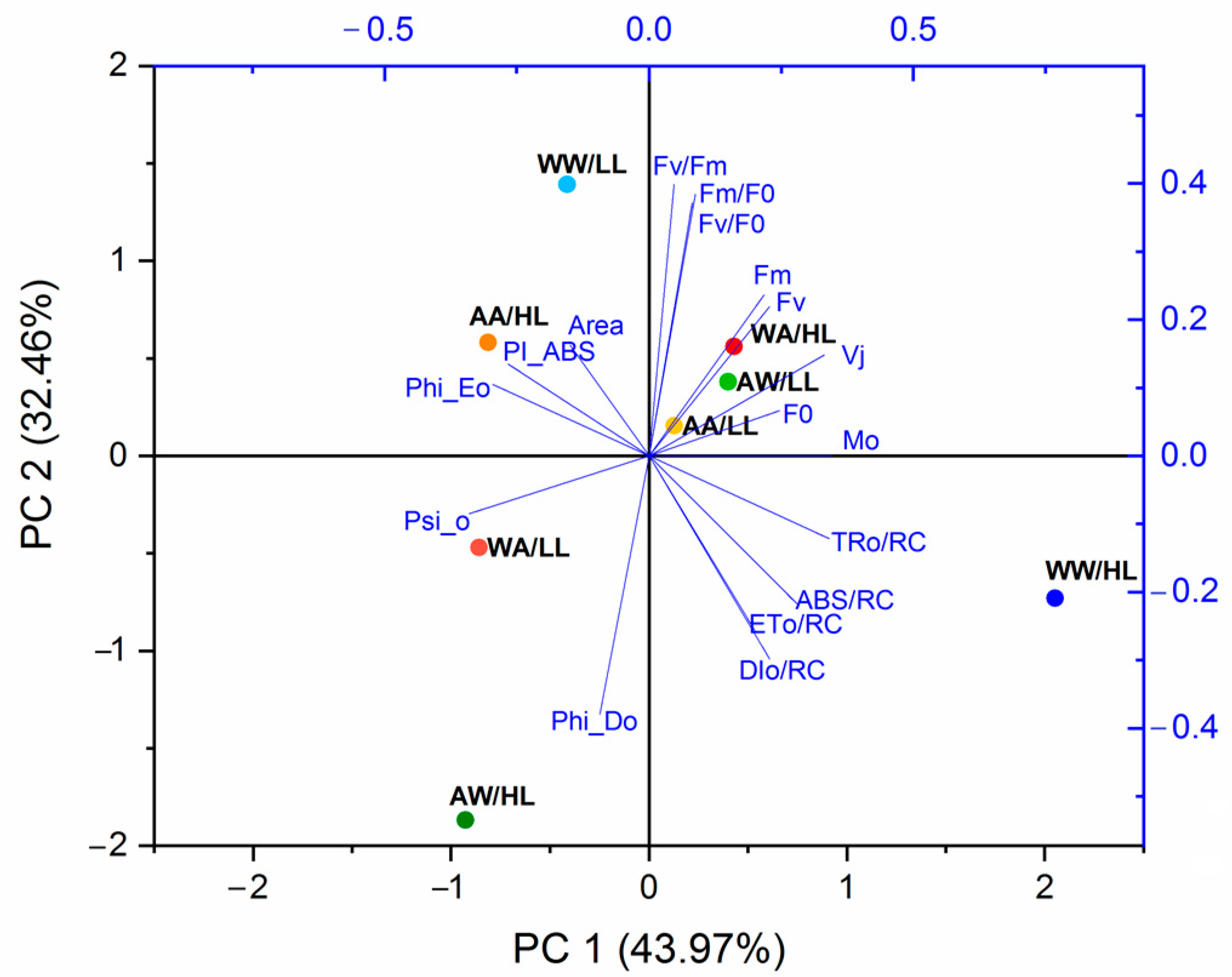
3.2. Fluorescence Parameters

The efficiency of the photosynthetic apparatus of microgreens was assessed by comparing changes in electron flow between PSII and PSI depending on the light intensity (HL and LL) under which they grew and the method of treating seeds or plants in the vegetative phase.
In Figure 2, the fluorescence parameters describe the energy flow rate, quantum yield, and energy flow through the reaction center (RC). On their basis, significant differences were found in the functioning of the photosynthetic apparatus in microgreens due to light intensity and treatment. A greater disruption of electron transport in PSII was observed in microgreens grown under HL. It was also found that plant treatment with water or algae solution was more important under HL than under LL. A large decrease in PSII activity and light stress was found in microgreens watered with water (AW and WW) in the vegetative phase and growing under HL. Significantly stronger light stress was found in WW microgreens than in other treatments. This stress was characterized by a reduced value of the PI and Area parameters with a simultaneous increase in the Vj, Mo, DIo, and TRo parameters. In the case of plants growing under LL, only AW plants had a reduced Area value and an increased Mo value. This may indicate a disturbance in the electron transport from RC to plastoquinone.
In the case of nonphotochemical parameters (NPQ, qP, and Rfd), there were two groups according to treatment (W or A) and these groups differed depending on the light intensity (Figure 3). Plants growing under HL and watered with water in the vegetation phase (WW and AW) had higher values of nonphotochemical parameters than other plants. However, watering with water in the seed phase (WW and WA) had a significant impact on the higher values of these parameters in plants growing under LL (Figure 3).
The results of the PCA analysis of fluorescence parameters and microgreens treatments under different light intensities showed that the highest values of parameters indicating the good condition of the photosynthetic apparatus (PI, Area, Phi_Eo) were observed in the AA/HL and WW/LL plants. However, photosynthetic parameters indicating a disturbance in the electron transport flow through PSII (TRo/RC, ABS/RC, DIo/RC, and ETo/RC) showed a negative correlation. In this case, WW/HL microgreens had the highest values (Figure 4).

3.3. Chemical Composition

Figure 5A,B show FT-Raman spectra recorded in the range from 400 to 3200 cm−1 for microgreens growing under different light intensities and treatments. Changes in chemical composition are identified based on specific bands characteristic of given chemical compounds. The differences observed in the spectrum indicate different contents of these components.
All presented Raman spectra had a visible carotenoid triplet, i.e., three bands at wavelengths 1525, 1159, 1005 cm−1 [32]. Another characteristic group was the bands at 1390, 1329, 1270, and 746 cm−1 derived from chlorophyll [33]. The occurrence of phenol bands at 1657 and 1390 cm−1 [34], as well as a band derived from lipids at 2930, 1657, 1440, and 1270 cm−1 [33,35], was also found. Moreover, a phospholipid band was detected around 870 cm−1, and the protein band was detected at 1602 cm−1. The analysis of the Raman spectra showed that in microgreens growing under HL, soaking the seeds with algae solution and later in the vegetation phase watering with water (AW) had a stimulated metabolism to produce more carotenoids, chlorophyll, proteins, and lipids. However, when the treatment method was not changed (WW and AA), the production of phospholipids and lipids were reduced (870, 915 and 1657 cm−1) (Figure 5A). However, watering with water during the vegetation phase (WW and AW) had the greatest influence on the chemical composition of microgreens growing under LL. These plants had a significantly higher amount of lipids and carotenoids (Figure 5B).
The PCA analysis of Raman spectra allowed for separation into two groups using PC1 and PC3. PCA showed that all microgreens growing under HL, regardless of treatment, have negative PC3 values, and plants with LL have positive values, which means that PC3, despite small variances (0.37%), distinguishes the groups from each other (Figure 5C). The loading plot indicated the Raman bands characteristic of HL microgreens (blue line, negative values) from the bands characteristic of LL plants (green line, positive values). Microgreens growing under HL can be distinguished from LL by the bands at 842, 881, 966, 1012, 1155, 1525, and 1745 cm−1, characteristic of HL plants (Figure 5D).

4. Discussion

The use of microalgae can stimulate plant growth and development, which can be observed in the improvement of plant traits such as fresh and dry weight, number of leaves, leaf area, and root length [36,37]. This is due to their positive effects on the soil environment and plant metabolism [38]. Among other things, algae assimilate and convert essential nutrients such as nitrogen, phosphorus, and potassium into forms readily available to plants [39]. Microalgae can also stimulate the growth of beneficial bacteria and fungi, thus promoting a healthy soil microbiome [36]. They also produce bioactive substances, including phytohormones, which have a direct effect on plant physiological processes and promote plant growth [40]. In the conducted experiment, no positive effect of algae on plant biometric traits was obtained separately for each light intensity. Often, plants not treated with algae had better parameters or there was no difference between the WW combination and the others. The lack of positive effects may have been due to the short growth period of the microgreens (14 days) as well as the optimal growing conditions. Also, in a study by Bumandalai and Tserennadmid [41] on tomato and cucumber seedlings, the Chlorella vulgaris suspension had a greater effect on root length than on shoot length at 12 days after emergence. The analysis of the effects of algal extracts (Planktochlorella nurekis) on seedlings of four microgreens (lettuce, wheat, radish, and broccoli) found that in most cases they had a negative or neutral effect on the tested species [42]. On the other hand, in a study on lettuce seedlings, spraying and sprinkling the plants with a Chlorella vulgaris extraction proved to be a promising treatment, enhancing fresh and dry matter at the level of the edible part only 4 days after the first treatment [28]. The neutral or negative effect obtained in the current experiment can be explained by the excessive concentration of phytohormones in the algal solution.
Phytohormones (among others such as gibberellins, cytokinins, auxins, brassinosteroids, ethylene, and abscisic acid) are very active components of algal extracts and at higher doses can inhibit important plant processes. They come from groups with different chemical structures and can affect plant metabolism in many ways [43,44]. In addition to phytohormones, algae contain many other biologically active components, such as proteins, amino acids, polysaccharides, antioxidants, and vitamins [45]. This large variety of active compounds can affect plant growth synergistically or antagonistically. They can have stimulatory or inhibitory effects on plants [42].
Fresh weight is an important attribute for the quality of microgreens growth. For all algal treatments, fresh weight was higher for HL. In the Hernández-Adasme et al. [46] study, the cultivation of beetroot microgreens at a high intensity (220 µmol m−2 s−1) resulted in a 22% reduction in yield compared to a low (120) and medium light intensity (160). Also, for broccoli microgreens, with an increase in intensity from 50 to 70 µmol m−2 s−1 and 70 to 90 µmol m−2 s−1, the fresh weight decreased by about 12.6% and 9.7%, respectively [11]. In contrast, in a study on mustard and mizuna, the fresh weight of microgreens increased with increasing PPFD from 105 to 315 µmol m−2 s−1 [14]. Light intensity is critical as it directly affects the transport of CO2 and H2O, so optimal light intensity can improve the rate of photosynthesis and increase plant productivity. Respiration exceeds photosynthesis below a certain light intensity, and plants become net oxygen consumers [15]. High light intensity can damage photosystem II and promote photoinhibition, reducing the rate of photosynthesis [47]. Based on the studies cited above, it can be concluded that each species has its level of optimal light intensity, which also depends on temperature and carbon dioxide levels in the air. The plant response observed in this study indicates that HL (230 μmol m−2 s−1) was not too high for pea microgreens and provided additional light for photosynthetic activity and consequently biomass accumulation. According to Wong [48], even though the artificial light under which leafy vegetables are grown is a fraction of full sunlight, they grow well under low light conditions.
In previous studies, an increase in LI increased the percentage of DW [13,14]. In our study, the differences in DW were small, which could be due to smaller differences in LI values compared to other research.
Low light intensity promotes elongated growth and may be beneficial for microgreens production [11]. In our study, taller plants were obtained for all combinations under HL compared to LL, with significant differences between either LI found only for the AA combination. Thus, it can be assumed that a light intensity of 115 (LL) µmol m−2 s−1 did not cause a shaded effect and consequently excessive elongation growth of the plants. The lack of excessive elongation of the pea shoots could also be related to an adequate (not too high) temperature during the plant growth period, which was adapted to the pea requirements and the light dose. According to Wang et al. [49], low light, high temperature, and high water potential promote hypocotyl elongation by inhibiting wall deposition, especially of pectin. Research by other authors has shown a tendency to reduce the length of the microgreens of different species at high light intensity [11,46,50]. Gerovac et al. [14] found that shoot elongation was influenced by light quality in addition to LI, including the proportion of blue light and the R/FR ratio.
The leaf parameters, i.e., width and length, were either larger with HL or the same regardless of PPFD. Also, a larger plant area was obtained for HL except for the combination in which no algae were used (WW). The results do not agree with previous studies, which showed that the leaf area of microgreens generally increased when grown under low LI conditions [14]. It is noteworthy that under LL, the differences between the WW combination and the others, i.e., with algae application, were significantly greater than under HL. For example, the area of plants grown with algae application (AA) compared to WW (without algae) was as much as 27% smaller for LL and only 13% smaller for HL.
SLA is characterized by a radiation-absorbing surface, so it indirectly influences the utilization of solar energy reaching the plants and thus biomass production. In addition, it shows a plastic response to light exposure through changes in leaf thickness [51]. The thickest leaves (lower SLA value) were obtained at LL, which was due to plant adaptation to low light. In a study by Silva et al. higher SLA and a thinner leaf blade were obtained for pepper (Capsicum chinense) leaves grown at a light dose reduced by 50%. According to these authors, the thinner leaf blade was due to thinner palisade and sponge crumb layers in shaded leaves compared to plants grown at 100% light. In our study, obtaining a lower SLA value at LL and treating the plants with algae may suggest that the algae stimulated the formation of more chlorophyll, and hence mesophyll, in leaves growing under poorer light conditions. For most plants, a decrease in SLA is observed with increasing leaf size [51]. In our study, SLA was positively correlated with plant area. In contrast, there was no correlation between SLA and DW, indicating that at this early stage of growth of very young leaves, the differences between combinations may have been due to morphological changes that improved light interception rather than better photosynthesis [52].
In addition to leaf characteristics, one of the most important parameters determining the purchase of microgreens is their color [11]. In pea microgreens, this color was partly dependent on the CCI (chlorophyll content index), which was highly dependent on both LI and algae application. The results show that LL stimulated chlorophyll formation similarly to algae application. Combinations where plants were sprayed and watered with algae resulted in the smallest differences in CCI between LI and a generally higher CCI value. The CCI value under HL for AA was 47% higher compared to WW. Low light results in a higher content of chloroplast pigments and electron carriers, while high light results in the degeneration of pigments that are involved in photoprotection [53]. However, at lower light levels (200 µmol m−2 s−1), light use efficiency is highest [54]. In our study, both light levels were at lower levels and the difference between the two was not very large. In another study, growing Brassica microgreens at lower light levels (110–330 µmol m−2 s−1) resulted in a significantly lower chlorophyll index compared to growing at high light levels (440–545 µmol m−2 s−1) [13].
High light intensity can disrupt the normal light reactions of photosynthesis. Then, there is a disturbance in electron transport and an increase in the number of closed reaction centers (RC) in photosystems, which in turn contributes to photoinhibition leading to the production of reactive oxygen species (ROS) [55]. The results obtained in this study regarding the activity of the photosynthetic apparatus showed that microgreens treated with algae had better efficiency in the photosynthetic process and thus higher fresh mass. However, in plants watered with water during the vegetation phase under HL, light stress and disturbances in electron transport in PSII were observed.
Leaf pigments, such as chlorophylls and carotenoids, may vary due to plant nutrition and light conditions [56]. Studies on strawberries and green cabbage showed that photosynthetic pigments (chlorophyll and carotenoids) changed under different light intensity conditions and presented significantly lower concentrations at high light intensity [57,58]. In these studies, the positive impact of algae treatment was detected on the chemical composition of leaves in the microgreens growing under HL. These leaves were characterized by a higher content of chlorophyll, carotenoids, and lipids. Broccoli microgreens grown at 50 μmol·m−2·s−1 had the highest fresh and dry mass, while the content of biologically valuable chemical compounds was the lowest. However, the content of valuable chemical compounds in broccoli microgreens improved with increasing light intensity [58]. In the study by Park et al. [59], it was shown that the production of metabolites in different plant species differed depending on how the biostimulant based on Chlorella vulgaris algae was administered. Numerous studies suggest that algae as biostimulants have beneficial properties in increasing the regulation of chlorophyll and carotenoid biosynthesis and delaying plant ageing [59]. The content of photosynthetic pigments in a plant is an important quality indicator that has a major impact on the consumer’s choice of product. The accumulation of chlorophylls and carotenoids is influenced not only by the physiological, biochemical, and genetic characteristics of plants, but also by environmental factors such as light, temperature, and fertilization [60]. As previously mentioned, algae extract contains many useful substances that can support the synthesis of photosynthetic pigments (chlorophyll and carotenoids) in plants. Previous studies showed a positive effect of using fresh algae cells on increased chlorophyll and carotenoid content in Swiss chard, tomatoes, and watermelons [61,62,63]. In our study, the use of algae at the seed stage (AW), regardless of LI, increased the carotenoid content compared to other variants. This fact may result from the double role of carotenoids that they play in plant leaves. On the one hand, they function as additional light-harvesting pigments, extending the light absorption range during photosynthesis when light intensity is low, which may have been the case with our AW-LL variant. On the other hand, carotenoids play a photoprotective role, protecting the light-harvesting complex, membranes, and proteins from excessive incident light by dissipating heat and removing free radicals. In the AW-HL variant, grown under higher light intensity, light stress and reduced photosynthetic efficiency were observed. Thus, the higher number of carotenoids in this case can be explained by their role in photoprotection. It should be noted that carotenoids, regardless of their role in plants, play an important role in human nutrition and health as antioxidants and an essential source of synthesis of the main visual pigment, vitamin A, which is required for several biological processes, including vision and the activation of the immune system.

5. Conclusions

The study evaluated the impact of algae and light intensity on pea microgreens’ yield and active substance content. The findings revealed that while algae did not show a significant positive effect on plant biometric traits, it did result in a significantly higher chlorophyll content index. Light intensity played a crucial role in pea growth, with plants grown under high light intensity (HL) exhibiting higher fresh weight, greater height, and greater plant area on average.
Overall, the results showed that the application of algae for microgreens growing under HL improved their growth and photosynthetic efficiency, as well as the chemical composition of the leaves. It should be remembered that the reactions of individual plants vary depending on the strain of microalgae and the method of their application. Therefore, before using algae as a biostimulant in production with artificial lighting, further research is necessary to optimize light conditions and algae dosage adjustment to improve the growth and quality of the obtained microgreens.

Author Contributions

Conceptualization, B.F. and M.K.-M.; methodology, B.F. and M.K.-M.; formal analysis, B.F. and C.L.; investigation, B.F. and C.L.; data curation, B.F. and C.L.; writing—original draft preparation, B.F. and M.K.-M.; writing—review and editing, B.F. and M.K.-M.; visualization, B.F. and M.K.-M.; supervision, B.F. All authors have read and agreed to the published version of the manuscript.

Funding

The study was supported by Dean’s Grant 2023 at Faculty of Agriculture, Horticulture, and Biotechnology, Poznań University of Life Sciences.

Institutional Review Board Statement

Not applicable.

Informed Consent Statement

Not applicable.

Data Availability Statement

Data are contained within the article.

Acknowledgments

We are grateful to “W.Legutko” company (Poland) for providing seeds for the experiment.

Conflicts of Interest

The authors declare no conflicts of interest.

References

  1. Xiao, Z.; Lester, G.E.; Luo, Y.; Wang, Q. Assessment of Vitamin and Carotenoid Concentrations of Emerging Food Products: Edible Microgreens. J. Agric. Food Chem. 2012, 60, 7644–7651. [Google Scholar] [CrossRef] [PubMed]
  2. Choe, U.; Yu, L.; Wang, T.T.Y. The Science Behind Microgreens as an Exciting New Food for the 21st Century. J. Agric. Food Chem. 2018, 66, 11519–11530. [Google Scholar] [CrossRef] [PubMed]
  3. Turner, E.R.; Luo, Y.; Buchanan, R.L. Microgreen nutrition, food safety, and shelf life: A review. J. Food Sci. 2020, 85, 870–882. [Google Scholar] [CrossRef]
  4. Frąszczak, B.; Kula-Maximenko, M. The Biometric Parameters of Microgreen Crops Grown under Various Light Conditions. Agriculture 2022, 12, 576. [Google Scholar] [CrossRef]
  5. Kyriacou, M.C.; El-Nakhel, C.; Graziani, G.; Pannico, A.; Soteriou, G.A.; Giordano, M.; Ritieni, A.; De Pascale, S.; Rouphael, Y. Functional quality in novel food sources: Genotypic variation in the nutritive and phytochemical composition of thirteen microgreens species. Food Chem. 2019, 277, 107–118. [Google Scholar] [CrossRef] [PubMed]
  6. Podsędek, A.; Frąszczak, B.; Sosnowska, D.; Kajszczak, D.; Szymczak, K.; Bonikowski, R. LED Light Quality Affected Bioactive Compounds, Antioxidant Potential, and Nutritional Value of Red and White Cabbage Microgreens. Appl. Sci. 2023, 13, 5435. [Google Scholar] [CrossRef]
  7. Wojdyło, A.; Nowicka, P.; Tkacz, K.; Turkiewicz, I.P. Sprouts vs. Microgreens as Novel Functional Foods: Variation of Nutritional and Phytochemical Profiles and Their In Vitro Bioactive Properties. Molecules 2020, 25, 4648. [Google Scholar] [CrossRef]
  8. Wu, M.C.; Hou, C.Y.; Jiang, C.M.; Wang, Y.T.; Wang, C.Y.; Chen, H.H.; Chang, H.M. A novel approach of LED light radiation improves the antioxidant activity of pea seedlings. Food Chem. 2007, 101, 1753–1758. [Google Scholar] [CrossRef]
  9. Despommier, D. Farming up the city: The rise of urban vertical farms. Trends Biotechnol. 2013, 31, 388–389. [Google Scholar] [CrossRef]
  10. Kopsell, D.A.; Pantanizopoulos, N.I.; Sams, C.E.; Kopsell, D.E. Shoot tissue pigment levels increase in “Florida Broadleaf” mustard (Brassica juncea L.) microgreens following high light treatment. Sci. Hortic. 2012, 140, 96–99. [Google Scholar] [CrossRef]
  11. Gao, M.; He, R.; Shi, R.; Zhang, Y.; Song, S.; Su, W.; Liu, H. Differential Effects of Low Light Intensity on Broccoli Microgreens Growth and Phytochemicals. Agronomy 2021, 11, 537. [Google Scholar] [CrossRef]
  12. Appolloni, E.; Pennisi, G.; Zauli, I.; Carotti, L.; Paucek, I.; Quaini, S.; Orsinia, F.; Gianquinto, G. Beyond vegetables: Effects of indoor LED light on specialized metabolite biosynthesis in medicinal and aromatic plants, edible flowers, and microgreens. J. Sci. Food Agric. 2021, 102, 472–487. [Google Scholar] [CrossRef] [PubMed]
  13. Samuoliené, G.; Brazaityte, A.; Jankauskiene, J.; Virsile, A.; Sirtautas, R.; Novickovas, A.; Sakalauskiene, S.; Sakalauskaite, J.; Duchovskis, P. LED irradiance level affects growth and nutritional quality of Brassica microgreens. Cent. Eur. J. Biol. 2013, 8, 1241–1249. [Google Scholar] [CrossRef]
  14. Gerovac, J.R.; Craver, J.K.; Boldt, J.K.; Lopez, R.G. Light intensity and quality from sole-source light-emitting diodes impact growth, morphology, and nutrient content of Brassica microgreens. HortScience 2016, 51, 497–503. [Google Scholar] [CrossRef]
  15. Ilieva, I.; Ivanova, T.; Naydenov, Y.; Dandolov, I.; Stefanov, D. Plant experiments with light-emitting diode module in Svet space greenhouse. Adv. Space Res. 2010, 46, 840–845. [Google Scholar] [CrossRef]
  16. Brazaitytė, A.; Sakalauskienė, S.; Samuolienė, G.; Jankauskienė, J.; Viršilė, A.; Novičkovas, A.; Sirtautas, R.; Miliauskiene, J.; Vaštakaite, V.; Dabašinskas, L.; et al. The effects of LED illumination spectra and intensity on carotenoid content in Brassicaceae microgreens. Food Chem. 2015, 173, 600–606. [Google Scholar] [CrossRef]
  17. Ronga, D.; Biazzi, E.; Parati, K.; Carminati, D.; Carminati, E.; Tava, A. Microalgal Biostimulants and Biofertilisers in Crop Productions. Agronomy 2019, 9, 192. [Google Scholar] [CrossRef]
  18. Metting, B.; Pyne, J.W. Biologically active compounds from microalgae. Enzym. Microb. Technol. 1986, 8, 386–394. [Google Scholar] [CrossRef]
  19. Olaizola, M. Commercial development of microalgal biotechnology: From the test tube to the marketplace. Biomol. Eng. 2003, 20, 459–466. [Google Scholar] [CrossRef]
  20. Priyadarshani, I.; Rath, B. Commercial and industrial applications of micro algae—A review. J. Algal Biomass Utln. 2012, 3, 89–100. [Google Scholar]
  21. El-Naggar, N.E.-A.; Hussein, M.H.; Shaaban-Dessuuki, S.A.; Dalal, S.R. Production, extraction and characterization of Chlorella vulgaris soluble polysaccharides and their applications in AgNPs biosynthesis and biostimulation of plant growth. Sci. Rep. 2020, 10, 3011. [Google Scholar] [CrossRef] [PubMed]
  22. Alvarez, A.L.; Weyers, S.L.; Goemann, H.M.; Peyton, B.M.; Gardner, R.D. Microalgae, soil and plants: A critical review of microalgae as renewable resources for agriculture. Algal Res. 2021, 54, 102200. [Google Scholar] [CrossRef]
  23. Garcia-Gonzalez, J.; Sommerfeld, M. Biofertilizer and biostimulant properties of the microalga Acutodesmus dimorphus. J. Appl. Phycol. 2015, 28, 1051–1061. [Google Scholar] [CrossRef] [PubMed]
  24. Lu, Y.; Xu, J. Phytohormones in microalgae: A new opportunity for microalgal biotechnology? Trends Plant Sci. 2015, 20, 273–282. [Google Scholar] [CrossRef]
  25. Renuka, N.; Guldhe, A.; Prasanna, R.; Singh, P.; Bux, F. Microalgae as multi-functional options in modern agriculture: Current trends, prospects and challenges. Biotechnol. Adv. 2018, 36, 1255–1273. [Google Scholar] [CrossRef]
  26. Alobwede, E.; Leake, J.R.; Pandhal, J. Circular economy fertilization: Testing micro and macro algal species as soil improvers and nutrient sources for crop production in greenhouse and field conditions. Geoderma 2019, 334, 113–123. [Google Scholar] [CrossRef]
  27. Németh, A.; Horváth, N.; Katona, S.; Molnár, Z. Review of the Use of Biostimulant Microalgae to Influence the Growth and Development of Ornamental Plants. Acta Agron. Óvár. 2024, 65, 36–55. [Google Scholar] [CrossRef]
  28. Puglisi, I.; La Bella, E.; Rovetto, E.I.; Stevanato, P.; Fascella, G.; Baglieri, A. Morpho-biometric and biochemical responses in lettuce seedlings treated by different application methods of Chlorella vulgaris extract: Foliar spray or root drench? J. Appl. Phycol. 2022, 34, 889–901. [Google Scholar] [CrossRef]
  29. Force, L.; Critchley, C.; Van Rensen, J.J.S. New fluorescence parameters for monitoring photosynthesis in plants. Photosynth. Res. 2003, 78, 17–33. [Google Scholar] [CrossRef]
  30. Dąbrowski, P.; Baczewska, A.H.; Pawluśkiewicz, B.; Paunov, M.; Alexantrov, V.; Goltsev, V.; Kalaji, M.H. Prompt chlorophyll a fluorescence as a rapid tool for diagnostic changes in PSII structure inhibited by salt stress in Perennial ryegrass. J. Photochem. Photobiol. B 2016, 157, 22–31. [Google Scholar] [CrossRef]
  31. Kalaji, H.M.; Schansker, G.; Brestic, M.; Bussotti, F.; Calatayud, A.; Ferroni, L.; Goltsev, V.; Guidi, L.; Jajoo, A.; Li, P.; et al. Frequently asked questions about chlorophyll fluorescence, the sequel. Photosynth. Res. 2017, 132, 13–66. [Google Scholar] [CrossRef] [PubMed]
  32. Baranska, M.; Schulz, H.; Baranski, R.; Nothnagel, T.; Christensen, L.P. In Situ simultaneous analysis of polyacetylenes, carotenoids and polysaccharides in carrot roots. J. Agric. Food Chem. 2005, 53, 6565–6571. [Google Scholar] [CrossRef] [PubMed]
  33. Schulz, H.; Baranska, M. Identification and quantification of valuable plant substances by IR and Raman spectroscopy. Vib. Spectrosc. 2007, 43, 13–25. [Google Scholar] [CrossRef]
  34. Andreev, G.; Schrader, B.; Schulz, H.; Fuchs, R.; Popov, S.; Handjieva, N. Non-destructive NIR-FT-Raman analyses in practice. Part 1. Analyses of plants and historic textiles. Fresenius J. Anal. Chem. 2001, 371, 1009–1017. [Google Scholar] [CrossRef] [PubMed]
  35. Muik, B.; Lendl, B.; Molina-Diaz, A.; Ayora-Canada, M.J. Direct monitoring of lipid oxidation in edible oils by Fourier transform Raman spectroscopy. Chem. Phys. Lipids 2005, 134, 173–182. [Google Scholar] [CrossRef]
  36. Gonçalves, J.; Freitas, J.; Fernandes, I.; Silva, P. Microalgae as Biofertilizers: A Sustainable Way to Improve Soil Fertility and Plant Growth. Sustainability 2023, 15, 12413. [Google Scholar] [CrossRef]
  37. Parmar, P.; Kumar, R.; Neha, Y.; Srivatsan, V. Microalgae as next generation plant growth additives: Functions, applications, challenges and circular bioeconomy based solutions. Front. Plant Sci. 2023, 14, 1073546. [Google Scholar] [CrossRef]
  38. Bulgari, R.; Franzoni, G.; Ferrante, A. Biostimulants Application in Horticultural Crops under Abiotic Stress Conditions. Agronomy 2019, 9, 306. [Google Scholar] [CrossRef]
  39. De Souza, M.H.B.; Calijuri, M.L.; Assemany, P.P.; Castro, J.d.S.; de Oliveira, A.C.M. Soil application of microalgae for nitrogen recovery: A life-cycle approach. J. Clean. Prod. 2019, 211, 342–349. [Google Scholar] [CrossRef]
  40. Guo, S.; Wang, P.; Wang, X.; Zou, M.; Liu, C.; Hao, J. Microalgae as Biofertilizer in Modern Agriculture. In Microalgae Biotechnology for Food, Health and High Value Products; Alam, M.A., Xu, J.-L., Wang, Z., Eds.; Springer: Singapore, 2020; pp. 397–411. [Google Scholar]
  41. Bumandalai, O.; Tserennadmid, R. Effect of Chlorella vulgaris as a biofertilizer on germination of tomato and cucumber seeds. Int. J. Aquat. Biol. 2019, 7, 95–99. [Google Scholar] [CrossRef]
  42. Karbarz, M.; Piziak, M.; Żuczek, J.; Duda, M. Influence of Microalgae Planktochlorella nurekis Clones on Seed Germination. Agronomy 2023, 13, 9. [Google Scholar] [CrossRef]
  43. Asif, R.; Yasmin, R.; Mustafa, M.; Ambreen, A.; Mazhar, M.; Rehman, A.; Umbreen, S.; Ahmad, M.; Asif, R.; Yasmin, R.; et al. Chapter 7: Phytohormones as Plant Growth Regulators and Safe Protectors against Biotic and Abiotic Stress. In Plant Hormones: Recent Advances, New Perspectives and Applications; Hano, C., Ed.; IntechOpen: London, UK, 2022. [Google Scholar] [CrossRef]
  44. Han, X.; Zeng, H.; Bartocci, P.; Fantozzi, F.; Yan, Y. Phytohormones and Effects on Growth and Metabolites of Microalgae: A Review. Fermentation 2018, 4, 25. [Google Scholar] [CrossRef]
  45. Kapoore, R.V.; Wood, E.E.; Llewellyn, C.A. Algae biostimulants: A critical look at microalgal biostimulants for sustainable agricultural practices. Biotechnol. Adv. 2021, 49, 107754. [Google Scholar] [CrossRef] [PubMed]
  46. Hernández-Adasme, C.; Palma-Dias, R.; Escalona, V.H. The Effect of Light Intensity and Photoperiod on the Yield and Antioxidant Activity of Beet Microgreens Produced in an Indoor System. Horticulturae 2023, 9, 493. [Google Scholar] [CrossRef]
  47. Youssef, R.; Kyriacou, M.C.; Petropoulos, S.A.; De Pascale, S.; Colla, G. Improving vegetable quality in controlled environments. Sci. Hortic. 2018, 234, 275–289. [Google Scholar] [CrossRef]
  48. Wong, C.E.; Teo, Z.W.N.; Shen, L.; Yu, H. Seeing the lights for leafy greens in indoor vertical farming. Trends Food Sci. Technol. 2020, 106, 48–63. [Google Scholar] [CrossRef]
  49. Wang, H.; Shang, Q. The combined effects of light intensity, temperature, and water potential on wall deposition in regulating hypocotyl elongation of Brassica rapa. Peer J. 2020, 8, e9106. [Google Scholar] [CrossRef]
  50. Vetchinnikov, A.; Filatov, D.A.; Olonina, S.I.; Kazakov, A.V.; Olonin, I.Y. Influence of the radiation intensity of LED light sources of the red-blue spectrum on the yield and energy consumption of microgreens. IOP Conf. Ser. Earth Environ. Sci. 2021, 723, 32–46. [Google Scholar] [CrossRef]
  51. Milla, R.; Reich, P.B. The scaling of leaf area and mass: The cost of light interception increases with leaf size. Proc. R. Soc. B 2007, 274, 2109–2115. [Google Scholar] [CrossRef]
  52. Cowden, R.J.; Markussen, B.; Ghaley, B.B.; Henriksen, C.B. The Effects of Light Spectrum and Intensity, Seeding Density, and Fertilization on Biomass, Morphology, and Resource Use Efficiency in Three Species of Brassicaceae Microgreens. Plants 2024, 13, 124. [Google Scholar] [CrossRef]
  53. Walters, R.G.; Shephard, F.; Rogers, J.J.M.; Rolfe, S.A.; Horton, P. Identification of mutants of Arabidopsis defective in acclimation of photosynthesis to the light environment. Plant Physiol. 2003, 131, 472–481. [Google Scholar] [CrossRef] [PubMed]
  54. Fu, W.; Li, P.; Wu, Y.; Tang, J. Effects of different light intensities on anti-oxidative enzyme activity, quality and biomass in lettuce. Hort. Sci. 2012, 39, 129–134. [Google Scholar] [CrossRef]
  55. Janeeshma, E.; Johnson, R.; Amritha, M.S.; Noble, L.; Aswathi, K.P.R.; Telesiński, A.; Kalaji, H.M.; Auriga, A.; Puthur, J.T. Modulations in chlorophyll a fluorescence-based on intensity and spectral variations of light. Int. J. Mol. Sci. 2022, 23, 5599. [Google Scholar] [CrossRef] [PubMed]
  56. Sumanta, N.; Imranul, C.; Nishika, J.; Suprakash, R. Spectrophotometric analysis of chlorophylls and carotenoids from commonly grown fern species by using various extracting solvents. Res. J. Chem. Sci. 2014, 25, 97–101. [Google Scholar] [CrossRef]
  57. Choi, H.G. Correlation among phenotypic parameters related to the growth and photosynthesis of strawberry (Fragaria × ananassa Duch.) grown under various light intensity conditions. Front. Plant Sci. 2021, 12, 647585. [Google Scholar] [CrossRef]
  58. Flores, M.; Hernández-Adasme, C.; Guevara, M.J.; Escalona, V.H. Effect of different light intensities on agronomic characteristics and antioxidant compounds of Brassicaceae microgreens in a vertical farm system. Front. Sustain. Food Syst. 2024, 8, 1349423. [Google Scholar] [CrossRef]
  59. Park, Y.J.; Park, J.-E.; Truong, T.Q.; Koo, S.Y.; Choi, J.-H.; Kim, S.M. Effect of Chlorella vulgaris on the Growth and Phytochemical Contents of “Red Russian” Kale (Brassica napus var. Pabularia). Agronomy 2022, 12, 2138. [Google Scholar] [CrossRef]
  60. Barickman, T.C.; Kopsell, D.A.; Sams, C.E. Abscisic acid impacts tomato carotenoids, soluble sugars, and organic acids. HortScience. 2016, 51, 370–376. [Google Scholar] [CrossRef]
  61. Abdel-Mawgoud, A.S.; Hafez, M.M.; Habib, A.M. Seaweed extract improves growth, yield and quality of different watermelon hybrids. Res. J. Agric. Biol. Sci. 2010, 6, 161–168. [Google Scholar]
  62. Djuric, M.; Mladenovic, J.; Pavlovic, R.; Zdravkovic, J. Application of bio-stimulator biocomplex 900 in producing tomato (Lycopersicon Essculentum Mill.) seedling. Acta Agric. Serb. 2014, 19, 97–103. [Google Scholar] [CrossRef]
  63. Hajnal-Jafari, T.; Seman, V.; Stamenov, D.; Đurić, S. Effect of Chlorella vulgaris on Growth and Photosynthetic Pigment Content in Swiss Chard (Beta vulgaris L. subsp. cicla). Pol. J. Microbiol. 2020, 69, 235–238. [Google Scholar] [CrossRef] [PubMed]
Figure 1. The layout of the experiment.
Agriculture 14 01665 g001
Figure 2. The spider plot presentation of selected fluorescence parameters describes the efficiency of microgreens’ photosynthetic apparatus from different light intensities (high light intensity (HL)—(A), low light intensity (LL)—(B)) and treatments.
Agriculture 14 01665 g002
Figure 3. Changes in the nonphotochemical fluorescence quenching (NPQ), the photochemical quenching in the relative reduction state of QA, reflecting the fraction of open PSII reaction centers (qP), and the vitality index of PSII indicative of interactions between the light-stage reactions activated by PAR absorption and the dark reactions of photosynthesis (Rfd) microgreens from different light intensities and treatments. Columns with different letters are statistically different among different treatments (p < 0.05).
Agriculture 14 01665 g003
Figure 4. Principal Component Analysis (PCA) of the variability of the fluorescence parameters in microgreens from different light intensities (high light intensity—HL, low light intensity—LL) and treatments. The abbreviations are described in Table 1.
Agriculture 14 01665 g004
Figure 5. Raman spectra for microgreens grown in high light intensity (A) and low light intensity (B), together with the marked positions of the peaks characteristic of the relevant functional groups (carotenoids, polysaccharides, chlorophyll, lipids, flavonoids, and proteins). PCA score plots (C) and loadings (D) for Raman range between 400–1800 cm−1 and 2700–3000 cm−1 for microgreens from different light intensities (high light intensity—HL, low light intensity—LL) and treatments.
Agriculture 14 01665 g005
Table 1. List and description of chlorophyll a fluorescence parameters [29,30,31].
ParametersDescription
Energy flow rate
FoMinimal fluorescence, when all PSII reaction centers (RCs) are open.
FmMaximal fluorescence, when all PSII RCs are closed.
FvMaximal variable fluorescence, which is measured after dark adaptation. The extent of Fv is related to the maximum quantum yield of PSII.
Vjinformation about the number of closed RCs in relation to the total number of RCs that can be closed.
Fm/FoThe ratio of maximum fluorescence to zero-time fluorescence. The low ratio Fm/Fo could indicate the destruction of PSII.
Fv/FoRatio of rate constants for photochemical reaction and nonphotochemical deactivation of PSII excitations.
Fv/FmMaximum photochemical quantum PSII after dark adaptation.
Moapproximated initial slope (in ms−1) of the fluorescence transient normalized on the maximal variable fluorescence Fv.
AreaThe area above the fluorescence induction curve is proportional to the pool size of electron acceptors in PSII.
Quantum yields and efficiencies
Psi_oThe probability of electron transport beyond QA-, i.e., the efficiency with which the exciton trapped using an RC drives the electron along ETC beyond QA-.
Phi_EoThe quantum efficiency of electron transfer from QA- to electron transport chain beyond QA-.
Phi_DoThe quantum efficiency of energy dissipation.
Performance indexes
PI absPerformance index (potential) for energy conservation from excitation to the reduction of intersystem electron acceptors.
Specific energy fluxes per reaction center
ABS/RCThe light energy absorbed using the PSII antenna photon flux per active reaction center.
TRo/RCTotal energy used to reduce QA by the unit reaction center of PSII per energy captured/trapped using a single active RC.
ETo/RCThe rate of electron transport through a single RC
DIo/RCTotal energy dissipated per reaction center (RC) as heat, fluorescence, and energy transfer to PSI.
Parameters of nonphotochemical and photochemical fluorescence quenching
NPQNonphotochemical quenching per reaction center of PSII. This parameter is associated with heat losses. For the majority of healthy plants, NPQ values are 0.5–3.5, although they differ in various species or plants cultivated at different growth conditions.
qPPhotochemical quenching. This is the fraction of light energy consumed by the open centers for photosynthetic reactions concerning the total amount of energy absorbed by PSII.
RfdVitality index of PSII. This parameter is indicative of interactions between the light-stage reactions activated by PAR absorption and the dark reactions of photosynthesis. This parameter is diminished when the balance between photochemical reactions in thylakoids and the rates of enzymatic reactions in the chloroplast stroma is disturbed.
Table 2. The effects of light intensity (LI) and algae treatment on the growth and morphology of pea microgreens.
TreatmentSeedsMean for
Light
Water (W)Algae (A)
Watering/Spraying
Water (WW)Algae (WA)Water (AW)Algae (AA)
Fresh mass [g plant−1]
Low light intensity (LL)0.94 abc *0.91 bc0.85 c0.83 c0.88 b
High light intensity (HL)1.07 a0.97 abc0.98 abc1.05 ab1.02 a
Mean for water/algae1.01 a0.94 a0.92 a0.94 a
Dry mass content [%]
Low light intensity (LL)7.92 ab7.96 ab7.75 b7.72 b7.84 a
High light intensity (HL)7.93 ab7.82 b8.39 a7.64 b7.95 a
Mean for water/algae7.93 ab7.89 ab 8.07 a7.68 b
Height [cm]
Low light intensity (LL)12.13 bc11.99 bc12.36 abc11.55 c12.01 b
High light intensity (HL)12.94 ab12.53 abc12.93 ab13.59 a13.00 a
Mean for water/algae12.53 a12.26 a12.64 a12.57 a
The leaf length [cm]
Low light intensity (LL)1.71 b1.74 b1.65 b1.64 b1.69 b
High light intensity (HL)1.95 a1.81 ab1.72 b1.84 ab1.83 a
Mean for water/algae1.83 a1.77 a1.69 a1.74 a
The leaf width [cm]
Low light intensity (LL)2.13 bc1.81 d1.85 d1.84 d1.91 b
High light intensity (HL)2.40 a2.30 ab2.04 cd2.17 abc2.23 a
Mean for water/algae2.27 a2.05 b1.94 b2.00 b
Plant area [cm2]
Low light intensity (LL)40.58 ab35.07 bc32.87 c31.95 c35.12 b
High light intensity (HL)44.94 a39.68 ab37.29 bc39.66 ab40.39 a
Mean for water/algae42.76 a37.37 b35.08 b35.80 b
Specific Leaf Area [SLA, m2 kg−1]
Low light intensity (LL)20.43 ab17.63 cd17.06 d16.44 d17.89 b
High light intensity (HL)22.84 a20.57 ab17.63 cd19.97 bc20.25 a
Mean for water/algae21.64 a19.1 b17.35 b18.21 b
Chlorophyll Content Index (CCI)
Low light intensity (LL)17.83 d20.20 c19.95 c23.45 a20.36 a
High light intensity (HL)15.70 e21.75 b17.25 d23.20 a19.48 b
Mean for water/algae16.76 d20.98 b18.60 c23.33 a
* Means with the same letter are not statistically different (p ≤ 0.05).
Disclaimer/Publisher’s Note: The statements, opinions and data contained in all publications are solely those of the individual author(s) and contributor(s) and not of MDPI and/or the editor(s). MDPI and/or the editor(s) disclaim responsibility for any injury to people or property resulting from any ideas, methods, instructions or products referred to in the content.

