Article

Melatonin Secretion in Regulating the Circadian Rhythms of Reproduction in Goose (Anser cygnoides)

1
Key Laboratory for Evaluation and Utilization of Poultry Genetic Resources of Ministry of Agriculture and Rural Affairs, Yangzhou University, Yangzhou 225009, China
2
Joint International Research Laboratory of Agriculture and Agri-Product Safety, The Ministry of Education of China, Yangzhou University, Yangzhou 225009, China
*
Author to whom correspondence should be addressed.
Agriculture 2023, 13(8), 1620; https://doi.org/10.3390/agriculture13081620
Submission received: 12 July 2023 / Revised: 3 August 2023 / Accepted: 14 August 2023 / Published: 17 August 2023
(This article belongs to the Section Farm Animal Production)

Abstract

:
Circadian rhythms affect the physiology and behavior of most organisms. The ovulation–laying cycle of poultry exhibits evident rhythmic patterns. However, the underlying biological mechanism has remained unclear. Herein, Yangzhou goose (Anser cygnoides) were selected at 6:00, 12:00, 18:00, and 24:00 (n = 6/timepoint) to investigate the regulation of circadian egg-laying through the light-driven melatonin secretion. Our study revealed that the laying rates displayed diurnal fluctuations, with a peak of 40% of eggs being laid between 4:00 and 7:00. The cosine analysis revealed that the expression of clock genes exhibited rhythmicities (p < 0.05). Relevantly, melatonin secretion also displayed circadian rhythmicity and sharply decreases with increasing amount of light (p < 0.001). The immunohistochemical analysis found that the melatonin receptor is highly expressed during the night period. Notably, tissue distribution analysis further revealed that the melatonin receptor genes showed a decreasing trend in the pineal gland and hypothalamic–pituitary–gonad (HPG) axis throughout the day. Concomitantly, the expression of reproduction-related genes at 12:00 was significantly higher than that at 24:00 (p < 0.01). Taken together, these data suggested cyclical secretion of melatonin in response to photoperiod, which acts as a neuroendocrine transducer of circadian rhythm and the time preference of reproduction in domestic geese.

1. Introduction

Circadian rhythms are endogenous adaptation mechanisms employed by living organisms to better adapt to the environment, and they play a fundamental characteristic in regulating various life activities. In order to survive and reproduce, the reproductive system of animals must be synchronized with favorable external stimuli and internal factors that enable or even optimize reproduction [1,2]. According to previous studies, the ovulation–laying cycle of female poultry exhibited a clear rhythm and environmental adaptability, especially regarding the time preference of laying eggs [3]. High-yielding hens have strict laying time preferences, mainly concentrated in the morning under natural photoperiod conditions [4]. It is still noteworthy that the cluster trait of hens was interrupted when the egg-laying time was delayed until the afternoon [5,6]. Moreover, a great number of studies have proved that poultry typically present with spontaneous ovulation, and the follicles need to go through a long process of growth and development to reach the mature state before ovulation occurs [7,8]. Follicles transform into hierarchical follicles and acquire the ovulatory potential under the synergistic action of reproductive endocrine hormones and various growth factors [9,10,11]. Subsequently, the gonadal axis induces ovulation of the mature follicle where the process of egg-laying is strictly dependent on ovulation. Among them, gonadotropin-releasing hormone (GnRH) and gonadotropins (Gths) play an important role in the process of follicular development and differentiation [12]. The Gths are classified as follicle-stimulating hormone (FSH) and luteinizing hormone (LH) [13]. Meanwhile, ovulation is triggered by a rapid increase in luteinizing hormone (LH) from the pituitary gland [14]. Studies have found that GnRH, secreted by the hypothalamus, enters the blood and stimulates the release of FSH, LH, and other glucocorticoids from the anterior pituitary through the hypophyseal portal system [15]. These hormones, via the bloodstream, act on the ovary to stimulate a series of functions related to ovulation by promoting the production of Estrogen (E2) and Progesterone (P4) [16]. Although the period of egg formation is closely related to reproductive endocrine activity, activation of the hypothalamic–pituitary–gonadal (HPG) axis is also controlled by the intrinsic timekeeping mechanism.
Light is a crucial exogenous environmental factor for controlling many physiological and behavioral processes in animals [17]. Unlike mammals, birds possess a singular endocrine gland, the pineal gland, which can detect and respond to external light stimuli [18]. In recent years, it has been confirmed in several studies that the pineal gland is responsible for converting light signals into hormone signals and plays a vital role in the regulation of reproductive processes [19]. Although many secretions of the pineal gland have been discovered in recent years, melatonin is considered the chief product. The regulation of melatonin release is governed by sympathetic afferentation to the pineal gland, mediating the suppressive impact of light on the secretion [19]. However, the hormone melatonin, which is synthesized by the pineal gland, plays a crucial role in the regulation of the circadian rhythm [20]. At present, multiple core clock genes have been confirmed in the pineal gland of birds, including Clock, Bmal (Bmal1 and Bmal2), Per (Per2 and Per3), and Cry (Cry1 and Cry2) genes [21,22]. The synthesis of melatonin is governed by the endogenous circadian clock of the pineal gland, resulting in rhythm oscillations on a near 24 h cycle [23]. Consequently, melatonin secretion exhibits a diurnal variation, with low levels during the day and high levels at night [24]. Therefore, the pineal gland of birds is characterized by the presence of both light input and melatonin output channels, indicating its dual functionality in terms of sympathetic innervation and circadian rhythm [25,26,27]. The pineal gland is also considered a dominant circadian oscillator, playing a crucial role in regulating the organism’s internal clock [20]. Notably, the melatonin secreted by the pineal gland also influences follicle development and ovulation via the hypothalamic–pituitary–gonad (HPG) axis. In addition, melatonin could also directly regulate the activity of the ovary through its interaction with specific receptors, thereby influencing the reproductive endocrine system of the organism [28,29,30]. There are three melatonin receptor subtypes, melatonin receptor 1A (MT1, Mel-1a), melatonin receptor 1B (MT2, Mel-1b), melatonin receptor 1C (MT3, Mel-1c), have been identified [31]. The presence of melatonin receptors 1A in the ovaries of African ostrich chicks suggested that melatonin serves as a significant physiological regulator in the maturation of ovarian granulosa cells [32]. For most species, it has been demonstrated that the light input and melatonin output pathways are closely related to internal circadian oscillators, however, the molecular mechanisms governing the regulation of the egg-laying circadian rhythm remain inadequately understood in geese [26,27].
Numerous studies have reported that geese exhibit a characteristic egg-laying time preference, but the precise involvement of the pineal gland in the modulation of reproduction rhythms remains ambiguous. In this study, Yangzhou geese (Anser cygnoides) during the egg-laying peak period were selected. On the basis of elucidating the egg-laying preference and the rhythmic expression of clock genes in the pineal gland, the circadian regulation of melatonin secretion was further explored. As a result, these data deepen the understanding of the physiological mechanism of circadian behavioral rhythms and provide a theoretical basis for improving egg production in domestic geese.

2. Materials and Methods

2.1. Ethics Statement

This study has been approved by Yangzhou University’s Ethics Committee on Animal Experiments for all animal experiments and protocols (Approval Code: 154-2020, Government of Jiangsu Province, China). Any experimentation involving geese was conducted in accordance with the Regulations for the Administration of Affairs Concerning Experimental Animals, as approved by the State Council of the People’s Republic of China.

2.2. Experimental Design and Sample Collection

This study included female Yangzhou geese raised in the conventional method of stocking in the Yangzhou Tiange Goose Industry Development Co., Ltd., Jiangsu Province of China (32°64′ N, 119°34′ E). This experiment was performed at 28 °C/14 °C (day/night), on a 13L: 11D natural lighting photoperiod. For recording reproductive performance under natural lighting photoperiod, lights were not turned on at the end of the light phase. The geese’ house was disinfected and segregated before the start of the experiment and breeding geese by means of flat farming. The general ventilation system was natural through two large doors and ventilation openings (2.0 m high) along the walls and along the roof. All animals were fed the same diet during the experiment (crude protein: 16%, metabolizable energy: 11.29 MJ/kg, Table 1). Feed and water were provided ad libitum. In the peak laying period, a total of 48 healthy Yangzhou geese 1R6 (68 weeks old) were selected for the experiments. They were tested at 6:00, 12:00, 18:00, and 24:00 for successive 2 days, representing different natural photoperiods, respectively. Each group was comprised of 6 individuals, specifically referred to as biological replicates. During each sampling period, the experimental subjects were female geese whose weights varied by no more than 3% of the average. Then serum was obtained through centrifugation at 3000× g for 15 min, the collected serum was stored at −80 °C. In this study, these geese were euthanized under sodium pentobarbital anesthesia and dissected to obtain the pineal gland, hypothalamus, pituitary, and ovary (six samples per composite were used for each test). These fresh tissues were immediately frozen in liquid nitrogen and stocked at −80 °C until RNA isolation. Finally, the collected pineal glands were subjected to further immunohistochemistry experiments. A summary of the sample collection and analysis process is presented in Figure 1.

2.3. Reproductive Performance and Immunohistochemical Observations

The global horizontal irradiations (GHI) and egg-laying rates were also continuously monitored during the two weeks prior to sampling. On a daily basis, egg-laying rates were calculated according to the formula: egg-laying rate (%) = (number of eggs divided by total number of geese) × 100%.
The immunohistochemistry procedure involved dewaxing the slides thrice in xylene for 15 min each and placing them in an EDTA buffer for microwave repair. Endogenous peroxidase activity was quenched with 3% H2O2 for 10 min. Then, the slides were incubated with primary antibodies overnight (Mtnr, 1:1000, ab87639; Abcam, Cambridge, UK). The WB experiments were performed to verify the specificity of the Mtnr antibody. After washing, the second antibody, rabbit anti-mouse IgG (ab6728; Abcam, Cambridge, UK), was added dropwise and incubated for 30 min. After washing again, freshly prepared 3,3′-diaminobenzidine (DAB) chromogenic reagent was added dropwise to the slides. After 10 min of chromogenic reaction, the reaction was terminated, followed by hematoxylin staining, alcohol dehydration (70%, 90%, and 100%), xylene clarification, and neutral resin mounting. The imaging system (Thermo Fisher Scientific, Waltham, MA, USA) was employed to capture both bright-field and fluorescence images (using a GFP filter) of the stained sections. The mean integrated optical density (IOD) of the immunohistochemical results was evaluated using Image-Pro Plus software (Image-Pro Plus, v 6.0).

2.4. RNA Extraction and Quantitative RT-PCR

Approximately 30 mg of tissue was used for RNA extraction. Total RNA was extracted with Trizol (Invitrogen, San Diego, CA, USA) in accordance with the manufacturer’s recommendations. The primers (Table 2) were synthesized by TSINGKE biological technology (Nanjing, China). Furthermore, the stably expressed reference gene, glyceraldehyde-3-phosphate dehydrogenase (GAPDH), was employed to standardize target gene expression. Subsequently, RT-PCR was performed with a Quant Studio 5 (Applied Biosystems, Thermo Fisher Scientific) using the SYBR Green Master Mix (ABclonal, Wuhan, China). This study utilized an RT-PCR thermal cycling program consisting of an initial denaturation step at 95 °C for 5 min, followed by 41 cycles of amplification at 95 °C for 20 s and 60 °C for 30 s. The relative amount of each mRNA was calculated by the DCt method. Each reaction was performed in triplicate, and the data represent the mean of three independent experiments.

2.5. Measurement of Serum Melatonin Hormone Concentrations

To estimate hormone levels, blood samples were subjected to centrifugation, and serum melatonin hormone (MT) levels were measured by ELISA kits (Qisong Biological, Beijing, China). The manufacturers’ protocols were adhered to during the execution of operations. ELISA tests were repeated three times. The ELISA assay exhibited a sensitivity of 1.55 pg/mL and did not exhibit any cross-reactivity with other structural analogues. Additionally, intra-assay and inter-assay variation coefficients were less than 15%. Finally, the absorbance of each well was measured at 450 nm. The ELISA data were calculated using a model as below.
y = A2 + (A1 − A2)/(1 + (x/x0) ^ p)
R2 values for all standard curves were higher than 0.98.

2.6. Statistical Analysis

All results were expressed as Mean ± standard error mean (SEM). These data underwent Kolmogorov–Smirnov (KS) analysis for normality of distribution, and nonnormal values were log-transformed for regression analysis. One-way ANOVA was performed, and multiple comparisons were made using the Duncan test before conducting the analysis. Significance was set at the 0.05 level (p < 0.05). In addition, non-linear regression was used to analyze the rhythmic expression of core clock genes, with the model equation formula as follows:
Y(t) = M + A × cos (ω × t − C)
Statistical analyses were conducted using the SPSS 20.0 software package (IBM Corp, Armonk, NY, USA). Spearman correlation tests are conducted utilizing the R programming language (version 4.2.0) to establish correlations.

3. Results

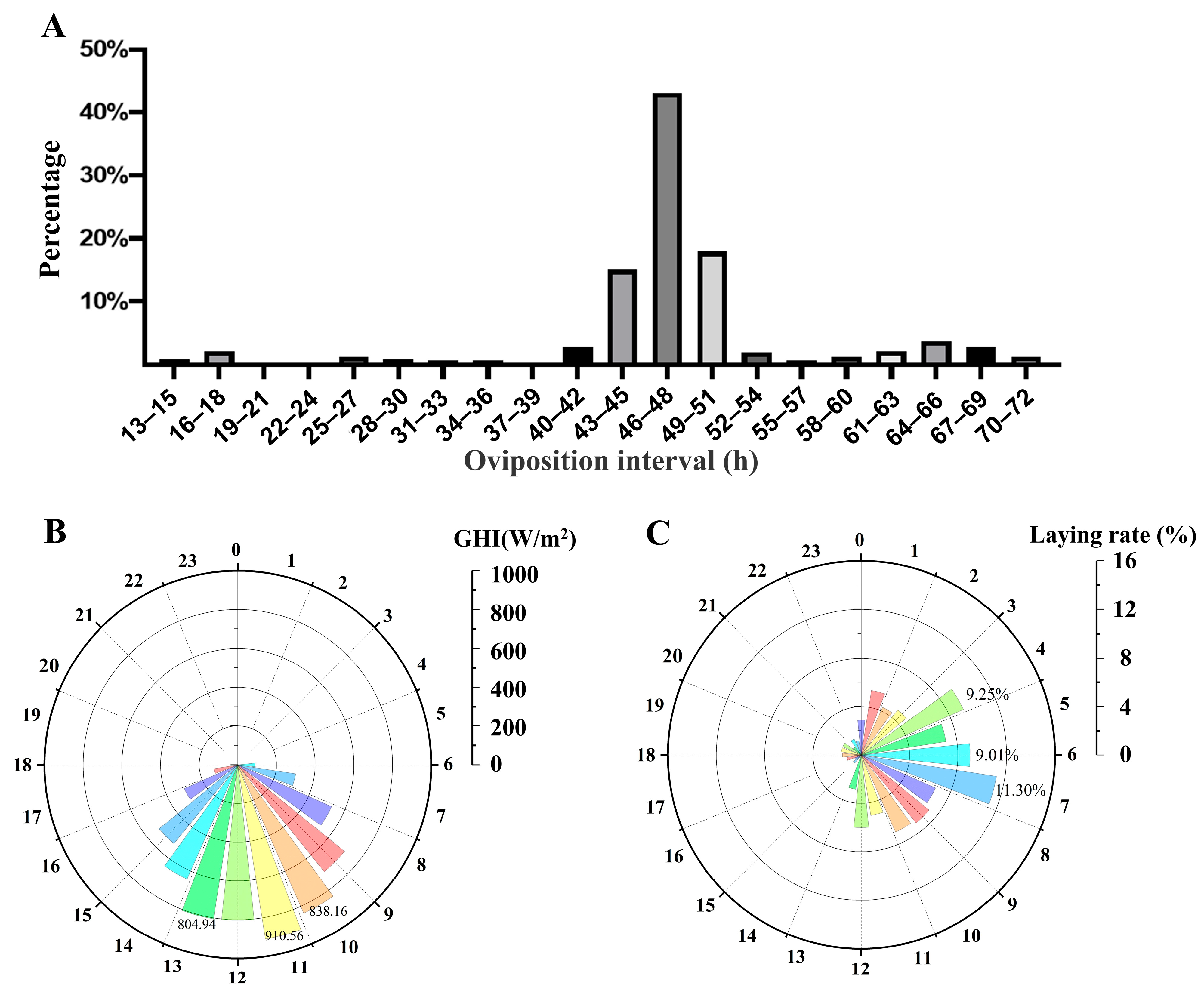
3.1. The Temporal Dynamics of Egg-Laying Preference in Goose

The characteristics of laying time in Yangzhou geese during the 14-day observation period were shown in Figure 2. Our study revealed that Yangzhou geese exhibited an average oviposition interval of approximately 47.74 h (Figure 2A). Among the individuals observed, approximately 43% oviposition interval within the time frame of 46–48 h, while around 76% oviposition interval within the range of 43–51 h. Moreover, the GHI was recorded throughout the day (Figure 2B). This result showed that the GHI appeared at 5:00 in the morning, then reached a peak value greater than 900 W/m2 at 11:00, and disappeared at 19:00. The time preferences of laying were also investigated (Figure 2C). Approximately 40% of the eggs were laid between 4:00 and 7:00. In contrast, only 7.9% of eggs were laid between 12:00 and 24:00.

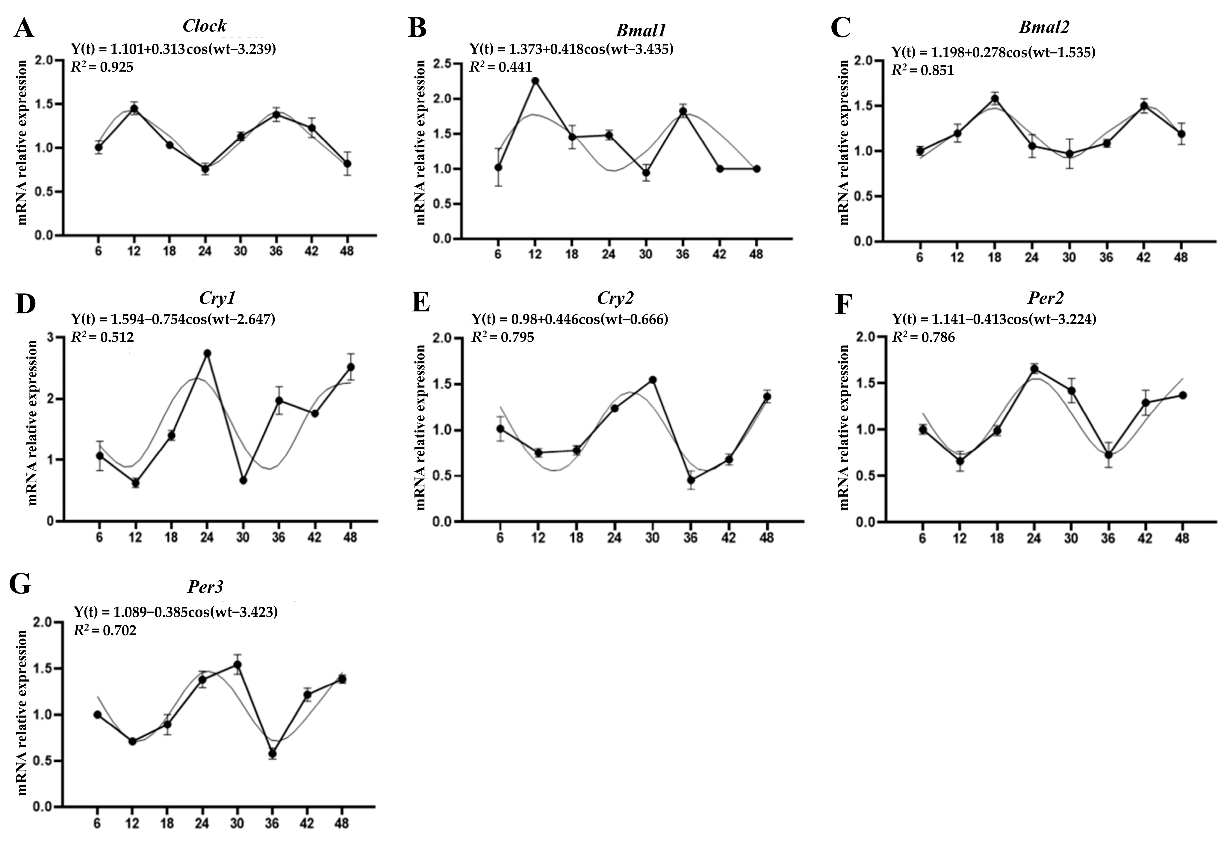
3.2. Circadian Expression of Clock Genes in Pineal Glands

To investigate the circadian rhythm of clock genes in the pineal gland, we examined their expression trends over a 48 h period. The results revealed that the pineal gland of Yangzhou geese exhibited expression of all clock genes, namely Clock, Bmal1, Bmal2, Cry1, Cry2, Per2, and Per3 (Figure 3, Table 3). Among them, the three positive regulatory genes Clock, Bmal1, and Bmal2 all showed an oscillation trend that increased during the day and decreased at night (Figure 3A–C). The cosine period regression analysis revealed the presence of a 24 h circadian rhythm in the cosine curve of Clock (R2 = 0.925, p = 0.001) and Bmal2 (R2 = 0.851, p = 0.0011), with discernible differences in amplitude. Notably, the amplitude of oscillation in Bmal2 expression was relatively smaller compared to that of Bmal1. Furthermore, the negative regulatory genes, Cry1, Cry2, Per2, and Per3, exhibited contrasting rhythmic expression patterns to that of positive regulatory genes, demonstrating a consistent decrease in expression during the day and elevating at night (Figure 3D–G). It is worth noting that these clock genes exhibit significant circadian oscillations (p < 0.05). Meanwhile, the cosine fitting of Cry2, Per2, and Per3 gene expression falls within the range of 0.7–0.8 (p = 0.003, p = 0.003, and p = 0.009, respectively). In addition, the peak times of expression for the clock genes were different. The negatively regulated genes reached their peak expression levels between 22:00 and 2:00, which was in contrast to the positively regulated genes.

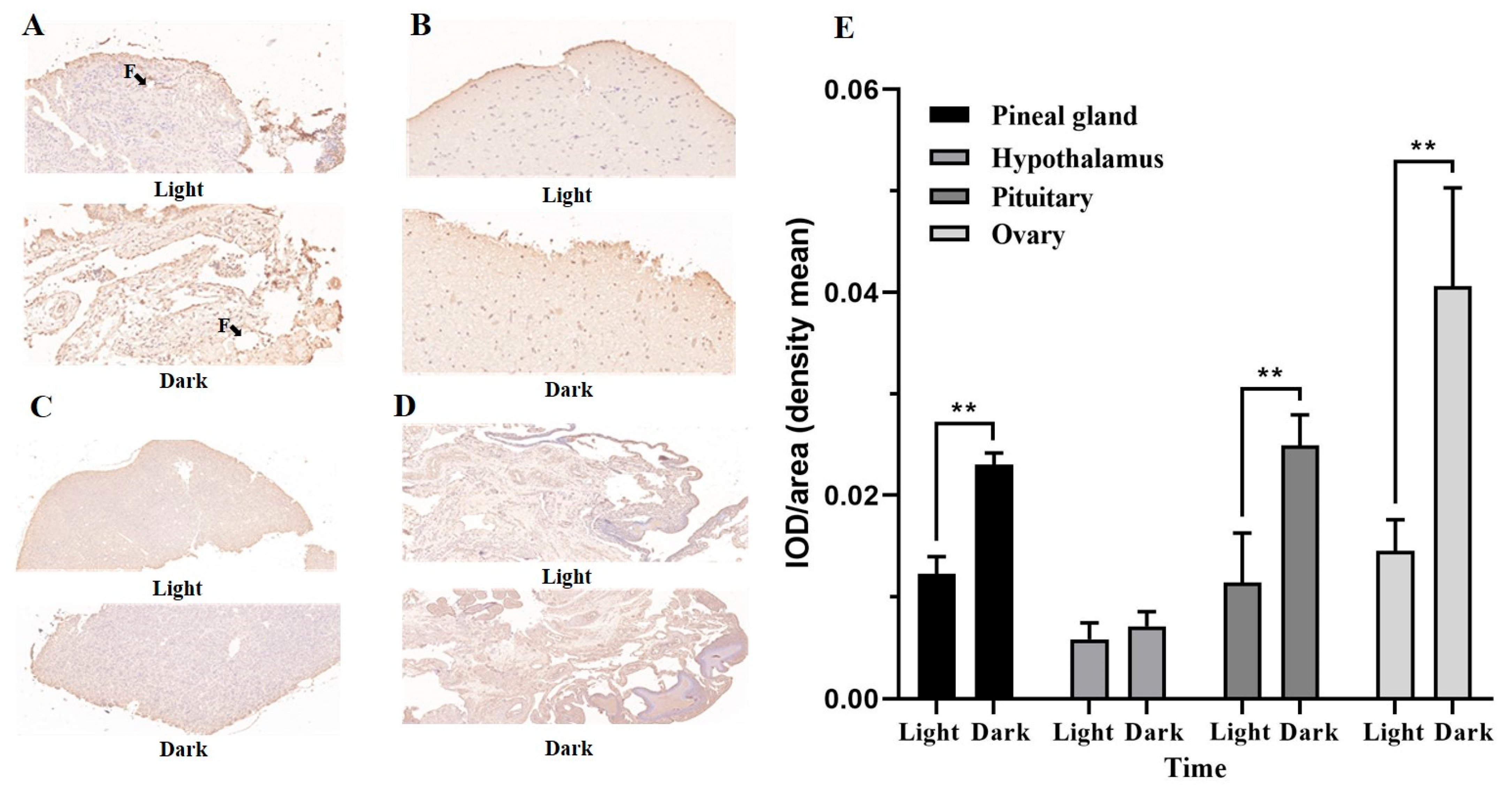
3.3. The Dynamics of Melatonin Secretion and Immunohistochemical Localization

The dynamic change of the melatonin hormone is shown in Table 4. Melatonin secretion exhibited a typical diurnal trend. The results showed that melatonin content was the highest at 24:00 (p < 0.001). Subsequently, as the duration of exposure to light increased, the concentration of melatonin decreased rapidly, resulting in a significantly lower concentration of melatonin at 12:00 compared to 6:00 (p < 0.001).
To further explore the expression of the Mtnr, immunohistochemical analysis was conducted on both the pineal gland and peripheral tissues at distinct times of light (12:00) and dark (24:00), respectively (Figure 4). Mtnr were widely expressed in different issues. In the pineal gland, the expression of Mtnr was observed to be widespread in the pineal organ, with the greatest level found in close proximity to the connective tissue capsule (Figure 4A,E). Furthermore, the expression level of Mtnr was significantly higher during the dark periods compared to periods of light (p < 0.001). Different from the pineal gland, while the expression of Mtnr was elevated during the dark period in the hypothalamus, no statistically significant differences in circadian were observed (p > 0.05, Figure 4B). The Mtnr exhibited abundant expression at the edge of the pituitary, with a significant increase in expression levels during the night as light exposure decreased (p < 0.001, Figure 4C). Similarly, the ovary, as evidenced by immunohistochemical analysis, indicated that the theca layer of ovarian follicles exhibited a light yellow or even brownish-yellow hue, signifying a significant expression of Mtnr (Figure 4D). Concurrently, the expression of Mntr during the dark period exhibited a significant increase compared to the light period (p < 0.001).

3.4. Tissue Distribution and Circadian Expression of Mtnrs Genes

Many physiological functions of melatonin are achieved by binding to receptors, so we explored the circadian regulation of Mtnrs genes in the pineal gland and peripheral tissue. The Mtnrs genes exhibited extensive distribution across various tissues, including the pineal gland, pituitary, hypophysis, and ovary (Figure 5). Real-time PCR analysis demonstrated that there is a significant circadian oscillation in the expression of Mel-1a and Mel-1c within the pineal gland (Figure 5A). Specifically, the expression levels at 24:00 exhibited a significant increase compared to the other three time points (p < 0.001). On the other hand, the expression level of Mel-1b in the pineal gland remained relatively constant and was notably lower than that of Mel-1a and Mel-1c (p < 0.001). In addition, in comparison to other tissues, the expression of Mel-1b was notably elevated in the hypothalamus and pituitary (p > 0.05, Figure 5B,C), with the most prominent expression observed at 24:00. However, the expression levels of Mel-1a and Mel-1c in the hypothalamus were relatively stable throughout the day (Figure 5B). In the interim, the Mel-1a expression within the pituitary persisted at a low level throughout the day and exhibited significantly lower in comparison to the other subtypes (p < 0.001, Figure 5C). At 24:00, the expression level of Mel-1b in the pituitary gland exhibited a minimum 5-fold increase in comparison to that at 12:00 (p < 0.001). Finally, the results indicate that the ovary exhibited the highest expression levels of the three melatonin receptor subtypes at 24:00, with Mel-1c demonstrating the largest circadian fluctuation (Figure 5D). Furthermore, the expression levels of Mel-1b and Mel-1c at 24:00 were significantly higher than those observed at 12:00 (p < 0.001).

3.5. Circadian Oscillation of the Reproduction-Related Genes in HPG Axis

A diagram showing the changes in reproduction-related genes with respect to circadian cycles can be found in Figure 6. The expression level of GnRH exhibited a rapid increase from 6:00 to 12:00 (p < 0.001), followed by a decline as the duration of light decreased, reaching its lowest point at 24:00 (Figure 6A). Correspondingly, the expression levels of FSHβ and LH showed similar circadian fluctuations, with a statistically significant increase observed at 12:00 in comparison to other times of the day (p < 0.001, Figure 6B,C). Notably, the expression level of LH exhibited large-amplitude fluctuations, whereby the expression level at 12:00 was approximately 30-fold higher than that at 24:00 (p < 0.001). In the ovary, the actions of P4 and E2 are mainly mediated through their respective nuclear receptors, namely the progesterone receptor (PGR) and estrogen receptor (ESR1) (Figure 6D,E). The study revealed that the levels of ESR1 expression did not exhibit a significant difference between 12:00 and 18:00 (p > 0.05), but were significantly elevated compared to other times of the day (p < 0.001). Furthermore, while PGR exhibited high levels at 12:00 (p < 0.001), its expression remained relatively stable across other time points.

3.6. Correlation Analysis of Melatonin Hormone and Reproductive-Related Genes

The correlation analysis between the melatonin hormone and reproductive-related genes was presented as a chordal graph (Figure 7A) and a correlation heatmap (Figure 7B). The present investigation revealed a negative association between melatonin hormone and reproductive-related genes, with a significant correlation observed between melatonin hormone and GnRH (R2 = −0.8, p = 0.044, Table S1). In addition, there were also some degree relationships between different reproductive-related genes. The results indicate that GnRH exhibited a positive association with FSH, LH, ESR1, and PGR1 (Figure 7A), with a statistically significant correlation observed between LH (p = 0.001) and ESR1 (p < 0.001). Similarly, FSH was significantly positively correlated with LH (p = 0.047). Furthermore, in comparison to other genes involved in reproduction, the positive associations observed between PGR and other reproductive-related genes did not reach statistical significance (p > 0.05, Figure 7B).

4. Discussion

To further reveal the regulatory mechanisms of circadian reproduction, we explored the expression of core clock genes and the secretory activity characterizations of the pineal gland under the normal day and night light cycles, with a particular emphasis on the regulation of the HPG axis. As shown in this study, the pineal gland in response to photoperiod regularly triggers ovulation and maintains the clear time preference of egg-laying in geese. In this way, the cyclical secretion of melatonin by the pineal gland provides the neuroendocrine control of reproduction with an internal molecular representation of changes in the photoperiod (Figure 8).
The circadian rhythm is a vital biological process that enables animals to acclimate to their external environment [35]. Studies have confirmed that the ovulation–laying cycle of poultry exhibited obvious rhythmicity and adaptability [36]. The egg production of poultry determines reproductive performance, which is an important economic characteristic. In this study, these geese were bred to get fertile eggs, and the probing of reproductive performance was of high significance. In this research, we observed Yangzhou geese during their peak laying period and discovered that they exhibited a distinct preference for laying duration. Unlike mammals, poultry differed in both anatomy and physiology, as their ovaries contain a great number of hierarchical follicles [7]. Among them, the process of egg formation in poultry involves multiple stages, including the development of the ovarian follicle, ovulation of the largest follicle, formation of the egg white and the eggshell membrane, calcification of the eggshell, and oviposition [10,11]. Animals synchronize their physiological state with external stimuli to accurately time their reproductive activities. Furthermore, previous studies have shown that high-yielding laying hens exhibit an egg-laying interval of 25–27 h under natural photoperiod conditions [37,38]. However, geese, unlike chickens, exhibited an egg-laying interval of approximately 46–48 h, potentially attributable to the longer duration of egg retention within the oviduct [39]. Meanwhile, our study also revealed that most female Yangzhou geese exhibited egg-laying preference in the morning. A previous observations found that nearly 10% of Sichuan white geese and Zhedong white geese laid eggs at 6:00 [40]. Similarly, in our study, we observed that the laying of eggs by Yangzhou geese predominantly occurred during the hours of 4:00 to 7:00. As such, it is recommended that breeders engage in a comprehensive cleaning of the goose house during the afternoon to mitigate any potential adverse effects on the egg-laying process of geese.
Furthermore, the circadian clock plays a crucial role in regulating behavioral regularities and physiological processes, including but not limited to endocrine hormone production, lipid metabolism, and maintenance of body temperature [36,41]. Studies have proved that light signals can influence the activity of the hypothalamus–pituitary–gonad axis (HPG axis) through the biological clock, thereby impacting the reproductive activities of animals [25,38]. Hence, investigating the rhythmic oscillation of circadian clocks is essential for comprehending the rhythmic patterns of egg production. Clock genes are crucial regulators of the biological rhythm, potentially linked to external environmental factors, including light, temperature, and other variables [42]. During the diurnal cycle, the pineal gland of White Leghorn chicken (Gallus gallus) exhibits higher expression levels of clock genes during the light compared to the night [43]. Moreover, the Rhythmic oscillation of the core circadian has also been shown in sparrows [44]. The present investigation revealed that the Clock and Bmal genes exhibited their highest expression levels at noon in the pineal gland, whereas the Cry and Per genes demonstrated their expression peaks in the late night and early hours of the morning. Some previous studies found that the transcription factors Bmal and Clock, belonging to the bHLH PAS family, activate the transcription of Per and Cry genes in vertebrates by binding to E-box promoter elements located in their respective promoters [44]. Meanwhile, the formation of a heterodimer between Per and Cry results in the repression of Per and Cry transcription by acting on the Clock-Bmal heterodimer [45]. The negative feedback loop comprised of these genes serves as the fundamental mechanism of the circadian clock in animals [46]. Our findings align with previous research conducted on mammals. Specifically, in rats exposed to a 12 h light and 12 h dark cycle, the expression of Clock was observed to be greater during the light than during the dark, suggesting that light cues may influence the expression of the core circadian clock [47].
The pineal gland, as a crucial core biological clock in poultry, plays an important role in endocrine regulation by sensing photoperiod information [19]. It was reported that a light regime could alter the morphology of the pineal gland [18]. Numerous studies have found that the synthesis and secretion of melatonin are closely related to light [19,48]. A study has confirmed that the pineal gland of reptiles, fish, and birds has the ability to respond to light stimulation, thereby regulating the release of hormones [49]. Moreover, the pineal gland further regulates growth and sexual maturation through the secretion of melatonin [50]. Pineal melatonin mostly acts as an endocrine agent, whereas melatonin performs not only autocrine functions but also functions in a paracrine manner [51,52]. Melatonin secretion follows a pattern of low levels during the day and high levels during the night in response to alterations in light duration [53]. The same observation was found in this research. Furthermore, as a short-day breeding species, most birds experience a suppression of gonadal activity due to the presence of melatonin [54]. Typically, melatonin exerts effects on the reproductive endocrine system by binding to the receptors [28]. The results of the immunohistochemical analysis in our study indicate that the expression of melatonin receptors was notably elevated during the dark phase in nearly all examined tissues, in contrast to the daytime. However, part of the staining is close to the pineal gland surface. It is likely that there is an edge effect for the immunohistochemical staining. Similarly, we examined the tissue distribution of the melatonin receptor gene. Three melatonin receptor subtypes are widely distributed in the pineal and HPG axis. This result was consistent with a study by Valent et al., (1999), which also demonstrated that multiple melatonin receptor subtypes expressed in most tissues and control gonadal development and sexual maturation [55].

5. Conclusions

In this study, our data demonstrate that the female Yangzhou geese have a distinct circadian preference for egg-laying, and the change of photoperiod signal was a significant regulator. As the central regulator of the biological clock, the pineal gland regulated the rhythmic release of melatonin in response to photic cues and effected on the hypothalamus. Afterward, the hypothalamus periodically releases GnRH to act on the pituitary gland and ovary, prompting follicle maturation and regular ovulation in poultry. In summary, our study provided basic information on circadian preference in reproduction. It also provided the inspiration for the relationship between melatonin and reproductive endocrine.

Supplementary Materials

The following supporting information can be downloaded at: https://www.mdpi.com/article/10.3390/agriculture13081620/s1, Table S1: Spearman coefficient of determination (R2).

Author Contributions

Conceptualization, Q.B., D.L. and Q.X.; methodology, Q.B. and G.C.; formal analysis, W.G. and Q.X.; data curation, Y.Z. (Yang Zhang) and Y.Z. (Yu Zhang); writing—original draft preparation, Q.B. and Z.C.; writing—review and editing, Q.B. and Y.G.; funding acquisition, G.C. All authors have read and agreed to the published version of the manuscript.

Funding

This research was funded by the Science and Technology Project of Jiangsu Province (BE2022350).

Institutional Review Board Statement

All animal experimentations and protocols were approved by the Ethics Committee on Animal Experiments of Yangzhou University (Approval Code: 154-2020, Government of Jiangsu Province, China). Any experimentation involving geese was conducted in accordance with the Regulations for the Administration of Affairs Concerning Experimental Animals, as approved by the State Council of the People’s Republic of China.

Informed Consent Statement

Not applicable.

Acknowledgments

We thank Home for Researchers (www.home-for-researchers.com, accessed on 3 May 2023) for their help with language and figures.

Conflicts of Interest

The authors declare no conflict of interest.

References

  1. Whylings, J.; Rigney, N.; de Vries, G.J.; Petrulis, A. Reduction in vasopressin cells in the suprachiasmatic nucleus in mice increases anxiety and alters fluid intake. Horm. Behav. 2021, 133, 104997. [Google Scholar] [CrossRef] [PubMed]
  2. Zhao, W.; Yuan, T.; Fu, Y.; Niu, D.; Chen, W.; Chen, L.; Lu, L. Seasonal differences in the transcriptome profile of the Zhedong white goose (Anser cygnoides) pituitary gland. Poult. Sci. 2021, 100, 1154–1166. [Google Scholar] [CrossRef] [PubMed]
  3. Bécot, L.; Bédère, N.; Coton, J.; Burlot, T.; Le Roy, P. Nest preference and laying duration traits to select against floor eggs in laying hens. Genet. Sel. Evol. GSE 2023, 55, 8. [Google Scholar] [CrossRef]
  4. Sharp, P.J.; Dawson, A.; Lea, R.W. Control of luteinizing hormone and prolactin secretion in birds. Comp. Biochem. Physiol. Part C Pharmacol. Toxicol. Endocrinol. 1998, 119, 275–282. [Google Scholar] [CrossRef] [PubMed]
  5. Patterson, P.H. The Relationship of Oviposition Time and Egg Characteristics to the Daily Light: Dark Cycle. J. Appl. Poult. Res. 1997, 6, 381–390. [Google Scholar] [CrossRef]
  6. Wolc, A.; Bednarczyk, M.; Lisowski, M.; Szwaczkowski, T. Genetic relationships among time of egg formation, clutch traits and traditional selection traits in laying hens. J. Anim. Feed Sci. 2010, 19, 452–459. [Google Scholar] [CrossRef]
  7. Zou, K.; Asiamah, C.A.; Lu, L.L.; Liu, Y.; Pan, Y.; Chen, T.; Zhao, Z.; Su, Y. Ovarian transcriptomic analysis and follicular development of Leizhou black duck. Poult. Sci. 2020, 99, 6173–6187. [Google Scholar] [CrossRef]
  8. Regan, S.L.P.; Knight, P.G.; Yovich, J.L.; Leung, Y.; Arfuso, F.; Dharmarajan, A. Involvement of Bone Morphogenetic Proteins (BMP) in the Regulation of Ovarian Function. Vitam. Horm. 2018, 107, 227–261. [Google Scholar] [CrossRef]
  9. Yang, W.; Chen, X.; Liu, Z.; Zhao, Y.; Chen, Y.; Geng, Z. Integrated transcriptome and proteome revealed that the declined expression of cell cycle-related genes associated with follicular atresia in geese. BMC Genom. 2023, 24, 24. [Google Scholar] [CrossRef]
  10. Lin, J.X.; Jia, Y.D.; Zhang, C.Q. Effect of epidermal growth factor on follicle-stimulating hormone-induced proliferation of granulosa cells from chicken prehierarchical follicles. J. Zhejiang Univ. Sci. B 2011, 12, 875–883. [Google Scholar] [CrossRef]
  11. Zhu, G.; Fang, C.; Li, J.; Mo, C.; Wang, Y.; Li, J. Transcriptomic Diversification of Granulosa Cells during Follicular Development in Chicken. Sci. Rep. 2019, 9, 5462. [Google Scholar] [CrossRef]
  12. Okubo, K.; Nagahama, Y. Structural and functional evolution of gonadotropin-releasing hormone in vertebrates. Acta Physiol. 2008, 193, 3–15. [Google Scholar] [CrossRef] [PubMed]
  13. Wu, J.; Xu, B.; Wang, W. Effects of luteinizing hormone and follicle stimulating hormone on the developmental competence of porcine preantral follicle oocytes grown in vitro. J. Assist. Reprod. Genet. 2007, 24, 419–424. [Google Scholar] [CrossRef] [PubMed]
  14. Mahoney, M.M.; Rossi, B.V.; Hagenauer, M.H.; Lee, T.M. Characterization of the estrous cycle in Octodon degus. Biol. Reprod. 2011, 84, 664–671. [Google Scholar] [CrossRef]
  15. Livadas, S.; Chrousos, G.P. Molecular and Environmental Mechanisms Regulating Puberty Initiation: An Integrated Approach. Front. Endocrinol. 2019, 10, 828. [Google Scholar] [CrossRef] [PubMed]
  16. Nimz, M.; Spitschak, M.; Schneider, F.; Fürbass, R.; Vanselow, J. Down-regulation of genes encoding steroidogenic enzymes and hormone receptors in late preovulatory follicles of the cow coincides with an accumulation of intrafollicular steroids. Domest. Anim. Endocrinol. 2009, 37, 45–54. [Google Scholar] [CrossRef] [PubMed]
  17. Li, X.; Rathgeber, B.; McLean, N.; MacIsaac, J. Providing colored photoperiodic light stimulation during incubation: 2. Effects on early posthatch growth, immune response, and production performance in broiler chickens. Poult. Sci. 2021, 100, 101328. [Google Scholar] [CrossRef] [PubMed]
  18. Wang, P.; Sun, Y.; Li, Y.; Fan, J.; Zong, Y.; Isa, A.M.; Shi, L.; Wang, Y.; Ni, A.; Ge, P.; et al. Monochromatic green light stimulation during incubation shortened the hatching time via pineal function in White Leghorn eggs. J. Anim. Sci. Biotechnol. 2021, 12, 17. [Google Scholar] [CrossRef]
  19. Csernus, V.; Mess, B. Biorhythms and pineal gland. Neuro Endocrinol. Lett. 2003, 24, 404–411. [Google Scholar]
  20. Wan, L.; Shi, X.Y.; Ge, W.R.; Sun, Y.L.; Zhang, S.; Wang, J.; Hu, L.Y.; Zou, L.P.; Yang, G. The Instigation of the Associations Between Melatonin, Circadian Genes, and Epileptic Spasms in Infant Rats. Front. Neurol. 2020, 11, 497225. [Google Scholar] [CrossRef]
  21. Nagy, A.D.; Kommedal, S.; Seomangal, K.; Csernus, V.J. Circadian expression of clock genes clock and Cry1 in the embryonic chicken pineal gland. Ann. N. Y. Acad. Sci. 2009, 1163, 484–487. [Google Scholar] [CrossRef] [PubMed]
  22. Martyniuk, K.; Hanuszewska, M.; Lewczuk, B. Metabolism of Melatonin Synthesis-Related Indoles in the Turkey Pineal Organ and Its Modification by Monochromatic Light. Int. J. Mol. Sci. 2020, 21, 9750. [Google Scholar] [CrossRef] [PubMed]
  23. Gwinner, E.; Brandstätter, R. Complex bird clocks. Philos. Trans. R. Soc. Lond. Ser. B Biol. Sci. 2001, 356, 1801–1810. [Google Scholar] [CrossRef] [PubMed]
  24. McMullan, C.J.; Schernhammer, E.S.; Rimm, E.B.; Hu, F.B.; Forman, J.P. Melatonin secretion and the incidence of type 2 diabetes. JAMA 2013, 309, 1388–1396. [Google Scholar] [CrossRef] [PubMed]
  25. Nishiwaki-Ohkawa, T.; Yoshimura, T. Molecular basis for regulating seasonal reproduction in vertebrates. J. Endocrinol. 2016, 229, R117–R127. [Google Scholar] [CrossRef] [PubMed]
  26. Natesan, A.; Geetha, L.; Zatz, M. Rhythm and soul in the avian pineal. Cell Tissue Res. 2002, 309, 35–45. [Google Scholar] [CrossRef]
  27. Bell-Pedersen, D.; Cassone, V.M.; Earnest, D.J.; Golden, S.S.; Hardin, P.E.; Thomas, T.L.; Zoran, M.J. Circadian rhythms from multiple oscillators: Lessons from diverse organisms. Nat. Rev. Genet. 2005, 6, 544–556. [Google Scholar] [CrossRef]
  28. Ciani, E.; Fontaine, R.; Maugars, G.; Mizrahi, N.; Mayer, I.; Levavi-Sivan, B.; Weltzien, F.A. Melatonin receptors in Atlantic salmon stimulate cAMP levels in heterologous cell lines and show season-dependent daily variations in pituitary expression levels. J. Pineal Res. 2019, 67, e12590. [Google Scholar] [CrossRef]
  29. Dubocovich, M.L.; Markowska, M. Functional MT1 and MT2 melatonin receptors in mammals. Endocrine 2005, 27, 101–110. [Google Scholar] [CrossRef]
  30. Yu, K.; Deng, S.L.; Sun, T.C.; Li, Y.Y.; Liu, Y.X. Melatonin Regulates the Synthesis of Steroid Hormones on Male Reproduction: A Review. Molecules 2018, 23, 447. [Google Scholar] [CrossRef]
  31. Feng, P.; Zhao, W.; Xie, Q.; Zeng, T.; Lu, L.; Yang, L. Polymorphisms of melatonin receptor genes and their associations with egg production traits in Shaoxing duck. Asian-Australas. J. Anim. Sci. 2018, 31, 1535–1541. [Google Scholar] [CrossRef]
  32. Wang, Y.; Peng, K.M.; Li, J.L.; Song, H.; Li, S.H.; Wei, L.; Wang, J.X. Ultrastructure and melatonin 1a receptor distribution in the ovaries of African ostrich chicks. Cytotechnology 2008, 56, 187–195. [Google Scholar] [CrossRef]
  33. Huang, Y.M.; Shi, Z.D.; Liu, Z.; Liu, Y.; Li, X.W. Endocrine regulations of reproductive seasonality, follicular development and incubation in Magang geese. Anim. Reprod. Sci. 2008, 104, 344–358. [Google Scholar] [CrossRef]
  34. Zhang, X.; Kang, B.; Zhang, L.N.; Guo, J.R.; Jiang, D.M.; Ji, H.; Zhen, L.; Yang, H.M. Gene expression profiles of LH, prolactin and their receptors in female Zi geese (Anser cygnoides) during development. Folia Biol. 2013, 61, 59–64. [Google Scholar] [CrossRef]
  35. Oh, G.; Koncevičius, K.; Ebrahimi, S.; Carlucci, M.; Groot, D.E.; Nair, A.; Zhang, A.; Kriščiūnas, A.; Oh, E.S.; Labrie, V.; et al. Circadian oscillations of cytosine modification in humans contribute to epigenetic variability, aging, and complex disease. Genome Biol. 2019, 20, 2. [Google Scholar] [CrossRef]
  36. Kobayashi, H.; Oishi, K.; Hanai, S.; Ishida, N. Effect of feeding on peripheral circadian rhythms and behaviour in mammals. Genes Cells 2004, 9, 857–864. [Google Scholar] [CrossRef]
  37. Tischkau, S.A.; Howell, R.E.; Hickok, J.R.; Krager, S.L.; Bahr, J.M. The Luteinizing Hormone Surge Regulates Circadian Clock Gene Expression in the Chicken Ovary. Chronobiol. Int. 2011, 28, 10–20. [Google Scholar] [CrossRef]
  38. Zhang, Z.C.; Wang, Y.G.; Li, L.; Yin, H.D.; Li, D.Y.; Wang, Y.; Zhao, X.L.; Liu, Y.P.; Zhu, Q. Circadian clock genes are rhythmically expressed in specific segments of the hen oviduct. Poult. Sci. 2016, 95, 1653–1659. [Google Scholar] [CrossRef]
  39. Khan, S.; Roberts, J.; Wu, S.B. Reference gene selection for gene expression study in shell gland and spleen of laying hens challenged with infectious bronchitis virus. Sci. Rep. 2017, 7, 14271. [Google Scholar] [CrossRef]
  40. Bao, Q.; Yao, Y.; Weng, K.; Zheng, M.; Zhang, Y.; Zhang, Y.; Chen, G.; Xu, Q. Research Note: Comparison on laying behavior and clutch traits among Zhedong white geese (Anser cygnoides), Sichuan white geese (Anser cygnoides), and Hungarian geese (Anser anser). Poult. Sci. 2022, 101, 101594. [Google Scholar] [CrossRef]
  41. Morris, A.R.; Stanton, D.L.; Roman, D.; Liu, A.C. Systems Level Understanding of Circadian Integration with Cell Physiology. J. Mol. Biol. 2020, 432, 3547–3564. [Google Scholar] [CrossRef] [PubMed]
  42. Lagishetty, V.; Parthasarathy, P.T.; Phillips, O.; Fukumoto, J.; Cho, Y.; Fukumoto, I.; Bao, H.; Cox, R., Jr.; Galam, L.; Lockey, R.F.; et al. Dysregulation of CLOCK gene expression in hyperoxia-induced lung injury. Am. J. Physiol. Cell Physiol. 2014, 306, C999–C1007. [Google Scholar] [CrossRef]
  43. Helfer, G.; Fidler, A.E.; Vallone, D.; Foulkes, N.S.; Brandstaetter, R. Molecular analysis of clock gene expression in the avian brain. Chronobiol. Int. 2006, 23, 113–127. [Google Scholar] [CrossRef] [PubMed]
  44. Fukada, Y.; Okano, T. Circadian clock system in the pineal gland. Mol. Neurobiol. 2002, 25, 19–30. [Google Scholar] [CrossRef]
  45. Keshvari, M.; Nejadtaghi, M.; Hosseini-Beheshti, F.; Rastqar, A.; Patel, N. Exploring the role of circadian clock gene and association with cancer pathophysiology. Chronobiol. Int. 2020, 37, 151–175. [Google Scholar] [CrossRef] [PubMed]
  46. Reppert, S.M.; Weaver, D.R. Coordination of circadian timing in mammals. Nature 2002, 418, 935–941. [Google Scholar] [CrossRef] [PubMed]
  47. Teo, C.H.; Soga, T.; Parhar, I.S. Social Isolation Modulates CLOCK Protein and Beta-Catenin Expression Pattern in Gonadotropin-Inhibitory Hormone Neurons in Male Rats. Front. Endocrinol. 2017, 8, 225. [Google Scholar] [CrossRef]
  48. Wojtulewicz, K.; Tomaszewska-Zaremba, D.; Herman, A.P. Endotoxin-Induced Inflammation Suppresses the Effect of Melatonin on the Release of LH from the Ovine Pars Tuberalis Explants-Ex Vivo Study. Molecules 2017, 22, 1933. [Google Scholar] [CrossRef]
  49. Matsuda, F.; Inoue, N.; Manabe, N.; Ohkura, S. Follicular growth and atresia in mammalian ovaries: Regulation by survival and death of granulosa cells. J. Reprod. Dev. 2012, 58, 44–50. [Google Scholar] [CrossRef]
  50. Owino, S.; Buonfiglio, D.D.C.; Tchio, C.; Tosini, G. Melatonin Signaling a Key Regulator of Glucose Homeostasis and Energy Metabolism. Front. Endocrinol. 2019, 10, 488. [Google Scholar] [CrossRef]
  51. Markus, R.P.; Cecon, E.; Pires-Lapa, M.A. Immune-pineal axis: Nuclear factor κB (NF-kB) mediates the shift in the melatonin source from pinealocytes to immune competent cells. Int. J. Mol. Sci. 2013, 14, 10979–10997. [Google Scholar] [CrossRef] [PubMed]
  52. Lee, J.G.; Woo, Y.S.; Park, S.W.; Seog, D.H.; Seo, M.K.; Bahk, W.M. The Neuroprotective Effects of Melatonin: Possible Role in the Pathophysiology of Neuropsychiatric Disease. Brain Sci. 2019, 9, 285. [Google Scholar] [CrossRef] [PubMed]
  53. Arendt, J. Melatonin and human rhythms. Chronobiol. Int. 2006, 23, 21–37. [Google Scholar] [CrossRef] [PubMed]
  54. Portugal, S.J.; White, C.R.; Frappell, P.B.; Green, J.A.; Butler, P.J. Impacts of “supermoon” events on the physiology of a wild bird. Ecol. Evol. 2019, 9, 7974–7984. [Google Scholar] [CrossRef] [PubMed]
  55. Valenti, S.; Thellung, S.; Florio, T.; Giusti, M.; Schettini, G.; Giordano, G. A novel mechanism for the melatonin inhibition of testosterone secretion by rat Leydig cells: Reduction of GnRH-induced increase in cytosolic Ca2+. J. Mol. Endocrinol. 1999, 23, 299–306. [Google Scholar] [CrossRef]
Figure 1. A summary of the sample collection and analysis process.
Agriculture 13 01620 g001
Figure 2. Characteristics of egg-laying preference in Yangzhou goose under natural photoperiod. (A) Oviposition interval. Recorded the duration between two consecutive ovipositions of geese during the 14-day observation period, and present these data as a percentage. (B) Global horizontal irradiations (GHI). The data were obtained from the European Centre for Medium-Range Weather Forecasts (ECMWF). (C) The observations of the egg-laying time. Each outer circle represents 24 h, and the vertical coordinates represent the laying rate. The columns of different colors correspond to the different time points.
Agriculture 13 01620 g002
Figure 3. The expression of clock gene in the pineal gland according to the continuous experiment time (6 h, 12 h, 18 h, 24 h, 30 h, 36 h, 42 h, and 48 h). The smooth curves are describing function fits. The R2 value was used to determine the degree of fit. The closer the R2 value is to 1, the better the fitting degree is, and vice versa.
Agriculture 13 01620 g003
Figure 4. Various tissue sections were subjected to immunohistochemical staining. (AD), the result of immunohistochemical staining; (E), average optical density; (A), pineal gland; (B), hypothalamus; (C), pituitary; (D), ovary; Light = 12:00, Dark = 24:00. Arrow indicates follicle-like structure (F). Asterisk (*) denotes significant differences, multiple asterisks indicate level of significance (** p < 0.01).
Agriculture 13 01620 g004
Figure 5. The tissue distribution and circadian expression of Mtnr genes in the pineal gland (A), hypothalamus (B), pituitary (C), and ovary (D). The vertical coordinates represent mRNA relative expression, and the data are shown with the mean ± SEM. Statistical analysis is conducted using two-way ANOVA, with different letters indicating statistically significant differences among groups (p < 0.05).
Agriculture 13 01620 g005
Figure 6. Relative mRNA expression of the reproduction-related genes. (A) GnRH mRNA expression in hypothalamus. (B,C) FSH and LH mRNA expression in the pituitary. (D,E) ESR1 and PGR expression mRNAs in the ovary. RNA levels are normalized by GAPDH, with mean + SEM. Statistically significant differences were indicated with different letters (p < 0.05 by one-way ANOVA).
Agriculture 13 01620 g006
Figure 7. Correlation Analysis of melatonin hormone and reproductive-related genes. (A) Chord diagram showing the correlation between the melatonin hormone and reproductive-related genes. (B) Heatmap showed the correlation. The value p is the p-value of observing the Spearman correlation. Significant correlations were filtered by p-value (p-value < 0.05).
Agriculture 13 01620 g007
Figure 8. Schematic summarizing the interaction mechanism between the cyclical secretion of melatonin and egg-laying behavior.
Agriculture 13 01620 g008
Table 1. Ingredient and nutrient levels of the experimental diet in geese (air-dry basis).
ItemContentItemContent
Metabolic energy (MJ/kg)11.29Zinc (mg/kg)33
Crude protein (%)16Calcium (g/kg)45
Corn powder (g/kg)403Manganese (mg/kg)55
Crushed wheat (g/kg)250Available phosphorus (g/kg)35
Bean cake powder (g/kg)135Vitamin A (IU/kg)4409
Green hay powder (g/kg)127Vitamin D3 (IU/kg)661
Salt (g/kg)5
Table 2. The primers used in a quantitative RT-PCR assay for gene expression.
Gene NameAccession NumberPrimer Sequence (5′-3′)PCR Product (bp)
Mel-1aXM_048078352.1F: TCATGCACGTTTGCACAGTC169
R: GTGGTCTCAGTCTGGGGTTG
Mel-1bXM_013178069.2F: CACGGTGGTGGACATCT139
R: CAGTGGGTATGGATACAAGG
Mel-1cXM_048075512.1F: CAGATAAGTGGGTTCCTGATGGG103
R: ACCGAAGGCTGTGGCAGATGTAG
ClockXM_048049947.1F: CAGAAAGACGGCGAGAAC155
R: GGAGGAAGCAGATGGTGT
Bmal1XM_013189028.2F: CCAGGTGGAACAAGAGC146
R: GATGTATCAGGCGGTGG
Bmal2XM_048050182.1F: GTTTTACGGATGGCTGTG127
R: CTGCTGCCCTAAGGATTA
Cry1XM_048057002.1F: TGGCTGTTTGTCTTGTCG145
R: CCGTGGATTGTTAGTCGC
Cry2XM_048065130.1F: GGCCATCATTAGCCGTAT106
R: CACATCATCGTGGTTCTCC
Per2XM_048062937.1F: GGCTTCTCCACCTTTATTCC141
R: TTGCTGCCTATGGCTCCT
Per3XM_048067384.1F: CGTCCTGTGAATGCTCTG114
R: CTTGTCCACGTAGTTCTGCT
GnRH aDQ023158F: GAAGATCTTGGTCGGTGTCCTCCTGT262
R: AATCTCCTTTCTTCTGGCTTCTCCTTC
FSHβXM_013177587.2F: CACCAGTATCATCCGTTCAGC153
R: CAGTGCTATCAGTGTCACAGGTC
LH bDQ023159F: GGTGTATCGCAGCCCTTTG133
R: TATCAGAGCCACGGGGAGG
ESR1XM_013178332.2F: GCAAAGAGAGTTCCAGGATTTGT103
R: AGCGCCAGACTAAGCCAATC
PGRXM_013178063.2F: GCGTCCTCTACAAGACCGAA227
R: GGATTCTGTATCTGGCCGCA
GAPDHXM_013199522.2F: GTGGTGCAAGAGGCATTGCTGAC86
R: GCTGATGCTCCCATGTTCGTGAT
a Primers from Huang, Y M., et al. (2008) [33]. b Primers from Zhang, X., et al. (2013) [34].
Table 3. Cosine characteristics of clock gene expression in pineal gland.
GeneMesor ± SEMAmplitudeAcrophase (hh:mm)Cosine p-Value
Clock1.10 ± 0.130.31312:220.001 *
Bmal11.37 ± 0.080.41813:02n.s.
Bmal21.19 ± 0.060.27817:480.001 *
Cry11.59 ± 0.030.75422:060.046 *
Cry20.98 ± 0.050.44602:320.003 *
Per21.14 ± 0.070.41300:190.003 *
Per31.08 ± 0.050.38501:050.009 *
Amplitude and Acrophase are calculated by nonlinear regression fit of a cosine function. * Cosinor analyses validated a statistically significant circadian rhythm (p < 0.05). n.s. indicates no significant level in cosine test (p > 0.05).
Table 4. The characteristics of melatonin hormone secretion.
Parameters6:0012:0018:0024:00
MT (pg/mL)5.093 ± 0.295 b4.103 ± 0.125 c3.997 ± 0.094 c6.146 ± 0.243 a
Note: The bloods of geese were collected every 6 h, and the content of melatonin was determined by ELISA. Mean ± SEM was used to represent all data, and different letters showed significant differences (p < 0.05).
Disclaimer/Publisher’s Note: The statements, opinions and data contained in all publications are solely those of the individual author(s) and contributor(s) and not of MDPI and/or the editor(s). MDPI and/or the editor(s) disclaim responsibility for any injury to people or property resulting from any ideas, methods, instructions or products referred to in the content.

