Abstract
Since interest in equine manual therapy and rehabilitation is constantly growing, the need for quantification of the horse’s postural response to used alternative therapy has increased. This study implemented geometric morphometrics (GM) for a dorsal profile comparison between the horse groups. The dorsal profile was represented by the centroid size and the centroid shape. The horse groups were defined using four classifiers: horse’s age, height at the withers, time lap in the massage session, and technique of the massage (dorsal, ventral, and dorso–ventral). Out of a total of 900 photographs of 20 horses, 180 photos were analyzed using GM, including thirty landmarks. Variation of the principal components (PCs) representing consecutive dorsal profiles were reported for the first three PSs as 59.50% for PC1, 14.36% for PC2, and 9.01% for PC3. The dorsal profiles differed depending on the classifier ‘height’ in terms of centroid size (p < 0.0001) as well as classifier ‘time’ (p < 0.0001) and classifier ‘technique’ (p < 0.0001) in terms of centroid shape, but not depending on the classifier ‘age’ (p > 0.05). GM allows visualizing the differences in the horses’ posture resulting from the selected manual rehabilitation techniques. The quantification of the horse’s body posture in the studied protocol indicates horses’ body posture after being warmed-up and massaged using the dorso–ventral technique is the most desirable.
1. Introduction
Interest in equine manual therapy and rehabilitation is constantly growing [1,2,3]. In recent decades, a lot of manual therapy techniques have been developed in the realm of the horse rehabilitation process [4]; however, it is considered that a comprehensive review of protocols for the application of specific rehabilitation methods has not been presented [1]. Manual therapy is a crucial specialization within the equine physiotherapy field and it consists of using passive or assisted active movements within the musculoskeletal system [5]. The main goal of both passive and assisted active movement techniques is assessing and supporting treatment and healing processes [4,5,6,7]. Since manual therapy represents a whole spectrum of techniques, physical therapy, kinesitherapy, massage, touch therapy, chiropractic, and osteopathy should be taken into account as representing different methodologies, purposes, and applicability to a horse’s behavioral level [6]. The important aspects of massage techniques are myofascial tone and function of connective tissue in supporting body parts such as optimal muscle, ligament, tendon, or joint. Furthermore, it can also provide crucial information about soft and osseous tissue status [7]. One may consider that the efficiency of some specific techniques of manual therapy is higher than of other ones [6,8]. In the present study, three different massage techniques based on existing manual techniques of a classic massage were used.
Classic massage techniques, including stroking, friction, petrissage, and squeezing, have a lot of positive functions, such as increasing or decreasing muscle tension, nutritional impacts, or increasing muscle mass [9]. One may observe the reduction in joint pain and stiffness as well as muscle hypertonicity after using the various types of classic massage such as joint mobilization or joint manipulation [10]. On the other hand, the specific type of classic massage, concerning spinal manipulative therapy, increased dorso–ventral displacement of the trunk, which is favorable for actively ridden horses as it produces increased passive spinal flexibility [11]. However, regardless of the classic massage technique used, the most visible reduction in horses’ stress level occurs when horses are massaged every day. Thus, one may conclude that classic massage is beneficial for horses’ welfare and performance [12].
Despite the growing interest in the use of classic massage techniques on horses, including spinal mobilization and manipulation, future studies are needed to establish the methods of quantification and qualitative evaluation of the massage effects [9]. One may observe that the detailed assessment of musculoskeletal condition is a crucial aspect to select suitable treatment and rehabilitation or massage method, which is also associated with horse’s welfare and health state [3,5]. Indicators such as pain, discomfort, stress, or fear can lead to contractions and tension of the back muscles, which compound musculoskeletal structures and biomechanical aspect of dorsal body outline [13,14,15]. Additionally, horses’ posture reflects their emotional and psychological condition [13,16]. It has been shown that the posture of “withdrawn horses” corresponds to human depression. In withdrawn horses, the neck is stretched and the angle between the jaw and neck is obtuse. In such a position, the neck and back are of a similar height. The posture of withdrawn horses significantly differed from the posture of horses that have observed the surrounding environment. These horses demonstrated posture with the neck higher than the back. Moreover, horses in good behavioral condition demonstrated posture with the rounder neck positioned higher than the back [17]. In horses, the roundness of the neck is also believed to partially reflect the condition of the back [13,18]. Therefore, in this study, geometric morphometrics (GM) was proposed to quantify the horses’ posture in the initial response to the first session of selected manual rehabilitation techniques. GM is a modern method of posture analysis recently adapted to equine medicine [13,19,20,21]. It was recently successfully applied to quantify and compare the horse’s dorsal line [19], which exemplifies the horse’s posture and represents the horse’s welfare and health state [13]. In the GM method, the landmarks in space and their coordinates are analyzed, and when the markers are positioned on the horse’s back, neck, and head, these landmarks refer to mentioned parts of the body [13,19,22]. As GM is focused not only on certain parts of the body but on the overall position of the body in space [13,19,23,24], recent research has applied GM to measuring several angles between different body parts [13,25,26], making measurements of animals posture less subjective and more adequate [13,19,23,27,28,29]. In this study, the effect of three basic massage techniques (dorsal, ventral, and dorso–ventral) on a dorsal line of a horse’s body was compared using the GM method.
We hypothesized that the initial horse’s postural response to the first session of the manual rehabilitation techniques will differ depending on the technique used after warm up—the dorsal, ventral, and dorso–ventral techniques. Moreover, we hypothesized that the massage technique-dependent differences in the initial horse’s postural response can be quantified using the GM method. Therefore, this study aimed to compare the horse’s dorsal profile between the horse groups. The centroid size and the centroid shape represented the dorsal profile, whereas the horse groups were defined using four classifiers: horse’s age, height at the withers, time lap in the massage session, and technique of the massage performed after warm up.
2. Materials and Methods
2.1. Animals
This study was conducted on twenty horses (n = 20) including eleven geldings and nine mares (mean age ± SD of 11.00 ± 5.08). Horses were mostly of the Polish warmblood breeds, such as a Polish halfbred horse (n = 9) and a Malopolska (n = 3). Moreover, Konik Polski (n = 4), Haflinger (n = 2), Connemara (n = 1), and Thoroughbred (n = 1) horses were investigated. According to the II Local Committee for Ethics in Animal Research of Warsaw University of Life Sciences-SGGW, this study does not fall under the legislation for the protection of animals used for scientific purposes (Dz. U. 2015 poz. 266—national decree-law and 2010-63-EU—European directive; letter of exemptions of 8 August 2022). Thus, the approval of the ethics committee is not required.
The Warsaw School of Life Sciences (WULS) was the owner of all horses, which were in daily use in the Didactic Stable of Horse Breeding Division (DSHBD) as school horses. Daily use included 1–2 h per day work under the saddle and work 5 days a week. Regardless of the work under saddle, the horses had access to paddocks for more than 6 h per day. The horses were kept in the same stable in individual stalls under constant, controlled environmental conditions and the same management. The horses’ feeding system was also unified by constant access to the fresh water and mineral salt block as well as three times a day access to hay, oats, and concentrate. The ration of hay, oats, and concentrate was individually calculated to each horse, following the individual nutritional requirements. None of the studied horses received any joint supplements or other nutrition-related anti-inflammatory products.
The following inclusion criteria for this study were considered: (1) the absence of episodes of exclusion from riding use in the month prior to photographing, (2) the absence of episodes of lameness and back pain in the month prior to photographing, (3) the lack of general and local administration of anti-inflammatory drugs in the month prior to photographing, (4) the lack of signs of disease in two examinations conducted before and after photographing, and (5) the 2 or 3 body condition score (BCS) in a 5-point scale. The data on the episodes of exclusion from riding, lameness, and back pain during use, as well as drug administration, were obtained from the official horse-working documents of the DSHBD. The data on the clinical signs of disease were obtained from two examinations: the basic clinical and a detailed orthopedic examination. The horses were examined twice in the free days of riding, the first time a week before the first photographing and the second time a week after the last photographing (MD). In basic clinical examination, the heart rate, the respiratory rate, the capillary refill time, rectal temperature, mucous membranes, and lymph nodes were examined [30]. In the detailed orthopedic examination, the protocol of the athletic horse lameness evaluation [31] as well as the protocol of the working horses back pain evaluation [32] were used. In evaluation detailed back examination, back muscle tension, lumps, abnormal hair, wear, and reaction to pain were evaluated. The BCS was assessed on a scale of 1 (poor) to 5 (obese) after palpation and a visual assessment of the horse. None of the selected horses were excluded.
2.2. Manual Rehabilitation Techniques
The rehabilitation sessions took place on free days to avoid any impact on the results; thus, during photographing, all animals were well rested. The weekly plan included work under the saddle per 5 days, one day of rest on the 6th day, and a massage on the 7th day. Thus, the three protocols were the three massage techniques—the dorsal technique, the ventral technique, and the dorso–ventral technique—were applied to all horses in the consecutive three massage days, with a six-day break between sessions. Between massage days, horses worked under saddle in their routine leisure usage, which was consistent between horses. The order of the techniques performed was randomized. Each protocol included a warm up and a massage session performed using the dorsal, ventral, or dorso–ventral techniques, respectively. On each massage day, the massage sessions were conducted at certain times of day, two hours after the second feeding. Between the first and second feedings, horses were on the sandy paddocks. One hour after massage sessions, horses went back to the sandy paddocks and were there until the third feeding, not less than 6 h per day.
All the other alternative therapies, including massage sessions and hydrotherapy, were discontinued for the duration of this study and one week prior to this study. Horses were familiar with massage techniques throughout their stay in the DSHBD, as the back and abdomen massage sessions were a part of good welfare practices applied to each horse once a month. Each horse was housed in the DSHBD for at least 2 years. The data on the good welfare practices were obtained from the official horse-working documents of the DSHBD.
Warm up was carried out by walking the familiar on a rope in the indoor riding area, with dimensions of 20 × 60 m, connected directly to the stable. Each horse was led by the same familiar handler (US) along the long and short walls of the hall with gentle curves. The warm up was standardized following the previously described protocol [33].
The massage sessions included three techniques—dorsal, ventral, and dorso–ventral—performed by the same massage therapist (MB), who had taken a graduated “Zoophysiotherapist of horses” course that finished with an exam that was passed on 14 February 2021 and confirmed by a MEN certificate. Only one technique was performed during each massage session.
The dorsal (DO) technique (manual mobilization of the back muscles) included massage of the back muscles: m. longissimus dorsi, m. latissimus dorsi, and m. trapezius pars thoracica. In the DO technique, using grips causes pain and tension reduction, which allows muscles to work in relaxation. The protocol of the dorsal technique for one side of the horse: stroking the surface of the back (1 min), rubbing the surface of the back except for the area right above the spine (3 min), petrissage along the muscle fibers of the back (3.5 min), petrissage across the muscle fibers of the back (1.5 min), and stroking the surface of the back (1 min). The whole procedure was repeated in the same way on both sides of the horse, for a total 20 min of massage.
The ventral (VE) technique (manual mobilization of the abdominal muscles) included massage of the abdominal muscles: m. pectoralis ascendens, m. obliquus externus abdominis, and m. rectus abdominis. In the VE technique, using grips causes pain reduction and an increase in positive muscle tension, which is necessary to lift the spine and obtain the correct posture. The protocol of the ventral technique for one side of the horse: stroking the surface of the abdomen (1 min), rubbing the surface of the abdomen (1.5 min), and squeezing the surface of the abdomen (2.5 min). The whole procedure was repeated in the same way on both sides of the horse, for a total 10 min of massage.
The dorso–ventral (DV) technique included massage of the back and abdominal muscles mentioned above. In the DV technique, using grips causes the combination of pain and tension reduction in back muscles with pain reduction and an increase in positive muscle tension of the abdominal muscles.
The protocol of the DV technique for one side of the horse: stroking the surface of the back (1 min), rubbing the surface of the back except for the area right above the spine (3 min), petrissage along the muscle fibers of the back (3.5 min), petrissage across the muscle fibers of the back (1.5 min), stroking the surface of the back (1 min), stroking the surface of the abdomen (1 min), rubbing the surface of the abdomen (1.5 min), and squeezing the surface of the abdomen (2.5 min). The whole procedure was repeated in the same way on both sides of the horse, for a total 30 min of massage.
The grips and time lengths were used following the bow and string theory [34] modified by Nickel et al. [35], which were a part of protocols used on the “Zoophysiotherapist of horses” course and supported by Wojtecka et al. [33]. The specific muscles were identified using palpation techniques supported by the bone anatomical points typical for the initial and final attachments of a given muscle [36].
2.3. Photographs Collection
The GM methodology was performed following previous research conducted on equids [13,19,20,21]. Firstly, seven markers, digitalized then as seven landmarks (LD), were positioned on one side of the horses’ bodies. The markers were made of a self-adherence medical patch intended for dressing horses. The markers were removed immediately after photographing the horses with no hair loss at the patch site. Photos were taken from one side of the horse, as the mane restricting the use of the GM method, was placed on the other side. The self-adhesive white and brown markers were applied on animal bodies. Anatomical points in the photographs were located with markers and positioned from hindquarters to head along the spine, on: the first Vertebra caudalis (1, LD 1), Articulatio lumbosacralis (2, LD 5), the first Vertebra lumbaris (3, LD 8), Articulatio intervertebralis between the last Vertebra thoracica and Processus transversus of the tenth Vertebra thoracic (4, LD 15), Arcus dorsalis of atlas (5, LD 27), Articulatio temporomandibularis (6, LD 28), and Tuber faciale corpi maxillae (7, LD 30). The markers were positioned on the skeleton structures considered as easy to palpate and thus easy to find.
The photographs were taken indoors, in the corridor of the stable covered with a hard surface, in the natural environment of the horses. An experimenter led a horse on a slack rope in a walk in the indoor riding area during warm up. Then, an experimenter turned the horse to the communication route connecting the stable corridor with the indoor area and continued leading through the corridor of the stables. Then, the horse was stopped and the spontaneous posture was achieved. The horses were free to hold their head and neck, yet standing still. One familiar experimenter (US) stood in front of the horse, with a slack rope, without body or voice signals. Five photographs were taken on the right side of the animal (an angle of the camera: 90°, a distance between horse and camera: 5 m). Photographs were taken by the familiar experimenter (MB) using an a digital wide-angle camera Canon EOS 5D Mk2 (26 mm f1.8; 12MP; 4032 × 3024; Canon Inc., Tokyo, Japan). The camera was positioned parallel to the ground so that the central beam, marked by the green light of the positioning laser, was positioned in the center of the horse’s trunk defined by two perpendicular lines: a horizontal line passing through the shoulder joint and a vertical line passing behind the withers through LD 15. The height of the camera was individually adjusted to each horses’ height at the withers. The zoom was adjusted manually, individually to each horses’ depending on horse’s height at the withers, in such a way as to cover the entire body of the horse, designating as the margin points: the tips of the ears, the atico-occipital joint, the base of the tail, the ends of the hooves, and the end of the snoring. The quality of photographs was assessed visually. The inclusion criteria for the photographs were: visually sharp and proper horse position with all four hooves on the ground. The exclusion criterion for the photographs was: resting horse position with not all four hooves on the ground.
All horses were photographed three times during each massage session: at rest directly before warm up, directly after warm up before massage, and directly after the massage session; which was consistent for each horse and each session. A total of 900 photographs were obtained, for which each horse (n = 20), each massage technique (n = 3) and each photographing time (n = 3) represented 5 images. Out of 900 photographs, 1 was selected, the best photo representing each horse and class (technique, time), so that a total of 180 photographs were selected for the further analysis. The selection criteria were: (1) visually sharp photographs, (2) all four hooves on the ground, (3) head and neck in a straight line, (4) and the ears placed forward. If more than one photograph passed these four criteria, one photograph was randomly selected.
2.4. Photographs Processing
The tpsUtil software (version 2.31) was used to build a TPS file from all photographs. The TPS file was opened in the tpsDig2 software (version 2.31) where the curve landmarking was started from 8 points related to anatomical points markers and the medial canthus of the eye. Then, the additional points were added to the curve to fit the curve to the horse’s back shape: 3 points between LDs 1 and 2, 2 points between LDs 2 and 3, 6 points between LDs 3 and 4, and 11 points between LDs 4 and 5. Then, the curve points coordinates were saved TPS file and the tpsUtil software was used to append the TPS curve to LDs so that 30 LDs coordinated were achieved (Figure 1). All LDs were digitized following the same order by the same experimenter (US).
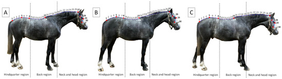
Figure 1.
The digitization of landmarks (LDs) using curve landmarking with 30 points, marked by numbers from 1 to 30. Big red points and bold font were related to anatomical points markers, whereas small red points were related to curve points added between those eight. The photographs represent three time points: at rest (A), after warm up (B), and after massage session (C). The dorsal profile was reflected by the blue curve. Dashed lines indicate the boundaries between regions.
Data in a TPS file were saved as a text file in the notebook, which contained: the number of digitized LDs (LM = 30), thirty consecutive two-dimensional numerical coordinates of each LD with a dot separator (e.g., 93.00000 302.00000), input photographs locations on disc (IMAGE), and photographs identifying (ID). The ID information of each photograph contained age (A, B, C, D), height (X, Y, Z), time (0, 1, 2), and technique (DO, VE, DV); and were used to extract new classifiers: age, height, time, and technique in MorphoJ software (version 2.0). In the text file, the 180 records were listed one after the other, so that each of them represented each of the 180 photographs selected for analysis. The text file prepared as an input file to the MorphoJ software.
2.5. Photographs Analysis
To evaluate the horses’ posture, four classifiers were introduced: age, height, time, and technique, as shown in Figure 2. Classifier age was represented by four classes: <5 years (A, including 5 horses); from ≥5 to <10 years (B, including 5 horses); from ≥10 to <15 years (C, including 5 horses); ≥15 years (D, including 5 horses). Classifier height was represented by three classes: <145 cm at the withers (X, including 7 horses); 145–160 cm at the withers (Y, including 7 horses); >160 cm at the withers (Z, including 7 horses). Classifier time was represented by three classes: photographing at rest (0, including 20 horses); photographing after warm up (1, including 20 horses); photographing after massage session (2, including 20 horses). Classifier massage technique was represented by three classes: the dorsal technique (DO, including 20 horses); the ventral technique (VE, including 20 horses), the dorso–ventral technique (DV, including 20 horses).
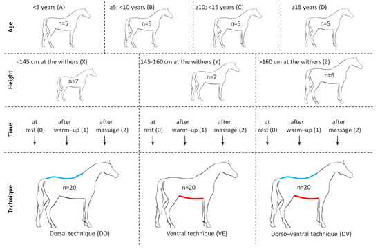
Figure 2.
The scheme of the study design including four studied classifiers: age (<5 years (A); ≥5; <10 years (B); ≥10; <15 years (C); ≥15 years (D)), height (<145 cm at the withers (X); 145–160 cm at the withers (Y); >160 cm at the withers (Z)), time (at rest (0); after warm–up (1); after massage (2)), and technique (dorsal technique marked by the blue line (DO); ventral technique marked by the red line (VE); dorso-ventral technique marked by the blue and red lines (DV)). For each classifier, the name and abbreviations of classes were provided. For each class, the number of horses included was provided.
The distribution of shape configurations corresponding to horses’ postures was visualized with the Generalized Procrustes Analysis (GPA, returning Procrutes coordinates), Covariance matrix (CovMatrix) generation, and principal component analysis (PCA). The classifiers’ (age, height, time, and technique) effect on the size and shape of centroid was determined using the Procrustes ANOVA. The significance level established as p < 0.05. Average observations for each class of two classifiers (time and technique) were executed and displayed as wireframe graphs. All analyses and visualizations were performed using MorphoJ software (Copyright 2008–2019 Christian Peter Klingenberg, Apache License, Version 2.0, https://morphometrics.uk/MorphoJ_guide/frameset.htm?index.htm, accessed on 10 April 2022).
3. Results
The dataset containing 180 observations for 30 LDs in 2 dimensions was analyzed to identify variations between the studied groups. The total variance was 0.0035, while the eigenvalue variance was 0.000000083. Scaling by total variance, the eigenvalue variance scaled by total variance solely was 0.0066. Scaling by total variance and the number of variables was 0.38. Then, eigenvalues, percentages of variance, and cumulative percentages were returned for the first three PCs as follows: PC1: 0.0021, 59.50%, 59.50%; PC2: 0.0005, 14.36%, 73.85%; PC3: 0.0003, 9.01%, 81.92%; PC4: 0.0002, 6.17%, 89.09%; PC5: 0.0001, 2.72%, 91.85% (Figure 3A). No eigenvalue passed the Kaiser rule (eigenvalues > 1) (Figure 3).
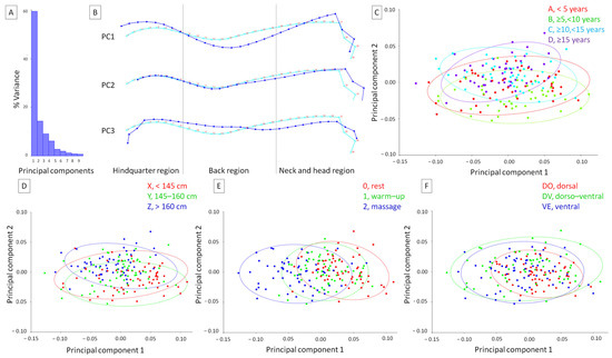
Figure 3.
The dorsal profiles of horses represented by (A) the histogram of variance, (B) the wireframe graph, and (C–F) the scatter plot of the principal components (PCs) scores. On the wireframe graph (B), light blue landmarks (marked by numbers from 1 to 30) and curves represent the consensus horse’s dorsal profile, whereas dark blue landmarks (marked by numbers from 1 to 30) and curves represent the curve deformation for PC1, PC2, and PC3, respectively. Boundaries between regions are indicated with dashed lines. For the general scatter plot of the PC scores (C–F) the color for each group was determined based on the classifier variables: (C) classifier age represented by A, <5 years; B, ≥5; <10 years; C, ≥10; <15 years; D, ≥15 years; (D) classifier height represented by X, <145 cm at the withers; Y, 145–160 cm at the withers; Z, >160 cm at the withers; (E) classifier time represented by 0, photographing at rest; 1, photographing after warm up; 2, photographing after massage session; (F) classifier massage technique represented by DO, the dorsal technique; DV, the dorso–ventral technique; VE, the ventral technique. The confidence ellipses were drawn using a 0.9 probability. The observations were grouped using a classifier as a criterion.
The PC1 curve deformation supported the following dorsal profile: an elevation of dorsal line in the hindquarter region, lowering of dorsal line in the back region, elevation of dorsal line in the neck and head region, dorsal shift of the dorsal arc of atlas, rostro-dorsally shift of the medial canthus of the eye. The PC2 curve deformation supported the following dorsal profile: shortening of dorsal line in the hindquarter region, lowering dorsal line in the back region (only in the withers area), and subtle lowering and elongation in the neck region with the temporomandibular joint, the medial canthus of the eye, and the facial tuberosity of the body of maxilla. The PC3 curve deformation supported the following dorsal profile: a broken and lowered dorsal line in the hindquarter region, elevated and lowered in the lumbar and withers area of back region, respectively, as well as a lowing of dorsal line in the neck region with the facial tuberosity of body of maxillae shifted rostrally (Figure 3B).
It is easy to notice that on the scatter plot of PCs, the scores are partially divided in the PC1 to PC2 orientation. This division reflect the categories of horses, vertically for height classifier (Figure 3D) and horizontally for time classifier (Figure 3E). More horses in a group above 160 cm at the withers represented the PC2-related dorsal profile, whereas more horses photographed at rest represented the PC1-related dorsal profile. A similar separation was not found for classifier age (Figure 3C) and technique (Figure 3F); therefore, the effect of the classifier was further quantified.
The effect of the classifiers on the horse’s dorsal profile indicators was reported in Table 1. The centroid size differed between groups of classifier height (p < 0.0001), and did not differ between groups of classifier age (p = 0.239), time (p = 0.470), and technique (p = 0.164). The shapes differed between groups of classifier time (p < 0.0001) and technique (p < 0.0001), whereas they did not differ between groups of classifier age (p = 0.470) and height (p = 0.402).

Table 1.
The classifier (age, height, time, and technique) effect on the equine dorsal profile indicators (centroid size and shape).
3.1. Dorsal Profiles Comparison within Subgroups of the Photographing Times
The database was divided to three subgroups—0, photographing at rest; 1, photographing after warm up; and 2, photographing after massage session. Each subgroup contains 60 observations. None of the observations were excluded. The remaining criteria effect on both horse’s dorsal profile indicators was tested within each subgroups and reported in Table 2, Table 3 and Table 4. The centroid size differed between groups of classifier height within subgroups 0 (Table 2, p = 0.042), 1 (Table 3, p = 0.021), and 2 (Table 4, p = 0.004), and did not differ between other groups of classifier within all compared subgroup (p > 0.05). The shapes differed between groups of classifier technique only within subgroup 2 (Table 4, p < 0.0001), and did not differ between other groups of classifier within all compared subgroup (p > 0.05).
Table 2.
The classifier (age, height, time, and technique) effect on the equine dorsal profile indicators (centroid size and shape) within subgroup 0 representing photographing at rest.
Table 3.
The classifier (age, height, time, and technique) effect on the equine dorsal profile indicators (centroid size and shape) within subgroup 1 representing photographing after warm up.
Table 4.
The classifier (age, height, time, and technique) effect on the equine dorsal profile indicators (centroid size and shape) within subgroup 2 representing photographing after massage session.
The average observations for the remaining classifiers were displayed in Figure 3, Figure 4 and Figure 5. Within subgroups 0 (Figure 4B), 1 (Figure 5B), and 2 (Figure 6B), the average horses in a group below 145 cm at the withers demonstrated the shortened dorsal line in the hindquarter region, whereas the average horses in a group above 160 cm at the withers were demonstrated elongated dorsal line in this region. Moreover, within subgroups 2 (Figure 6C), the average horses in a group massaged by the dorsal technique were represented by the elevating of the dorsal line in the hindquarter region and lowering dorsal line in the back region. On the other hand, the average horses in a group massaged by the DV technique demonstrated the lowered dorsal line in the hindquarter region and elevated in the back region.
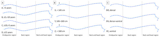
Figure 4.
Average observations of the horse’s dorsal profiles within subgroup 0 representing photographing at rest. The wireframe graphs displayed for the remaining classifier variables: (A) classifier age represented by A, <5 years; B, ≥5; <10 years; C, ≥10; <15 years; D, ≥ 15 years; (B) classifier height represented by X, <145 cm at the withers; Y, 145–160 cm at the withers; Z, >160 cm at the withers; (C) classifier massage technique represented by DO, the dorsal technique; DV, the dorso–ventral technique; VE, the ventral technique. On the wireframe graph, light blue landmarks (marked by numbers from 1 to 30) and curves represent the average dorsal profile of all horses, whereas dark blue landmarks (marked by numbers from 1 to 30) and curves represent the average dorsal profile for displayed classes. Dashed lines indicate the boundaries between regions. Arrows indicate regions where the differences reported in Table 2 are visible.
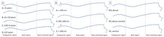
Figure 5.
Average observations of the horse’s dorsal profiles within subgroup 1 representing photographing after warm up. The wireframe graphs displayed for the remaining classifier variables: (A) classifier age represented by A, <5 years; B, ≥5; <10 years; C, ≥10; <15 years; D, ≥ 15 years; (B) classifier height represented by X, <145 cm at the withers; Y, 145–160 cm at the withers; Z, >160 cm at the withers; (C) classifier massage technique represented by DO, the dorsal technique; DV, the dorso–ventral technique; VE, the ventral technique. On the wireframe graph, light blue landmarks (marked by numbers from 1 to 30) and curves represent the average dorsal profile of all horses, whereas dark blue landmarks (marked by numbers from 1 to 30) and curves represent the average dorsal profile for displayed classes. Dashed lines indicate the boundaries between regions. Arrows indicate regions where the differences reported in Table 3 are visible.
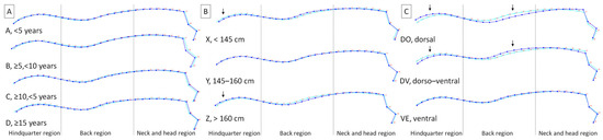
Figure 6.
Average observations of the horse’s dorsal profiles within subgroup 2 representing photographing after massage session. The wireframe graphs displayed for the remaining classifier variables: (A) classifier age represented by A, <5 years; B, ≥5; <10 years; C, ≥10; <15 years; D, ≥ 15 years; (B) classifier height represented by X, <145 cm at the withers; Y, 145–160 cm at the withers; Z, >160 cm at the withers; (C) classifier massage technique represented by DO, the dorsal technique; DV, dorso– the ventral technique; VE, the ventral technique. On the wireframe graph, light blue landmarks (marked by numbers from 1 to 30) and curves represent the average dorsal profile of all horses, whereas dark blue landmarks (marked by numbers from 1 to 30) and curves represent the average dorsal profile for displayed classes. Dashed lines indicate the boundaries between regions. Arrows indicate regions where the differences reported in Table 4 are visible.
3.2. Dorsal Profiles Comparison within Subgroups of the Massage Techniques
The database was divided to three subgroups representing the DO, DV, and VE techniques. Each subgroup contains 60 observations. None of the observations were excluded. The remaining criteria effect on both horse’s dorsal profile indicators was tested within each subgroups and reported in Table 5, Table 6 and Table 7. The centroid size differed between groups of classifier height within subgroups DO (Table 5, p = 0.029), DV (Table 6, p = 0.002), and VE (Table 7, p < 0.0001), and did not differ between other groups of classifier within all compared subgroup (p > 0.05). The shapes differed between groups of classifier time within subgroups DO (Table 5, p = 0.020), DV (Table 6, p = 0.025), and VE (Table 7, p = 0.030), and did not differ between other groups of classifier within all compared subgroup (p > 0.05).
Table 5.
The classifier (age, height, time, and technique) effect on the equine dorsal profile indicators (centroid size and shape) within subgroup DO representing the dorsal massage technique.
Table 6.
The classifier (age, height, time, and technique) effect on the equine dorsal profile indicators (centroid size and shape) within subgroup DV representing the dorso–ventral massage technique.
Table 7.
The classifier (age, height, time, and technique) effect on the equine dorsal profile indicators (centroid size and shape) within subgroup VE representing the ventral massage technique.
Average observations for the remaining classifiers were shown in Figure 7, Figure 8 and Figure 9. Within subgroups DO (Figure 7B), DV (Figure 8B), and VE (Figure 9B), the average horses in a group below 145 cm at the withers demonstrated the shorted dorsal line in the hindquarter region, whereas the average horses in a group above 160 cm at the withers demonstrated elongated dorsal line in the this region. Moreover, within subgroups DO (Figure 7C), the average horses in a group photographing after massage session demonstrated elevated dorsal line in the hindquarter region and lowered dorsal line in the back region. Within subgroups DV (Figure 8C), the average horses in a group photographing after massage session demonstrated lowered dorsal line in the hindquarter region and elevated dorsal line in the back region. Finally, within subgroups VE (Figure 9C), the average horses in a group photographing after massage session demonstrated slightly lowered dorsal line in the hindquarter region and slightly elevated dorsal line in the back region.
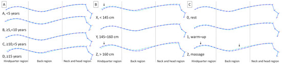
Figure 7.
Average observations of the horse’s dorsal profiles within subgroup DO representing the dorsal massage technique. The wireframe graphs displayed for the remaining classifier variables: (A) classifier age represented by A, <5 years; B, ≥5; <10 years; C, ≥10; <15 years; D, ≥ 15 years; (B) classifier height represented by X, <145 cm at the withers; Y, 145–160 cm at the withers; Z, >160 cm at the withers; (C) classifier time represented by 0, photographing at rest; 1, photographing after warm up; 2, photographing after massage session. On the wireframe graph, light blue landmarks (marked by numbers from 1 to 30) and curves represent the average dorsal profile of all horses, whereas dark blue landmarks (marked by numbers from 1 to 30) and curves represent the average dorsal profile for displayed classes. Dashed lines indicate the boundaries between regions. Arrows indicate regions where the differences reported in Table 5 are visible.
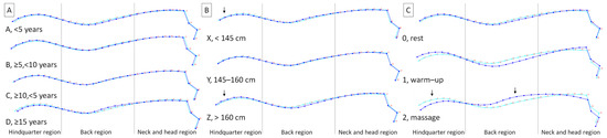
Figure 8.
Average observations of the horse’s dorsal profiles within subgroup DV representing the dorso–ventral massage technique. The wireframe graphs displayed for the remaining classifier variables: (A) classifier age represented by A, <5 years; B, ≥5; <10 years; C, ≥10; <15 years; D, ≥ 15 years; (B) classifier height represented by X, <145 cm at the withers; Y, 145–160 cm at the withers; Z, >160 cm at the withers; (C) classifier time represented by 0, photographing at rest; 1, photographing after warm up; 2, photographing after massage session. On the wireframe graph, light blue landmarks (marked by numbers from 1 to 30) and curves represent the average dorsal profile of all horses, whereas dark blue landmarks (marked by numbers from 1 to 30) and curves represent the average dorsal profile for displayed classes. Dashed lines indicate the boundaries between regions. Arrows indicate regions where the differences reported in Table 6 are visible.
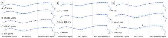
Figure 9.
Average observations of the horse’s dorsal profiles within subgroup VE representing the ventral massage technique. The wireframe graphs displayed for the remaining classifier variables: (A) classifier age represented by A, <5 years; B, ≥5; <10 years; C, ≥10; <15 years; D, ≥ 15 years; (B) classifier height represented by X, <145 cm at the withers; Y, 145–160 cm at the withers; Z, >160 cm at the withers; (C) classifier time represented by 0, photographing at rest; 1, photographing after warm up; 2, photographing after massage session. On the wireframe graph, light blue landmarks (marked by numbers from 1 to 30) and curves represent the average dorsal profile of all horses, whereas dark blue landmarks (marked by numbers from 1 to 30) and curves represent the average dorsal profile for displayed classes. Dashed lines indicate the boundaries between regions. Arrows indicate regions where the differences reported in Table 7 are visible.
4. Discussion
As the data on the massage proceeding mechanism are limited [9], all research on the postural response to the manual massage techniques should be considered preliminary. It has been proven that, among other things, muscle compliance increases, the level of hormones or other physiological parameters also compound, and the quality of circulation improves; however, the results are still inconclusive [9]. The neurological effects may be a result of mechanical stress by altering excitability; however, this has only been proven with the petrissage method [37]. Therefore, in the current study, the visual effect of the three massage techniques was quantified to shown the initial horse’s postural response to used basic equine manual rehabilitation techniques. It was shown that older horses exhibit a decline in body condition, muscle tone and general well-being [38,39]; therefore, classifier ‘age’ was included in the comparison. Moreover, it was shown that horses size affect the size of the geometric morphometrics measurements [21]; therefore, classifier ‘height’ was also considered. Finally, the effect of four classifiers ‘age’, ‘height’, ‘time’, and ‘technique’ on horses’ dorsal profiles was studied here, which seems to be the first equine application of its kind.
In the current study, no effect of the ‘age’ classifier on the geometric morphometrics measurements (the centroid size and shape) was found, which indicates a homogeneous group of the studied horses and their proper maintenance and riding use [19,38]. Since no age-related lowering of the dorsal line [38,39] was observed in older horses (≥15 years), further comparisons can be made on the entire study group of horses as representative. In the current study, a ‘height’ classifier effect on the centroid size but not shape was found. Currently presented results agree with recent findings, showing that the dorsal line of full size horses (mean height at withers 166.79 cm) differed in size with the dorsal line of ponies (mean height at withers 140.57 cm) [21], which should be considered in this and further welfare evaluations dependent on posture [13,19]. The dorsal line shortening effect in the hindquarter region (in a group below 145 cm at the withers) and the elongating of the dorsal line in the hindquarter region (in a group above 160 cm at the withers) was visible for all studied comparisons within subgroups of the photographing times as well as the massage techniques. In the current study, an effect of the ‘time’ classifier on the centroid shape but not size was found. Time-related differences in the horse’s dorsal profile were observed for subgroups DO, VE, and DV, thus regardless of the massage technique used. However, the technique-related differences in the horse’s dorsal profile were noted only for subgroup 2 representing photographing after massage session. The presented results are in line with the previous findings stated that regardless of the manual rehabilitation techniques [9,11,37]. Warm up is a crucial part of pre-training preparation, and is defined as every subdued exercise which precedes greater physical effort as a preparation for the muscle activity [40,41,42]. Warm up helps in muscle preparation and coordination which prevent developing injuries and traumas. It also affects the increase in muscle temperature and metabolism energy, synovial fluid production, blood flow improvement, or tissue elasticity expansion [40]. Warm up may be considered a protective mechanism for the muscle since after the warm up a greater length of stretching and greater strength are needed to produce injury [40]. Warm up is also necessary to obtain the visual massage effects visible in current work as a characteristic change in dorsal line. In the current study, these dorsal line changes were quantified as the ‘technique’ classifier effect on the centroid shape but not size. The horse’s dorsal profile after the dorsal massage technique was supported by elevating of dorsal line in the hindquarter and its lowering in the back region, whereas the horse’s dorsal profile after the dorso–ventral and ventral massage techniques was supported by dorsal line lowering in the hindquarter and its elevating in the back region was considerable and slight, respectively.
In recent studies, massage has been described as beneficial in 10–20 min sessions of various techniques (circular friction, muscle pressure, stroking, muscle shaking, effleurage skin manipulation, tapotement, cross-fiber massage, petrissage, and wringing) [5,6,7,8]. Those kinds of massage techniques decrease muscle hypertonicity and are favorable for soft tissue. After their application, soft tissue mobilization increases as well as soft tissue restrictions and pain decreases [7]. In the current study, the back massage was used to relax the back muscles and the abdominal massage was used to activate them. One may observe that coinstantaneous arousal of abdominal muscles and relaxing back muscles lead to the significant elevation of the horses’ dorsal line. The horses which underwent the dorso–ventral technique were taking the favorable posture with the lowering position of the neck, head, and hindquarter as well as enhancing back. The current results quantify for the first time the general belief of equine physiotherapists and clinicians that simultaneous using back and abdominal techniques of manual mobilization of muscles allow to obtain the most favorable position of the horse’s body [4,5,6,7,8,16,22].
In response to the gap in the quantitative and qualitative evaluation of the effects of massage on horses’ bodies [10], the initial quantification of a horse’s postural response to the first session of the selected massage techniques has been proposed here. The evaluation of horses’ dorsal profiles by the GM method is not complicated [13,19,20,21] and can be done using low-cost equipment. The simplified GM measurement protocol [20,21] requires only non-invasive application of markers in specific places on the horse’s back, taking pictures with any digital camera, and then processing the pictures in an easy to use and free MorphoJ software [43]. Pictures can be obtained immediately before and after the massage without the need to apply sensors (as in the surface electromyography (sEMG) method [44]) or move the horse (as in motion capture systems [45]), which could affect the measurement result. One may judge a MorphoJ software as the easiest standalone software for GM-based shapes and size evaluation [46], it could be introduced to the everyday equine practice. However, also other software packets such as R version 3.1.2 environment [13,19] or tpsUtil, tpsDig, tpsSmall, tpsRelw, and tpsPLS softwares [47] can be successfully used for the objective evaluation of the shape variations in the horses’ postures.
Regardless of the software used, if horse owners and breeders become familiar with the GM method, they will be able to use it in their own stables to evaluate their own horses, which will certainly benefit the equine welfare assessment and equine quality of live. However, one should note sEMG and motion capture remain the reference methods for the quantitative evaluation of the movement of the equine back [44,45,48], and the GM method has some limitations. Regardless of the massage technique used, one may remember that horses’ posture reflects also their emotional and psychological condition [13]. A horse’s specific dorsal profile, with flat or hollow back, may reflects a poor welfare condition and the “depressed-like” posture [13] Thus, the horse’s temporary emotional state could provide a limitation to the presented results. This effect cannot be excluded; however, it can be minimized by conducting the research in a short time-lap and under the same, controlled environmental condition as well as using best methods for the posture evaluation [19], as were done in the current study. One may observe, that the specific dorsal profile typical for poor welfare [13] was different from the specific dorsal profile related to massage, observed in the current study. Another limitation of the current studies that should be noted is the lack of consideration of the effect of horse breed. Although no study to date using GM to assess profile of horses’ back has considered breed as a variable of interest [13,19,21,47] it should be done because of the effect of horse height on centroid size [21]. As discussed above, these studies took into account the height of horses, but the breed groups were too small. Therefore, it should be highlighted that the obtained results represent the preliminary study on the initial horse’s postural response to massage.
The further research should include comparing postural response for the single and multiple massage sessions [10] and comparing horse groups representing different emotional states [13], different conditions of the horse’s back [13,18], different stages of dorsal pain and lack of dorsal pain demonstrated in more than a month of medical history [4,18,32], as well as different breeds and types of usage therein sport and school horses. Considering the second limitation of the study, the utilization of a relatively new research method, the further research mentioned above should also answer the question concerning the applicability of the used methodology.
5. Conclusions
Considering that geometric morphometrics is a relatively new research method, it can be carefully concluded that it allows visualizing the differences in the horses’ posture, which may result from the effect of manual rehabilitation techniques on the dorsal line of a horse’s body. The equine dorsal profiles depicted by GM are affected by classifier ‘height’ in terms of centroid size as well as the classifiers ‘time’ and ‘technique’ in terms of centroid shape. The used quantification of the horse’s body posture gives preliminary indication that horses initially respond to the massage using the dorso–ventral technique and exhibit elements of posture opposite to characteristics of poor welfare.
Author Contributions
Conceptualization, M.M. and M.D.; methodology, M.B., M.M. and M.D.; software, M.M. and M.D.; validation, M.M. and M.D.; formal analysis, M.B., A.Ś., U.S. and M.D.; investigation, M.B., A.Ś., U.S. and M.M.; resources, M.M. and M.D.; data curation, M.M. and M.D.; writing—original draft preparation, M.B., A.Ś, U.S., and M.D.; writing—review and editing, M.B., A.Ś., U.S., M.M. and M.D.; visualization, U.S. and M.D.; supervision, M.M. and M.D.; project administration, M.M. and M.D. All authors have read and agreed to the published version of the manuscript.
Funding
This research received no external funding.
Institutional Review Board Statement
The research, using the results of veterinary clinical examinations, does not fall under the legislation for the protection of animals used for scientific purposes, national decree-law (Dz. U. 2015 poz. 266 and 2010-63-EU directive). No ethical approval was needed according to the list of exemptions of the II Local Committee for Ethics in Animal Research of Warsaw University of Life Sciences-SGGW of 8 August 2022.
Informed Consent Statement
Not applicable.
Data Availability Statement
The data presented in this study are available on request from the corresponding author.
Conflicts of Interest
The authors declare no conflict of interest.
References
- Atalaia, T.; Prazeres, J.; Abrantes, J.; Clayton, H.M. Equine Rehabilitation: A Scoping Review of the Literature. Animals 2021, 11, 1508. [Google Scholar] [CrossRef]
- McGowan, C.M.; Cottriall, S. Introduction to Equine Physical Therapy and Rehabilitation. Vet. Clin. N. Am. Equine Pract. 2016, 32, 1–12. [Google Scholar] [CrossRef]
- Kaneps, A.J. Practical Rehabilitation and Physical Therapy for the General Equine Practitioner. Vet. Clin. N. Am. Equine Pract. 2016, 32, 167–180. [Google Scholar] [CrossRef]
- Haussler, K.K. The role of Manual Therapies in Equine Pain Management. Vet. Clin. N. Am. Equine Pract. 2010, 26, 579–601. [Google Scholar] [CrossRef]
- Goff, L.M. Manual Therapy for the Horse-A Contemporary Perspective. J. Equine Vet. Sci. 2009, 29, 799–808. [Google Scholar] [CrossRef]
- Haussler, K.K. Review of Manual Therapy Techniques in Equine Practice. J. Equine Vet. Sci. 2009, 29, 849–869. [Google Scholar] [CrossRef]
- Haussler, K.K. Equine Manual Therapies in Sport Horse Practice. Vet. Clin. N. Am. Equine Pract. 2018, 34, 375–389. [Google Scholar] [CrossRef] [PubMed]
- Triano, J. The theoretical basis for spinal manipulation. In Principles and Practice of Chiropractic, 3rd ed.; Haldeman, S., Ed.; McGraw-Hill: New York, NY, USA, 2005; pp. 361–381. [Google Scholar]
- Goff, L. Manual therapy and exercise for athletic horses. In Equine Sports Medicine and Surgery; Elsevier: Amsterdam, The Netherlands, 2014; pp. 1217–1224. [Google Scholar]
- Haussler, K.K.; Hesbach, A.L.; Romano, L.; Goff, L.; Bergh, A. A Systematic Review of Musculoskeletal Mobilization and Manipulation Techniques Used in Veterinary Medicine. Animals 2021, 11, 2787. [Google Scholar] [CrossRef]
- Haussler, K.; Martin, C.; Hill, A. Efficacy of spinal manipulation and mobilisation on trunk flexibility and stiffness in horses: A randomized clinical trial. Equine. Vet. J. 2010, 42 (Suppl. 38), 695–702. [Google Scholar] [CrossRef] [PubMed]
- Kędzierski, W.; Janczarek, I.; Stachurska, A.; Wilk, I. Comparison of effects of different relaxing massage frequencies and different music hours on reducing stress level in race horses. J. Equine Vet. Sci. 2017, 53, 100–107. [Google Scholar] [CrossRef]
- Sénèque, E.; Lesimple, C.; Morisset, S.; Hausberger, M. Could posture reflect welfare state? A study using geometric morphometrics in riding school horses. PLoS ONE 2019, 14, e0211852. [Google Scholar] [CrossRef] [PubMed]
- Ridgway, K.; Harman, J. Equine back rehabilitation. Vet. Clin. N. Am. Equine Pract. 1999, 15, 263–280. [Google Scholar] [CrossRef]
- Cook, W.R. Pathophysiology of bit control in the horse. J. Equine Vet. Sci. 1999, 19, 196–204. [Google Scholar] [CrossRef]
- Fureix, C.; Jego, P.; Henry, S.; Lansade, L.; Hausberger, M. Towards an ethological animal model of depression? A study on horses. PLoS ONE 2012, 7, e39280. [Google Scholar] [CrossRef] [PubMed]
- Hausberger, M.; Fureix, C.; Lesimple, C. Detecting horses’ sickness: In search of visible signs. Appl. Anim. Behav. Sci. 2016, 175, 41–49. [Google Scholar] [CrossRef]
- Lesimple, C.; Fureix, C.; De Margerie, E.; Sénèque, E.; Menguy, H.; Hausberger, M. Towards a postural indicator of back pain in horses (Equus caballus). PLoS ONE 2012, 7, e44604. [Google Scholar] [CrossRef]
- Sénèque, E.; Morisset, S.; Lesimple, C.; Hausberger, M. Testing optimal methods to compare horse postures using geometric morphometrics. PLoS ONE 2018, 13, e0204208. [Google Scholar] [CrossRef] [PubMed]
- Maśko, M.; Wierzbicka, M.; Zdrojkowski, Ł.; Jasiński, T.; Pawliński, B.; Domino, M. Characteristics of the Donkey’s Dorsal Profile in Relation to Its Functional Body Condition Assessment. Animals 2021, 11, 3095. [Google Scholar] [CrossRef]
- Maśko, M.; Wierzbicka, M.; Zdrojkowski, Ł.; Jasiński, T.; Sikorska, U.; Pawliński, B.; Domino, M. Comparison of Donkey, Pony, and Horse Dorsal Profiles and Head Shapes Using Geometric Morphometrics. Animals 2022, 12, 931. [Google Scholar] [CrossRef]
- Fureix, C.; Hausberger, M.; Sénèque, E.; Morisset, S.; Baylac, R.; Cornette, R.; Biquand, V.; Deleporte, P. Geometric morphometrics as a tool for improving the comparative study of behavioural postures. Naturwissenschaften 2011, 98, 583–592. [Google Scholar] [CrossRef]
- Xin, H. Assessing swine thermal comfort by image analysis of postural behaviors. J. Anim. Sci. 1999, 77, 1–9. [Google Scholar] [CrossRef]
- Schilder, M.B.; van der Borg, J.A. Training dogs with help of the shock collar: Short and long term behavioral effects. Appl. Anim. Behav. Sci. 2004, 85, 319–334. [Google Scholar] [CrossRef]
- Wilson, R.H. Agonistic postures and latency to the first interaction during initial pair encounters in the red jungle fowl, Gallus gallus. Anim. Behav. 1974, 22, 75–82. [Google Scholar] [CrossRef]
- Davies, W.G. Cluster analysis applied to the classification of postures in the Chilean Flamingo (Phoenicopterus chilensis). Anim. Behav. 1978, 26, 381–388. [Google Scholar] [CrossRef]
- Beerda, B.; Schilder, M.B.H.; Van Hooff, J.A.R.A.M.; De Vries, H.W.; Mol, J.A. Chronic stress in dogs subjected to social and spatial restriction. I. Behavioral responses. Physiol. Behav. 1999, 66, 233–242. [Google Scholar] [CrossRef]
- Huzzey, J.M.; von Keyserlingk, M.A.G.; Weary, D.M. Changes in feeding, drinking, and standing behavior of dairy cows during the transition period. J. Dairy Sci. 2005, 88, 2454–2461. [Google Scholar] [CrossRef] [PubMed]
- Krawczel, P.D.; Hill, C.T.; Dann, H.M.; Grant, R.J. Effect of stocking density on indices of cow comfort. J. Dairy Sci. 2008, 91, 1903–1907. [Google Scholar] [CrossRef] [PubMed]
- Costa, L.R. History and Physical Examination of the Horse. In Manual of Clinical Procedures in the Horse; Wiley-Blackwell: Hoboken, NJ, USA, 2017; pp. 27–58. [Google Scholar]
- Davidson, E.J. Lameness evaluation of the athletic horse. Vet. Clin. Equine Pract. 2018, 34, 181–191. [Google Scholar] [CrossRef]
- Martin, B.B., Jr.; Klide, A.M. Physical examination of horses with back pain. Vet. Clin. N. Am. Equine Pract. 1999, 15, 61–70. [Google Scholar] [CrossRef] [PubMed]
- Wojtecka, J.; Albera-Łojek, A.; Łojek, J. Masaż i stretching w treningu i rehabilitacji koni. Wiadomości Zootech. 2018, 56, 108–123. [Google Scholar]
- Slijper, E. Comparative biologic anatomical investigations on the vertebral column and spinal musculature of mammals. Tweede Sect. 1946, 17, 1–128. [Google Scholar]
- Nickel, R.; Schummer, A.; Seiferle, E.; Wilkens, H.; Wille, K.H.; Frewein, J. The Anatomy of the Domestic Animals—The Locomotor System of the Domestic Mammals; Verlag Paul Parey: Berlin, Germany, 1986; Volume 1. [Google Scholar]
- Dyce, K.M.; Sack, W.O.; Wensing, C.J.G. The neck, back, and vertebral column of the horse. In Dyce, Sack, and Wensing’s Textbook of Veterinary Anatomy, 5th ed.; Singh, B., Ed.; Saunders: Philadelphia, PA, USA, 2017. [Google Scholar]
- Weerapong, P.; Hume, P.; Kolt, G. The mechanisms of massage and effects on performance, muscle recovery and injury prevention. Sport. Med. 2005, 35, 235–256. [Google Scholar] [CrossRef]
- Burattini, B.; Fenner, K.; Anzulewicz, A.; Romness, N.; McKenzie, J.; Wilson, B.; McGreevy, P. Age-related changes in the behaviour of domestic horses as reported by owners. Animals 2020, 10, 2321. [Google Scholar] [CrossRef]
- Bollwein, A.; Hänichen, T. Age-related changes in the intervertebral disks of the cervical vertebrae of the horse. Tierarztl. Prax. 1989, 17, 73–76. [Google Scholar]
- Farinelli, F.; de Rezende, A.S.C.; Fonseca, M.G.; Lana, Â.M.Q.; Leme, F.D.O.P.; Klein, B.D.O.N.; Silva, R.H.P.; de Abreu, A.P.; de Jesus Damazio, M.; Melo, M.M. Influence of Stretching Exercises, Warm-Up, or Cool-Down on the Physical Performance of Mangalarga Marchador Horses. J. Equine Vet. Sci. 2021, 106, 103714. [Google Scholar] [CrossRef] [PubMed]
- Weineck, J.; Carvalho, B.M.R.; Barbanto, V.J. Treinamento Ideal: Instruções Téc- Nicas Sobre O Desempenho Fisiológico, Incluindo Considerações Específicas de Treinamento Infantil E Juvenil; Manole: Barueri, Brazil, 1999; Volume 1, pp. 1–740. [Google Scholar]
- Mcardle, W.D.; Katch, F.I.; Katch, V.L. Fisiologia Do Exercício: Nutrição, Energia E Desempenho Humano, 7th ed.; Giuseppe, T., Ed.; Guanabara Koogan: Rio Janeiro, Brazil, 2011; Volume 83, p. 3322. [Google Scholar]
- Klingenberg, C.P. MorphoJ: An integrated software package for geometric morphometrics. Mol. Ecol. Resour. 2011, 11, 353–357. [Google Scholar] [CrossRef] [PubMed]
- Gamucci, F.; Pallante, M.; Molle, S.; Merlo, E.; Bertuglia, A. A Preliminary Study on the Use of HD-sEMG for the Functional Imaging of Equine Superficial Muscle Activation during Dynamic Mobilization Exercises. Animals 2022, 12, 785. [Google Scholar] [CrossRef] [PubMed]
- Egan, S.; Brama, P.; McGrath, D. Research trends in equine movement analysis, future opportunities and potential barriers in the digital age: A scoping review from 1978 to 2018. Equine Vet. J. 2019, 51, 813–824. [Google Scholar] [CrossRef] [PubMed]
- Savriama, Y. A step-by-step guide for geometric morphometrics of floral symmetry. Front. Plant Sci. 2018, 9, 1433. [Google Scholar] [CrossRef]
- Raspa, F.; Roggero, A.; Palestrini, C.; Marten Canavesio, M.; Bergero, D.; Valle, E. Studying the shape variations of the back, the neck, and the mandibular angle of horses depending on specific feeding postures using geometric morphometrics. Animals 2021, 11, 763. [Google Scholar] [CrossRef]
- Pfau, T. Sensor-based equine gait analysis: More than meets the eye? UK-Vet Equine 2019, 3, 102–112. [Google Scholar] [CrossRef]


Disclaimer/Publisher’s Note: The statements, opinions and data contained in all publications are solely those of the individual author(s) and contributor(s) and not of MDPI and/or the editor(s). MDPI and/or the editor(s) disclaim responsibility for any injury to people or property resulting from any ideas, methods, instructions or products referred to in the content. |
© 2023 by the authors. Licensee MDPI, Basel, Switzerland. This article is an open access article distributed under the terms and conditions of the Creative Commons Attribution (CC BY) license (https://creativecommons.org/licenses/by/4.0/).