Article

Physiological and Ultrastructural Changes in Dendranthema morifolium Cultivars Exposed to Different Cadmium Stress Conditions

1
College of Landscape Architecture, Northeast Forestry University, Harbin 150040, China
2
Agricultural Research Station, Charsadda 24520, Pakistan
3
Laboratory of Phytochemistry, Department of Botany, University of São Paulo, São Paulo 05508-090, Brazil
4
Department of Zoology, College of Science, King Saud University, Riaydh P.O. Box 2455, Saudi Arabia
5
Department of Horticulture and Life Science, Yeungnam University, Gyeongsan 38541, Republic of Korea
*
Authors to whom correspondence should be addressed.
Agriculture 2023, 13(2), 317; https://doi.org/10.3390/agriculture13020317
Submission received: 2 January 2023 / Revised: 20 January 2023 / Accepted: 25 January 2023 / Published: 28 January 2023

Abstract

:
Ornamental plant species may vary substantially in their tolerance response to heavy metals. The aim of this research was to check chrysanthemum cultivars, namely Donglin Ruixue (C), Yellow (F), Red pocket (G), and New 9714 (I), which are commonly used as landscape plants to determine their levels of cadmium (Cd) tolerance at different cadmium concentrations through hydroponic cultures. Chrysanthemum cultivars were treated with five different Cd concentrations (0, 10, 20, 50, and 100 mg L−1) and different physiological, enzymatic, and ultra-structure traits were taken under consideration in vitro. The results showed that cadmium concentration significantly inhibited the total chlorophyll content, chlorophyll a, chlorophyll b, and carotenoid content. Chlorophyll contents were significantly reduced at higher Cd concentrations in all cultivars, but the reduction rates were higher in cultivar F (59.49%), G (40.41%), I (44.97%), and C (33.86%). Similarly, the chlorophyll b reduction was higher than that of chlorophyll a in I (73.33%), followed by G (58.06%), F (61.66%), and C (32.43%), under Cd stress conditions. Additionally, the relative conductivity was recorded in cultivars C (146.48%), F (223.66%), G (165.96%), and I (154.92%), respectively, at 100 mg L−1 Cd concentrations. Likewise, MDA was significantly increased with high Cd stress, at 155.56, 325.27, 173.91, and 322.18%, in C, F, G, and I cultivars at 100 mg L−1, but it was promoted with a greater increase in F and I cultivars. Similarly, SOD and CAT activities were increased with the increase in Cd stress, but reduced in F and I cultivars at higher stress levels of 100 mg L−1. In the same way, POD activity was significantly higher in the C and G cultivars. Additionally, ultrastructure changes also occurred with the increase in the Cd stress, i.e., 20 mg L−1 to 100 mg L−1, and these changes caused alterations in cell organelles, including in the chloroplast, grana, lamella, thylakoid, and stroma. They also caused noticeable damage to mitochondria at higher Cd concentrations. It was concluded that the higher levels of antioxidative defense of the C and G cultivars of chrysanthemum indicated their ability to tolerate high Cd stress conditions. These could, therefore, be used for their phytoremediation potential in Cd-contaminated areas.

1. Introduction

Due to the rapid modernization and development of agriculture sectors as well as the rise in chemical industries, soil contamination with heavy metals (HM) has become a serious challenge for the modern world [1,2,3]. It has been taken into consideration that the urban areas are highly prone to heavy metals as compared to rural due to unrestricted release of heavy metals into the natural environment. These HMs, released via the smelting, electroplating, and chemical industries, are one of the most serious threats to life in the 21st century owing to their toxic and potentially carcinogenic nature [4,5]. Studies have estimated that approximately 20.7–543.3 losses occur in the crop plants. Heavy metals are grouped into essential and nonessential micronutrients for normal plant growth [6]. Unlike other heavy metals, cobalt (Co), zinc (Zn), nickel (Ni), copper (Cu), lead (Pb), silver (Ag) and cadmium (Cd) are non-essential elements in plants nutrition. Among all of these, a few metals, such as Cd, are highly toxic to microbes, animals, plants, and human beings, even at very low concentrations [7,8,9]. Cadmium (Cd) is the most toxic element, ranked 7th among the top 20 toxic elements, and pollutes the environment more effectively due to its immense mobility and quick absorption in soil–plant systems [10,11].
Cadmium (Cd) negatively affects chlorophyll and carotenoid contents, due to which the photosynthetic ability of plants is reduced. This causes various diseases, such as chlorosis, which leads to a sharp reduction in plant growth and development [12,13,14]. In the same way, Cd not only affects the enzymatic, photosynthetic, and physiological traits, but also the leaf ultrastructure of plant species such as bush beans (Phaseolus vulgaris L.) [15], Elodea Canadensis vecchia, rocca [16], and pea plants (Pisum Sativum L.) [17]. The ultrastructural changes in leaf cells have been well-documented; these variations include an increased number of vacuoles and nucleoli, reduction in the mitochondrial cristae, and severe plasmolysis [18]. In the same way, changes are also being taken into consideration in the genetic make-up of different varieties of plants—for example, highly condensed chromatin materials and misarrangement of chromosomes. Other changes involve the enlargement of vacuoles and chloroplast structures [19,20,21], which has rarely been reported in chrysanthemum plants. Plant species are also reported to be extremely reactive and to immediately damage cell organelles through peroxidation [22]. In such conditions, plants must undergo various enzymatic changes and activate various mechanisms, such as binding sequester biomolecules or synthesizing antioxidant molecules, to avoid or minimize the stressful effects of these radicals, which are generated under stress conditions [23,24,25].
Among the other heavy metals (HM), Cd is one of the most toxic, and leads directly to the formation of reactive oxygen species (ROSs) [14,26]. These ROSs appear in the form of hydrogen peroxide (H2O2), singlet oxygen (1O2), hydroxyl radical (OH), and superoxide radicles (O2−) [27,28]. These ROSs cause adverse effects to different physiological, biochemical, enzymatic, anatomical, and molecular changes in plants [29]. Interestingly, plants have evolved various enzymatic and non-enzymatic mechanisms to scavenge such ROSs and thereby alleviate its deleterious effects. These protective enzymes include catalase (CAT), peroxidases (POD), and superoxide dismutase (SOD), while several molecules, such as glutathione, ascorbate, and carotenoids, provide non-enzymatic protection [30]. Among these, SOD is a major O2scavenger, and its enzymatic action results in H2O2 and O2 formation. In the same way, CAT and several classes of POD then scavenge the H2O2 that is produced. CAT dismutase H2O2 transforms into H2O and O2, and is found in peroxisomes, cytosol, and mitochondria [31,32]. POD decomposes H2O2 by oxidation of co-substrates such as phenolic compounds and/or antioxidants [33]. Under normal circumstances, the concentration of oxygen radicals remains low because of the activity of these antioxidative enzymes. Under stress conditions, the free radical species (forms of active oxygen) may be increased, which enhances the activities of these detoxifying enzymes [34].
Chrysanthemum indicum L. is an abundant ornamental bioresource around the globe, and its export was observed to increase from 0.5 billion in 1995 to 5.1 billion in 2005. It is expected to double again by 2025. China is the second leading country in chrysanthemum production, with a total area of 59,527 hectares. It grows in polluted areas surrounding cities [35], and important landscape species have been reported to grow in these polluted areas [36]. Moreover, it is an excellent ornamental plant for economical greening of the urban landscape, and possesses key traits that make it very attractive for use in metal-polluted sites [37], e.g., ease of cultivation, rapid growth, abundant and long flowering period, and short life cycle. It has been taken into consideration that chrysanthemum and its different varieties, e.g., Donglin Ruixue, Yellow, Red pocket, and New 9714, are potentially suitable and easy to grow for various purposes in China. Interestingly, there are few studies that have evaluated chrysanthemum regarding heavy metals, e.g., Pb [38], Cd [39], Cd-Ni [40], Cu [41], and Cd-Zn. However, the physiological and tolerance mechanisms that respond to different Cd levels among the chrysanthemum cultivars have rarely been explored. Therefore, the present research study was conducted with the aim to explore Cd’s effect on different physiological functions, enzymatic parameters, and ultra-structures, as well as to find the most efficient Cd-tolerant cultivars of chrysanthemum.

2. Materials and Methods

2.1. Plant Materials

The research experiment was performed in the greenhouse of the College of Landscape Architecture, Northeast Forestry University, Harbin, China. Four cultivars of chrysanthemum were properly purchased from local market, namely Donglin Ruixue (C), Yellow (F), Red pocket (G), and New 9714 (I), respectively, and used for the study, as they have differences in terms of their appearance and growth.
Chrysanthemum seedlings were raised in the experimental nursery of Northeast Forestry University, Harbin. Seedlings of uniform sizes were selected for the experiment; they were 10–15 cm in height, and were cultured for 3 days using distilled water in a 1L plastic box with a foam board for fixing. The plants were incubated with a light intensity of 5000 LX for 12 h per day, and the temperature was controlled at 28 and 18 °C during the day and night, respectively. After 3 days of pre-culture, the distilled water was adjusted to Hoagland nutrient solution and the pH value was adjusted to 5.8 and maintained for 24 h. The nutrient solution was changed after every three days without changing other conditions, and then collected into a container for recycling. The percent compositions of Hoagland solution used are given in Table 1. All the mineral nutrients used to analyze the pure chemical reagents were prepared in distilled water.

2.2. Cadmium Treatment

A total of 4 varieties were tested against 20 mg L−1 and subjected to cluster analysis, and four tolerant varieties were selected on the basis of their tolerance before the beginning of this experiment. The mineral salt CdCl2•2.5H2O was used to prepare different Cd concentration solutions. For this experiment, 5 cadmium concentration stresses were used, which ranged used from 0 to 100 mg L−1. There were five cadmium treatments in total, including 0, 10, 20, 50, and 100 mg L−1, respectively. Control plants were established under the same conditions, but received only Hoaglands nutrient solution. There were 20 seedlings for each treatment, and these were exposed to Cd doses at high concentrations for 10 days to avoid the death of the plants on the basis of their symptoms. The experiment was repeated three times for each treatment. Chrysanthemum seedlings treated with different Cd concentrations were harvested after 10 days. Then, 3–5 fully expanded leaves from plants of each variety were selected from top to bottom and rinsed with tap water, followed by deionized water, 3 times.

2.3. Effect of Cd on Physiological Attributes

2.3.1. Chlorophyll Determination

Based on Duxbury and Yentsch, [42] method, different photosynthetic attributes, such as Chlorophyll a, chlorophyll b, and carotenoid content, were analyzed. Fresh and fully expended leaves (0.2 g) were taken and homogenized in 2 mL of 80% acetone using a mortar and pestle. More solvent was added, and a final volume of up to 7 mL was reached. The samples were centrifuged at 14,000 rpm for 5 min at 4 °C. The supernatants were taken into consideration, and absorbance were measured at 480, 645, and 663 nm, respectively, using a spectrophotometer (Chemito UV, 2000). Chlorophyll a and b, as well as carotenoid content were calculated using the following formula.
Chlorophyll a (mg/g fresh weight) = 12.3D663 − 0.86D663𝑑 × 100 × w × v
Chlorophyll, b (mg/g fresh weight) = 12.3D663 – 0.86D645d × 1000 × w × v
Carotenoid (mg/g fresh weight) = 7.6D480 − 1.49D510d × 1000 × w× v.
where w = fresh weight, v = volume of filter solution, and D =dilution factor.

2.3.2. MDA Contents

Malondialdehyde (MDA) content was determined in the seedlings exposed to different Cd stress conditions [43]. Fully expended and fresh leaf samples of 0.2 g were properly homogenized in 10 mL of 10% trichloroacetic acid and centrifuged at 12,000 rpm for 10 min. After centrifugation, the 2 mL supernatants were taken and added to 2 mL of 0.6% thiobarbituric acid (TBA), then incubated in a water bath for 15 min at 100 °C. The mixture was then cooled down, and centrifugation was carried out at 12,000 rpm for 10 min. The supernatants were taken into consideration, and OD was measured at 532, 600, and 450 nm using a spectrophotometer (Chemito UV, 2000). The unities were expressed as mg g−1 (FW Wang and Zhou) [44].

2.3.3. Relative Electrical Conductivity (REC)

Fresh leaves were taken from both control and Cd-treated seedlings and rinsed with double distal water [45]. Five rounded sections were properly cut from the collected leaves and ultimately kept in autoclaved beaker with 30 mL of deionized water under vacuum conditions for 15 min. The first readings were taken into consideration and marked as S1. Additionally, leaf samples were then heated at 90 °C for 20 min and simultaneously cooled down at room temperature. In the same way, electrical conductivity was measured and marked as S2. By using the following formula, the relative conductivity (REC) was calculated:
REC = (S1/S2) × 100%

2.4. Antioxidant Enzymes in Chrysanthemum Seedlings under Cd Stress

Different antioxidant enzyme profiles, such as peroxidase (POD), catalase (CAT), and superoxide dismutase (SOD), were determined in the seedlings exposed to Cd stress condition by using a spectrophotometer (Chemito UV, 2000).
An SOD bioassay was taken into consideration according to the method described previously [46]. The reaction mixture consisted of 50 mmol/L Tris-HCl buffer (pH 7.8), 0.1 mmol/L EDTA, 0.1 mmol/L nitro blue tetrazolium (NBT), 13.37 mmol/L methionine, and 0.1 mmol/L riboflavin and enzyme extract. The reaction was initiated by adding the riboflavin. The mixture was first placed under light, then transferred into darkness immediately, and the absorbance recorded at 560 nm. One unit of SOD activity was defined as the amount of enzyme that inhibited 50% of NBT photo reduction.
Peroxidase dismutase activity was monitored [47]. The mixture was composed of phosphate buffer, 125 mM, with a pH of 6.8, pyrogallol 50 mM, H2O2 50 nM, and 1 mL of diluted enzyme extracts. The whole mixture was incubated for 5 min at 25 °C. The reaction was stopped by adding 0.5 mL of H2SO4 (5%). The amount of purpurogallin formed in the sample was taken into consideration at 420 nm using a spectrophotometer.
In the same way, moto catalase (CAT) activity was taken into consideration in the seedlings exposed to different Cd concentrations [48]. The whole reaction mixture was composed of 20 μL enzyme extract and a total of 0.980 mL of 50 mM potassium phosphate buffer, with pH 7.0 and 20 mM H2O2. The mixture was then incubated at 28 °C for 1 min, and the absorbance was taken into consideration at 240 nm.

2.5. Ultrastructure Characteristics of Chrysanthemum’ F Variety

Fresh leaves of each treatment were taken from top to bottom and washed with deionized water, then cut into 1 × 3 mm samples, avoiding the veins [49]. Samples were treated with 2.5% glutaraldehyde after 2 h; the samples were removed and rinsed 3 times with 0.1 M phosphate buffer solution (pH 6.8) for 15 min. The samples were then treated with 1% perosmic acid anhydride as the post-fixation solution for 2–3 h, followed by rinsing 3 times with 0.1 M phosphate buffer solution for 15 min. The prepared samples were then treated with ethanol 30 %, 70%, and 90%, one by one, for 15–20 min to dehydrate them, followed by treatment with 100% ethanol 2 times for 10 min. The samples were further dehydrated with a 1:1 mixture of alcohol and acetone for 10 min. All of these steps were carried out in a refrigerator at 4 °C. When the dehydrating agent changed, the samples were immediately put into the refrigerator to meet the temperature requirements each time. Finally, the samples were dehydrated with 100% acetone for 10 min at room temperature. The permeation was carried out with pure acetone: epoxy resin (epoxy resin 812) in a ratio of 1:1, 1:2, and 1:3 for 0.4–1 h, 2–5 h, and overnight, respectively. The prepared samples were soaked for a couple of hours and finally cut into 50–60 nm slices with a thin-slicing machine. Finally, samples were stained with uranyl acetate and lead citrate and were examined using a transmission electron microscope (Hitachi H-7650).

2.6. Statistical Analysis

Data were recorded in triplicate and statistical analyses were performed using SPSS software (Version 11.5 SPSS Inc., Chicago, IL, USA). The data were analyzed by one way analysis of variance (ANOVA), and their mean values were calculated from the three replicates. Their differences were determined using the least squares deviations (LSD) test [2].
Percent   inhibition   ( % ) = Control   Treatment   × 100 Control

3. Results

3.1. Chlorophyll and Carotenoid Contents

The chlorophyll and carotenoid content of the given cultivars were significantly affected by Cd concentrations (p ≤ 0.05) (Table 2). Chlorophyll, chlorophyll a, and carotenoid contents were significantly reduced in all cultivars with the increase in Cd concentration, except chlorophyll b in cultivar C, as compared to control. The chlorophyll contents were reduced in cultivar F (59.49%), G (40.41%), I (44.97%), and C (33.86%), respectively, at 100 mg L−1 Cd concentration. Interestingly, the chlorophyll content reduction was recorded as 33.86% for cultivar C, which indicated better Cd tolerance, while the F cultivar was relatively weak in terms of resisting Cd stress. Chlorophyll a content reductions were recorded in F (57.18%), C (34.4%), G (26.50%), and I (36.21%), respectively, at a 100 mg L−1 Cd concentration, as compared to control. On the other hand, the maximum reduction in chlorophyll b contents was recorded in cultivar I (73.33%), followed by G (58.06%), F (61.66%), and C (32.43%), as compared to the control. For carotenoids, the carotenoid contents of cultivars F and I increased gradually with the increase in Cd concentration, whereas it increased initially in the C and G cultivars, then decreased, and finally increased again. The chlorophyll a and b (a/b) ratios were higher, ranging from 0.99 to 3.24 at 100 mg L−1 Cd concentration indicated that the reduction in chlorophyll b was higher as compared to chlorophyll a; however, there were non-significant changes observed for C, F, and G cultivars.

3.2. Cadmium’s Effect on Physiological Traits

3.2.1. Relative Electrolytic Conductivity (REC)

The relative conductivity of the leaves of chrysanthemum cultivars was significantly affected by Cd concentration (p ≤ 0.05) (Figure 1A). The relative electric conductivity was recorded to be higher at high Cd concentrations as compared to the controls. The relative conductivity was recorded in cultivars C (146.48%), F (223.66%), G (165.96%), and I (154.92%), respectively, at 100 mg L−1 Cd concentrations.

3.2.2. MDA Contents

MDA content were significantly affected in cultivars by Cd, and showed an increasing trend with the increase in Cd concentration (p ≤ 0.05) (Figure 1A). The MDA content was recorded as the maximum in F and I cultivars, while it was relatively lower in C and G cultivars under high Cd concentrations. MDA content was recorded as 155.56, 325.27, 173.91, and 322.18% in the C, F, G, and I cultivars at 100 mg L−1 Cd as compared to the controls.

3.3. Cadmium Effect on Antioxidant Enzymes

3.3.1. Superoxide Dismutase Activity

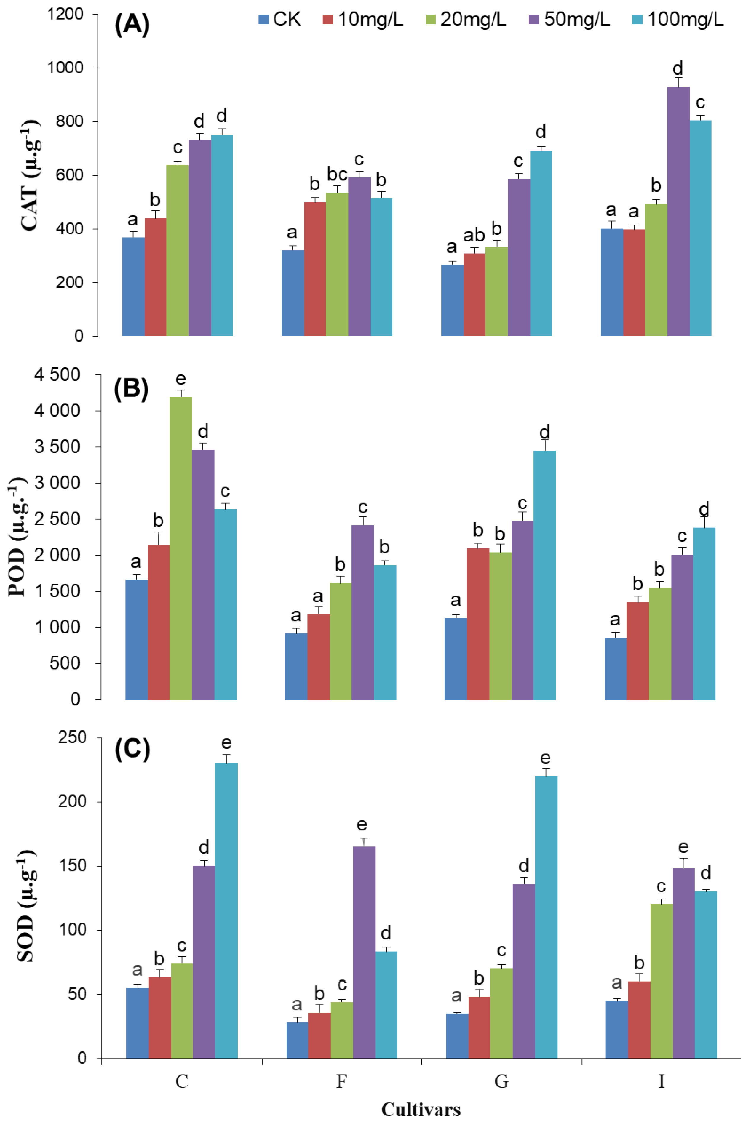
As the first line of defense for plant antioxidant systems, superoxide dismutase (SOD) plays an extremely important role in eliminating and maintaining relatively low levels of reactive oxygen species in plant cells to prevent and reduce the peroxidation process of the membranes and to improve their stress resistance. The SOD showed significant changes in cultivars with Cd treatment (p ≤ 0.05) (Figure 2A). Changes in SOD activity were increased initially, and then decreased with the increase in Cd concentration in the F and I cultivars, respectively, while a notable increase was recorded in the C and G cultivars with the increase in Cd concentration.

3.3.2. Peroxidase Enzyme Activity

Peroxidase (POD) is also an important plant protection enzyme. It has a synergistic effect with SOD, and widely exists in plants. Its activity is relatively high and is closely related to photosynthesis, respiration, and auxin oxidation. The POD activity significantly changed in cultivars with Cd treatment (p ≤ 0.05) (Figure 2B). The POD activity showed an increasing trend initially and then decreased with the increase in Cd concentration in C and F, while a remarkable increase was recorded for cultivars G and I, respectively. The POD activity was recorded at its highest level (4192.52) at 20 mg L−1 Cd for the C cultivar, while it was recorded at its highest (2416.13) for F at 50 mg L−1 Cd, but it decreased at 100 mg L−1. Similarly, the POD activity of the G (3449.73) and I (2378.67) cultivars increased with the increase in Cd concentration, and attained its highest values at 100 mg L−1 Cd. POD activity was recorded at higher levels in the C and G cultivars as a whole when compared with the I and F cultivars.

3.3.3. Catalase Enzyme Activity

Catalase (CAT) activity was significant altered in cultivars with Cd treatment (Figure 2C). The CAT activity was initially increased, but then decreased, in cultivars F and I, respectively, while CAT activity increased with the increase in Cd concentration for the C and G cultivars. CAT activity was recorded at its highest in the F (591.67) and I (928.19) cultivars at 50 mg L−1 Cd concentration, while it was recorded at its highest in the C (751.49) and G (691.98) cultivars, respectively, at 100 mg L−1 Cd concentration.

3.4. Cadmium Effect on Ultrastructure of F Variety

TEM pictures of the control leaves clearly demonstrate that the cell structures are properly arranged. However, there was a gradual increase in cadmium concentration, leading to the deformation of cell ultra-structures (Figure 3). From the TEM pictures (Figure 3), it is clear that distinct features of the cell are gradually distorted with cadmium concentrations from 20 mg L−1 to 100 mg L−1.
In the control group, the cells had intact organelles, such as nuclei, chloroplasts, and mitochondria, as well as a more tightly bound cell wall, a smooth and clear cell membrane, and more oval or fusiform chloroplasts distributed on the edge of the cells, close to the cell wall (Figure 3A).
The internal structure of a chloroplast is clear and complete. The thylakoid is abundant, while the lamellar structure is developed and arranged along the long axis of the chloroplast close to the cell membrane. The chloroplast contains little starch and few osmiophilic particles (Figure 3B). However, the mitochondria are more regularly distributed in the surrounding cells. The structure is complete, and the outer membrane and crest are visible (Figure 3C).
Under 50 mgL−1 Cd concentration, the nucleus was deformed. The nuclear membrane was still clear, but condensation of chromatin in the nucleus was observed, and organelles such as the chloroplast were distributed side by side (Figure 3D). The distribution of chloroplasts was close to the cell wall. With the increase in concentration, the matrix color became more profound, and a significant amount of starch grains appeared in the chloroplast. The chloroplast volume became larger, and the surrounding layers were squeezed (Figure 3E). Similarly, the grana lamellae and stroma were not arranged in order. The mitochondria showed a noticeably damaged and incomplete structure. The mitochondrial inner membrane and outer membrane were mostly dissolved (Figure 3F).
Under 100 mg L−1 Cd concentration, a significant amount of black layered material appeared on the cell wall. The nucleus was deformed and the cell volume was reduced. The chromatin, which was uniformly distributed in the nucleus, also coalesced. The organelles, such as the nuclei and chloroplasts, were no longer distributed along the cell edges. The cells were arched, moved, and suspended in the cytoplasm (Figure 3G). The chloroplasts further became nearly spherical, but the outer envelope remained clear and the matrix color became darker. The number and volume of starch grains increased significantly and occupied most of the chloroplast space, causing crushing (Figure 3I). The mitochondrial membrane structure disappeared, and disintegration and severe dissolution of the outer envelope and the internal crest could be clearly observed, as shown (Figure 3I).

4. Discussion

In the present study, chrysanthemum species exposed to cadmium treatment affected the different physiological and ultrastructure parameters. Chlorophyll and carotenoid contents are essential pigments of photosynthesis, and are synthesized in the chloroplast after photosynthesis. Their metabolism is closely related with chloroplast development [50]. The chlorophyll contents are usually determined in plants, which are considered a marker of the physiological state that is important for assessing the effect of environmental stress. Alterations in the pigment content are linked to plant illness and photosynthetic productivity [51,52]. In our study, chlorophyll content was reduced in all chrysanthemum cultivars after Cd treatment, which is in line with previously reported studies of Phaseolus vulgaris L. [53,54]. However, there were different alteration percentages in chlorophyll a and chlorophyll b in the chrysanthemum cultivars with 100 gm L−1 Cd treatment (Table 2). This alteration, as well as the tolerance differences across genotypes to Cd toxicity, has also been reported previously in species such as peas [55], barley [56], tomatoes [57], and Pisum sativum L. [55]. The chlorophyll content reduction percentage was at its minimum in the C (33.86%) and G (40.1%) cultivars, respectively. The chlorophyll a content was higher than that of chlorophyll b, but chlorophyll b was found to be more sensitive than chlorophyll a to cadmium toxicity. In contrast, Yang et al. reported a greater decrease in chlorophyll a than chlorophyll b in [58]. In our study, carotenoid content was mostly increased with the increase in Cd-treated cultivars (Table 2). It has been previously reported that carotenoid content does not show a set pattern in plants exposed to heavy metals, and it may either decrease or increase. The carotenoid content increase was reported in Cucumis satives L. and Nicotoniana tabacum L. [59,60]. In contrast, a decrease in carotenoid content was also reported in previous studies [61,62].
It has been found that in plants grown in stressed conditions, a number of ROSs are generated in excess and accumulated in the cells, the end product of which is MDA. The increased lipid peroxides are indicators of greater production of toxic oxygen species than normal [63]. In our study, chrysanthemum cultivars showed significant increases in MDA production with increased Cd treatment. However, the increase in MDA content was lower in the C and G cultivars as compared to the F and I cultivars, and not all cultivars showed significant increases at 10 mg L−1; C did not show an increase at 20 mg L−1 Cd (Figure 1A). The lower MDA content in the C and G cultivars implies that these cultivars are better protected from oxidative damage and can rapidly upregulate the antioxidative system, reducing H2O2 levels and collaboration of antioxidant enzyme activities (POD, SOD, and CAT). These results are in line with those previously reported in maize, barley, rice, and mangroves [32,64,65,66].
Environmental stresses induce various types of oxidative stress, thereby generating superoxide radicals, hydrogen peroxide, hydroxyl radicals, and single oxygen, which are collectively commonly known as ROS (reactive oxygen species) [67,68]. These ROSs are actually unstable entities that can rapidly attack all types of biomolecules, such as nucleic acids, amino acids, sugars, fatty acids, and lipid molecules in the cell, leading to irreparable metabolic dysfunction. As a result, cell death occurs [69,70,71]. Consequently, the induction of ant oxidative enzymes such as CAT, SOD, and POD is an important protective mechanism to minimize the oxidative damage in a stress environment. SOD is an important key cellular defensive enzyme against ROS. Its activity changes the relative amount of O2- and H2O2 and decreases the risk of OH radical formation. These are rigorously reactive and cause severe damage to proteins, cell membranes, and DNA. In our study, SOD activity response varies in the cultivars with cadmium treatment (Figure 2C). SOD activity of cultivars C and G increased with the increase in Cd, and their response was stronger, suggesting that this increase in the SOD leads to better protection against oxidant damage [72]. However, SOD activity of cultivars F and I sharply decreased at 100 mg kg−1. This decline in the SOD activity at 100 mg kg−1 indicated that the oxygen scavenging function of SOD was impaired, which is in line with the previously reported results [73,74]. Similarly, like other enzymes, POD plays a key role in the plant kingdom by eliminating various ROS under stressed conditions and catalyzing H2O2, depending on oxidation of the substrates. Our study results showed that the POD activity was higher in the C and G than the F and I cultivars, but the POD activity response showed an increasing and decreasing trend (Figure 2B). These differences in the enzyme’s activities are due to differences in the response of the species’ mechanisms to Cd stress [75,76]. Previous studies in other plants have reported increases, decreases, and lack of change in POD activity in response to heavy metal treatment in barley and garlic [32,65]. Furthermore, POD has the potential to directly participate in the biosynthesis of lignin, which helps in strengthening the physical barrier against various heavy metals such as Cd [65]. Interestingly, these POD activities can also increase in the C and G cultivars, which is also an indicator that they can efficiently avoid damage from heavy metals.
H2O2 is the product of SOD, which is formed due to Cd stress conditions in seedlings [77]. H2O2 is highly toxic and must be eliminated or converted to a reasonable form, such as H2O, in subsequent reactions [78,79]. Interestingly, there are a number of enzymes that regulate the H2O2 intracellular and intercellular levels; CAT, APX and GPX are listed as the most important among them. It was found that CAT actively eliminated H2O2 through cleaving it directly to form water and oxygen molecules [80]. However, this was less efficient than POD in H2O2 scavenging due its low potential substrate affinity [81]. In the current investigation, it was taken into consideration that CAT activity may reflect an increasing trend for C and G cultivars; however, this effect declined at higher Cd concentrations (100 mg/kg) in F and I cultivars, respectively. The higher levels of SOD and CAT activity in the C and G cultivars are a clue that their scavenging mechanisms are more effective than in the F and O cultivars. The reason for this could be the CAT potential, which coordinates with SOD activity. Together, they have a central protective role in O2− and H2O2 elimination.
Heavy metals’ effects on the tissues of living organisms include organelles such as chloroplasts, cell membranes, mitochondria, and nuclei [82,83]. When plants undergo Cd stress, they shows a series of abnormal physiological activities that are signs of ultrastructural changes in plants, but the changes and damages reported are dependent on Cd levels [84]. The nucleus is the genetic organizer for the continuity of traits through copying germplasms and cell division. It also controls cell action through the selective expression of genes [85]. Therefore, Cd toxicity damage to the nucleus is more serious than that of the chloroplast. In our study, the nucleus was deformed, cell volume was reduced, and chromatin also coalesced at higher concentrations. These effects are line with previously reported studies [86,87]. Similarly, the ultrastructure of the chloroplast had an obvious response to high Cd concentrations. Many starch granules appeared in the chloroplasts at higher Cd concentrations (50 and 100 mg L−1).
After photosynthesis, the synthesis and transformation of sugar, amino acids, and other essential components in the chloroplast are important sources of energy in plants for various physio-chemical reactions [88]. However, the reduction or complete stoppage of photosynthesis can occur under various heavy metal stress conditions [89,90]. In the same way, the accumulation of a number of starch granules in the chloroplast might be attributed due to the decline in photosynthetic functioning in the target seedlings under higher Cd concentrations. The chlorophyll content reduction and the consequent damage (shape and size) to the ultra-structures of the chloroplasts, i.e., disordered arrangement of grana and lamellae, have also been reported previously [91]. The mitochondria is the main site for intracellular oxidative phosphorylation and synthesis of adenosine triphosphate (ATP), impairing the internal electron transport system and the oxidative phosphorylation cycle. In our study, the mitochondria had noticeable damage and an incomplete structure. Additionally, the mitochondrial inner and outer membranes were dissolved (Figure 3f). The metabolism of aerobic glucose was obstructed, their normal leaf respiration was affected, and their normal photosynthesis deteriorated [92]. The plant mitochondria were more severely damaged at high Cd concentrations, which is inconsistent with previous studies [93].

5. Conclusions

It was taken into consideration that different levels of Cd could cause alterations in the traits of chrysanthemum cultivars, such as photosynthetic machinery and antioxidant metabolism. In the same way, the effect of Cd was also monitored, and it caused damage to the leaf cell ultrastructure in chrysanthemum plants. The intensity of the damage depended on the Cd concentration. The chlorophyll content reduction was significantly higher in the F and I cultivars as compared to the C and G cultivars. The C and G cultivars were equipped with superior antioxidative defenses to adapt to the oxidative stress as compared to the F and I cultivars when exposed to Cd levels. This exposure was associated with significantly higher SOD activity and POD activity in the leaves with Cd treatment. Moreover, the Cd application significantly increased CAT activity in the leaves of the C and G cultivars, which revealed that the antioxidative defense capability of the C and G cultivars might play an important role in Cd tolerance, and indicated their ability to alleviate high concentrations of Cd stress. It can be implied that chrysanthemum and its varieties can be cultivated in areas with high Cd concentration. The results of this investigation allow us to recommend that chrysanthemum can be used for an alternate strategy to remediate Cd contaminants via eco-friendly methods, in order to make the soil suitable for agricultural usage.

Author Contributions

L.M., S., S.A. and A.K.: investigation, methodology, writing—original draft. M.K., S., A.F.A. and L.M. arranged the data and conducted the formal analysis, investigation, and visualization. Y.Z., S.A. and M.H.: writing—original draft, supervision, methodology, editing final draft. All authors have read and agreed to the published version of the manuscript.

Funding

This research was funded by Researchers Supporting Project No. RSP2023R218, King Saud University, Riyadh, Saudi Arabia. And the Fundamental Research Funds for the Central University (2572017EA04) that supported this work.

Institutional Review Board Statement

Not applicable.

Data Availability Statement

Not applicable.

Conflicts of Interest

The authors declare no conflict of interest.

References

  1. Huang, T.; Xiong, B. Space comparison of agricultural green growth in agricultural modernization: Scale and quality. Agriculture 2022, 12, 1067. [Google Scholar] [CrossRef]
  2. Hayat, K.; Khan, J.; Khan, A.; Ullah, S.; Ali, S.; Salahuddin; Fu, Y. Ameliorative Effects of Exogenous Proline on Photosynthetic Attributes, Nutrients Uptake, and Oxidative Stresses under Cadmium in Pigeon Pea (Cajanus cajan L.). Plants 2021, 10, 796. [Google Scholar] [CrossRef] [PubMed]
  3. Hayat, K.; Khan, A.; Bibi, F.; Salahuddin Murad, W.; Fu, Y.; Batiha, G.E.; Alqarni, M.; Khan, A.; Al-Harrasi, A. Effect of Cadmium and Copper Exposure on Growth, Physio-Chemicals and Medicinal Properties of Cajanus cajan L.(Pigeon Pea). Metabolites 2021, 11, 769. [Google Scholar] [CrossRef] [PubMed]
  4. Mohiuddin, K.; Ogawa, Y.; Zakir, H.; Otomo, K.; Shikazono, N. Heavy metals contamination in water and sediments of an urban river in a developing country. Int. J. Environ. Sci. Technol. 2011, 8, 723–736. [Google Scholar] [CrossRef] [Green Version]
  5. Islam, M.S.; Ahmed, M.K.; Raknuzzaman, M.; Habibullah-Al-Mamun, M.; Islam, M.K. Heavy metal pollution in surface water and sediment: A preliminary assessment of an urban river in a developing country. Ecol. Indic. 2015, 48, 282–291. [Google Scholar] [CrossRef]
  6. Ali, I.; Khan, A.; Ali, A.; Ullah, Z.; Dai, D.-Q.; Khan, N.; Khan, A.; Al-Tawaha, A.R.; Sher, H. Iron and zinc micronutrients and soil inoculation of Trichoderma harzianum enhance wheat grain quality and yield. Front. Plant Sci. 2022, 13. [Google Scholar] [CrossRef]
  7. Andresen, E.; Küpper, H. Cadmium Toxicity in Plants; Springer: Dordrecht, The Netherlands, 2013; pp. 395–413. [Google Scholar]
  8. Shah, F.U.R.; Ahmad, N.; Masood, K.R.; Peralta-Videa, J.R.; Ahmad, F.U.D. Heavy Metal Toxicity in Plants; Springer: Dordrecht, The Netherlands, 2010; pp. 71–97. [Google Scholar]
  9. White, P.J.; Pongrac, P. Heavy-Metal Toxicity in Plants. In Plant Stress Physiology; Cabi: Wallingford, UK, 2017; pp. 300–331. [Google Scholar]
  10. Birke, M.; Reimann, C.; Rauch, U.; Ladenberger, A.; Demetriades, A.; Jähne-Klingberg, F.; Oorts, K.; Gosar, M.; Dinelli, E.; Halamić, J. GEMAS: Cadmium distribution and its sources in agricultural and grazing land soil of Europe—Original data versus clr-transformed data. J. Geochem. Explor. 2016, 173, 13–30. [Google Scholar] [CrossRef]
  11. Gill, S.S.; Khan, N.A.; Tuteja, N. Cadmium at high dose perturbs growth, photosynthesis and nitrogen metabolism while at low dose it up regulates sulfur assimilation and antioxidant machinery in garden cress (Lepidium sativum L.). Plant Sci. 2012, 182, 112–120. [Google Scholar] [CrossRef]
  12. Monteiro, M.S.; Santos, C.; Soares, A.M.V.M.; Mann, R.M. Assessment of biomarkers of cadmium stress in lettuce. Ecotoxicol. Environ. Saf. 2009, 72, 811–818. [Google Scholar] [CrossRef]
  13. Perveen, S.; Anis, M. In vitro morphogenic response and metal accumulation in Albizia lebbeck (L.) cultures grown under metal stress. Eur. J. For. Res. 2012, 131, 669–681. [Google Scholar] [CrossRef]
  14. Khan, A.; Ali, S.; Khan, M.; Hamayun, M.; Moon, Y.-S. Parthenium hysterophorus’s Endophytes: The Second Layer of Defense against Biotic and Abiotic Stresses. Microorganisms 2022, 10, 2217. [Google Scholar] [CrossRef] [PubMed]
  15. Barceló, J.; Vázquez, M.D.; Poschenrieder, C. Structural and ultrastructural disorders in cadmium-treated bush bean plants (Phaseolus vulgaris L.). New Phytol. 1988, 108, 37–49. [Google Scholar] [CrossRef]
  16. Vecchia, F.D.; Rocca, N.L.; Moro, I.; Faveri, S.D.; Andreoli, C.; Rascio, N.J.P.S. Morphogenetic, ultrastructural and physiological damages suffered by submerged leaves of Elodea canadensis exposed to cadmium. Plant Sci. 2005, 168, 329–338. [Google Scholar] [CrossRef]
  17. Sandalio, L.M.; Dalurzo, H.C.; Gómez, M.; Romero-Puertas, M.C.; del Río, L.A. Cadmium-induced changes in the growth and oxidative metabolism of pea plants. J. Exp. Bot. 2001, 52, 2115–2126. [Google Scholar] [CrossRef] [PubMed]
  18. Xu, S.; Li, J.; Zhang, X.; Wei, H.; Cui, L. Effects of heat acclimation pretreatment on changes of membrane lipid peroxidation, antioxidant metabolites, and ultrastructure of chloroplasts in two cool-season turfgrass species under heat stress. Environ. Exp. Bot. 2006, 56, 274–285. [Google Scholar] [CrossRef]
  19. Aravind, P.; Prasad, M.N.V. Cadmium-Zinc interactions in a hydroponic system using Ceratophyllum demersum L.: Adaptive ecophysiology, biochemistry and molecular toxicology. Braz. J. Plant Physiol. 2005, 17, 3–20. [Google Scholar] [CrossRef]
  20. Khan, M.D.; Mei, L.; Ali, B.; Chen, Y.; Cheng, X.; Zhu, S.J. Cadmium-Induced Upregulation of Lipid Peroxidation and Reactive Oxygen Species Caused Physiological, Biochemical, and Ultrastructural Changes in Upland Cotton Seedlings. BioMed Res. Int. 2013, 2013, 374063. [Google Scholar] [CrossRef]
  21. Liu, J.N.; Zhou, Q.X.; Sun, T.; Ma, L.Q.; Song, W. Growth responses of three ornamental plants to Cd and Cd–Pb stress and their metal accumulation characteristics. J. Hazard. Mater. 2008, 151, 261–267. [Google Scholar] [CrossRef]
  22. Gill, S.S.; Tuteja, N. Reactive oxygen species and antioxidant machinery in abiotic stress tolerance in crop plants. Plant Physiol. Biochem. 2010, 48, 909–930. [Google Scholar] [CrossRef]
  23. Riyazuddin, R.; Nisha, N.; Ejaz, B.; Khan, M.I.R.; Kumar, M.; Ramteke, P.W.; Gupta, R. A comprehensive review on the heavy metal toxicity and sequestration in plants. Biomolecules 2021, 12, 43. [Google Scholar] [CrossRef]
  24. Lyon, A.S.; Peeples, W.B.; Rosen, M.K. A framework for understanding the functions of biomolecular condensates across scales. Nat. Rev. Mol. Cell Biol. 2021, 22, 215–235. [Google Scholar] [CrossRef] [PubMed]
  25. Ahmad, S.U.; Ali, Y.; Jan, Z.; Rasheed, S.; Nazir, N.u.A.; Khan, A.; Rukh Abbas, S.; Wadood, A.; Rehman, A.U. Computational screening and analysis of deleterious nsSNPs in human p 14ARF (CDKN2A gene) protein using molecular dynamic simulation approach. J. Biomol. Struct. Dyn. 2022, 1–12. [Google Scholar]
  26. Khan, M.; Ali, S.; Al Azzawi, T.N.I.; Saqib, S.; Ullah, F.; Ayaz, A.; Zaman, W. The Key Roles of ROS and RNS as a Signaling Molecule in Plant–Microbe Interactions. Antioxidants 2023, 12, 268. [Google Scholar] [CrossRef]
  27. Laspina, N.V.; Groppa, M.D.; Tomaro, M.L.; Benavides, M.P. Nitric oxide protects sunflower leaves against Cd-induced oxidative stress. Plant Sci. 2005, 169, 323–330. [Google Scholar] [CrossRef]
  28. Khan, A.; Ali, S.; Murad, W.; Hayat, K.; Siraj, S.; Jawad, M.; Khan, R.A.; Uddin, J.; Al-Harrasi, A.; Khan, A. Phytochemical and pharmacological uses of medicinal plants to treat cancer: A case study from Khyber Pakhtunkhwa, North Pakistan. J. Ethnopharmacol. 2021, 281, 114437. [Google Scholar] [CrossRef]
  29. Huihui, Z.; Xin, L.; Zisong, X.; Yue, W.; Zhiyuan, T.; Meijun, A.; Yuehui, Z.; Wenxu, Z.; Nan, X.; Guangyu, S. Toxic effects of heavy metals Pb and Cd on mulberry (Morus alba L.) seedling leaves: Photosynthetic function and reactive oxygen species (ROS) metabolism responses. Ecotoxicol. Environ. Saf. 2020, 195, 110469. [Google Scholar] [CrossRef]
  30. Chen, F.; Wang, F.; Wu, F.; Mao, W.; Zhang, G.; Zhou, M. Modulation of exogenous glutathione in antioxidant defense system against Cd stress in the two barley genotypes differing in Cd tolerance. Plant Physiol Biochem. 2010, 48, 663–672. [Google Scholar] [CrossRef]
  31. Mckersie, B.D.; Leshem, Y.A.Y. Stress and Stress Coping in Cultivated Plants; Springer: Dordrecht, The Netherlands, 1994; pp. 79–103. [Google Scholar]
  32. Zhang, F.Q.; Wang, Y.-S.; Lou, Z.-P.; Dong, J.-D. Effect of heavy metal stress on antioxidative enzymes and lipid peroxidation in leaves and roots of two mangrove plant seedlings (Kandelia candel and Bruguiera gymnorrhiza). Chemosphere 2007, 67, 44–50. [Google Scholar] [CrossRef]
  33. Blokhina, O.; Virolainen, E.; Fagerstedt, K.V. Antioxidants, Oxidative Damage and Oxygen Deprivation Stress: A Review. Ann. Bot. 2003, 91, 179–194. [Google Scholar] [CrossRef] [Green Version]
  34. Lobo, V.; Patil, A.; Phatak, A.; Chandra, N. Free radicals, antioxidants and functional foods: Impact on human health. Pharmacogn. Rev. 2010, 4, 118. [Google Scholar] [CrossRef] [Green Version]
  35. Saxena, P.K.; Krishnaraj, S.; Dan, T.; Perras, M.R.; Vettakkorumakankav, N.N. Phytoremediation of Heavy Metal Contaminated and Polluted Soils. Chemosphere 2022, 303, 134788. [Google Scholar]
  36. Sajad, M.A.; Khan, M.S.; Bahadur, S.; Shuaib, M.; Naeem, A.; Zaman, W.; Ali, H. Nickel phytoremediation potential of some plant species of the Lower Dir, Khyber Pakhtunkhwa, Pakistan. Limnol. Rev. 2020, 20, 13–22. [Google Scholar] [CrossRef]
  37. Ball, V. Dendranthema. In Ball redbook, 16th ed.; Ball Publishing: Batavia, Indonesia, 1997; pp. 447–473. [Google Scholar]
  38. Mani, D.; Kumar, C.; Patel, N.K. Integrated micro-biochemical approach for phytoremediation of cadmium and lead contaminated soils using Gladiolus grandiflorus L cut flower. Ecotoxicol. Environ. Saf. 2016, 124, 435–446. [Google Scholar] [CrossRef]
  39. Kim, S.J.; Myung, N.Y.; Shin, B.G.; Lee, J.H.; So, H.S.; Park, R.K.; Um, J.Y.; Hong, S.H. Protective effect of a Chrysanthemum indicum containing formulation in cadmium-induced ototoxicity. Am. J. Chin. Med. 2011, 39, 587–600. [Google Scholar] [CrossRef] [PubMed]
  40. González-Chávez, M.D.C.A.; Carrillo-González, R. Tolerance of Chrysantemum maximum to heavy metals: The potential for its use in the revegetation of tailings heaps. J. Environ. Sci. (China) 2013, 25, 367–375. [Google Scholar] [CrossRef]
  41. Lan, W.; Luo, C.; Li, X.; Shen, Z. Copper Accumulation and Tolerance in Chrysanthemum coronarium L. and Sorghumsudanense L. Arch. Environ. Contam. Toxicol. 2008, 55, 238–246. [Google Scholar]
  42. Duxbury, A.C.; Yentsch, C.S. Plankton Pigment Nomographs. J. Fish. Res. Board Can. 1956, 22, 1575–1578. [Google Scholar]
  43. Classics Lowry, O.; Rosebrough, N.; Farr, A.; Randall, R. Protein measurement with the Folin phenol reagent. J. Biol. Chem. 1951, 193, 265–275. [Google Scholar] [CrossRef]
  44. Wang, M.E.; Zhou, Q.-X. Joint stress of chlorimuron-ethyl and cadmium on wheat Triticum aestivum at biochemical levels. Environ. Pollut. 2006, 144, 572–580. [Google Scholar] [CrossRef]
  45. Gichner, T.; Patková, Z.; Száková, J.; Demnerová, K. Cadmium induces DNA damage in tobacco roots, but no DNA damage, somatic mutations or homologous recombination in tobacco leaves. Mutat. Res. Genet. Toxicol. Environ. Mutagen. 2004, 559, 49–57. [Google Scholar] [CrossRef] [PubMed]
  46. Altschul, S.F.; Madden, T.L.; Schäffer, A.A.; Zhang, J.; Zhang, Z.; Miller, W.; Lipman, D.J. Gapped BLAST and PSI-BLAST: A new generation of protein database search programs. Nucleic Acids Res. 1997, 25, 3389–3402. [Google Scholar] [CrossRef] [PubMed] [Green Version]
  47. Gorin, N.; Heidema, F.T. Peroxidase activity in Golden Delicious apples as a possible parameter of ripening and senescence. J. Agric. Food Chem. 1976, 24, 200–201. [Google Scholar] [CrossRef] [PubMed]
  48. Goel, A.; Sheoran, I.S. Lipid Peroxidation and Peroxide-Scavenging Enzymes in Cotton Seeds Under Natural Ageing. Biol. Plant. 2003, 46, 429–434. [Google Scholar] [CrossRef]
  49. Stępiński, D. Functional ultrastructure of the plant nucleolus. Protoplasma 2014, 251, 1285–1306. [Google Scholar] [CrossRef]
  50. Yue, C.; Wang, Z.; Yang, P. The effect of light on the key pigment compounds of photosensitive etiolated tea plant. Bot. Stud. 2021, 62, 1–15. [Google Scholar] [CrossRef] [PubMed]
  51. Kalaji, M.; Guo, P. Chlorophyll fluorescence: A useful tool in barley plant breeding programs. Photochem. Res. Prog. 2008, 29, 439–463. [Google Scholar]
  52. Neves, N.R.; Oliva, M.A.; da Cruz Centeno, D.; Costa, A.C.; Ribas, R.F.; Pereira, E.G. Photosynthesis and oxidative stress in the restinga plant species Eugenia uniflora L. exposed to simulated acid rain and iron ore dust deposition: Potential use in environmental risk assessment. Sci. Total Environ. 2009, 407, 3740–3745. [Google Scholar] [CrossRef] [PubMed]
  53. Fang, Z.; Fang, Z.; Bouwkamp, J.; Bouwkamp, J.C.; Solomos, T.; Solomos, T. Chlorophyllase activities and chlorophyll degradation during leaf senescence in non-yellowing mutant and wild type of Phaseolus vulgaris L. J. Exp. Bot. 1998, 49, 503–510. [Google Scholar]
  54. Hashem, H.A. Cadmium toxicity induces lipid peroxidation and alters cytokinin content and antioxidant enzyme activities in soybean. Botanique 2013, 92, 1–7. [Google Scholar] [CrossRef]
  55. Metwally, A.; Safronova, V.I.; Belimov, A.A.; Dietz, K.J. Genotypic variation of the response to cadmium toxicity in Pisum sativum L. J. Exp. Bot. 2005, 56, 167–178. [Google Scholar] [CrossRef] [PubMed]
  56. Wu, F.; Zhang, G.; Dominy, P.J.E.; Botany, E. Four barley genotypes respond differently to cadmium: Lipid peroxidation and activities of antioxidant capacity. Environ. Exp. Bot. 2003, 50, 67–78. [Google Scholar] [CrossRef]
  57. Piotto, F.A.; Carvalho, M.E.A.; Souza, L.A.; Rabêlo, F.H.S.; Franco, M.R.; Batagin-Piotto, K.D.; Azevedo, R.A.J.E.S.; Research, P. Estimating tomato tolerance to heavy metal toxicity: Cadmium as study case. Environ. Sci. Pollut. Res. Int. 2018, 25, 27535–27544. [Google Scholar] [CrossRef] [PubMed]
  58. Young, A.J.; Britton, G. Carotenoids and Oxidative Stress; Springer: Dordrecht, The Netherlands, 1990; pp. 3381–3384. [Google Scholar]
  59. Burzyński, M.; Żurek, A. Effects of copper and cadmium on photosynthesis in cucumber cotyledons. Photosynthetica 2007, 45, 239–244. [Google Scholar] [CrossRef]
  60. Procházková, D.; Haisel, D.; Pavlíková, D.; Száková, J.i.; Wilhelmová, N.a. The impact of increased soil risk elements on carotenoid contents. Cent. Eur. J. Biol. 2014, 9, 678–685. [Google Scholar] [CrossRef]
  61. Hattab, S.; Dridi, B.; Chouba, L.; Ben, K.M.; Bousetta, H. Photosynthesis and growth responses of pea Pisum sativum L. under heavy metals stress. J. Environ. Sci. 2009, 21, 1552–1556. [Google Scholar] [CrossRef]
  62. Yang, H.; Shi, G.; Xu, Q.; Wang, H. Cadmium effects on mineral nutrition and stress in Potamogeton crispus. Russ. J. Plant Physiol. 2011, 58, 253–260. [Google Scholar] [CrossRef]
  63. Muradoglu, F.; Gundogdu, M.; Ercisli, S.; Encu, T.; Balta, F.; Jaafar, H.Z.; Zia-Ul-Haq, M. Cadmium toxicity affects chlorophyll a and b content, antioxidant enzyme activities and mineral nutrient accumulation in strawberry. Biol. Res. 2015, 48, 11. [Google Scholar] [CrossRef] [Green Version]
  64. Hassan, M.J.; Shao, G.; Zhang, G. Influence of Cadmium Toxicity on Growth and Antioxidant Enzyme Activity in Rice Cultivars with Different Grain Cadmium Accumulation. J. Plant Nutr. 2005, 28, 1259–1270. [Google Scholar] [CrossRef]
  65. Hegedüs, A.; Erdei, S.; Horváth, G. Comparative studies of H2O2 detoxifying enzymes in green and greening barley seedlings under cadmium stress. Plant Sci. 2011, 160, 1085–1093. [Google Scholar] [CrossRef]
  66. Neto, A.D.D.A.; Prisco, J.T.; Enéas-Filho, J.; Abreu, C.E.B.d.; Gomes-Filho, E. Effect of salt stress on antioxidative enzymes and lipid peroxidation in leaves and roots of salt-tolerant and salt-sensitive maize genotypes. Environ. Exp. Bot. 2006, 56, 87–94. [Google Scholar] [CrossRef]
  67. Ślesak, I.; Libik, M.; Karpinska, B.; Karpinski, S.; Miszalski, Z. The role of hydrogen peroxide in regulation of plant metabolism and cellular signalling in response to environmental stresses. Acta Biochim. Pol. 2007, 54, 39–50. [Google Scholar] [CrossRef]
  68. Tewari, R.K.; Kumar, P.; Sharma, P.N. Antioxidant responses to enhanced generation of superoxide anion radical and hydrogen peroxide in the copper-stressed mulberry plants. Planta 2006, 223, 1145–1153. [Google Scholar] [CrossRef] [PubMed]
  69. Mehta, R.A.; Fawcett, T.W.; Porath, D.; Mattoo, A.K. Oxidative stress causes rapid membrane translocation and in vivo degradation of ribulose-1,5-bisphosphate carboxylase/oxygenase. J. Biol. Chem 1992, 267, 2810–2816. [Google Scholar] [CrossRef] [PubMed]
  70. Luna, C.M.; González, C.A.; Trippi, V.S. Oxidative Damage Caused by an Excess of Copper in Oat Leaves. Plant Cell Physiol. 1994, 35, 11–15. [Google Scholar]
  71. Giridhar, G.; Thimann, K.V. The interaction of senescence and wounding in oat leaves. II. Chlorophyll breakdown caused by wounding in light. Plant Sci. 1998, 54, 133–139. [Google Scholar] [CrossRef]
  72. Takemura, T.; Hanagata, N.; Sugihara, K.; Baba, S.; Karube, I.; Dubinsky, Z. Physiological and biochemical responses to salt stress in the mangrove, Bruguiera gymnorrhiza. Aquat. Bot. 2000, 68, 15–28. [Google Scholar] [CrossRef]
  73. Schickler, H.; Caspi, H. Response of antioxidative enzymes to nickel and cadmium stress in hyperaccumulator plants of the genus Alyssum. Physiol. Plant. 1999, 105. [Google Scholar] [CrossRef]
  74. Zhang, H.; Jiang, Y.; He, Z.; Ma, M. Cadmium accumulation and oxidative burst in garlic (Allium sativum). J. Plant Physiol. 2005, 162, 977–984. [Google Scholar] [CrossRef]
  75. Bert, V.; Meerts, P.; Saumitou-Laprade, P.; Salis, P.; Gruber, W.; Verbruggen, N. Genetic basis of Cd tolerance and hyperaccumulation in Arabidopsis halleri. Plant Soil 2003, 249, 9–18. [Google Scholar] [CrossRef]
  76. Chaca, M.V.P.; Vigliocco, A.; Reinoso, H.; Molina, A.; Abdala, G.; Zirulnik, F.; Pedranzani, H. Effects of cadmium stress on growth, anatomy and hormone contents in Glycine max (L.) Merr. Acta Physiol. Plant. 2014, 36, 2815–2826. [Google Scholar] [CrossRef]
  77. Nasraoui-Hajaji, A.; Gouia, H.; Carrayol, E.; Haouari-Chaffei, C. Ammonium alleviates redox state in solanum seedlings under cadmium stress conditions. J. Environ. Anal. Toxicol 2012, 2, 116–120. [Google Scholar]
  78. Jiménez-Tototzintle, M.; Oller, I.; Hernández-Ramírez, A.; Malato, S.; Maldonado, M. Remediation of agro-food industry effluents by biotreatment combined with supported TiO2/H2O2 solar photocatalysis. Chem. Eng. J. 2015, 273, 205–213. [Google Scholar] [CrossRef]
  79. Rueda-Márquez, J.; Pintado-Herrera, M.G.; Martín-Díaz, M.; Acevedo-Merino, A.; Manzano, M. Combined AOPs for potential wastewater reuse or safe discharge based on multi-barrier treatment (microfiltration-H2O2/UV-catalytic wet peroxide oxidation). Chem. Eng. J. 2015, 270, 80–90. [Google Scholar] [CrossRef]
  80. Zhang, J.-Y.; Xia, C.; Wang, H.-F.; Tang, C. Recent advances in electrocatalytic oxygen reduction for on-site hydrogen peroxide synthesis in acidic media. J. Energy Chem. 2022, 67, 432–450. [Google Scholar] [CrossRef]
  81. An, J.; Feng, Y.; Zhao, Q.; Wang, X.; Liu, J.; Li, N. Electrosynthesis of H2O2 through a two-electron oxygen reduction reaction by carbon based catalysts: From mechanism, catalyst design to electrode fabrication. Environ. Sci. Ecotechnol. 2022, 11, 100170. [Google Scholar] [CrossRef]
  82. Asghar, M.; Habib, S.; Zaman, W.; Hussain, S.; Ali, H.; Saqib, S. Synthesis and characterization of microbial mediated cadmium oxide nanoparticles. Microsc. Res. Tech. 2020, 83, 1574–1584. [Google Scholar] [CrossRef]
  83. Shah, S.Q.; Khan, N.; Ahmad, S.U. The relationship of synonymous codon usage bias analyses of stress resistant and reference genes in significant species of plants. Pak. J. Bot. 2021, 53, 841–846. [Google Scholar] [CrossRef]
  84. Chen, Y.X.; He, Y.F.; Luo, Y.M.; Yu, Y.L.; Lin, Q.; Wong, M.H. Physiological mechanism of plant roots exposed to cadmium. Chemosphere 2003, 50, 789–793. [Google Scholar] [CrossRef]
  85. Zafar, I.; Iftikhar, R.; Ahmad, S.U.; Rather, M.A. Genome wide identification, phylogeny, and synteny analysis of sox gene family in common carp (Cyprinus carpio). Biotechnol. Rep. 2021, 30, e00607. [Google Scholar] [CrossRef] [PubMed]
  86. Jin, X.; Yang, X.; Islam, E.; Liu, D.; Mahmood, Q. Effects of cadmium on ultrastructure and antioxidative defense system in hyperaccumulator and non-hyperaccumulator ecotypes of Sedum alfredii Hance. J. Hazard. Mater. 2008, 156, 387–397. [Google Scholar] [CrossRef] [PubMed]
  87. Bhaduri, A.M.; Fulekar, M.H. Antioxidant enzyme responses of plants to heavy metal stress. Rev. Environ. Sci. Bio/Technol. 2012, 11, 55–69. [Google Scholar] [CrossRef]
  88. Sharma, R.K.; Bibi, S.; Chopra, H.; Khan, M.S.; Aggarwal, N.; Singh, I.; Ahmad, S.U.; Hasan, M.M.; Moustafa, M.; Al-Shehri, M. In Silico and In Vitro Screening Constituents of Eclipta alba Leaf Extract to Reveal Antimicrobial Potential. Evid. Based Complement. Altern. Med. 2022, 2022. [Google Scholar] [CrossRef] [PubMed]
  89. Appenroth, K.J.; Keresztes, á.; Sárvári, é.; Jaglarz, A.; Fischer, W. Multiple Effects of Chromate on Spirodela polyrhiza: Electron Microscopy and Biochemical Investigations. Plant Bio. 2003, 5, 315–323. [Google Scholar] [CrossRef] [Green Version]
  90. Qing, X.; Zhao, X.; Hu, C.; Wang, P.; Zhang, Y.; Zhang, X.; Wang, P.; Shi, H.; Jia, F.; Qu, C. Selenium alleviates chromium toxicity by preventing oxidative stress in cabbage (Brassica campestris L. ssp. Pekinensis) leaves. Ecotoxicol. Environ. Saf. 2015, 114, 179–189. [Google Scholar] [CrossRef] [PubMed]
  91. Suñe, N.; Sánchez, G.; Caffaratti, S.; Maine, M.A. Cadmium and chromium removal kinetics from solution by two aquatic macrophytes. Environ. Pollut. 2007, 145, 467–473. [Google Scholar] [CrossRef] [PubMed]
  92. Das, P.; Samantaray, S.; Rout, G.R. Studies on cadmium toxicity in plants: A review. Environ. Pollut. 1997, 98, 29–36. [Google Scholar] [CrossRef]
  93. Shi, G.X.; Du, K.H.; Xie, K.B.; Ding, X.Y.; Chen, G.X. Ultrastructural study of leaf cells damaged from Hg2+ and Cd2+ pollution in Hydrilla verticillata. Acta Bot. Sin. 2000, 42, 373–378. [Google Scholar]
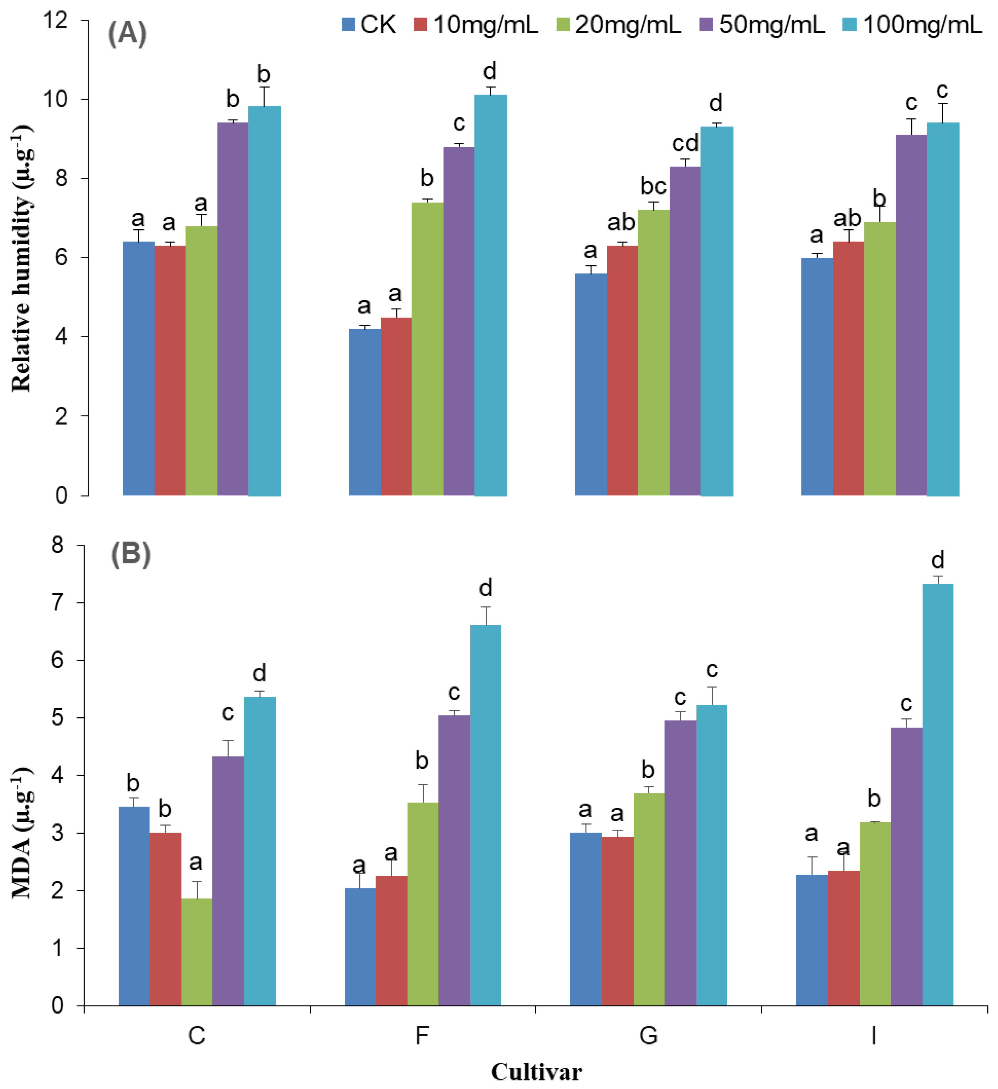
Figure 1. Relative conductivity (A) and MDA contents (B) of chrysanthemum cultivars subjected to different Cd concentrations. Each value represents the mean ± S.E from the three independent replicates (±, n = 3), and different statistical letters represent significant differences at the p < 0.05 level.
Agriculture 13 00317 g001
Figure 2. Antioxidant enzymes, i.e., CAT (A), POD (B), and SOD (C) contents of different chrysanthemum cultivars subjected to different Cd concentrations. Each value represents the mean ± S.E. from the three independent replicates (±, n = 3), and different statistical letters represent significant differences at the p ≤ 0.05 level.
Agriculture 13 00317 g002
Figure 3. Transmission electron micrographs of a chrysanthemum mesophyll cell and its organelles after exposure to different Cd stress levels. Figure (A) represents the control’s N, Ch, M, and V (nucleus, chlorophyll, mitochondria, and vacuole), which are arranged near the plasma membrane. With 10 mgL−1 of Cd, Figure (B) represents the complete structure of Ch (chloroplast), in which more thylakoid (black) and less S (starch) is present. (C) represents the Ch (chlorophyll) structure’s enlargement and the higher number of M (mitochondria) which appeared. Under 50 mgL−1 of Cd, in Figure (D), the size of the vacuole also increases and the nucleus is deformed, while the organelles are arranged on all sides. In (E), more S (starch) appeared in the Ch (chloroplast) near the CW (cell wall), while in (F), Ch (chlorophyll) was more affected compared to M (mitochondria). With 100 mgL−1 Cd treatment, as shown in Figure (G), black-colored material appeared on the cell wall, while the structure of the nucleus and the cell collapse. (H) Chl became more spherical and S (starch) increased significantly in size. In (I), the disappearance of mitochondria was noticed.
Agriculture 13 00317 g003
Table 1. Composition of Hoagland solution formulation.
NutrientConcentration mg/LNutrientConcentration mg/L
Ca(NO3)2·4H2O20.08FeSO4·7H2O0.15
KNO312.14MnSO4·H2O0.04
NH4NO31.6H3BO30.06
MgSO4·7H2O7.88ZnSO4·7H2O0.002
KH2PO45.98CuSO4·5H2O0.001
Na2EDTA0.2(NH4)6·Mo7·O24·4H2O0.002
Table 2. Effect of Cd treatment on the chlorophyll, chlorophyll a, chlorophyll b, and carotenoid content, as well as the chlorophyll (a/b) ratio, in the leaves of chrysanthemum cultivars.
CultivarsTreatment (mg L−1)Chlorophyll (g kg−1 F.W)Chlorophyll a (g kg−1 F.W)Chlorophyll b (g kg−1 F.W)Carotenoid (g kg−1 F.W)Chla/Chlb
Ratio
Donglin Ruixue (C)Control1.27 ± 0.15 a0.90 ± 0.07 a0.37 ± 0.09 a0.10 ± 0.04 a2.68 ± 0.41 a
10 mg L−11.02 ± 0.12 ab0.71 ± 0.04 ab0.31 ± 0.08 a0.11 ± 0.03 a2.75 ± 0.80 a
20 mg L−11.01 ± 0.15 ab0.72 ± 0.09 ab0.29 ± 0.06 a0.30 ± 0.01 a2.62 ± 0.21 a
50 mg L−10.91 ± 0.05 ab0.63 ± 0.02 b0.28 ± 0.03 a0.14 ± 0.01 a2.26 ± 0.16 a
100 mg L−10.84 ± 0.11 b0.59 ± 0.06 b0.25 ± 0.05 a0.31 ± 0.02 b2.44 ± 0.26 a
Yellow (F)Control1.58 ± 0.10 a0.98 ± 0.12 a0.60 ± 0.20 a 0.11 ± 0.10 b2.18 ± 0.84 a
10 mg L−11.09 ± 0.07 b0.77 ± 0.04 ab0.32 ± 0.04 ab0.26 ± 0.01 b2.47 ± 0.23 a
20 mg L−10.98 ± 0.04 b0.69 ± 0.06 b0.29 ± 0.03 ab0.30 ± 0.03 a2.42 ± 0.42 a
50 mg L−10.87 ± 0.11 bc0.59 ± 0.05 bc0.29 ± 0.08 ab0.30 ± 0.02 a2.37 ± 0.57 a
100 mg L−10.64 ± 0.05 c0.42 ± 0.04 c0.23 ± 0.06 b0.34 ± 0.01 a2.05 ± 0.42 a
Red pocket (G)Control1.46 ± 0.00 a0.83 ± 0.10 a0.62 ± 0.10 a0.09 ± 0.01 b1.47 ± 0.45 a
10 mg L−11.29 ± 0.09 a0.73 ± 0.01 ab0.57 ± 0.10 a0.10 ± 0.01 b1.38 ± 0.27 a
20 mg L−11.03 ± 0.07 b0.73 ± 0.04 ab0.30 ± 0.06 b0.29 ± 0.02 a2.60 ± 0.49 a
50 mg L−10.99 ± 0.10 b0.70 ± 0.01 ab0.29 ± 0.09 b0.13 ± 0.04 a2.97 ± 0.96 a
100 mg L−10.87 ± 0.06 b0.61 ± 0.07 b0.26 ± 0.03 b0.31 ± 0.02 a2.52 ± 0.61 a
New 9714 (I)Control1.49 ± 0.05 a0.74 ± 0.04 a0.75 ± 0.03 a0.07 ± 0.02 d0.99 ± 0.07 a
10 mg L−11.27 ± 0.07 b0.69 ± 0.03 a0.58 ± 0.04 b 0.15 ± 0.01 cd1.19 ± 0.02 c
20 mg L−11.11 ± 0.10 c0.72 ± 0.03 ab0.39 ± 0.07 c0.22 ± 0.06 bc1.97 ± 0.27 bc
50 mg L−10.90 ± 0.06 cd0.62 ± 0.02 b0.27 ± 0.06 cd0.30 ± 0.02 ab 2.49 ± 0.52 ab
100 mg L−10.82 ± 0.05 d0.62 ± 0.01 b0.20 ± 0.03 d0.34 ± 0.01 a 3.24 ± 0.42 a
Note. Each value represents the mean ± S.E. Means for each cultivars followed by the same letter are non-significantly different at (0.05) based on the LSD test.
Disclaimer/Publisher’s Note: The statements, opinions and data contained in all publications are solely those of the individual author(s) and contributor(s) and not of MDPI and/or the editor(s). MDPI and/or the editor(s) disclaim responsibility for any injury to people or property resulting from any ideas, methods, instructions or products referred to in the content.