Article

Influence of Osmotic, Salt, and Combined Stress on Morphophysiological Parameters of Chenopodium quinoa Photosynthetic Organs

by
Nina V. Terletskaya
1,2,*,
Malika Erbay
1,
Aigerim N. Zorbekova
1,
Maria Yu Prokofieva
3,
Luizat T. Saidova
3 and
Aigerim Mamirova
1,2
1
Faculty of Biology and Biotechnology and Faculty of Chemistry and Chemical Technology, Al-Farabi Kazakh National University, Al-Farabi 71, Almaty 050040, Kazakhstan
2
Institute of Genetic and Physiology, Al-Farabi 93, Almaty 050040, Kazakhstan
3
K.A. Timiryazev Institute of Plant Physiology of Russian Academy of Science, 127276 Moscow, Russia
*
Author to whom correspondence should be addressed.
Agriculture 2023, 13(1), 1; https://doi.org/10.3390/agriculture13010001
Submission received: 13 November 2022 / Revised: 15 December 2022 / Accepted: 16 December 2022 / Published: 20 December 2022

Abstract

:
Chenopodium quinoa Willd. is an annual facultative halophytic pseudocereal widely studied for its physiology and grain yield owing to its great tolerance to unfavorable growing conditions. However, the morphophysiological and anatomical characteristics of plants’ photosynthetic organs under various and combined abiotic stresses during the early stages of development have not been thoroughly studied. Therefore, the current study compared the influence of osmotic, salt, and combined stress at different intensities on the morphology and anatomy of photosynthetic organs in young quinoa plants. The main findings demonstrate that salt stress at an intensity between 100 and 200 mM NaCl is not critical for the growth of young quinoa plants and that the young plants can withstand salt stress at an intensity of 300 mM NaCl. However, it can be concluded that some adaptation mechanisms of the plants were already violated at a salt stress intensity of 200 mM NaCl, while significant changes in the water balance of the plants were observed at an intensity of 300 mM NaCl, possibly caused by damage to the cell structures.

1. Introduction

Quinoa (Chenopodium quinoa Willd.) is an annual, facultative halophytic pseudocereal of class C3 that belongs to the amaranth family and is known for its high adaptability to adverse growing conditions [1,2,3,4,5,6]. Quinoa is not considered a real halophyte since its productivity and development fluctuate greatly depending on salinity levels [7]. However, being a halophyte, quinoa is adaptable to a broad range of marginal environments, including high-salinity soils and drought-prone areas [8]. Moreover, quinoa seeds contain an excellent balance of carbohydrates, lipids, amino acids, and proteins that are beneficial to human nutrition [9,10]. Therefore, this species appears to be promising for introduction into agricultural production in arid regions. Increasing numbers of studies on the mechanisms of quinoa’s abiotic stress tolerance in the field and under laboratory conditions have recently been published [1,6,11,12,13,14,15].
Understanding the mechanisms of stress resistance is the most important means of subordinating marginal lands and breeding sustainable and commercially successful crops [16]. Although halophytes have a wide range of salt tolerance, they also suffer, to a greater or lesser extent, from excessive salt concentrations in the soil. Soil salinity is known to restrict plant growth in a manner comparable to osmotic stress in drought, impair nutrient (such as K, Ca, Mg, and P) uptake, and promote excessive Na+ accumulation, disrupting plant physiological processes, followed by the onset of ion toxicity [17]. Both osmotic and salt stress conditions cause root system water absorption capacity to diminish and increase water loss through leaves, justifying the consideration of salinity as hyperosmotic stress [9,18,19].
Although the physiology and grain yield of some quinoa cultivars under salt stress have been well studied, their drought tolerance has been less studied, and the combined influence of stress factors, typical of most marginal regions with a large proportion of saline and degraded soils, has practically not been considered at all [20,21]. The research indicates that when drought and salt stressors occur concurrently, the morphophysiological and molecular processes of plant struggle are analogous to some extent [22,23]. When compared to single stressors, combined stresses can sometimes elicit opposing, even antagonistic responses at various levels of plant organization [24,25]. However, in general, there currently exists a large vacuum in our knowledge concerning the consequences of the combined effects of multiple stressors on crops [20,26].
Both drought and salt stress have a detrimental impact on plants throughout the growing season, preventing seed germination, growth and development, flowering, and fruiting [27,28]. Stress resistance often rises with plant maturity. However, even in halophytes, the most severe effects of osmotic and salt stress on plant development are frequently found in young plants when their growth rate and sensitivity are at their peaks [29,30,31]. At the early stages of plant development, the cumulative effect of abiotic stresses is frequently more detrimental [32,33]. Therefore, it is crucial to evaluate the stress effects on young plants [31].
Literature data indicate several different morphological, physiological, biochemical, and molecular changes contributing to halophytic plants’ adaptation to salinity [34,35,36]. Thus, water deficiency can disrupt the basic organizational structure of a plant, damaging the photosynthetic apparatus and impeding regular gas exchange [37,38,39,40]. However, the present understanding of the physiological limitations of photosynthesis in crops under stress conditions, as well as the morphology and anatomy of immature plant photosynthetic organs under stress conditions, is still limited [7].
Therefore, the aim of this study was to comparatively investigate the effect of abiotic stress (osmotic, salt, and combined) at different intensities on the morphophysiological and anatomical parameters of photosynthetic organs of young quinoa plants. We aimed to determine whether (and how) tissue parameters of quinoa leaves and stems alter in response to varying levels of osmotic, salt, and combined stress, as well as whether there exists a link between tissue sizes and plant water content and tissue sizes and plant growth characteristics. Furthermore, we examine how the morphophysiological and anatomical characteristics of young quinoa plants under stress conditions are connected to alterations in plant tissue ion balance.

2. Materials and Methods

2.1. Plant Material

The Tajik variety of quinoa “Vahdat” was used in the current study; the seeds were obtained from the originator, the Centre for Genetic Resources of the Tajik Academy of Agricultural Sciences (CGR TAAS). For the experiment, 40-day-old young plants with no cotyledons and 4 rows of unfolded, functional leaves were employed, with 2 top unfolded leaves and a stem section in between collected for examination.

2.2. Growth Conditions

Plants were grown in a climatic chamber of the K.A. Timiryazev Institute of Plant Physiology, RAS (Moscow, Russia), under fluorescent lamps with a flux density of PAR quanta of 200 µmol m−2 s−1, a 16-h photoperiod, and a temperature of +25 °C. Seedlings were grown under circadian illumination (using commercial white light tubes) at 10 h (dark)/14 h (light) [200 µmol m−2 s−1 PAR, light meter LI-205 (Li-Cor, USA)] and 25 ± 5 °C temperature. Seeds were germinated in Petri dishes in distilled water over a period of 5 days. After that, the seedlings were transplanted to perlite in plastic pots of 24 cm length, 20 cm width, and 10 cm depth, with 20 seedlings per pot. Each plastic pot was placed on a separate plastic tray. The seedlings were cultivated for the following 26 days using the 50% Hoagland nutrient solution, which was applied to each plastic tray, and for the next 14 days with the addition of stress agents, for a total of 8 options, as shown in Table 1.

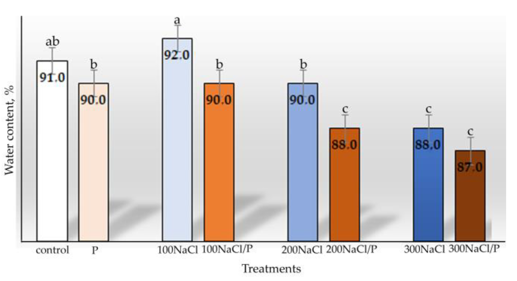
2.3. Water Content Determination

At the end of the 40-day trial, the effect of salinity on quinoa young plants’ survival, plant length, and leaf water content was assessed and calculated from the accumulation of leaf biomass according to the formula:
W C = ( a b a ) × 100 %
where a—biomass fresh weight, b—leaf dry weight.
The fresh biomass weight was determined with an analytical balance. After drying the leaves for 5 h at 105 °C (until the mass of the dry samples became constant after weighing), the dry weight of the biomass was calculated. Fixation of the plant material for anatomical studies was also carried out on the 40th day of the experiment.

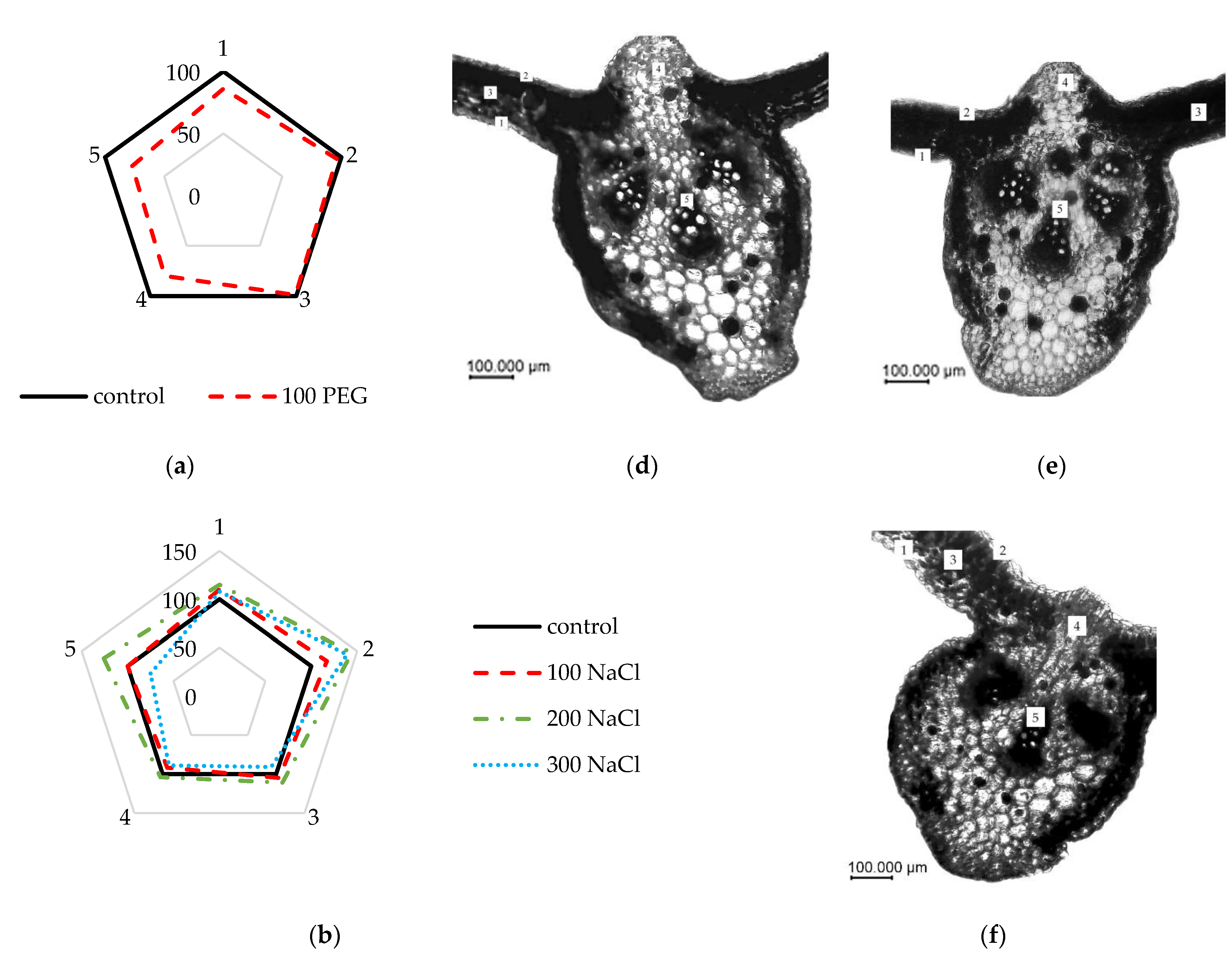
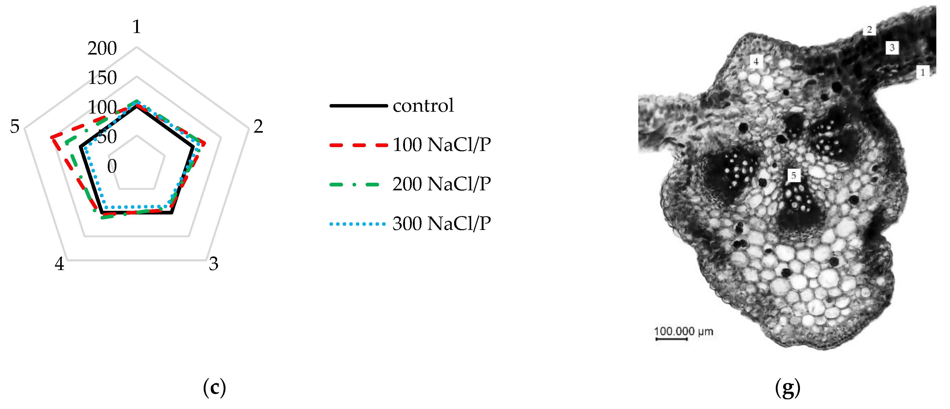
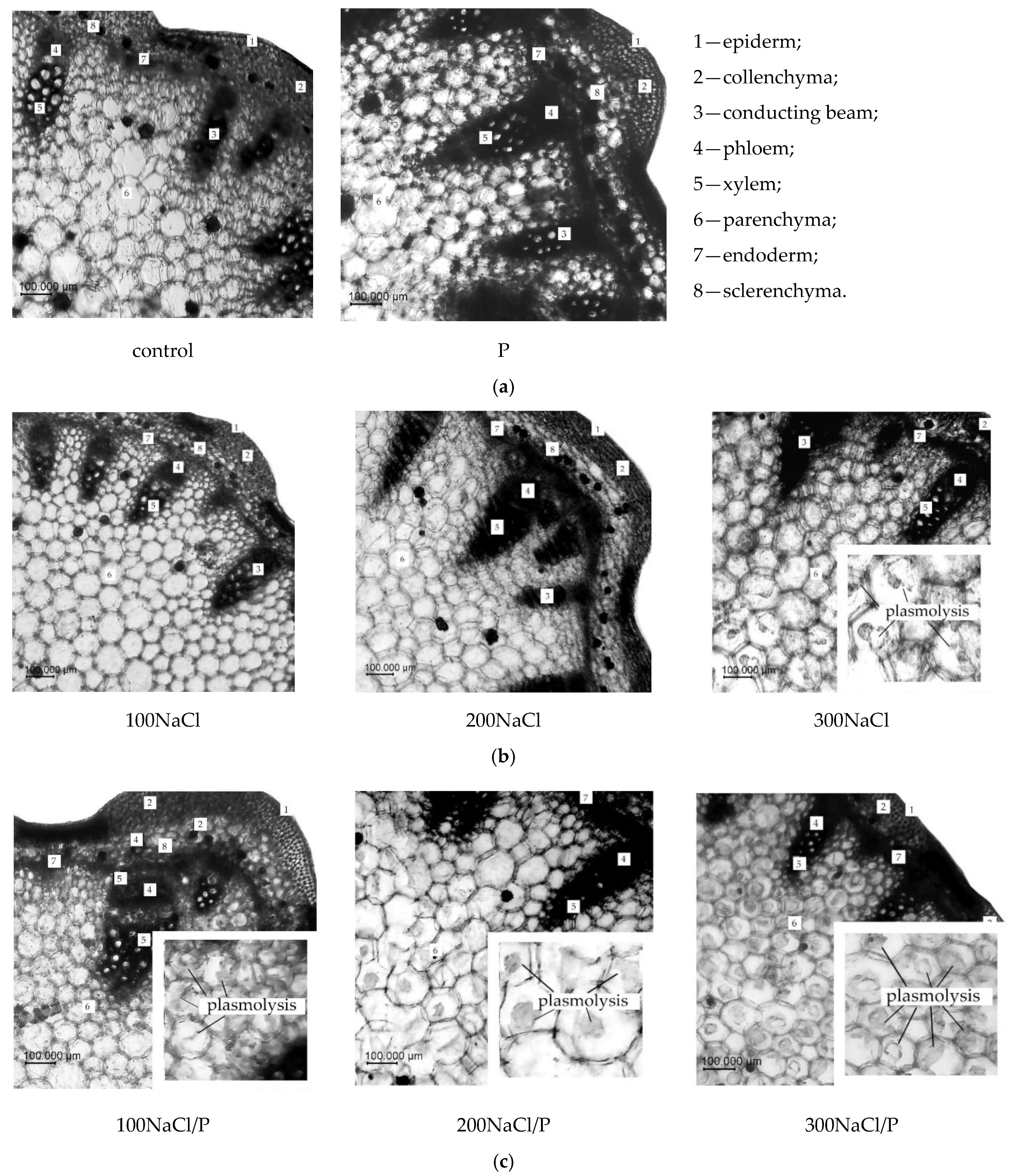
2.4. Analysis of Elements Changes in the Anatomical Structure

Plant tissues were fixed in 70% ethanol for 72 h and infused for at least 24 h in a Strasburger–Flemming mixture: 96% ethanol:glycerol:water at a 1:1:1 ratio [41]. The material was infused for 24 h. Anatomical sections were prepared using a microtome MZP-01 (“Technom”, Ekaterinburg, Russia) with a freezer OL-ZSO 30 (“Inmedprom”, Yaroslavl, Russia). The thickness of the anatomical sections varied between 10 and 15 microns. Microscopic images of the anatomical sections were taken on a microscope with a Micro Opix MX 700 (T) (West Medica, Brown Boveri-Strasse 6, B17-1 2351 Wiener Neudorf, Austria), and a CAM V1200C HD camera (West Medica, Brown Boveri-Strasse 6, B17-1 2351 Wiener Neudorf, Austria). The xylem area of the stem vessels was calculated using the formula for the area of an equilateral triangle:
S = 1 2 × A C × B H
where AC is the base of a triangular figure, equal in length to any of the sides (equilateral Δ), and BH is the height.
Plasmolysis was calculated as a percentage of the total number of cells in the microscope field of view.
All anatomical data were collected using a 40× objective in 3–5 replicates (5 plants each).

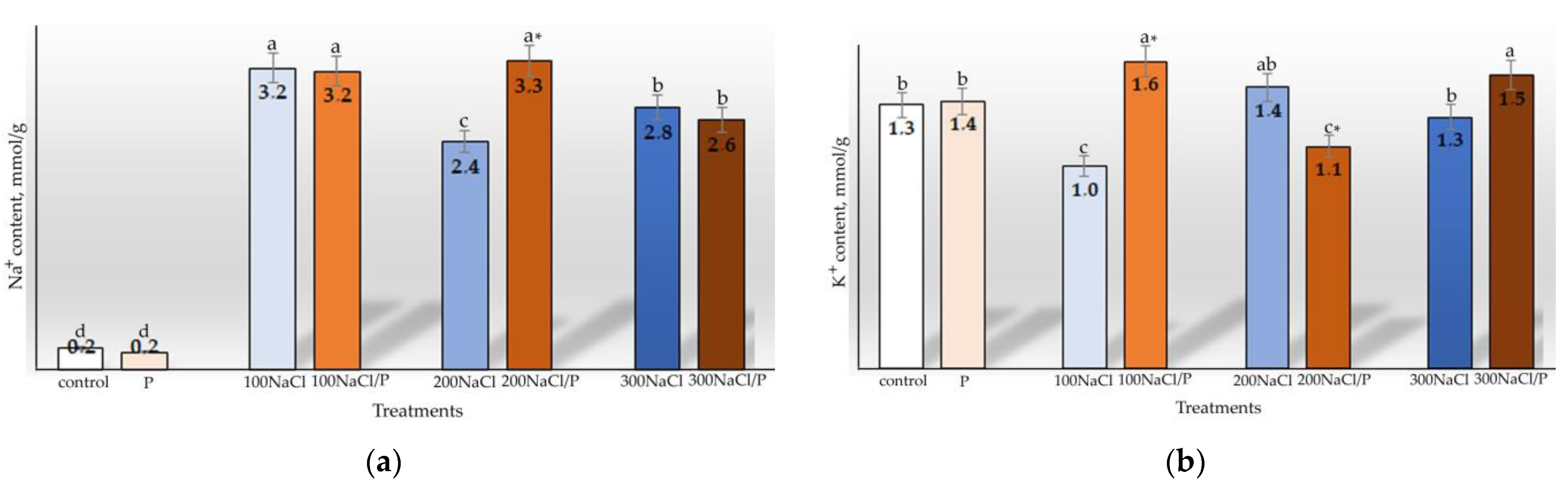
2.5. Determination of Ion Balance in Plant Tissues

The contents of Na+ and K+ in the shoots were determined in water extracts from 100 mg dry samples with the flame photometer FPA-2-01 (AOOT ZOMZ, Russia) and expressed as mmol g−1 DW.

2.6. Statistical Analysis
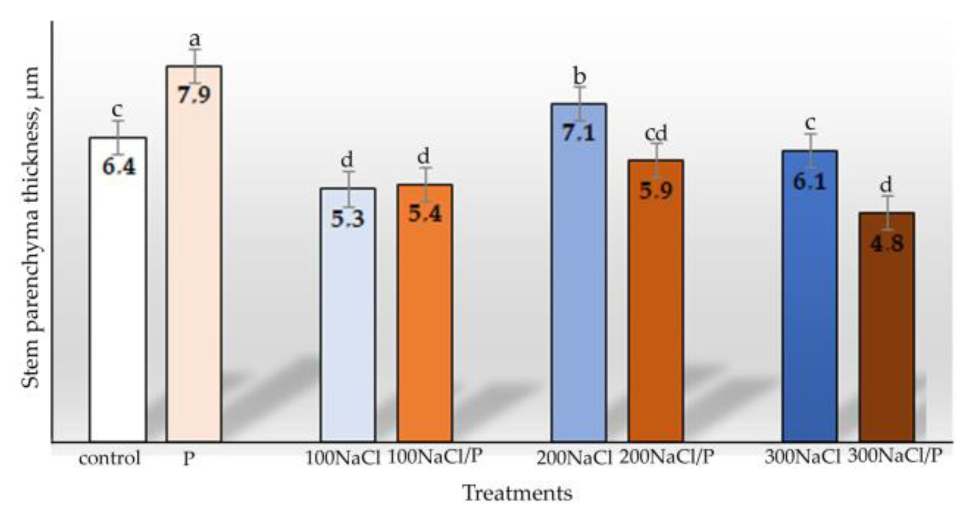
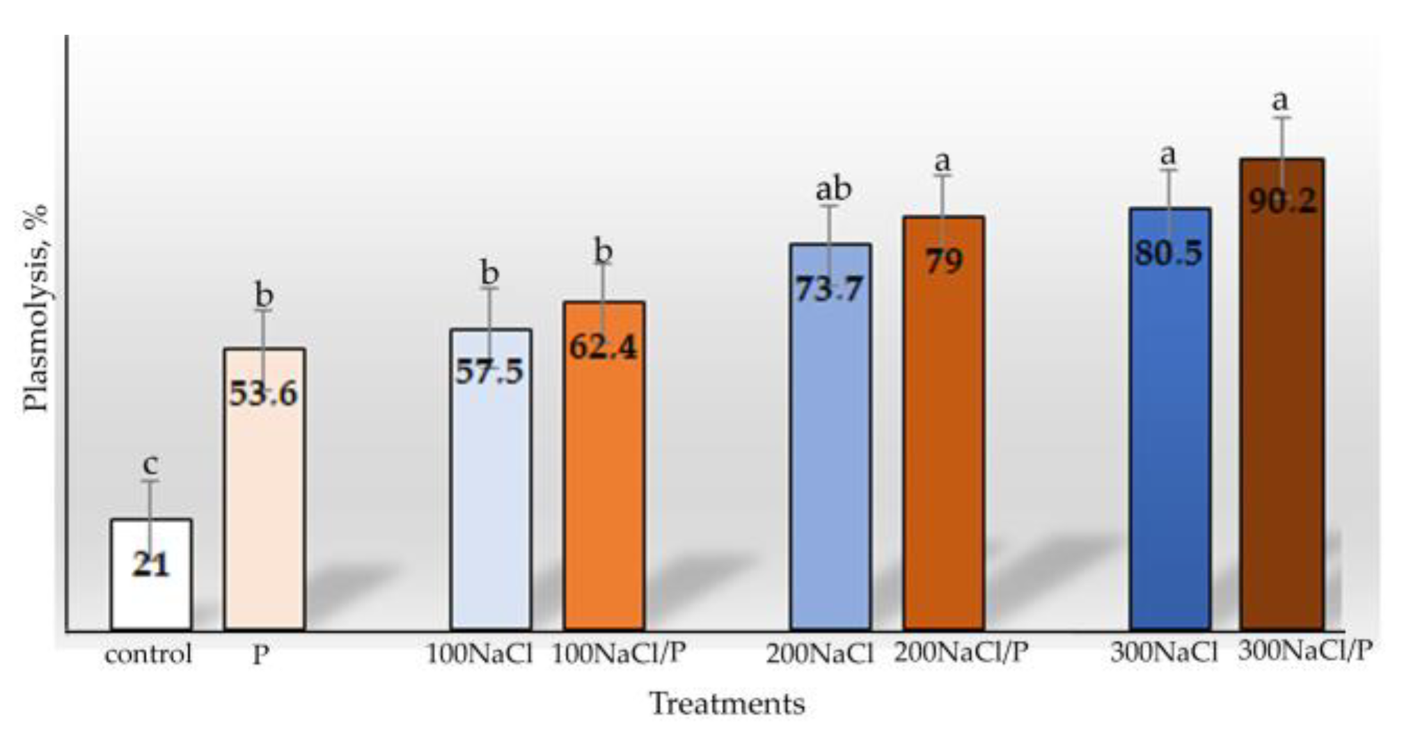
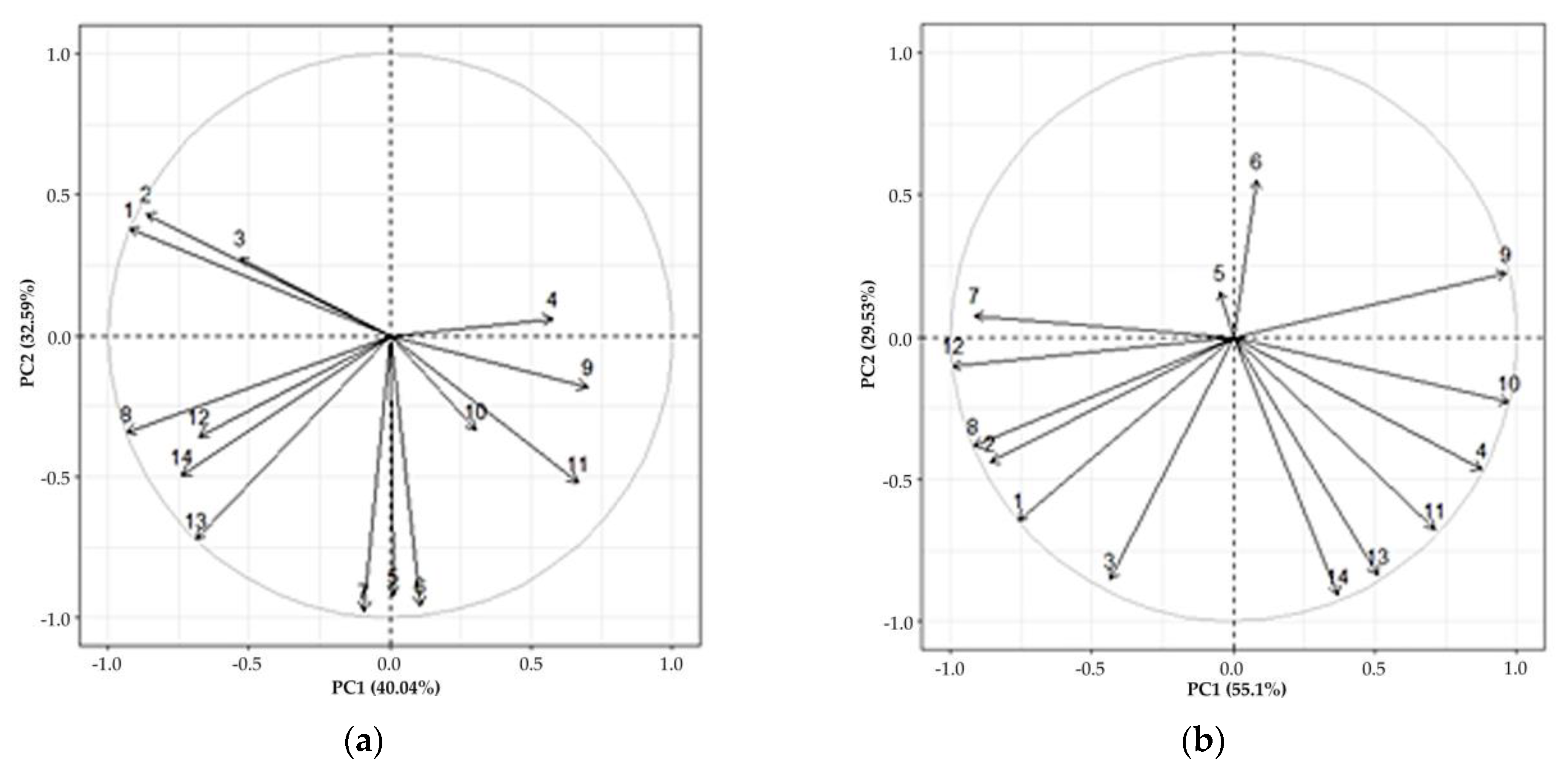
Data processing and graphical representation were conducted using Microsoft Excel (Microsoft Corp., Redmond, Washington, DC, USA). Atypical values were excluded from the data using t-tests, and the standard error of the average sample was calculated. Differences were considered significant at p < 0.05. The effects of the factors and their interaction were assessed using a two-way ANOVA on the SigmaPlot 12.5 analysis platform. Multiple factor principal component (correlation) analysis (PCA) was performed using R software (version 3.6.1).

3. Results

According to the results, the growth of the control sample was 24.2 cm after 40 days (Figure 1a). In terms of growth parameters, the samples exposed to the induced osmotic stress did not differ significantly from the control values. While the growth values at 100NaCl did not differ from the control values, the growth reduction at 200NaCl and 300NaCl was significantly different not only compared to the control plants but also to the plants exposed to lower-intensity salt stress. The combined stress results were nearly identical to those obtained under individual salt stress.
At an intensity of 100NaCl, the average weight of the quinoa leaf increased compared to the control, and only at 300NaCl did it decrease significantly. However, the individual osmotic stress and the combined stress of 200NaCl/P and 300NaCl/P significantly decreased the average leaf weight compared to the control plants (Figure 1b).
Analysis of the water content in the leaves of the experimental plants showed that quinoa has a high capacity to retain water under salt stress (Figure 2).
As can be seen in Figure 2, the data obtained under osmotic stress conditions corresponded to the results obtained under salt stress (100NaCl). Under combined stress, the water content of the leaves decreased faster than under salt stress. However, although it was significantly lower than that of the control, the leaf water content index under 300NaCl and 300NaCl/P stress was quite high (88%).
The sodium ion content in the leaves under salt stress rose more than 10-fold compared to the control and osmotic stress conditions (Figure 3a). Under combined stress, the content of sodium ions was significantly higher at a concentration of 200NaCl/P, than under salt stress.
For K+ ions, the trend was opposite to the accumulation of Na+ ions under salt stress. Such a clear pattern was not observed under combined stress (Figure 3b).
The ratio of K+/Na+ ions also showed a trend opposite to the accumulation of Na+ ions. However, it differed under salt (4.6, 8.1, and 6.7% of control under salt stress of 100NaCl, 200NaCl, and 300NaCl, respectively) and combined stress (7.2*, 5.1*, and 8.4% of control under combined stress of 100NaCl/P, 200NaCl/P, and 300NaCl/P, respectively) (asterisks indicate significant difference between values obtained under salt and combined stress at p ≤ 0.05). Consequently, as salt and combined stress levels rose, so did the K+/Na+ ratio. Nevertheless, a sharp increase in the ratio value was observed under salt stress of 200NaCl, while a slight decrease was observed under 300NaCl. In contrast, the combined stress of 200NaCl/P caused a marked decrease in the ratio value, followed by a sharp increase under 300NaCl/P.
The abiotic stress induced in the experiment affected the leaf anatomical structure, as can be seen in Figure 4. PEG-6000-induced osmotic stress resulted in statistically significant decreases in almost all parameters studied, including adaxial epidermis thickness (86% of control), central vein thickness (80% of control), and central vascular bundle diameter (77% of control).
Under salt stress conditions, the epidermal tissue of quinoa leaf blades thickened, particularly at 100NaCl and 200NaCl (105 and 111% of control, respectively); however, the thickness of leaf mesophyll decreased slightly at 300NaCl (91% of control). The diameter of the central vascular bundle increased at 200NaCl salinity and decreased at 300NaCl (126 and 75% of control, respectively). Although no statistically significant changes were observed in the thickness of the central leaf vein, there was a clear tendency for an increase at 200NaCl (104% of the control) and a decrease at 300NaCl (89% of the control).
Data obtained under combined stress conditions (Figure 4a) showed a significant increase in adaxial and abaxial epidermis thickness and a significant decrease in mesophyll thickness at all stress levels assessed. The thickness of the central vein tended to increase under the stress of 100NaCl/P and 200NaCl/P and was 105 and 112% of the control values, respectively (p > 0.05). At the same time, the thickness of the central vein decreased significantly under stress of 300NaCl/P and was 89% of the control value. The diameter of the central vascular bundle increased significantly under the combined stress of 100NaCl/P and 200NaCl/P and was 151 and 125% of the control value, respectively, while the diameter decreased significantly under the stress of 300NaCl/P and was 91% of the control value.
The data presented in Figure 5 and Figure 6 indicate that stress conditions affect the size of the stem parenchyma and vascular tissue, which in turn affects the stem growth of young plants.
The osmotic stress induced by PEG-6000 led to a significant increase in the thickness of the stem parenchyma tissue and the xylem area of the vascular bundles compared to the control. Salt stress of 100NaCl resulted in a decrease in these parameters, but the decrease in xylem area of the vascular bundle was not statistically significant. A significant difference in xylem area between the salt and combined stress variants was only found at a concentration of 100NaCl/P; it was greater under the combined stress conditions. However, under combined stress, a significant decrease in the thickness of stem parenchyma compared to salt stress was observed with increasing stress exposure.
The number of cells with marked plasmolysis in the plant tissue changed when quinoa was exposed to abiotic stressors of different intensity (Figure 7 and Figure 8).
It was found that osmotic stress induced by PEG-6000 increased the percentage of plasmolysed cells by 32.2% compared to control. Under both salt and combined stress, there was a tendency for plasmolysed cells to increase with enhancing stress exposure. However, a statistically significant difference between the salt and combined stress results was not observed. In the control samples and under osmotic and low salt stress, some cells with plasmolysis were detected in the peripheral parenchyma tissue.
The two-way analysis ANOVA showed that the osmotic, the salt and the combined stress acclimation demonstrated differences in the response of the C. quinoa young plants (Table 2).
Thus, for several parameters studied, there is a statistically significant interaction between osmotic, salinity, and combined stress.
Relationships with a fairly high degree of correlation were found for the parameters studied (Figure 9).
As shown in Figure 9a, growth parameters and water content were most strongly linked by a positive correlation, as were leaf anatomical parameters such as mesophyll thickness, central vein thickness, central vascular bundle diameter, and stem xylem area. Moreover, there was a positive dependence between these anatomical and growth parameters of the plant.
Significant correlations were found, showing that a decrease in leaf mesophyll thickness and an increase in epidermal tissue thickness and cell plasmolysis were due to Na+ ion content. Accordingly, cell plasmolysis and leaf epidermal thickness were negatively correlated with plant growth on both surfaces.
An increase in the K+/Na+ ratio, on the other hand, had a positive effect on the thickness of the mesophyll and other anatomical parameters of the leaf, as well as on the parenchyma tissue and the xylem area of the stem.
The data obtained under osmotic and combined stress with an enhanced osmotic component showed a different interaction of the parameters studied (Figure 9b). Thus, it was shown that not only the percentage of plasmolysis in the tissues and the thickness of the leaf epidermal cells, but also the thickness of the central vein and the diameter of the central vascular bundle of the leaf correlated positively with the accumulation of Na+ ions. In contrast, the K+/Na+ ratio had no positive influence on the anatomical parameters of quinoa observed under salt stress.
In addition, a strong positive dependence of parameters such as parenchyma tissue thickness and xylem area of the stem, leaf mesophyll thickness, leaf biomass, and plant growth on plant water content was observed.

4. Discussion

It is well known that the early stages of plant development are especially vulnerable to all types of stress, both individual and combined. Many studies report that at the seedling stage, combined stresses are more lethal than any form of single stress [26]. Recently, several studies have appeared on the mechanisms of field and laboratory resistance of quinoa to abiotic stress [1,12,13,14,15].
Quinoa has been shown to possess a combination of well-organized physiological properties that provide excellent salt tolerance [42,43,44]. The literature states that concentrations between 150 and 250 mM NaCl can delay the onset of seed germination [45,46,47]. However, literature data indicate that salt stress at concentrations of 100NaCl and 200NaCl did not negatively affect seed germination and growth, which is consistent with our results assessing leaf biomass and water content in young quinoa plants under stress. Consequently, a salt concentration ranging from 100 to 200 mM NaCl can be considered optimal for quinoa growth [42,48].
Health indicators for optimal plant growth include leaf area, plant height, RWC, and photosynthetic activity. As a genetically determined trait, they are also largely regulated by specific environmental conditions [45]. Both osmotic and salt stress can cause morphological and physiological changes in susceptible species [49,50,51]. Combined stress frequently exacerbates abnormalities in physiological processes, resulting in a decrease in optimal function [26,52]. The findings of our trials also corroborate this for a variety of factors.
Osmotic stress leads to plant-tissue dehydration to some extent, and the water content in plant tissue under induced stress is one of the indicators of stress resistance [53,54]. This is also evidenced by the correlations of the studied parameters with the water content in plant tissues, both under saline and (most strongly) under osmotic and combined stresses, as revealed by our experimental findings. However, the total plant biomass can be affected by ion accumulation in tissues [55], consequently becoming even higher under salt stress, which was confirmed in our study. Literature data indicate that quinoa salt tolerance depends on both external (anatomical features of the leaf surface) and internal (vacuoles of the mesophyll cells) Na+ sequestration. As plants mature, they increasingly rely on the mechanisms of Na+ sequestration in the vacuoles of the leaf mesophyll cells [56]. The K+/Na+ ratio in the cytosol, which tends to decrease under salt stress rather than the absolute Na+ concentration, has been proposed as an essential predictor of the degree of plant stress, and the K+/Na+ ratio in leaves is more informative than in main roots: the larger this ratio, the more sensitive the genotype [57,58]. Positive correlations between the K+/Na+ ratio and the number of leaf and stem anatomical parameters revealed in our experiment under salt stress are consistent with this conclusion. In addition, the tendency toward higher K+/Na+ ratio values, which was observed in our study with an increase in both salt and combined stress, shows a decline in adaption mechanisms, which was more severe with combined stress. At the same time, the concentration of 200NaCl/P under combined stress is not crucial for the survival of young quinoa plants, although the adaptation mechanisms are impaired at 300NaCl/P and 300NaCl.
An analysis of the anatomical parameters of the leaf and stem internal structure reveals the nuanced responses of young quinoa plants to different stress types. The literature describes the following morphological adaptation mechanisms: an increase in the total leaf area and thickness of the leaf and epidermis; strong sclerification; thickening of metaxylem vessels; an increase in cell size and vacuole volume; etc. [55,59]. The influence of abiotic stress factors can cause anatomical changes. For example, it is reported that the increase in leaf thickness was a successful characteristic of plant species growing under salt stress. Leaf thickening was considered a mechanism to increase water retention in the mesophyll tissue to counteract salt toxicity [60].
We know from the literature and our data that epidermis thickness, together with cuticle presence, is important in reducing leaf moisture loss [61,62,63]. It is assumed that the thicker epidermis can adapt to the conditions of limited water supply in drought and soil salinity. A large area of epidermal cells retains leaf moisture better, which is necessary for survival in harsh climates.
On the other hand, a thick mesophyll promotes higher conductivity and thus CO2 diffusion, which can increase the photosynthetic rate.
The maintenance of the parameters and the increase in mesophyll thickness can be observed as an adaptive trait of quinoa at 100NaCl and 200NaCl. This is due to an increase in the number of pneumatic cavities [7,64]. The decrease in mesophyll thickness, the main leaf photosynthetic tissue, observed in our study under severe salt stress (300NaCl) and all variants of the combined stress load can be explained by the negative effect on the division of the mesophyll cells, which reduces the leaf growth characteristics [55,62]. Photosynthesis occurs mainly in the palisade cells, and the increased thickness of the palisade parenchyma then provides higher photosynthetic activity as well as greater production of carbohydrates. However, stress can lead to a compaction of the mesophyll palisade layers as the intercellular spaces reduce under the influence of osmotic stress [65,66] or even to the deformation of both palisade and sponge tissues [67,68]. Al-Naggar et al. [63] reported a decrease in the area of quinoa leaves under drought conditions and found it might be caused by a decrease in the thickness of the columnar and spongy tissues.
The main function of water supply lies in large vascular bundles with well-developed xylem. In general, this function does not lose efficiency in young quinoa plants at salt concentrations of 100NaCl and 200NaCl under salt and combined stress, which is a distinct adaptive anatomical feature. The decrease in the main vascular bundle size observed under stress conditions, e.g., under osmotic stress and at the highest concentrations of 300NaCl under salt and combined stress, could be directly related to a decrease in the xylem area, responsible for the plant’s ability to take up water and conduct nutrients, by changing the vessel diameter [69].
The veins or nerves act as water carriers and mechanically support the leaf [70]. According to the literature, the thickness of the wheat midrib, the distance between the veins, and the number of vascular bundles decrease progressively as salinity increases [71]. In our study, a decrease in this parameter was only observed under osmotic and maximum combined stress; thus, the stability of midrib thickness is also an adaptive trait of quinoa.
In the case of stem anatomical characteristics, Munns [72] declared that the reduction in stem thickness and area could be because stress reduces the ability of plants to absorb water, which leads to a reduction in growth rate. For example, studies on quinoa have shown that when temperature increases, stem diameter decreases and plant height shortens [73,74]. Water transport from the root to the aboveground organs depends on processes such as root pressure, water potential, and the diameter and capillarity of the vessels. The presence of narrower metaxylem vessels reduces the axial conductivity of water but provides increased capillarity of the conducting elements (Yurin’s law) and thus more efficient water transport to the plant’s upper parts [75]. In addition, the smaller vessel diameter provides better protection against the effects of embolism that can occur under conditions of limited water exchange due to salinity. However, the reduced xylem area observed in our study under salt stress of 200NaCl and combined stress of 300NaCl/P provides greater resistance to water flow, requiring the plant to expend more energy to transport a given amount of water from the roots to the leaves, which may ultimately hinder the plant growth process [7]. Munns and Gilliham [76] demonstrated that stress-resistance mechanisms impose an extra energy cost on the plant to cope with osmotic stress under soil salinity, allowing growth at high salinity to be zero and the overall cost to the plant to be equal to any energy gain.
The two-way ANOVA revealed that the values of water content, leaf biomass, and percentage of plasmolysis in the cells obtained represent osmotic stress adaptability. With the exception of K+ ion content and mesophyll thickness, almost all of the characteristics analyzed may be used to identify adaptation to salt stress. In addition, leaf biomass, Na+ ion content, K+ ion content, K+/Na+ ratio, stem xylem area, and central leaf vein thickness all play a role in adaptation to combined stress. In general, the combined stress impact was not simply a summation of salt and osmotic factors but produced qualitatively unique stress responses.
Simultaneously, as evidenced by the correlation analysis results, a pronounced positive relationship was observed under salt stress between the growth parameters and water content, as well as between anatomical parameters such as mesophyll thickness, central vein thickness, central vascular bundle diameter, stem xylem area, and plant growth. Significant correlations were found, suggesting that a decrease in leaf mesophyll thickness and an increase in parenchyma tissue thickness and cell plasmolysis were conditional on Na+ ion content. A rise in the K+/Na+ ratio, in turn, had a favorable influence on the thickness of the mesophyll and other anatomical parameters of the leaf, as well as the parenchyma tissue and xylem area of the stem. As a result, cell plasmolysis and the thickness of the leaf’s parenchyma tissue are adversely associated with plant development.
The findings obtained under osmotic and combined stress demonstrated a slightly different interaction of the parameters studied. Thus, Na+ ion accumulation influenced not only the percentage of plasmolysis in the tissues and the thickness of the leaf’s parenchyma tissues but also the thickness of the central vein and diameter of the leaf’s central vascular bundle. Under salt stress, the K+/Na+ ratio no longer exerted a favorable influence on the morphological characteristics of quinoa. That is, the combined stress had the most detrimental impact on the morphophysiological parameters and adaptability of the quinoa seedlings investigated.
The revealed interactions between leaf anatomical parameters and growth parameters under single and combined stress factors contribute to a better understanding of the stress-resistance mechanisms in quinoa and demonstrate the multiple possibilities of adaptation at different levels of plant organization, as well as the importance of the balanced action of these mechanisms.

5. Conclusions

The high resistance of quinoa to biotic stresses has been confirmed. Salt concentrations between 100 and 200 mM NaCl are indeed optimal for growth of young plants, and at 300 mM NaCl, they can maintain viability. However, based on a number of indicators, it was found that at a salt concentration of 200 mM NaCl the adaptation mechanisms are violated, and a concentration of 300 mM NaCl leads to significant changes in the water balance of plants caused by damage to the cell structures. The negative effects of combined stress on the morphological and physiological properties and the anatomical structure of quinoa tissues are more pronounced than the effects of osmotic or salt stress. However, the combined stress had a qualitatively different effect than the combination of the effects of salt and osmotic factors.

Author Contributions

Conceptualization, N.V.T.; methodology, N.V.T.; formal analysis, N.V.T. and A.M.; investigation, M.E., A.N.Z., M.Y.P. and L.T.S.; data curation, N.V.T.; writing—original draft preparation, M.E. and A.N.Z.; writing—review and editing, N.V.T. and A.M.; visualization, M.E. and M.Y.P. All authors have read and agreed to the published version of the manuscript.

Funding

This research was conducted within the state assignment of the Ministry of Science and Higher Education of the Russian Federation (Theme No. 122042700044-6).

Institutional Review Board Statement

Not applicable.

Informed Consent Statement

Not applicable.

Data Availability Statement

Not applicable.

Conflicts of Interest

The authors declare no conflict of interest.

References

  1. Hinojosa, L.; González, J.A.; Barrios-Masias, F.H.; Fuentes, F.; Murphy, K.M. Quinoa Abiotic Stress Responses: A Review. Plants 2018, 7, 106. [Google Scholar] [CrossRef] [PubMed] [Green Version]
  2. Razzaghi, F.; Ahmadi, S.H.; Jacobsen, S.-E.; Jensen, C.R.; Andersen, M.N. Effects of Salinity and Soil–Drying on Radiation Use Efficiency, Water Productivity and Yield of Quinoa (Chenopodium quinoa Willd.). J. Agron. Crop Sci. 2012, 198, 173–184. [Google Scholar] [CrossRef]
  3. Toderich, K.N.; Mamadrahimov, A.A.; Khaitov, B.B.; Karimov, A.A.; Soliev, A.A.; Nanduri, K.R.; Shuyskaya, E.V. Differential Impact of Salinity Stress on Seeds Minerals, Storage Proteins, Fatty Acids, and Squalene Composition of New Quinoa Genotype, Grown in Hyper-Arid Desert Environments. Front. Plant Sci. 2020, 11, 607102. [Google Scholar] [CrossRef] [PubMed]
  4. Rezzouk, F.Z.; Shahid, M.A.; Elouafi, I.A.; Zhou, B.; Araus, J.L.; Serret, M.D. Agronomic Performance of Irrigated Quinoa in Desert Areas: Comparing Different Approaches for Early Assessment of Salinity Stress. Agric. Water Manag. 2020, 240, 106205. [Google Scholar] [CrossRef]
  5. Iqbal, S.; Maqsood Ahmed Basra, S.; Afzal, I.; Wahid, A. Exploring Potential of Well Adapted Quinoa Lines for Salt Tolerance. Int. J. Agric. Biol. 2017, 19, 933–940. [Google Scholar] [CrossRef]
  6. Adolf, V.I.; Jacobsen, S.-E.; Shabala, S. Salt Tolerance Mechanisms in Quinoa (Chenopodium quinoa Willd.). Environ. Exp. Bot. 2013, 92, 43–54. [Google Scholar] [CrossRef]
  7. Prado, F.E.; Hilal, M.B.; Albornoz, P.L.; Gallardo, M.; Ruíz, V. Anatomical and Physiological Responses of Four Quinoa Cultivars to Salinity at Seedling Stage. Indian J. Sci. Technol. 2017, 10, 1–12. [Google Scholar] [CrossRef]
  8. Bhargava, A.; Srivastava, S. Response of Amaranthus sp. to Salinity Stress: A Review. In Emerging Research in Alternative Crops; Hirich, A., Choukr-Allah, R., Ragab, R., Eds.; Environment & Policy; Springer: Cham, Switzerland, 2020; pp. 245–263. ISBN 978-3-319-90472-6. [Google Scholar]
  9. Asher, A.; Galili, S.; Whitney, T.; Rubinovich, L. The Potential of Quinoa (Chenopodium quinoa) Cultivation in Israel as a Dual-Purpose Crop for Grain Production and Livestock Feed. Sci. Hortic. 2020, 272, 109534. [Google Scholar] [CrossRef]
  10. Nanduri, K.R.; Hirich, A.; Salehi, M.; Saadat, S.; Jacobsen, S.E. Quinoa: A New Crop for Harsh Environments. In Sabkha Ecosystems: Volume VI: Asia/Pacific; Gul, B., Böer, B., Khan, M.A., Clüsener-Godt, M., Hameed, A., Eds.; Tasks for Vegetation Science; Springer: Cham, Switzerland, 2019; pp. 301–333. ISBN 978-3-030-04417-6. [Google Scholar]
  11. Zurita-Silva, A.; Fuentes, F.; Zamora, P.; Jacobsen, S.-E.; Schwember, A.R. Breeding Quinoa (Chenopodium quinoa Willd.): Potential and Perspectives. Mol. Breed. 2014, 34, 13–30. [Google Scholar] [CrossRef]
  12. Choukr-Allah, R.; Rao, N.K.; Hirich, A.; Shahid, M.; Alshankiti, A.; Toderich, K.; Gill, S.; Butt, K.U.R. Quinoa for Marginal Environments: Toward Future Food and Nutritional Security in MENA and Central Asia Regions. Front. Plant Sci. 2016, 7, 346. [Google Scholar] [CrossRef]
  13. Jacobsen, S.-E.; Mujica, A.; Jensen, C.R. The Resistance of Quinoa (Chenopodium quinoa Willd.) to Adverse Abiotic Factors. Food Rev. Int. 2003, 19, 99–109. [Google Scholar] [CrossRef]
  14. Ruiz, K.B.; Biondi, S.; Oses, R.; Acuña-Rodríguez, I.S.; Antognoni, F.; Martinez-Mosqueira, E.A.; Coulibaly, A.; Canahua-Murillo, A.; Pinto, M.; Zurita-Silva, A.; et al. Quinoa Biodiversity and Sustainability for Food Security under Climate Change. A Review. Agron. Sustain. Dev. 2014, 34, 349–359. [Google Scholar] [CrossRef] [Green Version]
  15. Ruiz, K.B.; Biondi, S.; Martínez, E.A.; Orsini, F.; Antognoni, F.; Jacobsen, S.-E. Quinoa—A Model Crop for Understanding Salt-Tolerance Mechanisms in Halophytes. Plant Biosyst. Int. J. Deal. Asp. Plant Biol. 2016, 150, 357–371. [Google Scholar] [CrossRef]
  16. Cuevas, J.; Daliakopoulos, I.N.; del Moral, F.; Hueso, J.J.; Tsanis, I.K. A Review of Soil-Improving Cropping Systems for Soil Salinization. Agronomy 2019, 9, 295. [Google Scholar] [CrossRef] [Green Version]
  17. Tebini, M.; Luu, D.T.; Mguis, K.; Ben Ahmed, H.; Meddich, A.; Zribi, F.; Chalh, A. Physiological Exploration of Intra-Specific Variability in Salinity Tolerance of Amaranth. Russ. J. Plant Physiol. 2022, 69, 59. [Google Scholar] [CrossRef]
  18. Munns, R.; Tester, M. Mechanisms of Salinity Tolerance. Annu. Rev. Plant Biol. 2008, 59, 651–681. [Google Scholar] [CrossRef] [PubMed] [Green Version]
  19. Shahid, S.; Shahbaz, M.; Maqsood, M.F.; Farhat, F.; Zulfiqar, U.; Javed, T.; Fraz, A.M.; Alhomrani, M.; Alamri, A.S. Proline-Induced Modifications in Morpho-Physiological, Biochemical and Yield Attributes of Pea (Pisum sativum L.) Cultivars under Salt Stress. Sustainability 2022, 14, 13579. [Google Scholar] [CrossRef]
  20. Sánchez-Bermúdez, M.; del Pozo, J.C.; Pernas, M. Effects of Combined Abiotic Stresses Related to Climate Change on Root Growth in Crops. Front. Plant Sci. 2022, 13, 918537. [Google Scholar] [CrossRef]
  21. Suzuki, N.; Rivero, R.M.; Shulaev, V.; Blumwald, E.; Mittler, R. Tansley review abiotic and biotic stress combinations. N. Phytol. 2014, 203, 32–43. [Google Scholar] [CrossRef]
  22. Mahmood, U.; Hussain, S.; Hussain, S.; Ali, B.; Ashraf, U.; Zamir, S.; Al-Robai, S.A.; Alzahrani, F.O.; Hano, C.; El-Esawi, M.A. Morpho-physio-biochemical and Molecular Responses of Maize Hybrids to Salinity and Waterlogging during Stress and Recovery Phase. Plants 2021, 10, 1345. [Google Scholar] [CrossRef]
  23. Hussain, S.; Mehmood, U.; Ashraf, U.; Naseer, M.A. Combined Salinity and Waterlogging Stress in Plants: Limitations and Tolerance Mechanisms. In Climate Change and Crop Stress; Academic Press: Cambridge, MA, USA, 2022; pp. 95–112. [Google Scholar] [CrossRef]
  24. Reynolds-Henne, C.E.; Langenegger, A.; Mani, J.; Schenk, N.; Zumsteg, A.; Feller, U. Interactions between Temperature, Drought and Stomatal Opening in Legumes. Environ. Exp. Bot. 2010, 68, 37–43. [Google Scholar] [CrossRef] [Green Version]
  25. Atkinson, N.J.; Urwin, P.E. The interaction of plant biotic and abiotic stresses: From genes to the field. J. Exp. Bot. 2012, 63, 3523–3543. [Google Scholar] [CrossRef] [PubMed] [Green Version]
  26. Shabbir, R.; Singhal, R.K.; Mishra, U.N.; Chauhan, J.; Javed, T.; Hussain, S.; Kumar, S.; Anuragi, H.; Lal, D.; Chen, P. Combined Abiotic Stresses: Challenges and Potential for Crop Improvement. Agronomy 2022, 12, 2795. [Google Scholar] [CrossRef]
  27. Quan, R.; Lin, H.; Mendoza, I.; Zhang, Y.; Cao, W.; Yang, Y.; Shang, M.; Chen, S.; Pardo, J.M.; Guo, Y. SCABP8/CBL10, a Putative Calcium Sensor, Interacts with the Protein Kinase SOS2 to Protect Arabidopsis Shoots from Salt Stress. Plant Cell 2007, 19, 1415–1431. [Google Scholar] [CrossRef] [PubMed] [Green Version]
  28. Park, H.J.; Kim, W.-Y.; Yun, D.-J. A Role for GIGANTEA. Plant Signal. Behav. 2013, 8, e24820. [Google Scholar] [CrossRef] [PubMed] [Green Version]
  29. Debez, A.; Ben Hamed, K.; Grignon, C.; Abdelly, C. Salinity Effects on Germination, Growth, and Seed Production of the Halophyte Cakile Maritima. Plant Soil 2004, 262, 179–189. [Google Scholar] [CrossRef]
  30. Gul, B.; Ansari, R.; Flowers, T.J.; Khan, M.A. Germination Strategies of Halophyte Seeds under Salinity. Environ. Exp. Bot. 2013, 92, 4–18. [Google Scholar] [CrossRef]
  31. Lambers, H.; Chapin III, F.S.; Pons, T.L. Plant Physiological Ecology, 2nd ed.; Springer: New York, NY, USA, 2008; ISBN 978-0-387-78340-6. [Google Scholar]
  32. Mittler, R. Abiotic Stress, the Field Environment and Stress Combination. Trends Plant Sci. 2006, 11, 15–19. [Google Scholar] [CrossRef]
  33. Pandey, P.; Ramegowda, V.; Senthil-Kumar, M. Shared and Unique Responses of Plants to Multiple Individual Stresses and Stress Combinations: Physiological and Molecular Mechanisms. Front. Plant Sci. 2015, 6, 723. [Google Scholar] [CrossRef]
  34. Poljakoff-Mayber, A. Morphological and Anatomical Changes in Plants as a Response to Salinity Stress. In Plants in Saline Environments; Poljakoff-Mayber, A., Gale, J., Eds.; Ecological Studies; Springer: Berlin/Heidelberg, Germany, 1975; pp. 97–117. ISBN 978-3-642-80929-3. [Google Scholar]
  35. Munns, R.; James, R.A. Screening Methods for Salinity Tolerance: A Case Study with Tetraploid Wheat. Plant Soil 2003, 253, 201–218. [Google Scholar] [CrossRef]
  36. Hameed, M.; Ashraf, M.; Naz, N. Anatomical Adaptations to Salinity in Cogon Grass [Imperata cylindrica (L.) Raeuschel] from the Salt Range, Pakistan. Plant Soil 2009, 322, 229–238. [Google Scholar] [CrossRef]
  37. Ali, Q.; Ashraf, M. Induction of Drought Tolerance in Maize (Zea mays L.) due to Exogenous Application of Trehalose: Growth, Photosynthesis, Water Relations and Oxidative Defence Mechanism. J. Agron. Crop Sci. 2011, 97, 258–271. [Google Scholar] [CrossRef]
  38. Ashraf, M.; Harris, P.J.C. Photosynthesis under Stressful Environments: An Overview. Photosynthetica 2013, 51, 163–190. [Google Scholar] [CrossRef]
  39. Mogazy, A.M.; Seleem, E.A.; Mohamed, G.F. Mitigating the Harmful Effects of Water Deficiency Stress on White Lupine (Lupinus albus L.) Plants using Algae Extract and Hydrogen Peroxide. J. Plant Prod. 2020, 11, 921–931. [Google Scholar] [CrossRef]
  40. Abobatta, W.F. Plant Responses and Tolerance to Combined Salt and Drought Stress. In Salt and Drought Stress Tolerance in Plants; Springer: Cham, Switzerland, 2020; pp. 17–52. [Google Scholar] [CrossRef]
  41. Barykina, R.; Veselova, T.; Devyatov, A.; Dzhalilova, K.K.; Ilyina, G.; Chubatova, N. Guide on Botanical Microtechique. In Basics Methods; MSU: Moscow, Russia, 2004; 312p. (In Russian) [Google Scholar]
  42. Hariadi, Y.; Marandon, K.; Tian, Y.; Jacobsen, S.-E.; Shabala, S. Ionic and Osmotic Relations in Quinoa (Chenopodium quinoa Willd.) Plants Grown at Various Salinity Levels. J. Exp. Bot. 2011, 62, 185–193. [Google Scholar] [CrossRef] [Green Version]
  43. Shabala, S. Learning from Halophytes: Physiological Basis and Strategies to Improve Abiotic Stress Tolerance in Crops. Ann. Bot. 2013, 112, 1209–1221. [Google Scholar] [CrossRef]
  44. Shabala, S.; Bose, J.; Hedrich, R. Salt Bladders: Do They Matter? Trends Plant Sci. 2014, 19, 687–691. [Google Scholar] [CrossRef]
  45. Gonzales, J.A.; Prado, F.E. Germination in Relation to Salinity and Temperature in Chenopodium quinoa (Willd.). Agrochim. Italy 1992, 36, 101–107. [Google Scholar]
  46. Orsini, F.; Accorsi, M.; Gianquinto, G.; Dinelli, G.; Antognoni, F.; Carrasco, K.B.R.; Martinez, E.A.; Alnayef, M.; Marotti, I.; Bosi, S.; et al. Beyond the Ionic and Osmotic Response to Salinity in Chenopodium quinoa: Functional Elements of Successful Halophytism. Funct. Plant Biol. 2011, 38, 818–831. [Google Scholar] [CrossRef]
  47. Prado, F.E.; Boero, C.; Gallardo, M.R.A.; González, J.A. Effect of NaCl on Growth Germination and Soluble Sugars Content in Chenopodium quinoa Willd. Seeds 2000, 41, 27–34. [Google Scholar]
  48. Eisa, S.S.; Eid, M.A.; Abd, E.-S.E.H.; Hussin, S.A.; Abdel Ati, A.A.; El Bordeny, N.E.; Ali, S.H.; Al Sayed Hanan, M.A.; Lotfy, M.E.; Masoud, A.M.; et al. “Chenopodium quinoa” Willd. A New Cash Crop Halophyte for Saline Regions of Egypt. Aust. J. Crop Sci. 2017, 11, 343–351. [Google Scholar] [CrossRef]
  49. El-Afry, M.M. Anatomical Studies on Drought-Stressed Wheat Plants (Triticum aestivum L.) Treated with Some Bacterial Strains. Acta Biol. Szeged. 2012, 56, 165–174. [Google Scholar]
  50. Fghire, R.; Anaya, F.; Ali, O.I.; Benlhabib, O.; Ragab, R.; Wahbi, S. Physiological and Photosynthetic Response of Quinoa to Drought Stress. Chil. J. Agric. Res. 2015, 75, 174–183. [Google Scholar] [CrossRef] [Green Version]
  51. Terletskaya, N.V.; Lee, T.E.; Altayeva, N.A.; Kudrina, N.O.; Blavachinskaya, I.V.; Erezhetova, U. Some Mechanisms Modulating the Root Growth of Various Wheat Species under Osmotic-Stress Conditions. Plants 2020, 9, 1545. [Google Scholar] [CrossRef]
  52. Rivero, R.M.; Mestre, T.C.; Mittler, R.O.; Rubio, F.; Garcia-Sanchez, F.; Martinez, V. The Combined Effect of Salinity and Heat Reveals a Specific Physiological, Biochemical and Molecular Response in Tomato Plants. Plant Cell Environ. 2014, 37, 1059–1073. [Google Scholar] [CrossRef]
  53. Munns, R.; James, R.A.; Läuchli, A. Approaches to Increasing the Salt Tolerance of Wheat and Other Cereals. J. Exp. Bot. 2006, 57, 1025–1043. [Google Scholar] [CrossRef] [Green Version]
  54. Naz, N.; Rafique, T.; Hameed, M.; Ashraf, M.; Batool, R.; Fatima, S. Morpho-Anatomical and Physiological Attributes for Salt Tolerance in Sewan Grass (Lasiurus scindicus Henr.) from Cholistan Desert, Pakistan. Acta Physiol. Plant. 2014, 36, 2959–2974. [Google Scholar] [CrossRef]
  55. Hameed, M.; Ashraf, M.; Ahmad, M.S.A.; Naz, N. Structural and Functional Adaptations in Plants for Salinity Tolerance. In Plant Adaptation and Phytoremediation; Ashraf, M., Ozturk, M., Ahmad, M.S.A., Eds.; Springer: Dordrecht, The Netherlands, 2010; pp. 151–170. ISBN 978-90-481-9370-7. [Google Scholar]
  56. Bonales-Alatorre, E.; Pottosin, I.; Shabala, L.; Chen, Z.-H.; Zeng, F.; Jacobsen, S.-E.; Shabala, S. Differential Activity of Plasma and Vacuolar Membrane Transporters Contributes to Genotypic Differences in Salinity Tolerance in a Halophyte Species, Chenopodium quinoa. Int. J. Mol. Sci. 2013, 14, 9267–9285. [Google Scholar] [CrossRef] [Green Version]
  57. Carden, D.E.; Walker, D.J.; Flowers, T.J.; Miller, A.J. Single-Cell Measurements of the Contributions of Cytosolic Na+ and K+ to Salt Tolerance. Plant Physiol. 2003, 131, 676–683. [Google Scholar] [CrossRef] [Green Version]
  58. Terletskaya, N.; Duisenbayeva, U.; Rysbekova, A.; Kurmanbayeva, M.; Blavachinskaya, I. Architectural Traits in Response to Salinity of Wheat Primary Roots. Acta Physiol. Plant. 2019, 41, 157. [Google Scholar] [CrossRef]
  59. Reinhardt, D.H.; Rost, T.L. Salinity Accelerates Endodermal Development and Induces an Exodermis in Cotton Seedling Roots. Environ. Exp. Bot. 1995, 35, 563–574. [Google Scholar] [CrossRef]
  60. Rashid, P.; Yasmin, F.; Karmoker, J. Effects of Salinity on Ion Transport and Anatomical Structure in Wheat (Triticum aestivum L. cv. Kanchan). Bangladesh J. Bot. 2001, 30, 65–69. [Google Scholar]
  61. Bahaji, A.; Mateu, I.; Sanz, A.; Cornejo, M.J. Common and Distinctive Responses of Rice Seedlings to Saline- and Osmotically-Generated Stress. Plant Growth Regul. 2002, 38, 83–94. [Google Scholar] [CrossRef]
  62. Terletskaya, N.; Kurmanbayeva, M. Change in Leaf Anatomical Parameters of Seedlings of Different Wheat Species under Conditions of Drought and Salt Stress. Pak. J. Bot. 2017, 49, 857–865. [Google Scholar]
  63. Al-Naggar, A.; Abd El-Salam, R.; Badran, A.; Boulos, S.; El-Moghazi Mai, M. Leaf Anatomical Characteristics and Its Heritability of Different Quinoa (Chenopodium auinoa Willd.) Genotypes as Influenced by Moderate and Severe Water Stress. Asian J. Biol. 2017, 4, 1–14. [Google Scholar] [CrossRef]
  64. Panfilova, O.V.; Golyaev, O.D. Adaptation of Currant to Action of a Drought and Abnormally High Temperatures (Review). Mod. Gard. 2015, 2, 88–98. [Google Scholar]
  65. Abd Elbar, O.H.; Farag, R.E.; Shehata, S.A. Effect of Putrescine Application on Some Growth, Biochemical and Anatomical Characteristics of Thymus vulgaris L. under Drought Stress. Ann. Agric. Sci. 2019, 64, 129–137. [Google Scholar] [CrossRef]
  66. Agami, R.A. Salicylic Acid Mitigates the Adverse Effect of Water Stress on Lettuce (Lactuca sativa L.). J. Appl. Sci. Res. 2013, 9, 5701–5711. [Google Scholar]
  67. Zhang, F.-J.; Zhang, K.-K.; Du, C.-Z.; Li, J.; Xing, Y.-X.; Yang, L.-T.; Li, Y.-R. Effect of Drought Stress on Anatomical Structure and Chloroplast Ultrastructure in Leaves of Sugarcane. Sugar Tech 2015, 17, 41–48. [Google Scholar] [CrossRef]
  68. Assem, N.; El Hafid, L.; Lamchouri, F. Impact of Water Stress on the Leaf Tissues of Two Moroccan Varieties of Durum Wheat (Triticum durum Desf). J. Mater. 2017, 8, 3583–3487. [Google Scholar]
  69. Ortega, L.; Fry, S.C.; Taleisnik, E. Why are Chloris gayana Leaves Shorter in Salt-Affected Plants? Analyses in the elongation zone. J. Exp. Bot. 2006, 57, 3945–3952. [Google Scholar] [CrossRef] [PubMed] [Green Version]
  70. Shepelov, V.V.; Malasai, V.M.; Penzev, A.F.; Kochmarsky, V.S.; Shelepov, A.V. Morphoobiology, Biology, Economic Value of Wheat; Mironovsky Institute of Wheat by V.N. Remeslo: Mironivka, Ukraine, 2004. [Google Scholar]
  71. Azmi, A.R.; Alam, S.M. Effect of Salt Stress on Germination, Growth, Leaf Anatomy and Mineral Element Composition of Wheat Cultivars. Acta Physiol. Plant. 1990, 12, 215–224. [Google Scholar]
  72. Munns, R. Comparative Physiology of Salt and Water Stress. Plant Cell Environ. 2002, 25, 239–250. [Google Scholar] [CrossRef] [PubMed] [Green Version]
  73. Peterson, A.J.; Murphy, K.M. Quinoa Cultivation for Temperate North America: Considerations and Areas for Investigation. In Quinoa: Improvement and Sustainable Production; John Wiley & Sons, Ltd.: Hoboken, NJ, USA, 2015; pp. 173–192. ISBN 978-1-118-62804-1. [Google Scholar]
  74. Yolcu, S. Determination of Sowing and Harvest Times on Hay Yield and Quality of Quinoa (Chenopodium quinoa Willd.) Plant Grown under Irrigated Conditions in Igdir. Ph.D. Thesis, Igdir University, Igdir, Turkey, 2018. [Google Scholar]
  75. Jurin, J., II. An Account of Some Experiments Shown before the Royal Society; with an Enquiry into the Cause of the Ascent and Suspension of Water in Capillary Tubes. Philos. Trans. R. Soc. Lond. 1718, 30, 739–747. [Google Scholar] [CrossRef]
  76. Munns, R.; Gilliham, M. Salinity Tolerance of Crops—What Is the Cost? New Phytol. 2015, 208, 668–673. [Google Scholar] [CrossRef]
Figure 1. Dependence of growth parameters of young Chenopodium quinoa on stress conditions: (a) plant height; (b) leaf fresh weight. The values shown are means (± SD). Different letters above the bars indicate a significant difference within the chart (between different stress intensities), whereas * indicates a significant difference between the single component and the combined stress at p ≤ 0.05.
Agriculture 13 00001 g001
Figure 2. Dependence of the water content in the leaves of young Chenopodium quinoa on the stress conditions. The values shown are means (±SD). Different letters above the bars indicate a significant difference at p ≤ 0.05.
Agriculture 13 00001 g002
Figure 3. Dependence of ion content in leaves of young Chenopodium quinoa on stress conditions: (a) sodium ions; (b) potassium ions. The values shown are means (± SD). Different letters above the bars indicate a significant difference within the chart (between different stress intensities), whereas * indicates a significant difference between the single component and the combined stress at p ≤ 0.05.
Agriculture 13 00001 g003
Figure 4. Morphometric parameters of leaves of young Chenopodium quinoa exposed to abiotic stress of varying intensity: (a) osmotic stress; (b) salt stress; (c) combined stress; (d) cross-sections through control quinoa leaf; (e) cross-sections through control quinoa leaf under P; (f) cross-sections through control quinoa leaf under 200NaCl; (g) cross-sections through quinoa leaf under 200NaCl/P; 1, the thickness of the adaxial epidermis; 2, the thickness of the abaxial epidermis; 3, the thickness of the mesophyll; 4, the thickness of the central vein; 5, the diameter of the central vascular bundle.
Agriculture 13 00001 g004aAgriculture 13 00001 g004b
Figure 5. Dependence of xylem area of stem vascular bundle of young Chenopodium quinoa on stress conditions. The values shown are means (±SD). Different letters above the bars indicate a significant difference at p ≤ 0.05.
Agriculture 13 00001 g005
Figure 6. Dependence of stem parenchymal tissue thickness of young Chenopodium quinoa on stress conditions. The values shown are means (±SD). Different letters above the bars indicate a significant difference within the chart (between different stress intensities).
Agriculture 13 00001 g006
Figure 7. Stem cross-sections of Chenopodium quinoa exposed to abiotic stress of varying intensity: (a) osmotic stress; (b) salt stress; (c) combined stress.
Agriculture 13 00001 g007
Figure 8. Dependence of plasmolysis development in stem cells of young Chenopodium quinoa on stress conditions. The values shown are means (±SD). Different letters above the bars indicate a significant difference at p ≤ 0.05.
Agriculture 13 00001 g008
Figure 9. Influence of morphophysiological and anatomical parameters on correlations under salt and combined stress of different intensities. (a) Salt stress; (b) Osmotic and combined stress. 1, Plant growth; 2, Water content (from raw biomass); 3, Leaf biomass; 4, Na+ ion content; 5, K+ ion content; 6, K+/Na+ ratio; 7, Thickness of stem parenchyma; 8, Area of stem xylem; 9, Percentage of plasmolysis in cells; 10, Thickness of adaxial leaf epidermis; 11, Thickness of abaxial leaf epidermis; 12, Thickness of mesophyll; 13, Thickness of central leaf vein; 14, Diameter of central leaf vascular bundle.
Agriculture 13 00001 g009
Table 1. Experimental growth conditions of Chenopodium quinoa seedlings.
12345678
ControlP100NaCl200NaCl300NaCl100 NaCl/P200NaCl/P300NaCl/P
14 days, solution 50% Hoagland10 days per solution 50% Hoagland
+ 4 days PEG-6000 at a concentration of 12.5% (m/v)
14 days, solution 50% Hoagland + 100 mM NaCl14 days, solution 50% Hoagland + 200 mM NaCl14 days, solution 50% Hoagland + 300 mM NaCl10 days per solution 50% Hoagland
+ 100 mM NaCl
+ 4 days PEG-6000
10 days per solution 50% Hoagland
+ 200 mM NaCl
+ 4 days PEG-6000
10 days per solution 50% Hoagland
+ 300 mM NaCl
+ 4 days PEG-6000
Table 2. A two-way ANOVA shows the effect of the main factors (salinity and osmotic stress) and their interaction on the morphophysiological parameters of Chenopodium quinoa photosynthetic organs.
Variable and Source of VariationdfFpVariable and Source of VariationdfFp
PlantgrowthArea of stem xylem
Osmotic stress13.0440.100Osmotic stress11.5600.230
Salt stress314.720<0.001Salt stress312.930<0.001
Combined stress30.3810.768Combined stress33.9680.027
Water contentPercentage of plasmolysis in cells
Osmotic stress17.1490.017Osmotic stress16.7230.020
Salt stress37.6240.002Salt stress317.373<0.001
Combined stress30.3370.799Combined stress31.6910.209
Leaf biomassThe thickness of the adaxial leaf epidermis
Osmotic stress1123.183<0.001Osmotic stress12.9090.107
Salt stress386.311<0.001Salt stress34.0480.026
Combined stress33.4720.041Combined stress30.4650.711
Na+ ion contentThe thickness of the abaxial leaf epidermis
Osmotic stress13.1200.096Osmotic stress13.2070.092
Salt stress3192.443<0.001Salt stress33.2570.049
Combined stress36.1760.005Combined stress31.1060.376
K+ ion contentThickness of mesophyll
Osmotic stress14.2890.055Osmotic stress12.3620.144
Salt stress30.8890.468Salt stress30.6680.584
Combined stress311.302<0.001Combined stress30.5240.672
K+/Na+ ratioThe thickness of the central leaf vein
Osmotic stress11.7420.205Osmotic stress10.0100.923
Salt stress36.5170.004Salt stress35.5460.008
Combined stress321.531<0.001Combined stress33.6500.035
The thickness of stem parenchymaDiameter of the leaf central vascular bundle
Osmotic stress10.1580.696Osmotic stress12.0710.169
Salt stress32.3370.112Salt stress310.2710.001
Combined stress31.3490.294Combined stress34.7350.015
Note: the main significant factors are bold.
Disclaimer/Publisher’s Note: The statements, opinions and data contained in all publications are solely those of the individual author(s) and contributor(s) and not of MDPI and/or the editor(s). MDPI and/or the editor(s) disclaim responsibility for any injury to people or property resulting from any ideas, methods, instructions or products referred to in the content.

