Article

Modification of Cuticular Wax Composition and Biosynthesis by Epichloë gansuensis in Achnatherum inebrians at Different Growing Periods

1
State Key Laboratory of Grassland Agro-Ecosystems, Key Laboratory of Grassland Livestock Industry Innovation, Ministry of Agriculture and Rural Affairs, College of Pastoral Agriculture Science and Technology, Lanzhou University, Lanzhou 730020, China
2
Institute of Horticulture, Ningxia Academy of Agricultural and Forestry Sciences, Yinchuan 750002, China
3
Grasslands Research Centre, Private Bag 11-008, Palmerston North 4442, New Zealand
4
Xining Center of Natural Resources Comprehensive Survey, CGS (China Geological Survey), Xining 810000, China
*
Author to whom correspondence should be addressed.
Agriculture 2022, 12(8), 1154; https://doi.org/10.3390/agriculture12081154
Submission received: 14 June 2022 / Revised: 30 July 2022 / Accepted: 31 July 2022 / Published: 4 August 2022
(This article belongs to the Special Issue Plant–Soil–Microorganism Interaction in Grassland Agroecosystem)

Abstract

:
Cuticular wax plays a critical role as a plant protectant against various environmental stresses. We predicted that the presence of the mutualistic fungal endophyte Epichloë gansuensis in Achnatherum inebrians would change both the composition of leaf cuticular wax as plants aged during the growing season and the gene expression levels associated with the wax biosynthesis pathway. Endophyte-infected (EI) and endophyte-free (EF) A. inebrians plants were established for a four-month pot experiment. In agreement with our prediction, the presence of E. gansuensis can change the composition of leaf cuticular wax at different growing periods, particularly the proportion of esters, fatty acids and hydrocarbons. The proportion of fatty acids in EI plants was lower than that in EF plants. The proportion of hydrocarbons increased and esters decreased as plants grew. Furthermore, we found 11 DEGs coding for proteins involved in cuticular wax biosynthesis, including FabF, FAB2, ECR, FAR, CER1, ABCB1 and SEC61A. The present study highlights the significant contribution of E. gansuensis to leaf cuticular wax composition and biosynthesis in A. inebrians plants.

1. Introduction

Cuticular wax, as the first physical barrier, plays a crucial role in protecting all land plants from nonstomatal water loss, UV radiation, pathogens and herbivores [1,2,3,4]. Cuticular wax is a mixture of very long-chain fatty acids (VLCFAs), primary alcohols, esters, aldehydes, hydrocarbons, secondary alcohols, ketones, phenols, triterpenoids, sterols and flavonoids, with carbon chain lengths ranging from C20 to C40 [5,6]. The formation of cuticular wax is completed in two stages. The first stage is the biosynthesis of C16 and C18 fatty acyl-acyl carrier proteins (ACPs) in the plastids, which is the same as other lipid biosynthetic pathways [7]. Then, long-chain acyl-ACPs are esterified to long-chain acyl-Coenzyme As (CoAs) and translocated to the endoplasmic reticulum (ER) [8]. The second stage is the elongation of C16 and C18 acyl-CoAs and the formation of derivatives in the ER. The extension is mediated by fatty acid elongase (FAE) complexes including the condensing enzyme β-ketoacyl-CoA synthase (KCS), β-keto acyl reductase (KCR), β-hydroxyacyl-CoA dehydratase (HCD) and enoyl-CoA reductase (ECR), which add two carbons per cycle [9]. The biosynthesis of derivatives is divided into two different pathways. One pathway produces primary alcohols and esters while another synthesizes aldehydes, alkanes, secondary alcohols and ketones [10]. Finally, the wax mixture is exported from the ER, through the plasma membrane, across the cell wall, and to the cuticle by adenosine triphosphate (ATP)-binding cassette (ABC) transporters and lipid transfer proteins (LTPs) [11].
The cuticular wax composition varies from species to species, as well as the growing periods [12]. For example, in leaves of Arabidopsis thaliana, alkanes were reported as the most dominant wax compound [10]. Primary alcohols were found to be the most abundant components in wheat (Triticum aestivum) [13], maize (Zea mays) [14], barley (Hordeum vulgare) [15] and Poa pratensis [16]. However, few studies have focused on the wax composition in different growing periods. Early research found that in wheat, the major components were octacosanol and β-diketone at 50 and 66 days, respectively, and the content of fatty acids and esters increased from 66 to 100 days [17]. In maize, primary alcohols were reported as the most plentiful components in young leaves, while esters were the most dominant wax components in mature leaves [18]. After leaf unfolding, a dynamic biosynthesis of wax components started, especially aldehydes and β-amyrenyl acetate [19]. Recent studies showed that in wheat the quantities of alkanes, fatty acids and aldehydes increased gradually from 50 to 230 days, while the quantity of alcohols first increased and reached a maximum at 100 days, and then continuously decreased during further leaf development [20]. As leaves developed, the cuticular wax compositions in arabidopsis were found to shift from fatty acid to alkane constituents [21]. Though the genes involved in the biosynthesis of cuticular wax are not well characterized for grasses compared with arabidopsis, progress has been recently made. TaFAR1, TaFAR2, TaFAR3, TaFAR4 and TaFAR5 genes were reported as homologs of arabidopsis CER4 encoding active alcohol-forming fatty acyl-coenzyme A reductase (FAR), which is involved in the synthesis of primary alcohol in wheat leaf in response to environmental stresses [22,23,24]. At least 18 Glossy genes can affect the synthesis and composition of cuticular wax in maize leaves [25]. OsGL1-1, OsGL1-2 and OsGL1-3 genes, homologs of arabidopsis CER3, were reported to be involved in cuticular wax biosynthesis in rice (Oryza sativa) [26,27,28].
Epichloë (syn. Neotyphodium) endophytes that are symbiotic with many cool season grasses spend all or nearly all of their life cycles within the hosts [29,30]. In the stable symbioses of Epichloë endophytes with hosts, growth of both partners is fully synchronized [31]. Host grasses provide shelter, nutrition, and dissemination through seeds, while endophytes protect against herbivory, which is attributable to bioactive alkaloids of fungal origin [32,33,34]. Nearly 100% of Achnatherum inebrians plants, a widespread perennial bunchgrass, are infected by E. gansuensis or E. inebrians in the arid and semi-arid grasslands of northwest China [35,36,37]. Epichloë gansuensis can confer to A. inebrians plants the ability to resist various adverse environmental conditions, such as pests, pathogens, drought, heavy metals, low temperature and salt, to improve the competitive edge of hosts [38,39,40,41,42,43,44]. However, studies to see if endophytes can change the composition and biosynthesis of cuticular wax have not been reported yet.
Here, we predicted that the presence of the endophyte E. gansuensis would change the composition of leaf cuticular wax and that changes in composition would continue over the growing season. In addition, we predicted that the gene expression levels associated with the wax biosynthesis pathway in A. inebrians plants would also change. In order to verify these predictions, we set up endophyte-infected (EI) and endophyte-free (EF) A. inebrians plants for a four-month pot experiment in the greenhouse. The composition and proportion of leaf cuticular wax and the expression levels of genes related to the cuticular wax biosynthesis pathway were determined by gas chromatography−mass spectrometry (GC−MS) and transcriptome analysis, respectively.

2. Materials and Methods

2.1. Plant Materials and Treatment

EI and EF seeds originated from a single EI and EF A. inebrians plant in the field of the College of Pastoral Agriculture Science and Technology, Yuzhong Campus of Lanzhou University (104°39′ E, 35°89′ N, Altitude 1653 m) in 2013. The seeds were stored at 4 °C for further study. Before sowing, the seeds were stained with aniline blue as described by Li et al. [45] to determine that the frequency of fungal endophytes of EI and EF seeds were 100% and 0%. In June 2020, a four-month pot experiment was carried out in the greenhouse (temperature: 26 ± 2 °C; moisture: 42 ± 2%) of the College of Pastoral Agriculture Science and Technology, Yuzhong Campus of Lanzhou University. The seeds were sown in 100 plastic pots (diameter: 24 cm; height: 15 cm; 50 pots for EI plants and 50 pots for EF plants) with 200 g sterilized vermiculite (180 °C sterilization for 2 h). Three seeds were sown in each pot initially, but only one plant was chosen and kept in each pot after seed germination. These pots were placed at random in different positions and watered appropriately every day. Hoagland’s solution was rationed to the pots every 7 days after the second fully unfolded leaf appeared. Leaf blade samples from different growing periods were collected at 1, 2, 3 and 4 months after sowing (S1, S2, S3 and S4, respectively), which came from the same part of equal-sized A. inebrians plants (6 EI plants and 6 EF plants each period). After collecting, samples were instantly frozen in liquid nitrogen and then kept at −80 °C in a refrigerator until analysis.

2.2. Leaf Cuticular Wax Extraction

Leaf cuticular waxes were extracted by soaking leaf blades, which were cut into 10 cm lengths, for 45 s in 10 mL chloroform (Tianjin Guangfu Fine Chemical Research Institute, Tianjin, China) at room temperature and containing 20 μL n-tetracosane (1 μg/μL, Sigma-Aldrich Corp., St. Louis, MO, USA) as an internal standard. Then, concentrate the solution to 1 mL under a stream of nitrogen and treat it with 20 µL of N, O-bis(trimethylsilyl)-trifluoroacetamide (GC Derivatization reagent, ≥98.0%, Sigma-Aldrich Corp., St. Louis, MO, USA) and 20 µL of pyridine (Standard for GC, >99.9%, Alfa Aesar, Ward Hill, MA, USA) for 1 h at 70 °C. Through organic filtration (0.45 μm), the solution was transferred into GC sample bottles (1.5 mL), blown dry under nitrogen gas again, and redissolved in 500 µL of chloroform for GC−MS analysis.

2.3. Chemical Analysis

The composition of cuticular wax was analyzed by GC−MS (6890N-5975C, Agilent Technologies Inc., Palo Alto, CA, USA) equipped with a DB−1 MS capillary column (0.25 µm film thickness, 0.25 mm inner diameter, 30 m length, Agilent Technologies Inc., Palo Alto, CA, USA). GC was performed with temperature-programmed on-column injection and oven temperature set at 50 °C for 2 min, increased by 40 °C/min to 200 °C, maintained at 200 °C for 2 min, increased by 3 °C/min to 320 °C, and maintained at 320 °C for 30 min. Inlet temperature, MS quadrupole temperature and transfer line temperature were set at 280 °C, 150 °C and 250 °C, respectively. Helium was used as a carrier gas at a flow rate of 1.2 mL/min. Individual wax components were identified via comparing their mass spectra with data in the literature through the National Institute of Standards and Technology (NIST) 2008 library [46]. Quantification was based on comparison of peak areas with those of the internal standard.

2.4. Transcriptome Analysis

The transcriptomes were characterized by sequencing total RNAs on leaves of EI and EF plants at four months after sowing. The RNA extraction was conducted with the TRIzol reagent (Invitrogen, Carlsbad, CA, USA) and cleaned using the RNeasy Plant Mini Kit (Qiagen, Valencia, CA, USA). The integrity, purities and concentration of RNA were assessed with RNA Nano 6000 assays (Agilent Bioanalyzer 2100 system, Agilent Technologies, Palo Alto, CA, USA), NanoPhotometer® spectrophotometers (IMPLEN, Calabasas, CA, USA) and Qubit® 2.0 fluorometers (Life Technologies, Carlsbad, CA, USA), respectively. The Biomarker Technologies Company (Beijing, China) performed transcriptome analysis. Transcriptome libraries were generated by the NEBNext® Ultra™ RNA Library Prep Kit for Illumina® (NEB, Ipswich, MA, USA) and then sequenced using the Illumina HiSeq 2000 platform. The library fragments were amplified by PCR, using Phusion high-fidelity DNA polymerases (New England Biolabs, Ipswich, MA, USA) and purified with the AMPure XP system (Beckman Coulter, Brea, CA, USA).
Annotation of gene functions was based on several databases, namely, GO (gene Ontology), KEGG (Kyoto Encyclopedia of Genes and Genomes), KOG/COG/eggNOG (Clusters of Orthologous Groups of proteins), NR (NCBI non-redundant protein sequences), Pfam (Protein family) and Swiss-Prot (a manually annotated and reviewed protein sequence database). The expression level of each annotated gene was normalized based on the transcript lengths and the library size. The normalized gene expression levels were then reported as FPKM (fragments per kilobase per million mapped fragments). Differentially expressed genes (DEGs) were assigned to unigenes using the DESeq2 package in the R software with fold change (FC) values greater than or equal to 2 and with false discovery rate (FDR) values less than 0.05.
All raw sequence data utilized in the present study were deposited in the Sequence Read Achieve (SRA) of the NCBI database and the accession number was PRJNA748183.

2.5. Statistical Analysis

The effects of the presence of Epichloë endophytes and the different growing periods on the wax composition of the leaves of A. inebrians plants were analyzed by two-way ANOVA using SPSS Statistics 22.0 software (SPSS Inc., Chicago, IL, USA). The significance of differences between EI and EF plants at different growing periods was determined by an independent samples t-test. The values shown in the result all correspond to means + standard errors. Statistical significance was defined at the 95% confidence level.

3. Results

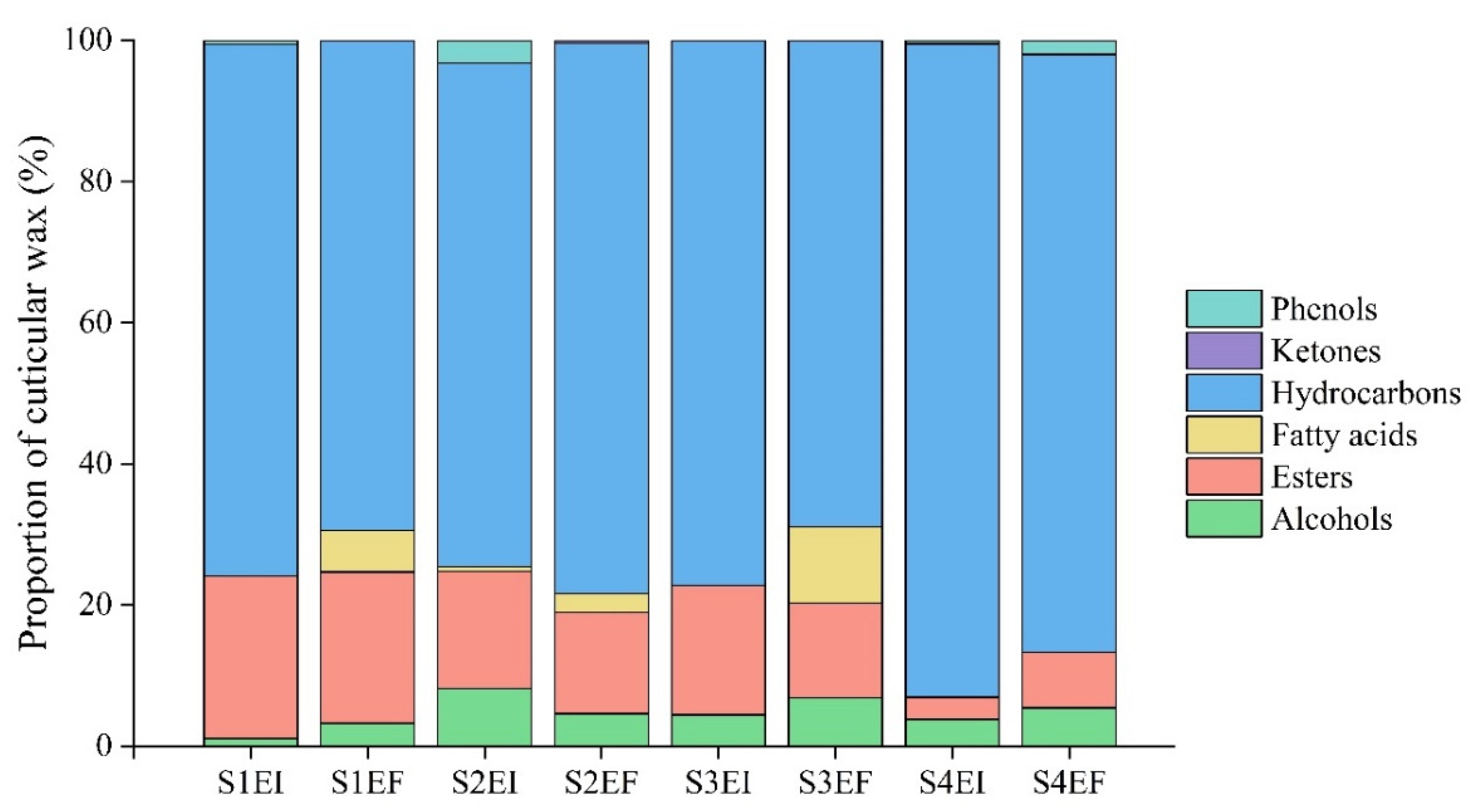
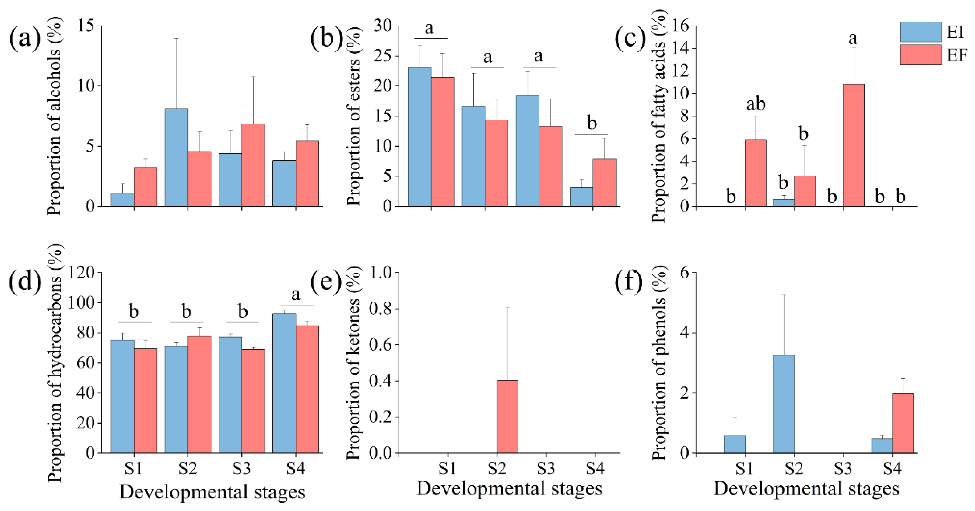
3.1. Composition of Leaf Cuticular Wax

The leaf cuticular wax composition under eight treatments (S1EI, S1EF, S2EI, S2F, S3EI, S3EF, S4EI and S4EF) was identified and relatively quantified by GC−MS. A total of 47 different wax compounds were detected, with 23, 23, 24, 29, 23, 24, 16, 14 compounds in the eight treatments, respectively (Table S1 of the Supplementary Materials). The wax mixture contained six identifiable compound classes including hydrocarbons (from minimum to maximum of 69.4392.61%), esters (from minimum to maximum of 3.11–23.03%), alcohols (from minimum to maximum of 1.07–8.12%), fatty acids (from minimum to maximum of 0–10.85%), phenols (from minimum to maximum of 0–3.25%) and ketones (from minimum to maximum of 0–0.40%; Figure 1 and Figure 2).
Two–way ANOVA analysis indicated that the status of E. gansuensis endophytes, the different growing periods and interaction between the two factors had significant (p < 0.05) effects on the proportion of fatty acids (Table 1). Fatty acids were detected in EI plants only at S2. At S4, fatty acids were not identified in either EI or EF plants. Within EF plants, the proportion of fatty acids at S3 was significantly (p < 0.05) higher than that at S2 (Figure 2c). In addition, the different growing periods had significant (p < 0.05) effects on the proportion of esters and hydrocarbons (Table 1). The proportion of esters at S4 was significantly lower than that at S1, S2 and S3 (Figure 2b). However, the proportion of hydrocarbons at S4 was significantly higher than at S1, S2 and S3 (Figure 2d). Ketones were only found in EF plants at S2 (Figure 2e).

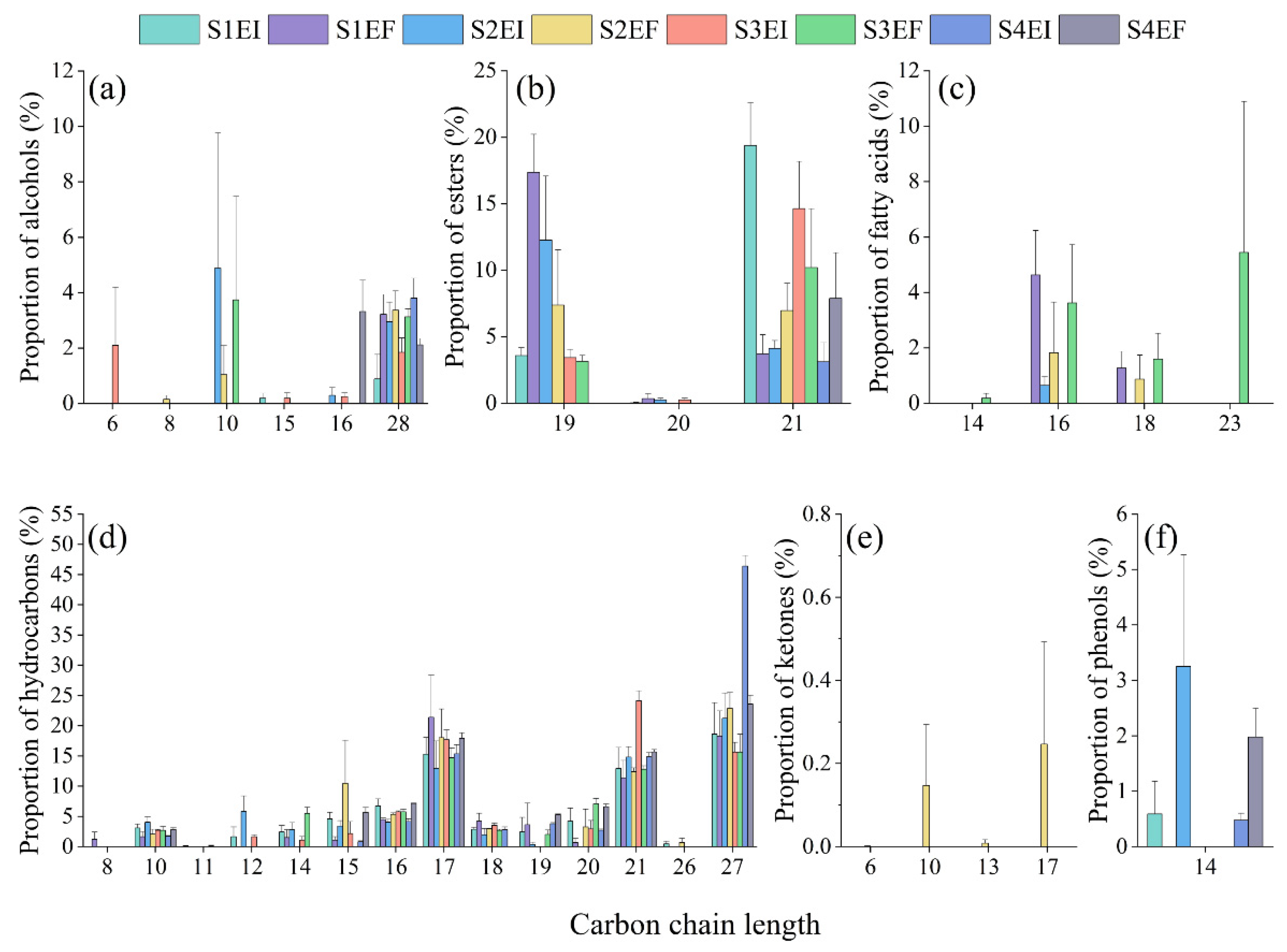
3.2. Carbon Chain Length of Leaf Cuticular Wax

The carbon chain length distribution suggested that alcohols ranged from mannitol (C6) to octacosanol (C28), and octacosanol was the most abundant (Figure 3a, Table S1). C19 and C21 were the dominant esters (Figure 3b, Table S1). Four fatty acids were detected in this study including tetradecanoic acid (C14), hexadecanoic acid (C16), octadecanoic acid (C18) and nordeoxycholic acid (C23; Figure 3c, Table S1). The carbon chain length of hydrocarbons was the longest of all components, and ranged from 1,3,7-octatrien-5-yne (C8) to n-heptacosane (C27), and the prominent components were C17, C21 and C27 (Figure 3d, Table S1). C10 and C17 were the abundant ketones—these ranged from C6 to C17 (Figure 3e, Table S1). Furthermore, 2,4-di-tert-butylphenol (C14) was the only phenol detected (Figure 3f, Table S1).

3.3. Differentially Expressed Gene (DEG) Analysis

Upon comparison with the EF plants, the unigenes with gene expression FC values greater than or equal to 2 and with FDR values less than 0.05 were defined as DEGs. Based on these strict criteria, we found 11 (1 upregulated and 10 downregulated) DEGs between EF plants versus EI plants.

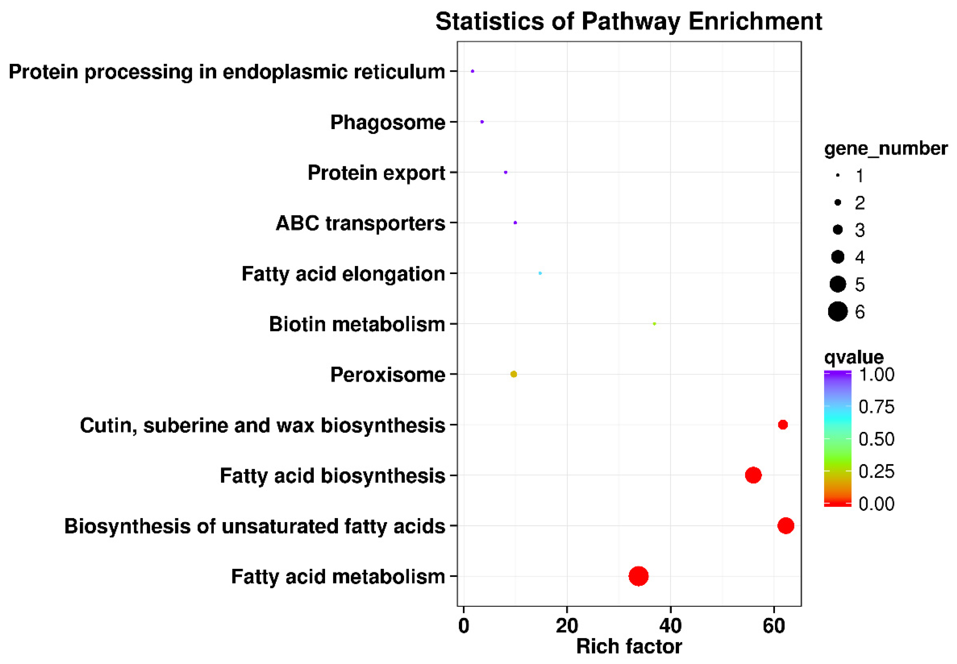
3.4. KEGG Pathway Enrichment Analysis of the DEGs

To characterize the complex biological behavior of the transcriptome, the 11 DEGs were subjected to a KEGG pathway enrichment analysis (Figure 4). The “Fatty acid metabolism (ko01212)”, “Biosynthesis of unsaturated fatty acids (ko01040)”, “Fatty acid biosynthesis (ko00061)” and “Cutin, suberine and wax biosynthesis (ko00073)” categories were significantly enriched.

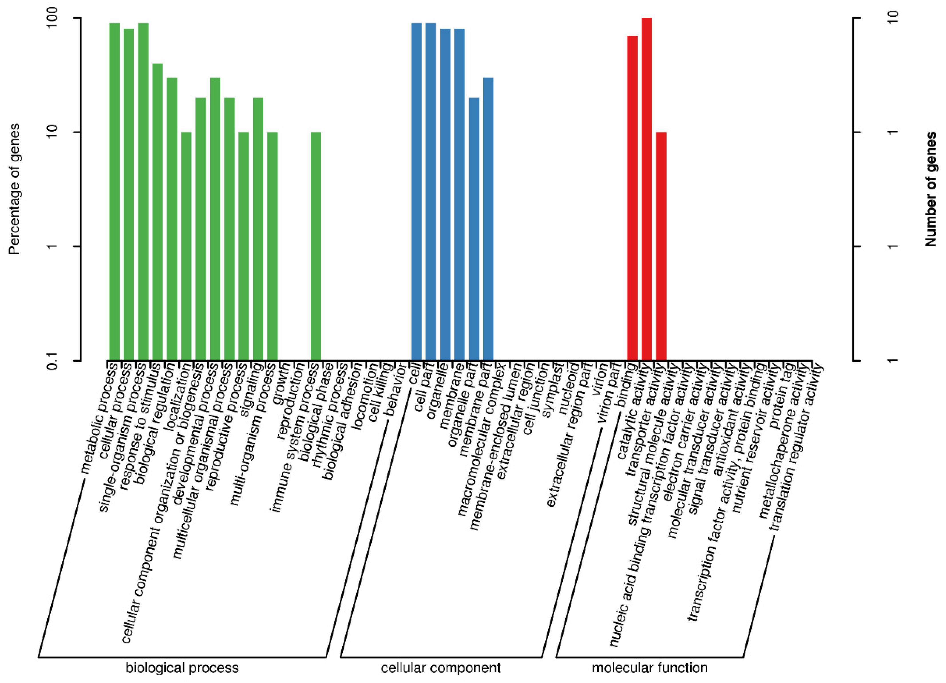
3.5. GO Functional-Enrichment Analysis of the DEGs

To clarify the functions of the detected 11 DEGs, we performed the GO annotation. A total of 22 GO categories were assigned to the 11 DEGs (Figure 5). GO term enrichment analysis was conducted to categorize the annotated sequences into three main categories: biological process, cellular component and molecular function. The most abundant subcategory under the biological process category was "metabolic process," which was followed by "single-organism process" and "cellular process". In the cellular component category, “cell” and “cell part” were the dominant groups, followed by “organelle” and “membrane”. As for the molecular function category, “catalytic activity” was the most dominant subcategory, followed by “binding” and “transporter activity”.

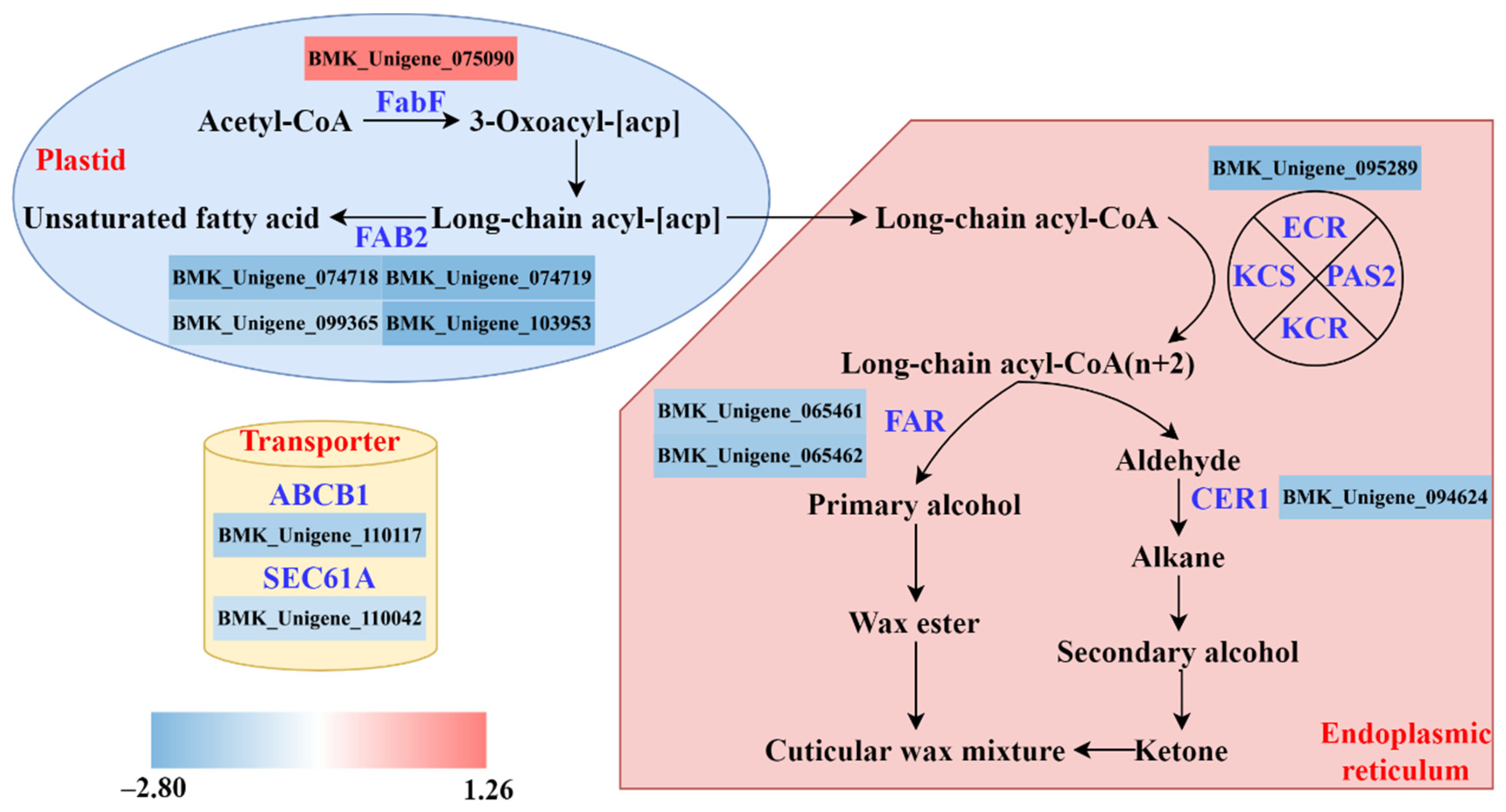
3.6. Biosynthesis Pathway of Cuticular Wax

A four-part model of cuticular wax biosynthesis pathway was proposed including fatty acid biosynthesis, fatty acid elongation, wax biosynthesis and transporters (Figure 6). Only one gene (BMK_Unigene_075090) coding for FabF protein expression level was upregulated by the Epichloë endophyte. The expression levels of 10 genes were downregulated by the Epichloë endophyte, including four genes (BMK_Unigene_074718, BMK_Unigene_074719, BMK_Unigene_099365, BMK_Unigene_103953) coding for FAB2 protein, one gene (BMK_Unigene_095289) coding for ECR protein, one gene (BMK_Unigene_094624) coding for CER1 protein, two genes (BMK_Unigene_065461, BMK_Unigene_065462) coding for FAR protein, one gene (BMK_Unigene_110117) coding for ABCB1 protein and one gene (BMK_Unigene_110042) coding for SEC61A protein.

4. Discussion

In this study, we have established a hypothesis that both the composition of leaf cuticular wax at different growing periods and the expression levels of genes related to the wax biosynthesis pathway in EI A. inebrians plants would change compared with that in EF plants. Our results through GC−MS indicated that hydrocarbons were the most abundant components of cuticular wax in both EI and EF plants. The proportion of esters was reduced as plants grew. By contrast, the proportion of hydrocarbons increased with plant growth. Compared with EF plants, the leaf cuticular wax of EI plants had fewer fatty acids. In agreement with our hypothesis, by transcriptome analysis, we found 11 DEGs coding for proteins involved in cuticular wax biosynthesis, including FabF, FAB2, ECR, FAR, CER1, ABCB1 and SEC61A.
Cuticular wax plays a critical role as a plant protectant against various environmental stresses [12,47]. In the present study, cuticular wax was dominated by hydrocarbons in A. inebrians plants. It is consistent with the results in arabidopsis [10], but different from the results in other grasses. Several studies about the leaf cuticular wax of grasses indicated that primary alcohols are the predominant components in maize [14], barley [15,48], wheat [13,49], and Poa pratensis [16] In addition, the composition of the cuticle changes at different growing periods [12]. In our study of A. inebrians plants, we found that the proportions of hydrocarbons and esters differed in reverse ways from each other. Although hydrocarbons were always the major components at different sampling times, the proportion of hydrocarbons and esters increased and decreased at 4 months after sowing, respectively. Previous studies showed that the cuticular wax content of esters increased from 66 to 100 days in wheat [17]. Clearly, our results are inconsistent with this study, which may be a characteristic of the different species. Epichloë endophyte-grass associations are generally considered as mutualistic associations and almost certainly have been exposed to the selection pressures of long-term coevolution [50]. Previous studies indicated that disease symptoms caused by the powdery mildew fungus Blumeria graminis were reduced by the presence of endophyte Epichloë within A. inebrians plants [41]. Powdery mildew, a common fungal disease, causes serious damage to a variety of cereal crops with a broad host range including 634 species belonging to the Poaceae [51]. To cause infection, conidia of B. graminis produce appressoria and germ tubes that penetrate through the cuticle and walls of epidermal cells. Fatty acid biosynthesis in plants provides nutritional support to powdery mildew and promotes infection [52]. Therefore, according to the above evidence, we can infer that endophytic Epichloë is likely to enhance the resistance of grasses to powdery mildew by reducing the content of fatty acids. Our results showing that the proportion of fatty acids in the cuticle of EI plants was lower than in EF plants support this inference. Although advances have been made in gene characterization associated with the biosynthesis of cuticular wax in grasses, more studies are needed to obtain the understanding that is now available for arabidopsis [53]. In this study, we proposed a possible model for cuticular wax biosynthesis in mutualistic symbiosis using transcriptome analysis. The FabF gene encoding β-ketoacyl-ACP synthases (KAS) II enzymes has been demonstrated to regulate temperatures of fatty acid synthesis [54,55]. In the plastid, a FabF gene was upregulated by Epichloë endophyte according to our results, which may lead to the difference in the proportion of fatty acids. ECR/CER10 catalyzes the final step of VLCFA elongation required for the synthesis of all the VLCFA and plays an essential role in cell expansions [56]. We found that one DEG coding for ECR protein was downregulated in ER in the present study. In the alkane synthesis pathway, there was also a downregulated DEG coding for CER1 protein, which catalyzes the redox-dependent synthesis of alkanes from acyl-CoAs [57]. In the primary alcohol synthesis pathway, two DEGs coding for the FAR protein were downregulated. Wang et al. found that the TaFAR5 gene, coding for FAR, contributes significantly to producing primary alcohols in wheat leaf blades [24]. Additionally, wax mixtures must be exported to the plant surface once synthesized. The mechanisms behind the transport of cuticular wax remain poorly understood. Previous studies showed that in arabidopsis, ABCG11 and CER5/ABCG12 proteins are required for wax transport [58,59]. In our study, we found one DEG coding for the ABCB1 protein was downregulated by the presence of Epichloë endophytes, which may play the same role as proteins of the ABCG subfamily. However, validation of the gene function still needs to be carried out in subsequent studies. In the protein export pathway, a gene belonging to the SEC61A subfamily was differentially expressed, which plays an important role in protein translocation across the ER [60].

5. Conclusions

In the present study, an objective was to investigate the contribution of the mutualistic E. gansuensis endophyte to leaf cuticular wax composition and the biosynthesis of A. inebrians plants. Our results suggested that the presence of Epichloë endophytes can change the composition of leaf cuticular wax at different growing periods, particularly the proportion of esters, fatty acids and hydrocarbons. In addition, we found 11 DEGs involved in wax biosynthesis, most of which were downregulated. However, validation of the gene function related to cuticular wax of endophyte-symbiotic plants is required.

Supplementary Materials

The following supporting information can be downloaded at: https://www.mdpi.com/article/10.3390/agriculture12081154/s1, Table S1: Qualitative GC-MS analysis of the leaf extract of endophyte-infected Achnatherum inebrians under different growing periods, elucidation of empirical formulas and putative identification (where possible) of the compounds (S1: 1 month after sowing, S2: 2 months after sowing, S3: 3 months after sowing, S4: 4 months after sowing, EI: endophyte-infected and EF: endophyte-free). Data are the means of three replications. ND, not detected.

Author Contributions

Conceptualization, X.Z. and Z.N.; Methodology, M.K. and M.T.; Investigation, P.Z., M.K. and Z.Z.; Writing—Original Draft, Z.Z.; Writing—Review & Editing, M.J.C. and Z.Z.; Funding Acquisition, X.Z.; Resources, P.Z., M.K. and Z.Z.; Supervision, M.K., X.Z. and Z.N. All authors have read and agreed to the published version of the manuscript.

Funding

This work was financially supported by the National Nature Science Foundation of China (32061123004 and 31772665), the National Basic Research Program of China (2014CB138702), and the Leading Fund Project of Science and Technology Innovation of Ningxia Academy of Agricultural and Forestry Sciences (NKYG-22-03).

Institutional Review Board Statement

Not applicable.

Informed Consent Statement

Not applicable.

Data Availability Statement

All data supporting the findings of this study are available within the paper and within its Supplementary Materials published online. The RNA-seq used in this study have been deposited in the Sequence Read Achieve (SRA) of the NCBI database under the accession number PRJNA748183.

Acknowledgments

We wish to thank the editor and anonymous reviewers for their valuable comments.

Conflicts of Interest

The authors declare no competing interests.

Abbreviations

UV: ultraviolet; VLCFAs: very long-chain fatty acids; ACPs: acyl-acyl carrier proteins; CoAs: Coenzyme As; ER: endoplasmic reticulum; FAE: fatty acid elongase; KCS: β-ketoacyl-CoA synthase; KCR: β-keto acyl reductase; HCD: β-hydroxyacyl-CoA dehydratase; ECR: enoyl-CoA reductase; ABC transporters: adenosine triphosphate-binding cassette transporters; LTPs: lipid transfer proteins; FAR: fatty acyl-coenzyme A reductase; EI: endophyte-infected; EF: endophyte-free; GC−MS: gas chromatography−mass spectrometry; S1: 1 month after sowing; S2: 2 months after sowing; S3: 3 months after sowing; S4: 4 months after sowing; NIST: National Institute of Standards and Technology; NR: NCBI non-redundant protein sequences; Pfam: Protein family; KEGG: Kyoto Encyclopedia of Genes and Genomes; GO: gene Ontology; DEGs: differentially expressed genes; FC: fold change; FDR: false discovery rate; SRA: Sequence Read Achieve; ANOVA: analysis of variance; FabF: 3-oxoacyl-[acyl-carrier-protein] synthase II; FAB2: acyl-[acyl-carrier-protein] desaturase; CER1: aldehyde decarbonylase; ABCB1: ATP-binding cassette, subfamily B, member 1; SEC61A: protein transport protein SEC61 subunit alpha; KAS: β-ketoacyl-ACP synthases; ABCG: ATP-binding cassette, subfamily G.

References

  1. Kosma, D.; Bourdenx, B.; Bernard, A.; Parsons, E.P.; Lü, S.; Joubès, J.; Jenks, M.A. The impact of water deficiency on leaf cuticle lipids of Arabidopsis. Plant Physiol. 2009, 151, 1918–1929. [Google Scholar] [CrossRef] [Green Version]
  2. Krauss, P.; Markstadter, C.; Riederer, M. Attenuation of UV radiation by plant cuticles from woody species. Plant Cell Environ. 1997, 20, 1079–1085. [Google Scholar] [CrossRef]
  3. Pfündel, E.E.; Agati, G.; Cerovic, Z.G. Optical properties of plant surfaces. In Annual Plant Reviews Volume 23: Biology of the Plant Cuticle; Riederer, M., Müller, C., Eds.; Blackwell: Oxford, UK, 2006; pp. 216–249. [Google Scholar]
  4. Wink, M. Plant breeding: Importance of plant secondary metabolites for protection against pathogens and herbivores. Theor. Appl. Genet. 1988, 75, 225–233. [Google Scholar] [CrossRef]
  5. Javelle, M.; Vernoud, V.; Rogowsky, P.M.; Ingram, G.C. Epidermis: The formation and functions of a fundamental plant tissue. New Phytol. 2011, 189, 17–39. [Google Scholar] [CrossRef]
  6. Lemieux, B. Molecular genetics of epicuticular wax biosynthesis. Trends Plant Sci. 1996, 1, 312–318. [Google Scholar] [CrossRef]
  7. Li-Beisson, Y.; Shorrosh, B.; Beisson, F.; Andersson, M.X.; Arondel, V.; Bates, P.D.; Baud, S.; Bird, D.; DeBono, A.; Durrett, T.P.; et al. Acyl-Lipid Metabolism; The American Society of Plant Biologists: Rockville, MD, USA, 2013; Volume 11. [Google Scholar]
  8. Samuels, L.; Kunst, L.; Jetter, R. Sealing plant surfaces: Cuticular wax formation by epidermal cells. Annu. Rev. Plant Biol. 2008, 59, 683–707. [Google Scholar] [CrossRef] [Green Version]
  9. Kunst, L.; Samuels, A.L. Biosynthesis and secretion of plant cuticular wax. Prog. Lipid Res. 2003, 42, 51–80. [Google Scholar] [CrossRef]
  10. Bernard, A.; Joubès, J. Arabidopsis cuticular waxes: Advances in synthesis, export and regulation. Prog. Lipid Res. 2013, 52, 110–129. [Google Scholar] [CrossRef]
  11. Kunst, L.; Samuels, L. Plant cuticles shine: Advances in wax biosynthesis and export. Curr. Opin. Plant Biol. 2009, 12, 721–727. [Google Scholar] [CrossRef]
  12. Post-Beittenmiller, D. Biochemistry and molecular biology of wax production in plants. Annu. Rev. Plant Physiol. Plant Mol. Biol. 1996, 47, 405–430. [Google Scholar] [CrossRef] [Green Version]
  13. Koch, K.; Barthlott, W.; Koch, S.; Hommes, A.; Wandelt, K.; Mamdouh, W.; De-Feyter, S.; Broekmann, P. Structural analysis of wheat wax (Triticum aestivum, c.v. ‘Naturastar’ L.): From the molecular level to three dimensional crystals. Planta 2006, 223, 258–270. [Google Scholar] [CrossRef] [PubMed]
  14. Javelle, M.; Vernoud, V.; Depège-Fargeix, N.; Arnould, C.; Oursel, D.; Domergue, F.; Sarda, X.; Rogowsky, P.M. Overexpression of the epidermis-specific homeodomain-leucine zipper IV transcription factor OUTER CELL LAYER1 in maize identifies target genes involved in lipid metabolism and cuticle biosynthesis. Plant Physiol. 2010, 154, 273–286. [Google Scholar] [CrossRef] [PubMed] [Green Version]
  15. Avato, P.; Mikkelsen, J.D.; Wettstein-knowles, P.V. Synthesis of epicuticular primary alcohols and intracellular fatty acids by tissue slices from cer-j59 barley leaves. Carlsberg Res. Commun. 1982, 47, 377–390. [Google Scholar] [CrossRef]
  16. Yao, L.H.; Ni, Y.; Guo, N.; He, Y.J.; Gao, J.H.; Guo, Y.J. Leaf cuticular waxes in Poa pratensis and their responses to altitudes. Acta Prataculturae Sin. 2018, 27, 97–105. [Google Scholar]
  17. Tulloch, A.P. Composition of leaf surface waxes of Triticum species: Variation with age and tissue. Phytochemistry 1973, 12, 2225–2232. [Google Scholar] [CrossRef]
  18. Avato, P.; Bianchi, G.; Pogna, N. Chemosystematics of surface lipids from maize and some related species. Phytochemistry 1990, 29, 1571–1576. [Google Scholar] [CrossRef]
  19. Gülz, P.-G.; Prasad, R.B.N.; Müller, E. Surface structure and chemical composition of epicuticular waxes during leaf development of Tilia Tomentosa moench. Z. Nat. C 1991, 46, 743–749. [Google Scholar] [CrossRef]
  20. Wang, Y.; Wang, J.H.; Chai, G.Q.; Li, C.L.; Hu, Y.G.; Chen, X.H.; Wang, Z.H. Developmental changes in composition and morphology of cuticular waxes on leaves and spikes of glossy and glaucous wheat (Triticum aestivum L.). PLoS ONE 2015, 10, e0141239. [Google Scholar] [CrossRef]
  21. Busta, L.; Hegebarth, D.; Kroc, E.; Jetter, R. Changes in cuticular wax coverage and composition on developing Arabidopsis leaves are influenced by wax biosynthesis gene expression levels and trichome density. Planta 2017, 245, 297–311. [Google Scholar] [CrossRef] [PubMed]
  22. Wang, M.L.; Wang, Y.; Wu, H.Q.; Xu, J.; Li, T.T.; Hegebarth, D.; Jetter, R.; Chen, L.T.; Wang, Z.H. Three TaFAR genes function in the biosynthesis of primary alcohols and the response to abiotic stresses in Triticum aestivum. Sci. Rep. 2016, 6, 25008. [Google Scholar] [CrossRef] [Green Version]
  23. Wang, Y.; Wang, M.L.; Sun, Y.L.; Hegebarth, D.; Li, T.T.; Jetter, R.; Wang, Z.H. Molecular characterization of TaFAR1 involved in primary alcohol biosynthesis of cuticular wax in hexaploid wheat. Plant Cell Physiol. 2015, 56, 1944–1961. [Google Scholar] [CrossRef] [PubMed] [Green Version]
  24. Wang, Y.; Wang, M.L.; Sun, Y.L.; Wang, Y.T.; Li, T.T.; Chai, G.Q.; Jiang, W.H.; Shan, L.W.; Li, C.L.; Xiao, E.S.; et al. FAR5, a fatty acyl-coenzyme A reductase, is involved in primary alcohol biosynthesis of the leaf blade cuticular wax in wheat (Triticum aestivum L.). J. Exp. Bot. 2015, 66, 1165–1178. [Google Scholar] [CrossRef] [PubMed] [Green Version]
  25. Neuffer, M.G.; Coe, E.H.; Wessler, S.R. Mutants of maize. Q. Rev. Biol. 1998, 73, 207. [Google Scholar]
  26. Islam, M.A.; Du, H.; Ning, J.; Ye, H.; Xiong, L. Characterization of Glossy1-homologous genes in rice involved in leaf wax accumulation and drought resistance. Plant Mol. Biol. 2009, 70, 443–456. [Google Scholar] [CrossRef]
  27. Qin, B.-X.; Tang, D.; Huang, J.; Li, M.; Wu, X.-R.; Lu, L.-L.; Wang, K.-J.; Yu, H.-X.; Chen, J.-M.; Gu, M.-H.; et al. Rice OsGL1-1 Is Involved in Leaf Cuticular Wax and Cuticle Membrane. Mol. Plant 2011, 4, 985–995. [Google Scholar] [CrossRef] [Green Version]
  28. Zhou, X.Y.; Li, L.Z.; Xiang, J.H.; Gao, G.F.; Xu, F.X.; Liu, A.L.; Zhang, X.W.; Peng, Y.; Chen, X.B.; Wan, X.Y. OsGL1-3 is involved in cuticular wax biosynthesis and tolerance to water deficit in rice. PLoS ONE 2015, 10, e116676. [Google Scholar] [CrossRef]
  29. Siegel, M.R.; Latch, G.C.M.; Johnson, M.C. Fungal endophytes of grasses. Annu. Rev. Phytopathol. 1987, 25, 293–315. [Google Scholar] [CrossRef]
  30. Leuchtmann, A.; Bacon, C.W.; Schardl, C.L.; White, J.J.F.; Tadych, M. Nomenclatural realignment of Neotyphodium species with genus Epichloë. Mycologia 2014, 106, 202–215. [Google Scholar] [CrossRef]
  31. Christensen, M.; Voisey, C. The biology of the endophyte/grass partnership. NZGA Res. Pract. Ser. 2007, 13, 123–133. [Google Scholar] [CrossRef]
  32. Bacon, C.W.; Porter, J.K.; Robbins, J.D.; Luttrell, E.S. Epichloë typhina from toxic tall fescue grasses. Appl. Environ. Microbiol. 1977, 34, 576–581. [Google Scholar] [CrossRef] [Green Version]
  33. Schardl, C.L.; Grossman, R.B.; Nagabhyru, P.; Faulkner, J.R.; Mallik, U.P. Loline alkaloids: Currencies of mutualism. Phytochem. 2007, 68, 980–996. [Google Scholar] [CrossRef] [PubMed]
  34. Schardl, C.L.; Leuchtmann, A.; Spiering, M.J. Symbioses of grasses with seed borne fungal endophytes. Annu. Rev. Plant Biol. 2004, 55, 315–340. [Google Scholar] [CrossRef] [PubMed]
  35. Nan, Z.B.; Li, C.J. Neotyphodium in Native Grasses in China and Observations on Endophyte/Host Interactions. In Proceedings of the 4th International Neotyphodium/Grass Interactions Symposium, Soest, Germany, 27–29 September 2000; pp. 41–50. [Google Scholar]
  36. Chen, L.; Li, X.Z.; Li, C.J.; Swoboda, G.A.; Young, C.A.; Sugawara, K.; Leuchtmann, A.; Schardl, C.L. Two distinct Epichloë species symbiotic with Achnatherum inebrians, drunken horse grass. Mycologia 2015, 107, 863–873. [Google Scholar] [CrossRef] [Green Version]
  37. Li, C.J.; Nan, Z.B.; Paul, V.H.; Dapprich, P.D.; Liu, Y. A new Neotyphodium species symbiotic with drunken horse grass (Achnatherum inebrians) in China. Mycotaxon 2004, 90, 141–147. [Google Scholar]
  38. Kou, M.Z.; Bastías, D.; Christensen, M.; Zhong, R.; Nan, Z.B.; Zhang, X.X. The plant salicylic acid signalling pathway regulates the infection of a biotrophic pathogen in grasses associated with an Epichloë endophyte. J. Fungi 2021, 7, 633. [Google Scholar] [CrossRef] [PubMed]
  39. Wang, J.; Tian, P.; Christensen, M.J.; Zhang, X.; Li, C.; Nan, Z. Effect of Epichloë gansuensis endophyte on the activity of enzymes of nitrogen metabolism, nitrogen use efficiency and photosynthetic ability of Achnatherum inebrians under various NaCl concentrations. Plant Soil 2018, 435, 57–68. [Google Scholar] [CrossRef]
  40. Xia, C.; Christensen, M.J.; Zhang, X.X.; Nan, Z.B. Effect of Epichloë gansuensis endophyte and transgenerational effects on the water use efficiency, nutrient and biomass accumulation of Achnatherum inebrians under soil water deficit. Plant Soil 2018, 424, 555–571. [Google Scholar] [CrossRef]
  41. Xia, C.; Li, N.N.; Zhang, X.X.; Feng, Y.; Christensen, M.J.; Nan, Z.B. An Epichloë endophyte improves photosynthetic ability and dry matter production of its host Achnatherum inebrians infected by Blumeria graminis under various soil water conditions. Fungal Ecol. 2016, 22, 26–34. [Google Scholar] [CrossRef]
  42. Zhang, X.; Li, C.; Nan, Z. Effects of cadmium stress on growth and anti-oxidative systems in Achnatherum inebrians symbiotic with Neotyphodium gansuense. J. Hazard. Mater. 2010, 175, 703–709. [Google Scholar] [CrossRef]
  43. Zhang, X.X.; Li, C.; Nan, Z.B.; Matthew, C. Neotyphodium endophyte increases Achnatherum inebrians (drunken horse grass) resistance to herbivores and seed predators. Weed Res. 2012, 52, 70–78. [Google Scholar] [CrossRef]
  44. Chen, N.; He, R.L.; Chai, Q.; Li, C.J.; Nan, Z.B. Transcriptomic analyses giving insights into molecular regulation mechanisms involved in cold tolerance by Epichloë endophyte in seed germination of Achnatherum inebrians. Plant Growth Regul. 2016, 80, 367–375. [Google Scholar] [CrossRef]
  45. Li, C.J.; Nan, Z.B.; Liu, Y.; Paul, V.H.; Peter, D. Methodology of Endophyte Detection of Drunken Horse Grass (Achnatherum Inebrians). In Proceedings of the Annual Meeting of Chinese Society for Plant Pathology, Guangzhou, China, 21–27 July 2008; pp. 129–132. [Google Scholar]
  46. Linstrom, P.J.; Mallard, W.G. NIST Chemistry WebBook, NIST Standard Reference Database Number 69; National Institute of Standards and Technology: Gaithersburg, MD, USA, 2001. Available online: https://webbook.nist.gov/chemistry/ (accessed on 24 May 2022).
  47. Lewandowska, M.; Keyl, A.; Feussner, I. Wax biosynthesis in response to danger: Its regulation upon abiotic and biotic stress. New Phytol. 2020, 227, 698–713. [Google Scholar] [CrossRef] [PubMed] [Green Version]
  48. Wang, C.; Wang, J.H.; Wang, Y.; Wang, Z.h.; Quan, L. Analysis of cuticular wax components and crystal structures of barley. J. Triticeae Crops 2018, 38, 693–700. [Google Scholar]
  49. Zhang, Y.Y.; Li, T.T.; Sun, Y.L.; Wang, Y.T.; Wang, M.L.; Wang, Y.; Shi, X.; Quan, L.; Li, C.L.; Wang, Z.H. Analysis on composition and content of leaf cuticular waxes of wheat (Triticum Aestivum) detected by GC-MS. J. Triticeae Crops 2014, 34, 963–968. [Google Scholar]
  50. Saikkonen, K.; Wäli, P.; Helander, M.; Faeth, S.H. Evolution of endophyte-plant symbioses. Trends Plant Sci. 2004, 9, 275–280. [Google Scholar] [CrossRef]
  51. Inuma, T.; Khodaparast, S.A.; Takamatsu, S. Multilocus phylogenetic analyses within Blumeria graminis, a powdery mildew fungus of cereals. Mol. Phylogenetics Evol. 2007, 44, 741–751. [Google Scholar] [CrossRef]
  52. Jiang, Y.; Wang, W.; Xie, Q.; Liu, N.; Liu, L.; Wang, D.; Zhang, X.; Yang, C.; Chen, X.; Tang, D.; et al. Plants transfer lipids to sustain colonization by mutualistic mycorrhizal and parasitic fungi. Science 2017, 356, 1172–1175. [Google Scholar] [CrossRef] [Green Version]
  53. Lee, S.B.; Suh, M.C. Advances in the understanding of cuticular waxes in Arabidopsis thaliana and crop species. Plant Cell Rep. 2015, 34, 557–572. [Google Scholar] [CrossRef]
  54. Edwards, P.; Nelsen, S.J.; Metz, J.G.; Dehesh, K. Cloning of the fabF gene in an expression vector and in vitro characterization of recombinant fabF and fabB encoded enzymes from Escherichia coli. FEBS Lett. 1997, 402, 62–66. [Google Scholar] [CrossRef] [Green Version]
  55. Magnuson, K.; Carey, M.R.; Cronan, J.E. The putative fabJ gene of Escherichia coli fatty acid synthesis is the fabF gene. J. Bacteriol. 1995, 177, 3593–3595. [Google Scholar] [CrossRef] [Green Version]
  56. Zheng, H.Q.; Rowland, O.; Kunst, L. Disruptions of the Arabidopsis Enoyl-CoA reductase gene reveal an essential role for very-long-chain fatty acid synthesis in cell expansion during plant morphogenesis. Plant Cell 2005, 17, 1467–1481. [Google Scholar] [CrossRef] [Green Version]
  57. Bernard, A.; Domergue, F.; Pascal, S.; Jetter, R.; Renne, C.; Faure, J.-D.; Haslam, R.P.; Napier, J.A.; Lessire, R.; Joubès, J. Reconstitution of plant alkane biosynthesis in yeast demonstrates that Arabidopsis ECERIFERUM1 and ECERIFERUM3 are core components of a very-long-chain alkane synthesis complex. Plant Cell 2012, 24, 3106–3118. [Google Scholar] [CrossRef] [PubMed] [Green Version]
  58. Bird, D.; Beisson, F.; Brigham, A.; Shin, J.; Greer, S.; Jetter, R.; Kunst, L.; Wu, X.M.; Yephremov, A.; Samuels, L. Characterization of Arabidopsis ABCG11/WBC11, an ATP binding cassette (ABC) transporter that is required for cuticular lipid secretion. Plant J. 2007, 52, 485–498. [Google Scholar] [CrossRef] [PubMed]
  59. Pighin, J.A.; Zheng, H.Q.; Balakshin, L.J.; Goodman, I.P.; Western, T.L.; Jetter, R.; Kunst, L.; Samuels, A.L. Plant cuticular lipid export requires an ABC transporter. Science 2004, 306, 702–704. [Google Scholar] [CrossRef] [PubMed] [Green Version]
  60. Rapoport, T.A. Protein translocation across the eukaryotic endoplasmic reticulum and bacterial plasma membranes. Nature 2007, 450, 663–669. [Google Scholar] [CrossRef] [PubMed]
Figure 1. Composition and proportion of leaf cuticular wax in Achnatherum inebrians according to the status of Epichloë gansuensis endophytes and in different growing periods (S1: 1 month after sowing, S2: 2 months after sowing, S3: 3 months after sowing, S4: 4 months after sowing, EI: endophyte-infected and EF: endophyte-free).
Agriculture 12 01154 g001
Figure 2. The proportion of alcohols (a), esters (b), fatty acids (c), hydrocarbons (d), ketones (e) and phenols (f) of leaf cuticular wax in Achnatherum inebrians according to the status of Epichloë gansuensis endophytes and in different growing periods (S1: 1 month after sowing, S2: 2 months after sowing, S3: 3 months after sowing, S4: 4 months after sowing, EI: endophyte-infected and EF: endophyte-free). Values are mean, with standard error bars (n = 3). Columns with non-matching letters indicate a significant difference at p < 0.05.
Agriculture 12 01154 g002
Figure 3. The carbon chain length distribution detected in the cuticular wax of Achnatherum inebrians according to the status of Epichloë gansuensis endophytes and in different growing periods: alcohols (a), esters (b), fatty acids (c), hydrocarbons (d), ketones (e), phenols (f). Values are mean, with standard error bars (n = 3, S1: 1 month after sowing, S2: 2 months after sowing, S3: 3 months after sowing, S4: 4 months after sowing, EI: endophyte-infected and EF: endophyte-free).
Agriculture 12 01154 g003
Figure 4. KEGG pathway enrichment of differentially expressed genes (DEGs) associated with Achnatherum inebrians according to the status of Epichloë endophytes and in different growing periods. The q-value ranges from 0 to 1, and a q-value closer to 0 indicates greater enrichment.
Agriculture 12 01154 g004
Figure 5. GO class of differentially expressed genes (DEGs) associated with Achnatherum inebrians according to the status of Epichloë endophytes and in different growing periods.
Agriculture 12 01154 g005
Figure 6. Proposed model for cuticular wax biosynthesis. Color bars ranging from blue to red represent downregulation and upregulation in transcript expression. Gene ID is shown.
Agriculture 12 01154 g006
Table 1. Two–way ANOVA analysis showing the effects of the status of Epichloë gansuensis endophytes and different growing periods on the wax composition of the leaves of Achnatherum inebrians (E: the status of endophytes, S: developmental stages, E × S: the interaction between endophyte status and developmental stages).
AlcoholsEstersFatty AcidsHydrocarbonsKetonesPhenols
TreatmentdfFPFPFPFPFPFP
E10.120.7340.1370.71615.8120.0012.1580.16110.3321.1720.295
S30.9020.4626.3170.0053.7410.0338.5520.00110.4182.0070.154
E × S30.540.6620.5640.6474.0730.0251.8130.18510.4183.3720.045
Publisher’s Note: MDPI stays neutral with regard to jurisdictional claims in published maps and institutional affiliations.

