Article

Effect of Watermelon (Citrullus lanatus) Extract on Carbohydrates-Hydrolyzing Enzymes In Vitro

1
Applied Science and Technology, North Carolina Agricultural and Technical State University, Greensboro, NC 27411, USA
2
Department of Food Science, University of Arkansas, Fayetteville, AR 72704, USA
3
Food and Nutritional Sciences, Department of Family and Consumer Sciences, North Carolina Agricultural and Technical State University, Greensboro, NC 27411, USA
*
Author to whom correspondence should be addressed.
Agriculture 2022, 12(6), 772; https://doi.org/10.3390/agriculture12060772
Submission received: 22 April 2022 / Revised: 16 May 2022 / Accepted: 25 May 2022 / Published: 27 May 2022
(This article belongs to the Section Agricultural Product Quality and Safety)

Abstract

:
Hindering the absorption of glucose through inhibition of intestinal carbohydrate-hydrolyzing enzymes is an efficient strategy for reducing hyperglycemia. The purpose of this study was to examine the effect of watermelon flesh extracts (WFE), rind extract (WRE), skin extract (WSE), and citrulline on intestinal carbohydrate-hydrolyzing enzymes and to identify their bioactive compounds. WSE showed higher bioactive compounds and total phenolic content than WFE and WRE. WFE, WRE, and WSE demonstrated dose-dependent inhibition against carbohydrate-hydrolyzing enzymes. WFE, WRE, and WSE inhibited α-glucosidase by 40~45% at a concentration of 60 mg/mL whereas 80 mg/mL citrulline showed a similar inhibitory effect. WRE and citrulline showed IC50 values of 0.02 and 0.01 mg/mL for maltase and sucrase, respectively. Citrulline at 20 mg/mL exhibited higher glucoamylase and pancreatic α-amylase inhibition than WFE, WRE, and WSE at the same concentration. Citrulline and WRE showed similar IC50 values for glucoamylase and α-amylase compared to 1 mg/mL acarbose. This study suggests that watermelon, including its byproduct parts possibly due to citrulline, has the potential for carbohydrate-hydrolyzing enzyme inhibition that is beneficial to reducing postprandial hyperglycemia.

1. Introduction

Diabetes is a metabolic disorder that is characterized by hyperglycemia. Hyperglycemia contributes to the development of other metabolic complications over time that increase the mortality rate, indicated by the seventh leading cause of death in the U.S. [1]. One way to prevent the onset of diabetes or reduce diabetic conditions is to prevent postprandial hyperglycemia by inhibiting intestinal carbohydrate-hydrolyzing enzymes such as α-glucosidases and pancreatic α-amylase that break down polysaccharides into monosaccharides, glucose [2]. There are several antidiabetic drugs, such as acarbose, but long-term usage of acarbose has shown unpleasant gastrointestinal side effects such as diarrhea and bloating [3].
Bioactive compounds, secondary metabolites in plants such as fruits and vegetables, have shown inhibitory effects on intestinal carbohydrate-hydrolyzing enzymes [4,5]. Luteolin and Kaempferol, bioactive compounds in broccoli and guava, are non-competitive and mixed-type inhibitors of α-glucosidase enzyme activities respectively [5,6].
Watermelon (Citrullus Lanatus) is a fruit rich in nutrients and bioactive compounds [7]. The red flesh is commonly recognized as an edible part, while the rind, seeds, and skin are discarded as by-products. Watermelon flesh shows various health benefits including its antioxidant, anti-obesity, anti-diabetic and anti-cancer effects because it contains bioactive compounds such as lycopene, β-carotene, citrulline, arginine, and phenolic compounds [8,9,10,11]. The sweetness of watermelon gives people the misconception that it causes high blood glucose levels. However, most of the sugar in watermelon is fructose which does not affect blood glucose levels and watermelon has a low glycemic load, even while it has a high glycemic index [12]. This allows people with diabetes to consume a moderate amount of watermelon. Overweight and obese adults who consumed two cups of watermelon daily for four weeks did not had increased blood glucose and insulin levels [8]. Diabetic mice fed either watermelon juice or watermelon flesh powder showed reduced fasting blood glucose levels by regulating hepatic glucose transporter and enzymes involved in glycolysis and gluconeogenesis [2,13,14,15]. Moreover, the rind and skin, edible byproducts of watermelon, also have similar bioactive compounds as the flesh part [16,17]. Therefore, not only the flesh part of watermelon but also the rind and skin, may have the potential to improve diabetic conditions. The purpose of this study was to examine the inhibitory effect of watermelon flesh, rind, skin extracts, and citrulline, a major bioactive compound of watermelon on intestinal carbohydrate-hydrolyzing enzymes and to further identify their bioactive compounds.

2. Materials and Methods

2.1. Watermelon Extract Preparation

Fresh watermelon was purchased from a local market, washed, dried, and cut to separate the flesh, rind, and skin. The flesh, rind, and skin were sliced and then dried in a vacuum oven at 40 °C. The dried flesh, rind, and skin slices were ground with a blender into fine powder. Each powder was homogenized in 80% aqueous ethanol (v/v) for 5 min, sonicated for 15 min at room temperature (22 °C) in an ultrasonic bath (Ultrasonic Cleaner FS60, Thermo Fisher Scientific, Waltham, MA, USA) at a frequency of 40 kHz, and then stirred for 5 h at room temperature (22 °C). The extracts were filtered through a Büchner funnel fitted with Whatman No. 2 filter paper (Whatman International Limited, Kent, UK). The filter residue was re-extracted three times following the procedure as described above, and the filtrates were combined. Watermelon flesh extract (WFE), rind extract (WRE), and skin extract (WSE) were obtained by completely evaporating the solvent using a Rotavapor R-100 (BUCHI Corp., New Castle, DE, USA) at 40 °C. WFE, WRE and WSE were stored at −20 °C for further use.

2.2. Measurement of Total Phenolic Compound (TPC)

TPC was measured using Folin–Ciocalteu’s phenol reagent [18]. Briefly, 10 μL of WFE, WRE, and WSE were mixed with 130 μL of deionized water in a 96-well plate. The wells were then filled with 10 µL Folin–Ciocalteu’s phenol reagent and 100 µL of 7% sodium carbonate. The plate was covered with aluminum foil and incubated at room temperature (22 °C) for 90 min at 250 rpm on an orbital shaker (MaxQ 2000, Thermo Fisher Scientific). The absorbance was measured at 765 nm using SpectraMax M3 Microplate Reader with SoftMax Pro 7.0.3 software (Molecular Devices, San Jose, CA, USA). The TPC of the samples was calculated by plotting a standard curve with different concentrations of gallic acid (10, 30, 60, 100, and 200 mg/L). The result was expressed as mg gallic acid equivalents (GAE) per g of dry watermelon extract. The experiments were performed in triplicates.

2.3. Identification and Quantification of Watermelon Bioactive Compounds

2.3.1. Carotenoid and Chlorophyll Analysis

Ethyl ether (10 mL) was added to 10 mL of the reconstituted extract and vortexed for 30 s. The ether and aqueous phases were allowed to separate for 10 min. The aqueous phase was re-extracted as before and then discarded. The ether phase was dried under nitrogen and reconstituted with 0.5 mL methanol. Carotenoids and chlorophyll were analyzed and detected at 445 nm using Acquity ultra-performance liquid chromatography (UPLC) (Waters Ltd., ON, Canada) equipped with an ACQUITY UPLC® BEH C18 (100 × 2.1 mm, 1.7 μm) column and a photodiode array detector [19]. The mobile phase consisted of 0.1% trifluoroacetic acid in water (solvent A) and a mixture of methanol/acetonitrile/isopropyl alcohol (54/44/2, v/v/v) (solvent B). The linear gradient was as follows: 85% B to 95% B in 1 min, 95% B to 99% B in 1 min, 99% B to 99% B in 3 min, and 99% B to 95% B in 1 min. The flow rate was 0.6 mL/min and the injection volume was 1.0 μL.

2.3.2. Citrulline Analysis

Extracts were mixed with o-phthaldildehyde and filtered through a 0.45 μm and 4 mm polyethersulfone filter. Identification and quantification of citrulline (Sigma-Aldrich, St. Louis, MO, USA) were conducted on high-performance liquid chromatography (HPLC) (Shimadzu Scientific Instruments, Columbia, MD, USA) comprising a YMC-ODS, C18, 250 × 4.6 mm column (YMC America, Inc., Devens, MA, USA) and a photodiode array detector [20]. Mobile phase A (0.1 M sodium acetate, pH 7.2) and B (100% methanol) were used as following gradient: 15 min at 14% B, 15–20 min at 30% B, 20–24 min at 35% B, 24–24 min at 47% B, 26–34 min at 50% B, 34–38 min at 70% B, and 38–40 min at 100% B. The flow rate was 1.0 mL/min. Citrulline peak was identified at 338 nm.

2.4. Rat α-Glucosidase and α-Amylase Inhibition Assays

To examine the inhibitory effects of WFE, WRE, WSE, and citrulline on intestinal α-glucosidase, α-glucosidase enzyme solution was prepared by dissolving rat-intestinal acetone powder into 0.1 M sodium phosphate buffer (pH 6.9), sonicating for 30 s (12 times), centrifuging at 10,000× g for 15 min at 4 °C, and filtering using 0.45 µm filters [21]. The α-glucosidase enzyme solution was mixed with different concentrations of samples at a 1:1 ratio (v/v) and then incubated at 37 °C for 10 min. As substrates for α-glucosidase, maltase, sucrase, and glucoamylase activity, 5 mM p-nitrophenyl-α-d-glucopyranoside (pNPG), 100 mM maltose, 200 mM of sucrose, and 1% starch were added respectively and incubated at 37 °C for 30 min. The released glucose was detected using a glucose oxidase kit (Sigma-Aldrich) according to a manufacturer’s protocol. The absorbance of pNP and detected glucose were measured at 540 nm and 405 nm respectively using SpectraMax M3 Microplate Reader (Molecular Devices).
Porcine pancreas α-amylase (0.5 mg/mL) was mixed with 50 µL of samples and 1% (w/v) starch solution (in 0.02 M sodium phosphate buffer) and incubated at 25 °C for 10 min [22]. The reaction was terminated with 100 µL of dinitrosalicylic acid and placed in a boiling water bath for 5 min. After cooling to room temperature (22 °C), the mixture was transferred to 96-well plates. The absorbance was measured at 540 nm using a SpectraMax M3 Microplate Reader (Molecular Devices). Acarbose was used as a positive control. The experiments were performed in triplicates. The percentage inhibitory activity was calculated as the difference between the absorbance of the control and the absorbance of the sample divided by the absorbance of control multiplied by 100. To calculate IC50 values, 0.05, 0.1, 0.5, 1, 2 mg/mL of acarbose was tested whereas extracts and citrulline were tested at 5, 20, 40, 60, 80, and 100 mg/mL.

2.5. Statistical Analysis

All data were presented as mean  ±  standard deviation (SD) and analyzed using GraphPad Prism 6.0 software (GraphPad Software, Inc., San Diego, CA, USA). Statistical significance was analyzed using a one-way analysis of variance with Tukey’s post hoc test.

3. Results

3.1. TPC and Bioactive Compounds of Watermelon Extracts

TPC values of WFE, WRE, and WSE were 15.99, 5.25, and 16.23 mg GAE/g respectively. As shown in Table 1, WSE had a higher abundance of chlorophyll than WFE and WRE. WSE had 240- and 114-times higher amounts of lutein than WFE and WRE, respectively. β-carotene was high in WSE compared to WFE and WRE. Citrulline was abundant in WSE, WRE, and WFE in order.

3.2. Inhibitory Effect of Watermelon Extract and Citrulline on α-Glucosidase Activity

As shown in Figure 1A and Table 2, 1 mg/mL of acarbose inhibited α-glucosidase by 44.33% and exhibited IC50 values of 0.03 mg/mL. WFE and WRE at 40, 60, and 80 mg/mL exerted 41.27~44.59% and 36.18~45.31% inhibitory activity against α-glucosidase, with IC50 values of 8.12 mg/mL and 11.95 mg/mL, respectively. WSE at 40, 60, and 80 mg/mL showed slightly lower inhibitory effects (30.16~39.28%) compared to WRE and WRE with an IC50 value of 5.68 mg/mL. A major bioactive compound of watermelon, citrulline at 80 mg/mL exhibited a 43.66% inhibitory effect and the IC50 value was 43.49 mg/mL. The inhibition percentage of 1 mg/mL acarbose on maltase was 71.37 % with an IC50 value of 0.02 mg/mL (Figure 1B). WFE at 60 and 80 mg/mL showed 65.30 and 68.53% inhibitory activity against maltase. WRE and WSE inhibited maltase in a dose-dependent manner. WFE, WRE and WSE at 100 mg/mL inhibited maltase by 96.5, 100, and 100% with IC50 values of 26.58, 0.02, and 44.30 mg/mL respectively. As shown in Figure 1B, 20 mg/mL citrulline exhibited over 50% inhibition for maltase. However, above 40 mg/mL citrulline that showed 78.84% inhibition, there was no additional inhibitory effect. The IC50 value of citrulline for maltase was 6.48 mg/mL (Table 2). In Figure 1C and Table 2, 1 mg/mL acarbose inhibited 79.76% sucrase enzyme activity with an IC50 value of 0.02 mg/mL. WFE and WRE at 5 mg/mL showed approximately 50% inhibitory effect against sucrase while WSE at the same concentration had 32.44% inhibition. WFE, WRE, and WSE at 60 mg/mL and citrulline above 80 mg/mL exerted a similar inhibitory effect as 1 mg/mL acarbose had. IC50 values of WFE, WRE, WSE, and citrulline for sucrase were 10.19, 13.00, 16.00, and 0.01 mg/mL. Glucoamylase inhibition of 1 mg/mL of acarbose was 94.11% with an IC50 value of 0.01 mg/mL (Figure 1D and Table 2). WFE, WRE, WSE, and citrulline at 5 mg/mL showed over 40% inhibitory effect for glucoamylase. WFE at 80 mg/mL, WRE at 60 mg/mL, and citrulline at 40 mg/mL attained each maximum inhibitory activity against glucoamylase by 82.37%, 89.67%, and 90.96%, respectively. The IC50 value of citrulline for glucoamylase showed the same value as acarbose had as 0.01 mg/mL.

3.3. Inhibitory Effect of Watermelon Extract and Citrulline on Pancreatic α-Amylase Activity

Pancreatic α-amylase was inhibited by acarbose with an IC50 value of 0.07 mg/mL (Figure 1E and Table 2). WFE showed inhibition for α-amylase enzyme activity in a dose-dependent manner. WFE at 100 mg/mL exhibited a similar inhibitory activity as 1 mg/mL acarbose had. WRE and WSE showed lower inhibitory activity than WFE at the same concentrations. There was no difference between the IC50 value of acarbose and WRE. Citrulline at 5 mg/mL inhibited almost 50% α-amylase and 20 mg/mL concentration reached the maximum inhibitory effect of 85.54%.

4. Discussion

Our study examined the effects of extracts of watermelon’s flesh, rind, and skin and citrulline on intestinal carbohydrate-hydrolyzing enzymes and further identified and quantified bioactive compounds in watermelon extracts. In the present study, WFE, WRE, and WSE at 60 mg/mL showed a similar inhibitory effect on α-glucosidase as acarbose had. WFE, WRE, and WSE at lower concentrations showed a higher inhibitory effect than citrulline. This may result from the synergistic effect of several bioactive compounds, supported by higher IC50 in citrulline than watermelon extracts. Lutein purified from green alga Chlorella ellipsoidea inhibited α-glucosidase enzyme with an IC50 of 70 µmol/L by non-competitive inhibition [23]. Jabril et al. reported that 70% aqueous ethanol flesh and rind extract of yellow seeded watermelon cultivar showed a strong α-glucosidase inhibitory activity [24].
While maltase releases glucose by hydrolyzing the α-1,4 glycosidic bond of maltose [25], sucrase hydrolyzes the α-1,2 glycosidic linkages of sucrose to release glucose and fructose [26,27]. Maltase and sucrase were inhibited in a dose-dependent manner by WFE, WRE, and WSE, but citrulline had maximum inhibition from 60 mg/mL. This may be due to the synergistic effect of various bioactive compounds in the extracts. Although WSE had the highest amount of chlorophyll, lutein, β-carotene, and citrulline, WRE had the same IC50 value as acarbose had for maltase inhibition and WRE had lower IC50 than citrulline, a single bioactive compound. WRE had higher amount of lutein, β-carotene, and citrulline than WFE, but not chlorophyll. This suggests that a combination of bioactive compounds has a better inhibitory effect on maltase and chlorophyll may be a competitive inhibitor for other compounds on maltase. This strong inhibitory potential of WRE may be due to polyphenols that have an -OH group that can form H-bonds with the amino acid residing at the binding site of the enzyme, and thus increase their affinities to the enzymes and impairing the binding of the substrate [28,29]. Watermelon rind extract contained various polyphenols such as gallic acid (3.21–5.12 µg/g of extract), caffeic acid (18.01–135.42 µg/g of extract) quercetin (4.69–171.27 µg/g of extract), vanillic acid (26.13–2317.01 µg/g of extract) and chlorogenic acid (115.60–1611.04 µg/g of extract) [30]. Chlorogenic acid had a non-competitive inhibition on maltase, sucrase, and glucoamylase enzyme activities by binding to the several aromatic amino acids surrounding the catalytic pockets of these enzymes, which leads to conformational changes in the enzymes and affects their substrate-binding capabilities or catalytic mechanism [31]. In the present study, citrulline inhibited sucrase with a similar IC50 value as acarbose did, and extracts having a higher amount of chlorophyll, lutein, β-carotene, and citrulline showed higher IC50, indicating citrulline, not extracts were the best molecule to inhibit sucrase. WFE at 80 mg/mL, WRE at 60 mg/mL, WSE at 100 mg/mL, and citrulline at 20 mg/mL had a similar inhibitory effect as 1 mg/mL acarbose had in glucoamylase, and citrulline had the same IC50 as acarbose. This indicates that citrulline may be a major compound to inhibit glucoamylase. WFE at 100 mg/mL and citrulline at 20 mg/mL had a similar α-amylase inhibitory effect as 1 mg/mL acarbose. Although WFE had less amount of lutein, β-carotene, and citrulline than WRE and WSE, WFE showed better α-amylase enzyme inhibition than WRE and WSE at the same concentrations. This suggests that bioactive compounds in WFE that were not identified in our study may have the potential to inhibit α-amylase and some bioactive compounds may compete with citrulline for binding the active site in α-amylase. Abu-Reidah et al. showed that methanol extract of watermelon flesh had protocatechuic acid glucoside, p-coumaric acid, ferulic acid hexoside, rutin, isorientin, isovitexin and luteolin-o-hexoside [32]. Isorientin and isovitexin from Phyllostachys edulis leaf extract inhibited α-amylase by fitting their C6-glycosides linear structure into the hydrophobic active site of α-amylase [33].
Twenty mg/mL citrulline inhibited the activities of maltase, sucrase, glucoamylase, and α-amylase as 1 mg/mL acarbose did, whereas α-glucosidase was inhibited by 80 mg/mL of citrulline and 80 mg/mL of WFE. The recommended daily consumption of watermelon is about 1 cup (152 g) per day [34]. 1 g watermelon includes 0.7 to 3.6 mg citrulline [34]. Although citrulline was measured in extracts, not fresh samples in the present study, 80 mg of WFE contains 54.76 µg of citrulline, indicating a very small amount of watermelon intake may inhibit all intestinal carbohydrate-hydrolyzing enzymes. Moreover, WRE has a higher amount of citrulline than WFE and showed strong inhibition for carbohydrate-hydrolyzing enzymes. Therefore, as the edible part of the watermelon, the rind may, like watermelon flesh, also be a good food source.

5. Conclusions

Watermelon flesh, rind, and skin have the potential to reduce the postprandial hyperglycemia, possibly due to citrulline by inhibiting intestinal carbohydrate hydrolyzing enzymes, which may reduce the onset of type 2 diabetes. Further studies are warranted to investigate the effect of watermelon bioactive compounds on glucose absorption and their bioavailability.

Author Contributions

H.W.K. worked on the conceptualization. O.B. and D.O. worked on data curation, and visualization. O.B., H.W.K., C.R.B. and S.-O.L. worked on the methodology and validation. O.B., D.O. and C.R.B. worked on the formal analysis and investigation. O.B. worked on writing—original draft preparation. H.W.K. worked on the writing—review and editing. H.W.K. and S.-O.L. worked on funding acquisition. All authors have read and agreed to the published version of the manuscript.

Funding

This work was supported by the National Watermelon Promotion Board.

Conflicts of Interest

The authors declare no conflict of interest.

References

  1. Ceriello, A. Postprandial hyperglycemia and diabetes complications: Is it time to treat? Diabetes 2005, 54, 1–7. [Google Scholar] [CrossRef] [Green Version]
  2. Ajiboye, B.O.; Shonibare, M.T.; Oyinloye, B.E. Antidiabetic activity of watermelon (Citrullus lanatus) juice in alloxan-induced diabetic rats. J. Diabetes Metab. Disord. 2020, 19, 343–352. [Google Scholar] [CrossRef]
  3. Kast, R. Acarbose related diarrhea: Increased butyrate upregulates prostaglandin E. Inflamm. Res. 2002, 51, 117–118. [Google Scholar] [CrossRef]
  4. Kumar, S.; Narwal, S.; Kumar, V.; Prakash, O. α-glucosidase inhibitors from plants: A natural approach to treat diabetes. Pharmacogn. Rev. 2011, 5, 19. [Google Scholar] [CrossRef] [Green Version]
  5. Peng, X.; Zhang, G.; Liao, Y.; Gong, D. Inhibitory kinetics and mechanism of kaempferol on α-glucosidase. Food Chem. 2016, 190, 207–215. [Google Scholar] [CrossRef]
  6. Yan, J.; Zhang, G.; Pan, J.; Wang, Y. α-Glucosidase inhibition by luteolin: Kinetics, interaction and molecular docking. Int. J. Biol. Macromol. 2014, 64, 213–223. [Google Scholar] [CrossRef]
  7. Zamuz, S.; Munekata, P.E.S.; Gullón, B.; Rocchetti, G.; Montesano, D.; Lorenzo, J.M. Citrullus lanatus as source of bioactive components: An up-to-date review. Trends Food Sci. Technol. 2021, 111, 208–222. [Google Scholar] [CrossRef]
  8. Lum, T.; Connolly, M.; Marx, A.; Beidler, J.; Hooshmand, S.; Kern, M.; Liu, C.; Hong, M.Y. Effects of fresh watermelon consumption on the acute satiety response and cardiometabolic risk factors in overweight and obese adults. Nutrients 2019, 11, 595. [Google Scholar] [CrossRef] [Green Version]
  9. Oseni, O.; Odesanmi, O.; Oladele, F. Antioxidative and antidiabetic activities of watermelon (Citrullus lanatus) juice on oxidative stress in alloxan-induced diabetic male Wistar albino rats. Niger. Med. J. J. Niger. Med. Assoc. 2015, 56, 272. [Google Scholar] [CrossRef] [Green Version]
  10. Elkahoui, S.; Bartley, G.E.; Yokoyama, W.H.; Friedman, M. Dietary supplementation of potato peel powders prepared from conventional and organic russet and non-organic gold and red potatoes reduces weight gain in mice on a high-fat diet. J. Agric. Food Chem. 2018, 66, 6064–6072. [Google Scholar] [CrossRef]
  11. Aderiye, B.; David, O.; Fagbohun, E.; Faleye, J.; Olajide, O. Immunomodulatory and phytomedicinal properties of watermelon juice and pulp (Citrullus lanatus Linn): A review. GSC Biol. Pharm. Sci. 2020, 11, 153–165. [Google Scholar] [CrossRef]
  12. Bantle, J.P. Dietary fructose and metabolic syndrome and diabetes. J. Nutr. 2009, 139, 1263s–1268s. [Google Scholar] [CrossRef] [Green Version]
  13. Ahn, J.; Choi, W.; Kim, S.; Ha, T. Anti-diabetic effect of watermelon (Citrullus vulgaris Schrad) on Streptozotocin-induced diabetic mice. Food Sci. Biotechnol. 2011, 20, 251–254. [Google Scholar] [CrossRef]
  14. Becraft, A.R.; Sturm, M.L.; Mendez, R.L.; Park, S.H.; Lee, S.I.; Shay, N.F. Intake of watermelon or its byproducts alters glucose metabolism, the microbiome, and hepatic proinflammatory metabolites in high-fat–fed male C57BL/6 J mice. J. Nutr. 2020, 150, 434–442. [Google Scholar] [CrossRef]
  15. Wong, S.Y.; Wu, L.; Lu, P.; Ojo, B.; Tang, M.; Clarke, S.; Keirns, B.; Lucas, E.A.; Smith, B.; Chowanadisai, W. Drinking watermelon juice shift the gut microbiome in diabetic mice (P20-025-19). Curr. Dev. Nutr. 2019, 3, nzz040.P020-025-019. [Google Scholar] [CrossRef]
  16. Angeline, C.; Krishnakumari, S. Qualitative phtochemistry profile of watermelon (Citrullus vulgaris schrad) rind extracts with different solvent. Asian J. Pharm. Clin. Res. 2015, 8, 62–65. [Google Scholar]
  17. Edori, O.; Marcus, A. Phytochemical screening and physiologic functions of metals in seed and peel of Citrullus lanatus (Watermelon). Int. J. Green Herb. Chem. B 2017, 6, 35–46. [Google Scholar]
  18. Otieno, D.; Lee, E.J.; Lee, S.G.; Richard, C.; Kang, H.W. Optimizing process of brewing onion peel tea using a response surface methodology. NFS J. 2020, 20, 22–27. [Google Scholar] [CrossRef]
  19. Van Hung, P.; Hatcher, D.W.; Barker, W. Phenolic acid composition of sprouted wheats by ultra-performance liquid chromatography (UPLC) and their antioxidant activities. Food Chem. 2011, 126, 1896–1901. [Google Scholar] [CrossRef]
  20. Wu, G.; Meininger, C.J. Analysis of citrulline, arginine, and methylarginines using high-performance liquid chromatography. Methods Enzymol. 2008, 440, 177–189. [Google Scholar]
  21. Malunga, L.N.; Eck, P. Inhibition of intestinal α-glucosidase and glucose absorption by feruloylated arabinoxylan mono-and oligosaccharides from corn bran and wheat aleurone. J. Nutr. Metab. 2016, 2016, 1932532. [Google Scholar] [CrossRef] [Green Version]
  22. Kwon, Y.-I.I.; Vattem, D.A.; Shetty, K. Evaluation of clonal herbs of Lamiaceae species for management of diabetes and hypertension. Asia Pac. J. Clin. Nutr. 2006, 15, 107. [Google Scholar]
  23. Qi, J.; Kim, S.M. α-Glucosidase inhibitory activities of lutein and zeaxanthin purified from green alga Chlorella ellipsoidea. J. Ocean Univ. China 2018, 17, 983–989. [Google Scholar] [CrossRef]
  24. Jibril, M.M.; Abdul-Hamid, A.; Ghazali, H.M.; Dek, M.S.P.; Ramli, N.S.; Jaafar, A.H.; Karrupan, J.; Mohammed, A.S. Antidiabetic antioxidant and phytochemical profile of yellow-fleshed seeded watermelon (Citrullus lanatus) extracts. J. Food Nutr. Res. 2019, 7, 82–95. [Google Scholar]
  25. Lee, B.-H.; Hamaker, B.R. Maltase has most versatile α-hydrolytic activity among the mucosal α-glucosidases of the small intestine. J. Pediatr. Gastroenterol. Nutr. 2018, 66, S7–S10. [Google Scholar] [CrossRef]
  26. Simsek, M.; Quezada-Calvillo, R.; Nichols, B.L.; Hamaker, B.R. Phenolic compounds increase the transcription of mouse intestinal maltase-glucoamylase and sucrase-isomaltase. Food Funct. 2017, 8, 1915–1924. [Google Scholar] [CrossRef]
  27. Zheng, Y.-G.; Shentu, X.-P.; Shen, Y.-C. Inhibition of porcine small intestinal sucrase by valienamine. J. Enzym. Inhib. Med. Chem. 2005, 20, 49–53. [Google Scholar] [CrossRef]
  28. Lo Piparo, E.; Scheib, H.; Frei, N.; Williamson, G.; Grigorov, M.; Chou, C.J. Flavonoids for controlling starch digestion: Structural requirements for inhibiting human α-amylase. J. Med. Chem. 2008, 51, 3555–3561. [Google Scholar] [CrossRef]
  29. Zhu, J.; Chen, C.; Zhang, B.; Huang, Q. The inhibitory effects of flavonoids on α-amylase and α-glucosidase. Crit. Rev. Food Sci. Nutr. 2020, 60, 695–708. [Google Scholar] [CrossRef]
  30. Mushtaq, M.; Sultana, B.; Bhatti, H.N.; Asghar, M. RSM based optimized enzyme-assisted extraction of antioxidant phenolics from underutilized watermelon (Citrullus lanatus Thunb.) rind. J. Food Sci. Technol. 2015, 52, 5048–5056. [Google Scholar] [CrossRef] [Green Version]
  31. Simsek, M.; Quezada-Calvillo, R.; Ferruzzi, M.G.; Nichols, B.L.; Hamaker, B.R. Dietary phenolic compounds selectively inhibit the individual subunits of maltase-glucoamylase and sucrase-isomaltase with the potential of modulating glucose release. J. Agric. Food Chem. 2015, 63, 3873–3879. [Google Scholar] [CrossRef] [PubMed]
  32. Abu-Reidah, I.M.; Arráez-Román, D.; Segura-Carretero, A.; Fernández-Gutiérrez, A. Profiling of phenolic and other polar constituents from hydro-methanolic extract of watermelon (Citrullus lanatus) by means of accurate-mass spectrometry (HPLC–ESI–QTOF–MS). Food Res. Int. 2013, 51, 354–362. [Google Scholar] [CrossRef]
  33. Yang, J.-P.; He, H.; Lu, Y.-H. Four flavonoid compounds from Phyllostachys edulis leaf extract retard the digestion of starch and its working mechanisms. J. Agric. Food Chem. 2014, 62, 7760–7770. [Google Scholar] [CrossRef] [PubMed]
  34. Maoto, M.M.; Beswa, D.; Jideani, A.I. Watermelon as a potential fruit snack. Int. J. Food Prop. 2019, 22, 355–370. [Google Scholar] [CrossRef] [Green Version]
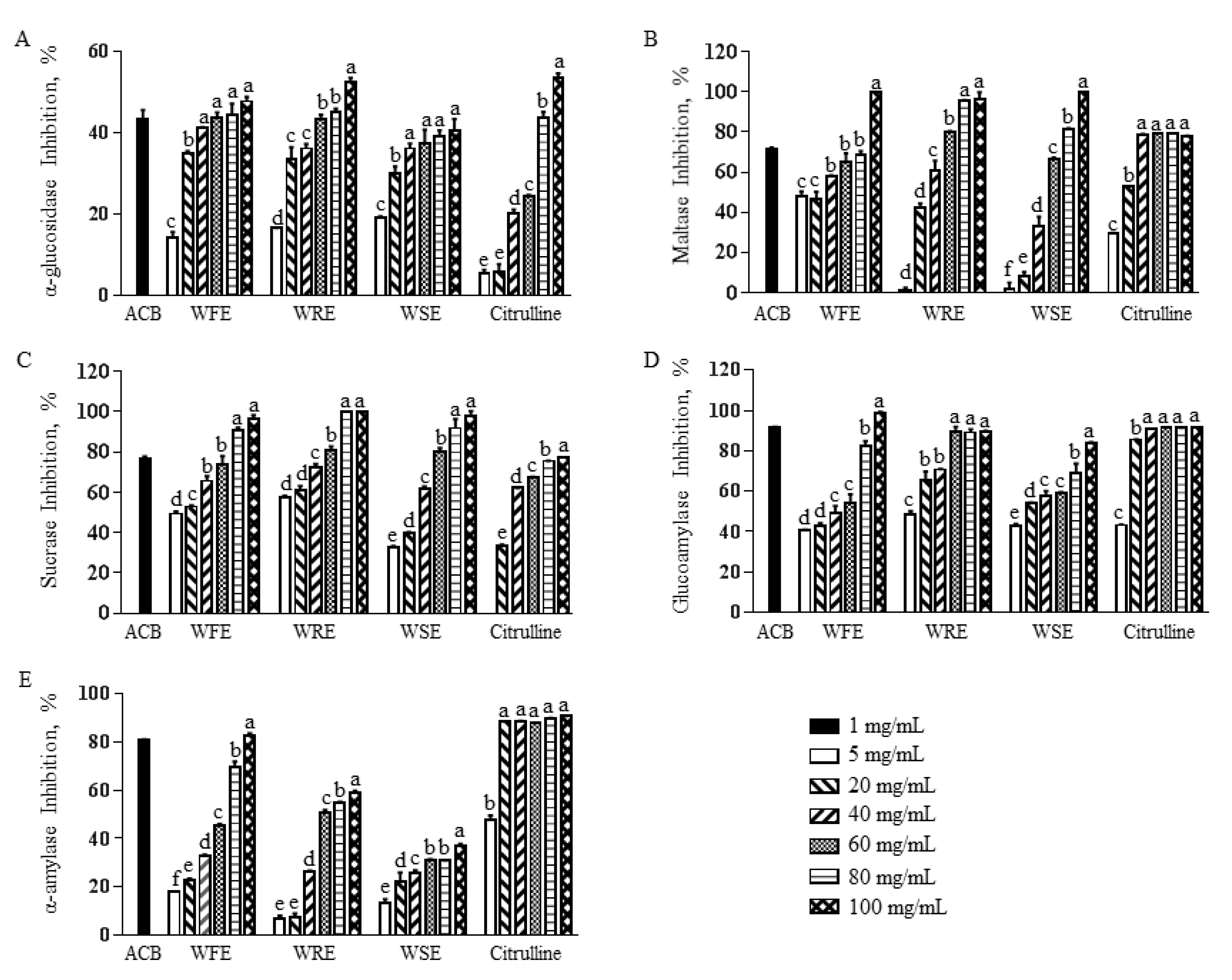
Figure 1. Effect of watermelon extracts and citrulline on carbohydrate-hydrolyzing enzymes. (A) α-glucosidase, (B) maltase, (C) sucrase, (D) glucoamylase, and (E) α-amylase activities of WFE, WRE, WSE, and citrulline at concentrations of 0, 5, 20, 40, 60, 80, and 100 mg/mL were determined. Acarbose (ACB, 1 mg/mL) was used as a positive control. Alphabet lower case letters indicate a statistically significant difference (p < 0.05).
Agriculture 12 00772 g001
Table 1. Bioactive compounds in watermelon extracts (µg/g dry extract for chlorophyll, lutein, β- carotene, and citrulline).
WFEWREWSE
Chlorophyll83.7 ± 1.6 b26.4 ± 0.3 b7880.4 ± 132.2 a
Lutein5.3 ± 0.1 b11.2 ± 0.1 b1273.0 ± 11.1 a
β-carotene1.2 ± 0.1 b2.3 ± 0.1 b20.7 ± 0.8 a
Citrulline684.4 ± 2.8 c1017.1 ± 64.3 b3101.0 ± 5.5 a
Alphabet lower case letters indicate a statistically significant difference (p < 0.05).
Table 2. The IC50 values of watermelon extracts on carbohydrate-hydrolyzing enzymes (mg/mL).
AcarboseWFEWREWSECitrulline
α-glucosidase0.03 ± 0.00 c8.12 ± 0.95 b11.95 ± 2.80 b5.68 ± 0.94 b43.49 ± 2.57 a
Maltase0.02 ± 0.00 d26.58 ± 0.46 b0.02 ± 0.00 d44.30 ± 1.17 a6.48 ± 0.32 c
Sucrase0.02 ± 0.00 d10.19 ± 0.13 c13.00 ± 1.00 b16.00 ± 0.55 a0.01 ± 0.00 d
Glucoamylase0.01 ± 0.00 d23.00 ± 0.70 a5.03 ± 0.21 c10.11 ± 0.59 b0.01 ± 0.00 d
α-amylase0.07 ± 0.00 d32.82 ± 0.79 a0.03 ± 0.01 d12.15 ±1.75 b3.20 ± 0.76 c
Alphabet lower case letters indicate a statistically significant difference (p < 0.05).
Publisher’s Note: MDPI stays neutral with regard to jurisdictional claims in published maps and institutional affiliations.