Article

Indirect Somatic Embryogenesis: An Efficient and Genetically Reliable Clonal Propagation System for Ananas comosus L. Merr. Hybrid “MD2”

by
Argelys Kessel-Domini
1,
Daisy Pérez-Brito
2,
Adolfo Guzmán-Antonio
1,
Felipe A. Barredo-Pool
3,
Javier O. Mijangos-Cortés
4,
Lourdes Georgina Iglesias-Andreu
5,
Alberto Cortés-Velázquez
2,
Adriana Canto-Flick
1,
Susana A. Avilés-Viñas
1,
Yaritza Rodríguez-Llanes
1 and
Nancy Santana-Buzzy
1,*
1
Unidad de Bioquímica y Biología Molecular de Plantas, Centro de Investigación Científica de Yucatán (CICY), Calle 43 No. 130, Chuburná de Hidalgo, Mérida 97205, Mexico
2
GeMBio, Grupo de Estudios Moleculares Aplicados a la Biología, Centro de Investigación Científica de Yucatán (CICY), Calle 43 No. 130, Chuburná de Hidalgo, Mérida 97205, Mexico
3
Unidad de Biotecnología, Centro de Investigación Científica de Yucatán (CICY), Calle 43 No. 130, Chuburná de Hidalgo, Mérida 97205, Mexico
4
Unidad de Recursos Naturales, Centro de Investigación Científica de Yucatán (CICY), Calle 43 No. 130, Chuburná de Hidalgo, Mérida 97205, Mexico
5
Instituto de Biotecnología y Ecología Aplicada, Campus para la Cultura, Universidad Veracruzana, las Artes y el Deporte. Av. de las Culturas Veracruzanas No. 101. Colonia Emiliano Zapata, Xalapa-Enríquez 91090, Mexico
*
Author to whom correspondence should be addressed.
Agriculture 2022, 12(5), 713; https://doi.org/10.3390/agriculture12050713
Submission received: 29 March 2022 / Revised: 11 May 2022 / Accepted: 12 May 2022 / Published: 18 May 2022
(This article belongs to the Special Issue Advances in Agricultural Engineering Technologies and Application)

Abstract

:
The objective of this study was to establish an efficient—direct or indirect—regeneration system for pineapple (Ananas comosus L.) plants, with a high rate of multiplication and that would preserve the genetic identity of the donor genotype (Hybrid ‘MD2’) in the regenerated plants. Ten treatments, with different concentrations of 2,4-Dichlorophenoxyacetic (2,4-D) and Picloram (P), in the absence or presence of 6-Benzylaminopurine (BAP), were used for in vitro morphogenesis induction, as well as histological and molecular techniques, in order to characterize the morphogenic responses induced. Significant differences between treatments tested, to induce callus and buds, were assessed by the Kruskal Wallis method and the Mann–Whitney U-tests. Different pineapple regeneration routes were identified, showing the high regeneration potential of this species. The medium containing 2 mg L−1 2,4-D and 2 mg L−1 BAP, where indirect somatic embryogenesis occurred, was selected as the most efficient treatment, with an average of 120 somatic embryos per explant, differing significantly from the rest of the treatments. It was also demonstrated that the pineapple plants regenerated in vitro preserved the genetic identity of the donor genotype, which represents a high degree of confidence for the application of indirect somatic embryogenesis for A. comusus clonal propagation.

Graphical Abstract

1. Introduction

Pineapple (Ananas comosus L. Merr.) is a monocotyledonous plant native to tropical America, specifically South America, belonging to the Bromeliaceae family, and is one of the most important tropical species in the international market, due to the great acceptance of its fruit by consumers [1]. The fruit is recognized as one of the finest in tropical regions, and is considered the queen of all fruits, because it is highly appreciated for its high nutritional value. It is rich in carbohydrates and saturated fats, free of cholesterol, low in sodium, and has high contents of vitamins A, C, and minerals. It is also a highly digestible food because it contains the enzyme bromelain [2]. It is currently the second tropical crop in terms of production volume, only surpassed by the banana (Musa acuminata). It is produced in many tropical regions of the world and is exported mainly to the United States of America and the European Union [3,4]. Seventy percent of its commercial production is consumed fresh, and the rest is intended for canning in syrup, so the hybrid ‘MD2’, known as “Extra Sweet”, “Golden Sweet”, or “honey pineapple”, is the most demanded in the international market, since when compared with other cultivars, it stands out for: its sweetness (values from 13 to 18° Brix), its uniform golden color, and especially for its long shelf life (more than 30 days) [2,5,6].
This plant is described as a perennial herb that grows between one and one-and-a-half meters high. Prior to fruiting, this plant generates around two hundred flowers, which, when fertilized, combine to form a polyfruit, which is known as a pineapple. Each plant generates between seventy and eighty leaves, which are like pods or sheets arranged in a spiral, usually in layers. Some cultivars have thorns on the edges of their leaves. Its flowers have foliar scales that have the property of retaining water. The embryo of the plant has only one leaf, or cotyledon, and is a self-incompatible species [1], so its main form of propagation is vegetative, based on three types of tillers: crowns, cocks, and spikes [3,7]. The crowns, located in the upper part of the fruit, have a slow but uniform development, with a low percentage of natural flowering, and their cycle can be considered late. The propagules, or tillers, that grow and develop from buds located on the peduncle, and the base of the fruit are known as roosters and the number and vigor per plant vary, with the most occurring during the first harvest and being of intermediate vigor. Nails are considered scions, and grow and develop from axillary buds, located throughout the stem, and their cycle is considered the earliest, because they are more vigorous and are the most commonly used tillers for the establishment of plantations [8].
All these species characteristics imply a very low growth and multiplication rate through vegetative propagation, since the interval between planting and fruiting can be up to two years, and only two to three uniform propagules are produced per plant, which in turn drastically limits the availability of suitable planting material to establish large areas of the crop [9]. On the other hand, the incidence of pests and diseases is higher, and this condition is aggravated by the low genetic variability present in their populations, since traditional genetic improvement is delayed and inefficient [8]. Given this problem, it is necessary to use biotechnological tools that allow the rapid and massive production of uniform plants with high phytosanitary quality, in a rapid and massive way, thus producing larger fruits with higher agricultural yields. Technologies based on plant tissue culture provide us with very valuable tools for the massive and uniform propagation of this crop in very short cycles of time and at any time of the year [10,11]. However, it should be noted that the conditions required for tissue culture can result in genomic changes in plants, resulting from in vitro culture [7,12,13,14].
Tissue culture conditions, such as the use of growth regulators, successive transfers of explants, certain temperatures, and culture medium pH, sometimes lead to alterations at the cellular level, which result in genomic changes in plants from in vitro culture [7,12] which are not favorable in micropropagation. Authors such as Akdemir et al. [15] found that when high concentrations of cytokinins are used in long-term pineapple cultivation, somaclonal variations occur, which can be detected by molecular markers. In another study, differences in morphological and physiological characters between regenerants and mother plants were observed, when Naphthaleneacetic acid (NAA) and 6-Benzylaminopurine (BAP) were used for plant regeneration [7]. Years later, in another study using these same regulators, DNA methylation increase was found in the dedifferentiation process (callus formation) and differentiation (seedling regeneration), and it was concluded that these changes lead to somaclonal variation in pineapple [13].
Therefore, the establishment of a tissue culture technology in this valuable fruit plant requires reproducible protocols for each of the process stages, and for this, it is necessary to define the structures that originate in the different morphogenic routes; as well as to analyze if the change that originated in the genetic program of these cells leads to somaclonal variation, since a fundamental requirement in the commercial industry when working with elite genotypes is to maintain their relevant agronomic characteristics [11]. For this reason, the present research hypothesizes that with the application of somatic embryogenesis, many genetically stable plants are obtained, and the multiplication rate of this fruit species is increased in a short period of time. Therefore, the objective of this work is to present the advances achieved in the search for a highly efficient and genetically reliable mass propagation technology, in which vigorous “MD2” pineapple in vitro plants with a high survival rate are generated.

2. Materials and Methods

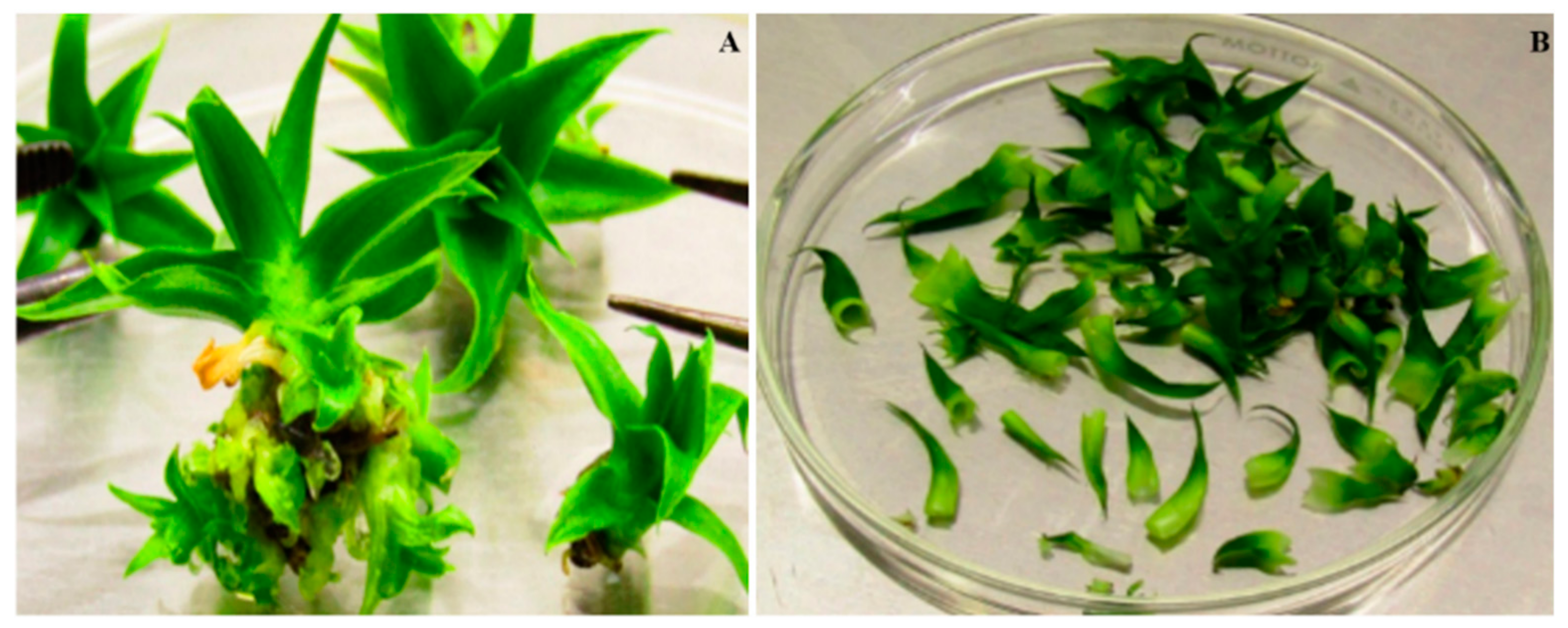
2.1. Explant Selection

For the induction of pineapple morphogenic structures, the leaf base from young (4–6-week-old) in vitro plants of the ‘MD2’ hybrid undergoing in vitro vegetative growth (Figure 1) was used as plant material. Leaves explants were placed horizontally with their adaxial surface on the culture medium.
Four treatments were established and tested to induce morphogenesis, in which different concentrations of naphthaleneacetic acid (NAA) and benzylaminopurine (BAP) were used in Murashige and Skoog (MS) [14] medium, supplemented with sucrose (30 g L−1), Myoinositol (100 mg L−1), and Thiamine (5 mg L−1). The explants remained under conditions of 16 h of light and 8 h of darkness, and a temperature of 27–30 °C. The shoots obtained were subcultured on the same medium every 45 days until shoots were obtained. For callus regeneration, six treatments were established and evaluated. Where an MS medium was used, which was supplemented with sucrose (30 g L−1), Myoinositol (100 mg L−1), Thiamine (1 mg L−1), and different concentrations of regulators: 2,4-Dichlorophenoxyacetic (2,4-D) and Picloram (P), in the absence or presence of BAP (Table 1). In this case, the explants were incubated in total darkness at 25–28 °C and subsequently, the calluses obtained were subcultured in the same medium (MS) every 45 days. From the second subculture (90 days), they were transferred to conditions of a photoperiod of 16 h of light and 8 h of darkness, and a temperature of 27–30 °C, to induce the formation of morphogenic structures (embryos or shoots) in an MS medium without auxin, which contained BAP at different concentrations (0.5, 1, and 2 mg L−1), in which they remained 145 days, with refreshing passes every 45 days. The shoots obtained were transferred to a rooting medium [MS + (NAA 1 mg L−1/1 mg L−1 BAP)] until reaching seedlings of 6 to 8 cm in height.
In all treatments, the media were solidified with Gelrite (3000 mg L−1), and 40 mL of it were distributed in magentas and sterilized in an autoclave at 121 °C for 15 min. Five explants were planted per magenta, and a completely randomized experimental design was used, with three replications, for which a total of 45 explants were planted per treatment, and when more than 50% of the explants presented morphogenic structures, the following features were evaluated: the number of explants with morphogenic structures, the type of morphogenic response (calluses, shoots, and direct and indirect somatic embryos), number of embryos per treatment, number of shoots per treatment, and number of plants obtained.

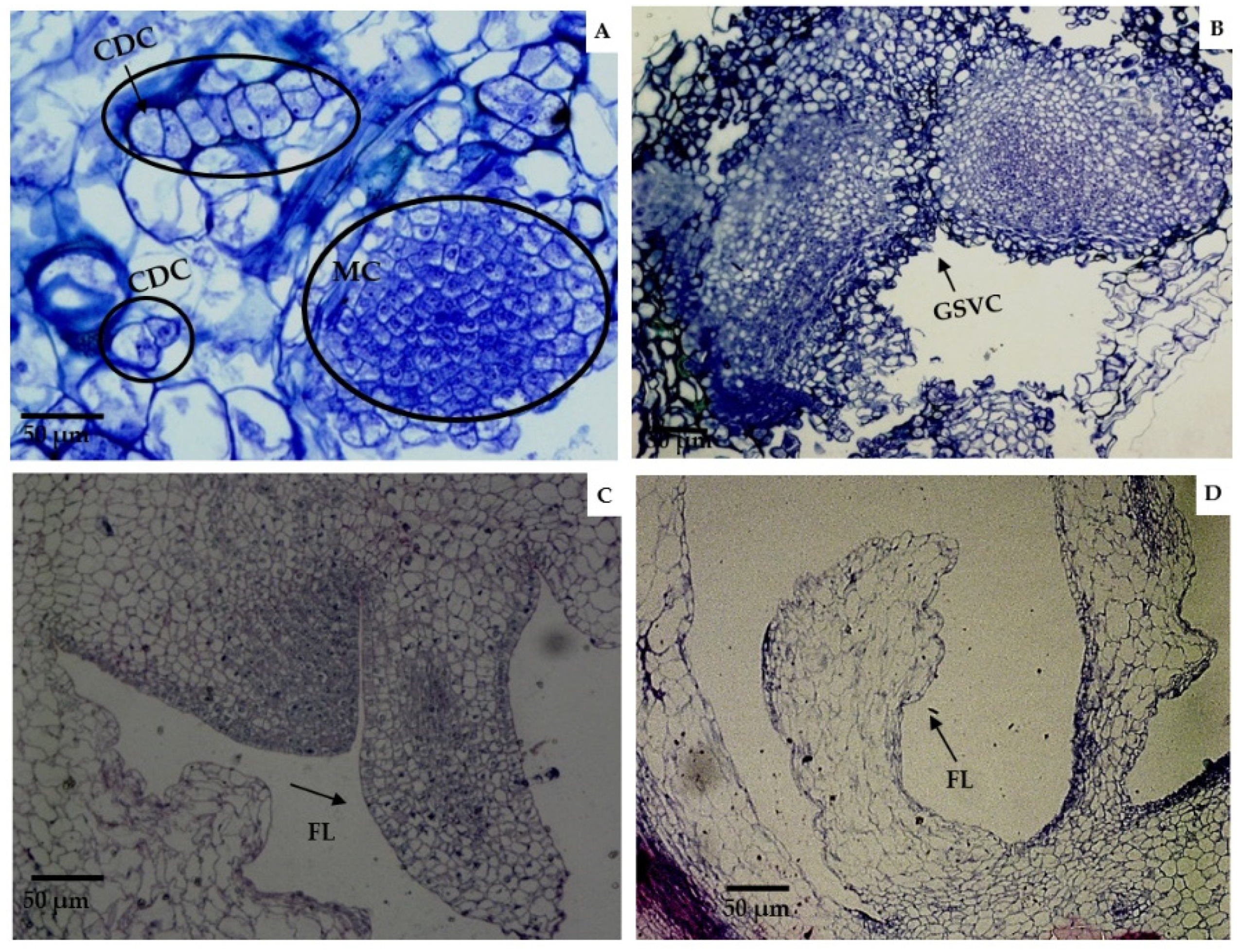
2.2. Histological Analysis of Morphogenic Structures That Originated in In Vitro Regeneration

For histological processing, samples of the different morphogenic structures formed in the different treatments were taken. The methodology used was that described by Piven et al. [16], though slightly modified. Biological samples were taken in small portions of approximately 0.5–1.0 cm2, containing callus mass, embryos, and shoots. Three samples per treatment, taken at random, were fixed for 96 h in a FAA solution containing formaldehyde (4%), glacial acetic acid (95%), ethanol (95%), and distilled water, in a ratio of 2:1:10:7, respectively. After fixation, the tissues were washed three times (30 min each) with distilled water to remove fixative residues. Subsequently, the washed samples were subjected to dehydration treatments in different concentrations of ethanol (C2H6O), in a gradual and ascending manner (30%, 50%, and 70%) [17]. The samples were then left to stand in the last ethanol concentration (70%) for 24 h, and the dehydration process was continued with 85%, 96%, and absolute or anhydrous ethanol. Two alcohol changes were made for each concentration, with an immersion time of one hour for each change. The tissues were then embedded in JB-4 plastic resin according to the manufacturer’s instructions (Embedding Solution Poly-sciences, Los Angeles, CA, USA). The embedded tissues were immersed in solution A + catalyst (Benzoyl Peroxyde Plasticized, Polysciences, Warrington, PA, USA ) and maintained for one week at room temperature (20 to 25 °C). At the end of resin infiltration, casts were made, including the tissues for transverse and tangential sectioning on an HM 325 rotary microtome (Microm, CA, USA) with a thickness of 2–4 µm. Histological sections were serially performed and spread in distilled water and collected on a 26 mm × 76 mm slide. To visualize the cellular organization, the histological sections were stained with Toluidine Blue and Paz stain, and observations were made on an Axioplan Zeiss microscope, equipped with AxioCam ICc 5 (Jena, Germany).

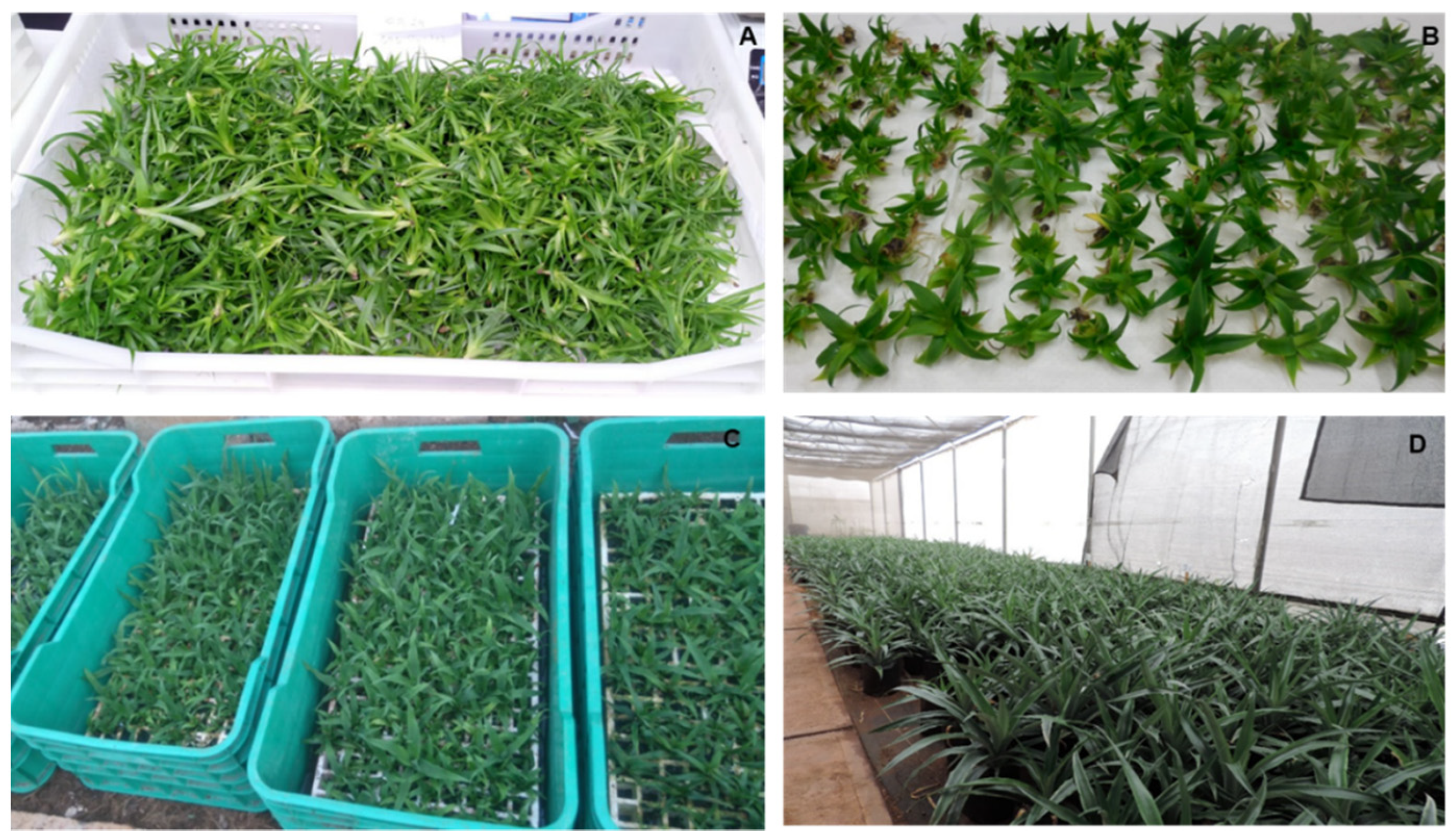
2.3. Acclimatization of Plants In Vitro

Seedlings with a height of 6–8 cm were extracted from the culture containers, washed with abundant running water, and placed in plastic trays for transfer to the greenhouse, where they were established in trays containing a mixture of red soil + coconut fiber + perlite (1:1:1) as substrate, following the methodology described by Guzmán-Antonio et al. (data in process of publication). After 45 days, the seedlings were transplanted into black plastic bags (22 cm × 26 cm), where they remained for about 16–20 weeks, for subsequent transfer to the field.

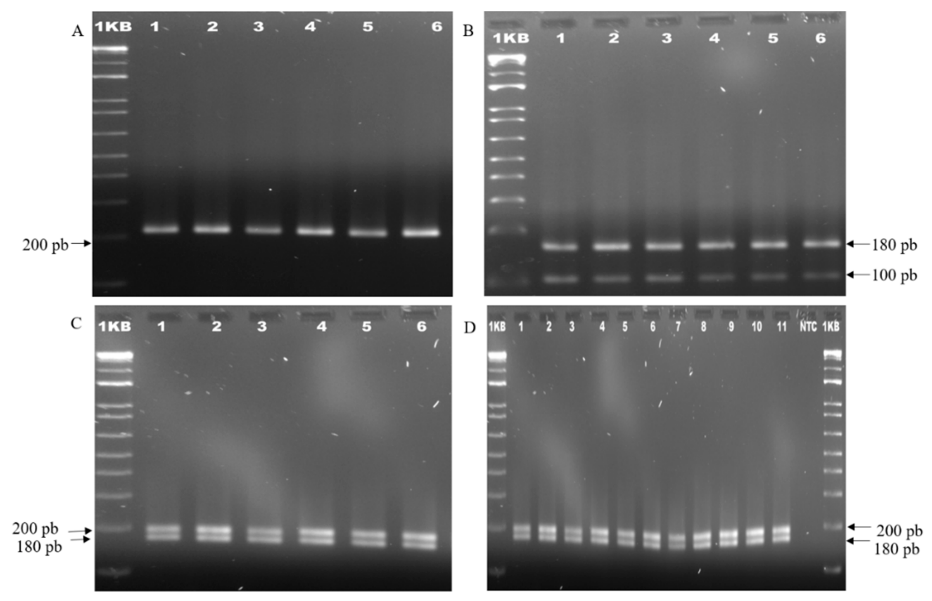
2.4. Analysis of the Genetic Stability of the Plants Obtained from In Vitro Regeneration

Genomic DNA extraction. For DNA extraction, leaf samples were taken from 10 plants from the different treatments. Total genomic DNA was extracted according to the method reported by Tapia et al. [18]. The quality and integrity of the DNA was determined by electrophoresis in a 0.8% (w/v) agarose gel, and the concentration and purity of the genomic DNA was determined by reading absorbance at 260 and 280 nm in a spectrophotometer (Nanodrop 2000, Madrid, Spain), according to the methodology described above. Once the DNA was obtained, we proceeded to the analysis to determine the genetic stability with the SSR and ISSR markers.
Amplification by SSR. To determine the appropriate alignment temperature, each SSR primer was tested at 50 °C, 55 °C, and 60 °C. Amplifications were developed in a final volume of 25 µL for a reaction, containing 40 ng of template genomic DNA, 0.3 µM of each SSR primer, 0.15 mM of dNTP mix (Invitrogen, Carslbad, CA, USA), 1.5 mM of magnesium chloride (Invitrogen, Carslbad, CA, USA), 1× buffer, and 1.2 U of Taq polymerase (recombinant) (Invitrogen, Carslbad, CA, USA). Twenty SSR primers reported by Wang et al. [19] were evaluated. For each amplification reaction, three samples from each treatment with three replicates per primer were analyzed.
PCR reactions were performed on a Thermal cycler 2720 (Applied Biosystems, CA, USA). The amplification program began with a 2.30 min cycle at 94 °C, followed by 35 cycles at 94 °C at 45 s. The alignment temperatures were 55 °C, 60 °C (45 s), and 72 °C (1 min), with an 8-min final extension cycle at 72 °C. PCR products were separated on 2% agarose gels in a 0.1× TBE buffer (0.04 M Tris hydroxymethyl aminomethane (Tris-HCl); 1 mM ethylenediamine tetra acetic acid (EDTA); 0.04 M boric acid; pH 8) at 100 v (constant voltage). Staining was performed with ethidium bromide and gels were visualized in a U.V. light transilluminator. (BIO-RAD Gel Doc Ez Imager, Hercules, CA, USA). By comparing the size of the amplified fragments to a 1 Kb plus DNA molecular weight marker (Invitrogen, Carslbad, CA, USA), the size of the amplified fragments was determined.
Amplification by ISSR. Amplifications were made in a final volume of 25 µL for a reaction containing 20 ng of template genomic DNA, 1 µM ISSR primer, 0. 25 mM dNTP mix (Invitrogen), 2.5 mM magnesium chloride (Invitrogen, Carslbad, USA), 1× buffer (Invitrogen, Carslbad, CA, USA), and 1.0 U Taq polymerase (recombinant). Five ISSR primers (IS01, IS13, IS14, IS17, and IS19), reported by Weiguo et al. [20], were evaluated. In each amplification reaction, 10 samples per previously described treatment were analyzed, and three repetitions of the amplifications per primer were performed. PCR reactions were run on a thermal cycler 2720 (Applied Biosystems, California, USA), using the following amplification program: an initial cycle at 94 °C for 2 min, followed by 45 cycles at 94 °C (45 s), where the alignment temperature was 54 °C (45 s) and 72 °C (1 min and 30 s), and a final extension cycle at 72 °C for 7 min. PCR products were analyzed on 2% agarose gels in a 0.1X TBE buffer (0.04 M Tris hydroxymethyl aminomethane (Tris-HCl); 1 mM ethylenediamine tetra acetic acid (EDTA); 0.04 M boric acid; pH 8) at 100 v (constant voltage). The bands were visualized by ethidium bromide staining on a U.V. light transilluminator. (BIO-RAD, Gel Doc Ez Imager, Hercules, CA, USA). The size of the amplified fragments was determined by comparison with a 1 Kb plus DNA molecular weight marker (Invitrogen). The Polymorphic Information Content was calculated by the methodology cited by Anderson et al. [21].

2.5. Statistical Analysis

The SPSS program (V.17.0) was used to analyze the data. The Kruskal–Wallis method was used, and the quantitative variables that influenced the variation observed among the different treatments established were identified. Paired t-tests (post hoc to Mann–Whitney U) were performed to test for differences between treatments. For the molecular analysis, the percentage of monomorphism obtained by each SSR and ISSR primer was determined, considering the total number of monomorphic bands generated for each of them, over the sum of the total number of bands detected with all the primers, times 100, and this value was considered to determine the genetic stability of each treatment.

3. Results

3.1. Evaluation of the Treatments Established for In Vitro Regeneration of the Pineapple Hybrid ‘MD2′

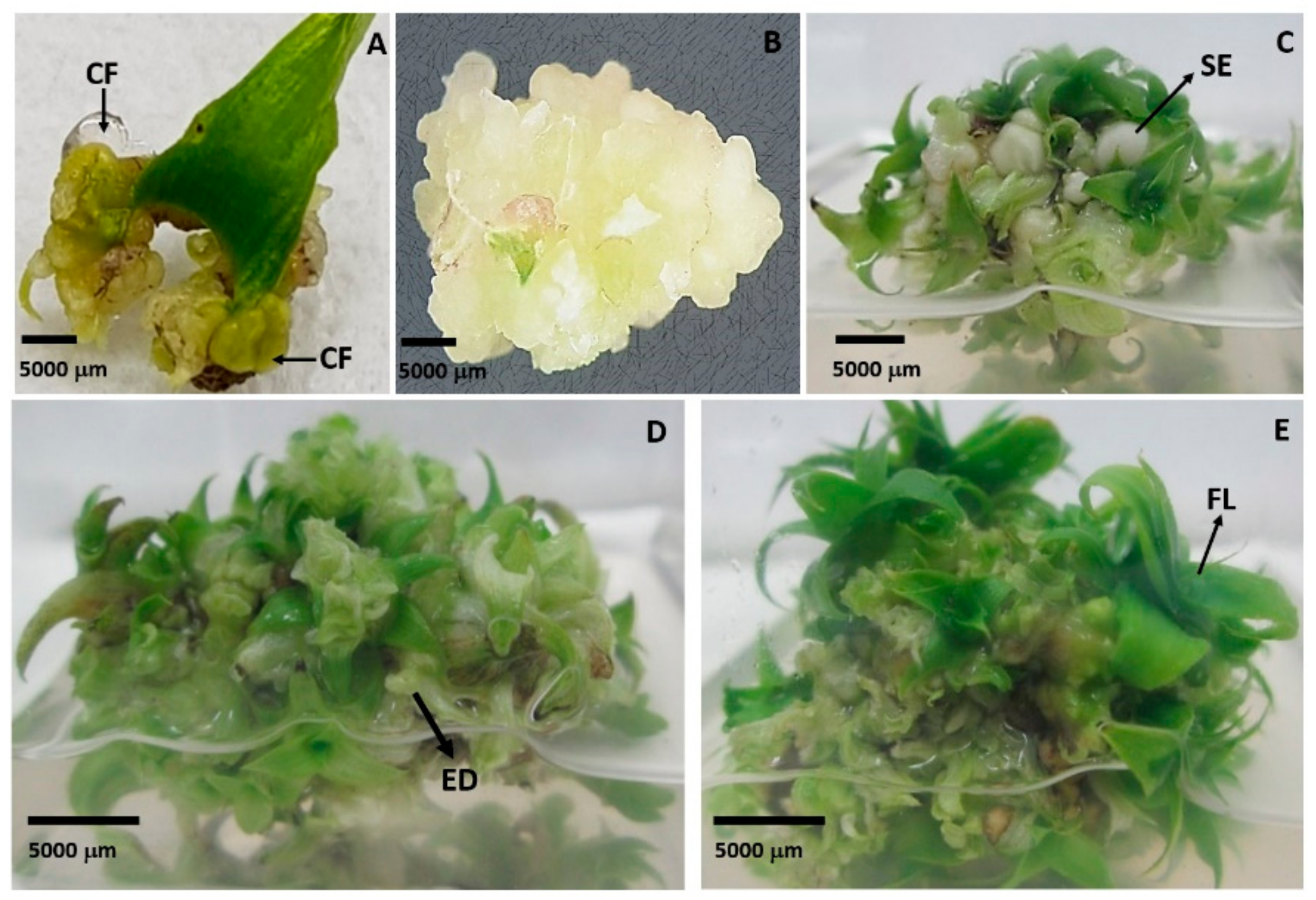
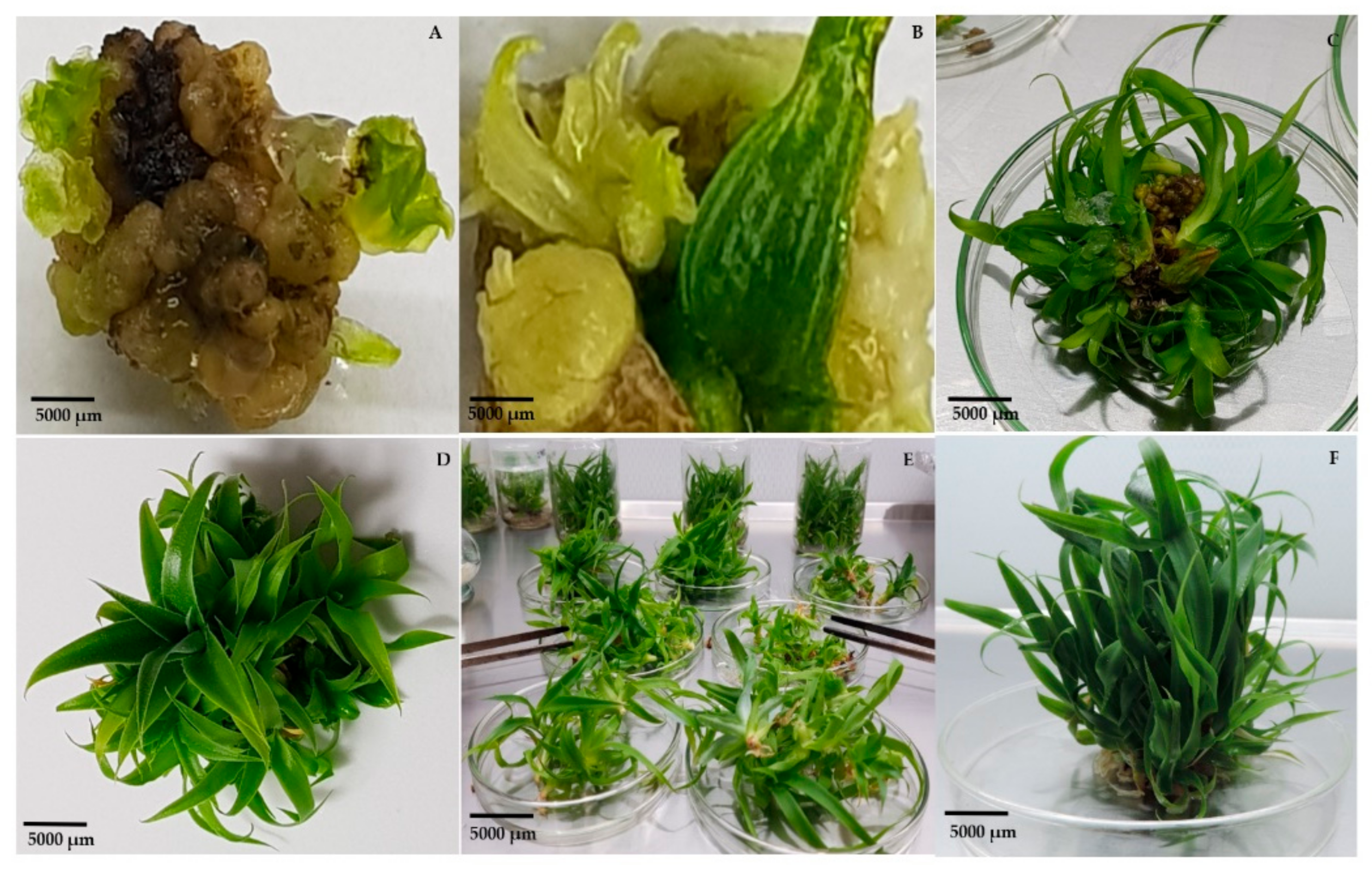
3.1.1. Direct Somatic Embryogenesis (DSE)

Results obtained in the treatments established for the in vitro regeneration of the ‘MD2’ hybrid showed that depending on the balance of BAP/NAA to which the explants were exposed, the morphogenic response varied (Table 2). Direct Somatic Embryogenesis occurred in some of the explants in the treatment M3 used since the formation of abundant globular structures directly from the explant was observed (Figure 2A,B). Unlike the rest of the treatments that contained these two growth regulators, the structures formed evolved rapidly at advanced stages of their development, which is shown in Figure 2B,C, as well as their conversion to plants, so M3 was identified as an adequate medium for the direct induction and proliferation of ‘MD2’ pineapple embryos from leaf segments. In the rest of the treatments, in which BAP and NAA were used, no embryogenic structures formation was observed, but there was formation of leaf primordia directly from the explant and multiple budding.
From the shoots obtained in all the treatments, the same number of plants was obtained in the rooting medium; this means that there was a 100% conversion of shoots to plants.

3.1.2. Indirect Somatic Embryogenesis (ISE)

By analyzing the response of leaf explants to the different treatments, it could be observed that in all treatments containing 2,4-D, alone or in combination with BAP, there was callus formation, although only explants exposed to the treatment with 2 mg L−1 of 2,4-D and 2 mg L−1 of BAP (M7) showed the formation of an embryogenic callus that rapidly evolved to form somatic embryos. At 30 days of culture, this callus was characterized by showing a creamy white coloration and a semicompact consistency (Figure 3A,B). In the treatments in which Picloram was present, the formation of a compact, slow-growing, beige-colored callus was observed, turning dark brown rapidly. After 60 days, isolated and minute outgrowths were visible on the periphery of the callus. Only in medium M7 (2 mg L−1 of 2,4-D/2 mg L−1 of BAP) was embryogenic callus formation observed, and 93.3% of the explants responded to the treatment (Table 2). In this treatment (M7), the callus started in the basal zone of the leaf and, six weeks later, the first embryogenic structures (globular) became visible from a primary, vitreous white, incipient, semicompact callus. These first embryos, of a faint green color, emitted a leaf primordium, typical of monocotyledonous species, which allowed for the inferring that they had a well-defined apical meristem (Figure 3A). After the first subculture (six weeks), a callus of semifriable consistency proliferated on the callus and formed 30 days after it was placed in the explant in the medium (Figure 3B), which evolved to form an abundant and granular embryogenic callus, revealing numerous embryogenic structures at early and advanced stages of their development (Figure 3C). These structures gave way to the appearance of numerous, tiny, pale green apices, which evolved into small buds located throughout the periphery of the callus, while in other areas of the callus there was an intense proliferation of embryogenic structures in early stages (globular), which shows an asynchrony typical of somatic embryogenesis (Figure 3D). The high embryogenic capacity (120 SE/Explant) of the callus induced in the M7 medium (2 mg L−1 of 2,4-D and 2 mg L−1 of BAP) was observed, differing significantly from the rest of the treatments evaluated. (Figure 3E). The rapid evolution of somatic embryos from globular to an advanced stage, characteristic of somatic embryogenesis in monocot species, became evident.

3.1.3. Indirect Organogenesis (IO) and Shoot Development

The results obtained showed that in the treatment in which 2.0 mg L−1 NAA was combined with 5.0 mg L−1 BAP (M2), there was a high responsiveness to regeneration from the explant cells’ leaf segment, so it showed a fast and efficient response to shoot formation (Figure 4). It is worth noting that in this treatment, an average of 30 shoots per explant were formed, which was significantly higher than in the rest of the treatments in which regeneration occurred via organogenesis, both directly and indirectly (Table 2). The formed shoots were characterized by their vigor and rapid development, which preserved the vascular connection with the maternal tissue during the first generation of shoots. Figure 4 shows the evolution of this regeneration process.
On the other hand, it was observed that the shoots formed in the M4 treatment developed and became suitable seedlings for transfer to the acclimatization phase in less time compared with the rest of the evaluated treatments. Therefore, this result allowed us to recommend this medium for the final stage of development of all the shoots obtained in the different regeneration pathways, either by direct and indirect somatic embryogenesis or by the direct organogenesis pathway. In treatments M5, M6, M8, M9, and M10—in which indirect organogenesis (IO) occurred via callus formation—the callus formed was very slow growing with a compact consistency that quickly turned dark, so none of these treatments had embryogenic structures because only tiny shoots were formed in certain areas of the callus, which subsequently evolved favorably in plants. However, it should be noted that plant regeneration was very slow in these treatments.

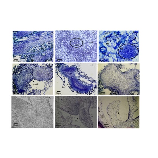
3.2. Histological Analysis

The histological analyses of the morphogenic structures that originated from callus in the M7 treatment showed an intense cell division activity in callus cells, clearly distinguishing the formation of abundant, well-defined meristematic centers (Figure 5A) as a prelude to somatic embryogenesis. These cells were characterized by being small and isodiametric, with a dense cytoplasm and a prominent vacuole. An intense asymmetric cell division was observed, the cells began to divide longitudinally and transversally, giving way to the histogenesis process (Figure 5A), in which asynchronously, globular somatic embryos could be observed in the same callus and in an advanced stage of their development (Figure 5B). There was not a vascular connection to maternal tissue. Through the longitudinal section of the embryo (Figure 5C), the development of the central cylinder could be seen in the innermost part of the scutelum, where later the formation of leaf primordia can be seen (Figure 5D).

3.3. Acclimatization

Once the developed in vitro plants reached 6–8 cm in height, they were isolated (Figure 6A,B) and transferred to greenhouse conditions, where they were placed in trays containing a previously determined substrate mixture of red soil + coconut fiber + perlite (1:1:1) (data in process of publication) (Figure 6C). The in vitro plants, after the hardening stage in the trays (45 days), were transplanted into bags where they remained for 16–20 weeks. After this time, the in vitro plants showed optimal development with long, thick, dark green, shiny, and turgid leaves, and thick and vigorous stems, which demonstrated an adequate physiological state (Figure 6D). Survival of the in vitro plants exceeded 98% at the time of transfer to the field.

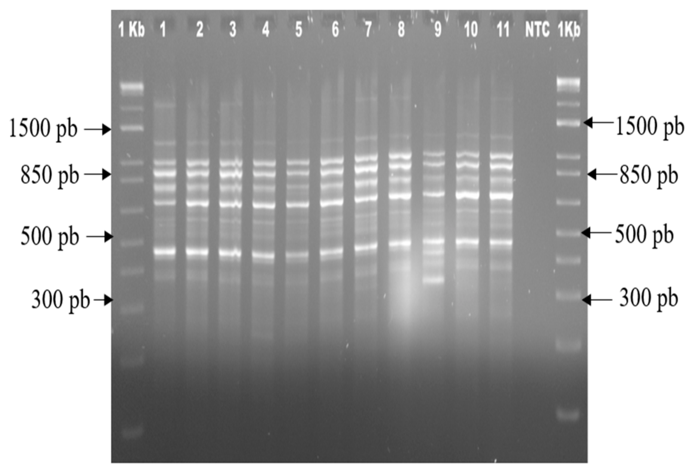
3.4. Genetic Stability of the Plants Obtained from In Vitro Regeneration

SSR and ISSR markers were used to assess the genetic stability of pineapple plants that were regenerated in various treatments. Of the 20 SSR primers used for molecular analysis, 15 produced amplification products with adequate resolution and reproducibility to evaluate the genetic stability of the regenerated plants. Table 3 shows a total of 21 monomorphic bands for each primer, obtaining 100% monomorphism. The number of amplified fragments for each primer was between 1 and 2 bands, for an average of 1 fragment per primer among all the treatments evaluated. The height of the fragments obtained was in the range of 100 and 300 bp, and 100% monomorphism was observed in all treatments (Figure 7). This monomorphism was also corroborated by the analysis of randomly selected individual plants from the treatment where indirect embryogenesis M7 (2 mg L−1 2,4-D/2 mg L−1 BAP) occurred (Figure 7A–C) and the genetic stability of the pineapple in vitro plants regenerated in the 10 treatments evaluated for this marker could be appreciated.
Of the five ISSR primers used for the analysis, all were appropriate to observe the monomorphism patterns with adequate resolution and reproducibility. Table 4 shows the total number of bands obtained by each primer, as well as the percentage of monomorphism. Of these 35 monomorphic bands, representing 89.74% of monomorphism, primers IS14 and IS17 were the ones that presented 100% monomorphic bands.
The number of amplified fragments for each primer ranged from 4 to 12 bands, for an average of seven fragments per primer, among all treatments evaluated. The height of the fragments obtained was in the range of 250 to 2000 bp. Figure 8 shows the band profiles obtained with primer IS19, which showed the highest number of bands.
With respect to the monomorphism detected (Table 5), 100% could be seen with this marker in treatments M3 (2 mg L−1 BAP/2 mg L−1 NAA), M4 (1 mg L−1 BAP/1 mg L−1 NAA), M5 (1 mg L−1 2,4-D), M6 (1 mg L−1 2,4-D/1 mg L−1 BAP), and M7 (2 mg L−1 2,4-D/2 mg L−1 BAP). At the same time, it was visualized that the treatment with the lowest stability, 94.26%, was the one in which the highest concentration of Picloram, M10 (5 mg L−1 P/0.05 mg L−1 BAP), was applied. When the PIC values were calculated, they were very low; an overall value of 0.298 was obtained, which indicates that these are genetically stable plants (Table 4).

4. Discussion

Conceptually, the differences between somatic embryogenesis and organogenesis basically distinguish one from the other by the presence of both poles in the somatic embryo (caulinar and radical) and not so in organogenesis; in addition, somatic embryos have no vascular connection with the mother tissue and multiply adventitiously (secondary embryogenesis). In the present investigation, morphogenesis occurred only in the basal segments of the pineapple leaf and demonstrated a structure that was not directly connected to the callus, which subsequently emitted leaves and roots without requiring induction of the process. However, a radical pole is appreciated, apparently structurally rudimentary compared to that of the somatic embryo of other species. Figure 2F,G shows the intense multiplication of the structures, which allows maintaining the cultures at different stages of development: callus proliferation, multiplication of morphogenic structures, and plant development. In the study, different types of tissues were observed: nodular and creamy, compact and brown, and friable and brown, and it was noted that in the treatment with 2,4-D in the presence of BAP (M7), there was a rapid formation of callus and a greater number of these, which may be related to the fact that 2,4-D is a synthetic regulator that stimulates a rapid synchronized cell division [22,23,24]. It was also observed that when the concentrations of 2,4-D and BAP were increased, callus texture was favored and embryogenic callus was obtained, which coincides with that reported by other researchers [25], who recommended combining cytokinin and auxin to induce embryogenesis in pineapple.
It is worth noting that several years ago, other auxin origin regulators were highlighted to induce callogenesis in pineapple, since many authors considered that in this crop, 2,4-D sometimes inhibited the induction of callus [7,26,27]. However, in the present research, the efficiency of 2,4-D in the induction of embryogenic structures that gave rise to many plants with respect to Picloram was perceived, which disagrees with what was cited by the authors. Therefore, it is not possible to generalize methodologies or work protocols, since the culture media and the selected strategies must be specific for each particular situation, and at the same time, it must be considered that not all cultivars respond in the same way to in vitro morphogenesis because there must be an interaction or synergic action between the exogenous treatments and the particular endogenous conditions of the plant in question to favor the formation of competent organogenetic meristematic centers in particular areas of the callus and, subsequently, their determination for the development of specific organs [28]. It can be inferred from in vitro regeneration of pineapple plants via somatic embryogenesis that the pattern concerning in vitro regeneration of pineapple plants, in the opinion of several authors, is not well defined [23,24]. Some consider embryogenic callus to be yellowish in color and nodular in appearance, despite the fact that no embryos can be distinguished, and use the term “shoots” for plants regenerated from embryogenic tissues [24]. Similarly, if somatic embryos are considered to develop from different structures with a closed vascular system, the term “seedlings” should be the most appropriate, according to other authors [28].
In the organogenetic system, shoot regeneration was achieved in two ways: shoots were regenerated directly from the bases of the treatment leaves (M1 and M4), or structures of nodular and globular appearance were formed (adventitious organogenesis) (M2), by which multiple sprouting was obtained. It should be appreciated that a group of authors who have worked with species of the Bromeliaceae family [29] demonstrated that the basal region of bromeliad leaves presents vascular elements as intercalary axillary meristems that attribute a high regenerative potential to the cells in dedifferentiation when activated by growth regulators and that the induction and development of monopolar structures generates adventitious shoots with high frequencies and that these structures, when isolated from a single explant, can respond and generate adventitious shoots from nodule cultures. Specifically, in the case of the Vriesea species, several lines of evidence suggest that the in vitro regenerative pathway follows a specific pattern associated with nodule cluster cultures [30] as competent morphogenic structures that are globular in shape, translucent to yellowish in color, and compact in texture. They also posit that, under suitable conditions, these nodule clusters develop into a series of small (0.5 cm long) buds called microhotels, which elongate to give rise to shoots. This terminology is used by them in in vitro regenerative systems in bromeliads and suggests that there are probably some species in this family that exhibit a third morphogenic route, intermediate between somatic embryogenesis and organogenesis, and that probably exceeds the morphogenic potential of the two best known routes [29,30], so that future research in culture could value a more in-depth study in this regard.
Although SSR primers have been used to assess the genetic diversity of species [19,31], their application in the analysis to detect the incidence of somaclonal variation has not been reported. In turn, ISSRs proved to be very useful in the analysis of the genetic stability of micropropagated plants by different breeding methods [32]. Hence, in the present investigation, a set of SSR and ISSR primers were used to try to analyze the entire pineapple genome, and the levels of monomorphism that were detected by both methods were evaluated, which was very useful because the application of only one type of molecular marker can underestimate the levels of monomorphism and genetic stability [33]. In the evaluation of the results obtained with each marker, ISSRs were shown to be more informative because they provided a greater number of bands than SSRs and were able to detect certain percentages of polymorphism in three of the treatments evaluated: M1 (5 mg L−1 BAP), M2 (5 mg L−1 BAP/2 mg L−1 NAA), and M10 (5 mg L−1 P/0.05 mg L−1 BAP). However, when the PIC value was analyzed, the polymorphism was found to be negligible, which could be considered as monomorphism. The explanation for these differences in the resolution of the two marker systems could be related to the specificity of the species with each primer used [34] and the characteristic of ISSRs to express many bands compared to SSRs that express few, which consequently influences greater polymorphism. However, using both techniques to analyze the genetic stability in the established regeneration protocols was extremely beneficial because it allowed us to show that they are suitable for clonal propagation of this valuable fruit tree.
The present research is necessary and of great benefit since the response of A. comosus to growth regulators with respect to plant regeneration is considered by many researchers as variable, so that similar studies are necessary in the crop to deepen the more precise knowledge of its relationship with the different types of growth regulators, their combinations, and doses capable of stimulating a regenerative process at high frequency. From the different treatments established, it was possible to systematically characterize by optical microscopy the formation and development of morphogenic structures that gave rise to embryogenic and organogenetic tissues that can be propagated and used continuously as a source for the regeneration of new plants. In addition, when the multiplication coefficient was analyzed, a protocol of high efficiency in short-term regeneration was obtained, since an average of 120 plant shoots representing clones of the hybrid “MD2” was estimated. It was demonstrated that they are genetically homogeneous plants, so it is expected that the relevant characteristics, such as its high vitamin C and total soluble solids content, its low acidity, the striking color of its pulp, and its long shelf life, which distinguish it from the rest of the pineapple cultivars and make it the most demanded in the international market, will be maintained.

5. Conclusions

In vitro regeneration of pineapple hybrid ‘MD2’ was achieved in different ways: shoots regenerated directly from the bases of the leaves (M1 and M4), adventitious organogenesis (M2), indirect organogenesis occurred via callus formation (M5, M6, M8, M9, and M10) direct somatic embryogenesis (M3), and indirect somatic embryogenesis (M7). The most efficient treatment was M7 (2 mg L−1 2,4-D and 2 mg L−1 BAP) because indirect somatic embryogenesis occurred, with an average of 120 somatic embryos per explant. The application of ISSR and SSR to analyze the genetic stability in the different established regeneration protocols demonstrated that they are suitable for the clonal propagation of this valuable fruit tree. Pineapple plants regenerated in vitro were shown to maintain the genetic identity of the donor genotype, which means that indirect somatic embryogenesis can be applied for clonal propagation of A. comosus.

Author Contributions

Conceptualization, N.S.-B. and A.K.-D.; methodology, A.C.-F., F.A.B.-P., A.G.-A., D.P.-B. and J.O.M.-C.; investigation, A.K.-D., S.A.A.-V., Y.R.-L. and A.C.-V.; writing—original draft preparation, A.K.-D. and N.S.-B.; writing—review and editing, L.G.I.-A., J.O.M.-C., D.P.-B. and N.S.-B.; project administration, N.S.-B. and A.C.-F.; funding acquisition, N.S.-B. All authors have read and agreed to the published version of the manuscript.

Funding

This research was funded by Consejo Nacional de Ciencia y Tecnología de México (CONACYT), grant number PN/2016/3953.

Acknowledgments

To Yasmin A. Sánchez-Rodríguez, Anuar A. Magaña-Alvarez, and Hernán Morffi-Mestre for technical support.

Conflicts of Interest

The authors declare no conflict of interest.

References

  1. Jaji, K.; Man, N.; Nawi, N.M. Factors affecting pineapple marker supply in Johor, Malaysia. Int. Food Res. J. 2018, 25, 365–371. [Google Scholar]
  2. Abdul, N.A.; Chin, B.; Razik, M.; Madon, M.; Khalid, N.; Syafawati, N. Abscisic acid and salinity stress induced somaclonal variation and increased histone deacetylase (HDAC) activity in Ananas comosus var. MD2. Plant Cell Tiss. Org. 2018, 133, 123–135. [Google Scholar] [CrossRef]
  3. Blanco, H.A.; Vargas, T.C.; García, T. Regeneración in vitro de plantas de piña (Ananas comosus, L. Merr.). Rev. Colomb. Biotechnol. 2017, 19, 7–20. [Google Scholar] [CrossRef]
  4. Hikal, W.M.; Mahmoud, A.A.; Hussein, A.H.; Ahl, S.A.; Bratovcic, A.; Tkachenko, K.G.; Kakaniova, M.; Maldonado, R. Pineapple (Ananas comosus L. Merr.) waste streams, characterization and valorization: An overview. Open J. Ecol. 2021, 11, 610–634. [Google Scholar] [CrossRef]
  5. Russel, A. Piña miel: Un nuevo producto para aumentar la competitividad. 40 casos de éxitos. SAGARPA 2017, 1–18. Available online: https://www.redinnovagro.in/casosexito/2017/Pi%C3%B1a_Amador_Russell.pdf (accessed on 22 November 2021).
  6. Mercado, J.N.; Tortoledo, O.; García, J.M.; Báez, R.; García, B.Y.; Ávila, J.; Corella, D.A.; Cruz, M.C.; Velásquez, D.; Zuñiga, B.S. Calidad comercial de piña MD2 (Ananas comosus L.) tratada en postcosecha con ácido 2-hidroxibenzoico. Rev. Iberoam. Tec. Poscosecha 2019, 20, 1–15. Available online: https://www.redalyc.org/journal/813/81361553004/html/ (accessed on 22 November 2021).
  7. Nouri, F.; Farahani, F.; Noormohammadi, Z. Somaclonal variation in the in vitro regenerated pineapple (Ananas comosus): Investigation of the cellular characteristics, biochemical specificities and ISSR markers. Phytol. Balc. 2017, 23, 73–83. [Google Scholar]
  8. Garcidueñas, J.A. Caracterización Morfológica Y Molecular de Piña (Ananas Comosus, (L.) Merr.) Híbrido MD2 Y su Establecimiento in vitro. Master’s Thesis, Universidad Autónoma de Chapingo, Chapingo, Mexico, 2013. [Google Scholar]
  9. Guzmán, N.M. Obtención de Plantas de Piña (Ananas comosus, (L.) Merr) por Cultivo in vitro a partir de Yemas Axilares de La Corona. Master’s Thesis, Universidad Nacional Autónoma de México, Ciudad de México, Mexico, 1988. [Google Scholar]
  10. Souza, W.C.; Nascimento, M.; Oliveira, M.; Porcino, H.; Silva, A. Genetic diversity of Fusarium spp. in pineapple ‘Pérola’ cultivar. Fungal Biol. UK 2017, 120, 265–278. [Google Scholar] [CrossRef]
  11. Shamin, M.D.; Kumar, M.; Ranjan, T.; Ranjan, R.; Kumar, A.; Kumar, P.; Kumar, V.; Kumar, P. Importance of micropropagation in pineapple for disease free plantlets and rapid multiplication. J. Pharmacogn. Phytochem. 2016, 5, 359–362. [Google Scholar]
  12. Dhurve, L.; Kumar, K.A.; Bhaskar, J.; Sobhana, A.; Francies, R.M.; Mathew, D. Wide variability among the ‘Mauritius’ somaclones demonstrates somaclonal variation as a promising improvement strategy in pineapple (Ananas comosus L.). Plant Cell Tiss. Org. 2021, 145, 701–705. [Google Scholar] [CrossRef]
  13. Lin, W.; Xiao, X.; Zhang, H.; Li, Y.; Liu, S.; Sun, W.; Zhang, W.; Wu, Q. Whole-Genome Bisulfite Sequencing Reveals a Role for DNA Methylation in Variants from Callus Culture of Pineapple (Ananas comosus L.). Genes 2019, 10, 877. [Google Scholar] [CrossRef] [Green Version]
  14. Murashige, T.; Skoog, F. A revised medium for rapid growth and bioassays with tobacco tissue culture. Physiol. Plant 1962, 15, 473–497. [Google Scholar] [CrossRef]
  15. Akdemir, H.; Suzerer, V.; Tilkat, E.; Onay, A.; Ozden, Y. Detection of variation in long term micro propagated mature pistachio via DNA based molecular markers. Appl. Biochem. Biotechnol. 2016, 180, 1301–1312. [Google Scholar] [CrossRef] [PubMed]
  16. Piven, N.M.; Barredo, F.A.; Borges, I.A.; Herrera, M.A.; Mayo, A.; Herrera, J.L.; Robert, M.L. Reproductive Biology of Henequén (Agave fourcroydes) and its wild ancestor Agave angustifolia (Agavaceae). I. Gametophyte development. Am. J. Bot. 2001, 88, 1966–1976. [Google Scholar] [CrossRef] [PubMed]
  17. Ruzin, S.E. Plant Microtechnique and Microscopy; Oxford University Press: New York, NY, USA, 1999; Volume 198, p. 322. [Google Scholar]
  18. Tapia-Tussell, R.; Quijano-Ramayo, A.; Rojas-Herrera, R.; Larque-Saavedra, A.; Perez-Brito, D. A fast, simple and reliable high-yielding method for ADN extraction from different plant species. Mol. Biotechnol. 2005, 31, 137–139. [Google Scholar] [CrossRef]
  19. Wang, J.S.; He, J.H.; Chen, J.R.; Chen, Y.Y.; Qiao, F. Genetic Diversity in Various Accessions of Pineapple (Ananas comosus (L.) Merr.) using ISSR and SSR Markers. Biochem. Genet. 2017, 55, 347–366. [Google Scholar] [CrossRef]
  20. Weiguo, Z.; Zhihua, Z.; Xuexia, M.; Sibao, W.; Lin, Z.; Yile, P.; Yongping, H. Genetic relatedness among cultivated and wild mulberry (Moraceae: Morus) as revealed by inter-simple sequence repeat analysis in China. Can. J. Plant Sci. 2005, 86, 251–257. [Google Scholar] [CrossRef]
  21. Anderson, J.A.; Churcill, G.A.; Autrique, J.E.; Tanksley, S.D.; Sorrels, M.E. Optimizing parental selection for genetic linkage maps. Genome 1992, 36, 181–186. [Google Scholar] [CrossRef]
  22. Yancheva, S.D.; Golubowicz, S.; Fisher, E.; Lev-Yadun, S.; Flaishmanm, A. Auxin type and timing of application determine the activation of the developmental program during in vitro organogenesis in apple. Plant Sci. 2003, 165, 299–309. [Google Scholar] [CrossRef]
  23. Sripaoraya, S.; Power, B.J.; Davey, M.R. Plant regeneration by somatic embryogenesis and organogenesis in commercial pineapple (Ananas comosus L.). In Vitro Cell. Dev. Biol.-Plant 2003, 39, 450–454. [Google Scholar] [CrossRef]
  24. Firoozabady, E.; Moy, Y. Regeneration of pineapple plants via somatic embryogenesis and organogenesis. In Vitro Cell. Dev. Biol.-Plant 2004, 40, 67–74. [Google Scholar] [CrossRef]
  25. Kouadio, O.; Kouadio, S.; Sopie, Y.; Kouassi, K.; Oumar, S.; Koffi, F.; Kouakou, T. Improved callogenesis and somatic embryogenesis using amino acids and plant growth regulators combination in pineapple [Ananas comosus (L.) Merr. (Bromeliaceae). Eur. J. Biotechnol. Biosci. 2017, 5, 6–16. [Google Scholar]
  26. Cisnero, A.; Daquinta, M.; Rodriguez, Y.; Escalona, M.; Luna, I.; Borroto, C. Influencia de los reguladores del crecimiento clorinados en la embriogénesis somática en piña. Rev. Bras. Frutic. 1996, 18, 269–274. [Google Scholar]
  27. Roostika, I.; Khumaida, N.; Wahyuning, S. RAPD analysis to detect somaclonal variation of pinneaple in vitro cultures during micropropagation. Biotropia 2015, 22, 109–119. [Google Scholar] [CrossRef] [Green Version]
  28. Bennici, A.; Mori, B.; Tani, C.; Bussi, B. Callogenesis and organogenesis in pineapple: A histological and ultrastructural study of developing callus and morphogenic processes. Adv. Hortic. Sci. 2007, 21, 19–27. [Google Scholar]
  29. Paola, J.; Schmidt, C.E.; Guerra, M.P.; Bouzon, Z.; Dal Vesco, L.L.; Pescador, R. Histodifferentiation and ultrastructure of nodular cultures from seeds of Vriesea friburgensis Mez var. paludosa L.B. Smith and leaf explants of Vriesea reitzii Leme and A. Costa (Bromeliaceae). J. Microsc. Ultra. 2015, 3, 200–209. [Google Scholar]
  30. Paola, J.; De Conti, D.; Guerra, M.P.; Dal Vesco, L.L.; Pescador, R. Dynamics of proteins, carbohydrates and global DNA methylation patterns during induction of nodular cluster cultures from seeds of Vriesea reitzii. Acta Scientiarum. Agron. 2020, 42, 442–448. [Google Scholar] [CrossRef]
  31. Makaranga, A.; Songelael, M.; Ndee, A.; Mneney, E.; Mbwambo, G.; Lema, K.; Godfrey, A.; Kachiwile, M.; Mrema, M.; Theodosy, J. Diversity and genetic identity of pineapple [Ananas comosus (L.) Merr.] in Tanzania based on microsatellite markers. Afr. J. Biotechnol. 2018, 17, 811–817. [Google Scholar] [CrossRef]
  32. Leite, R.; Fortes, C.; Da Silva, C.A.; Hilo, E.; Henrique, P.; Pereira, M.A.; Duarte, F.V. Viability and genetic stability of pineapple germplasm after 10 years of in vitro conservation. Plant Cell Tiss. Org. 2016, 127, 123–133. [Google Scholar] [CrossRef]
  33. Borsai, O.; Harța, M.; Szabo, K.; Kelemen, C.D.; Andrecan, F.A.; Codrea, M.M.; Clapa, D. Evaluation of genetic fidelity of in vitro propagated blackberry plants using RAPD and SRAP molecular markers. Hortic. Sci. 2020, 47, 21–27. [Google Scholar] [CrossRef]
  34. Norhayati, S.I.; Abdul, S.A.; Ab, S.F.; Zainal, R.A.; Mohd, M.F.; Zubir, N.M.; Zaynol, R. Genetic Diversity of Pineapple (Ananas comosus) Germplasm in Malaysia Using Simple Sequence Repeat (SSR) Markers. Trop. Life Sci. Res. 2020, 31, 15. [Google Scholar] [CrossRef]
Figure 1. (A) Aseptic ‘MD2’ pineapple in vitro plants (4–6 weeks old) used as a source of explants to induce direct and/or indirect morphogenesis; (B) leaves isolated from the in vitro plants used as initial explant.
Agriculture 12 00713 g001
Figure 2. Somatic embryos formed directly from ‘MD2’ hybrid pineapple explants in M3 treatment. (A,B) Direct formation of globular structures (GS) from leaf explants, (ED) advance stage of embryo development. (C) Asynchronous development of formed embryos; FLP: foliar leaf primordia.
Agriculture 12 00713 g002
Figure 3. Indirect somatic embryogenesis (ISE) from young leaves of ‘MD2’ pineapple grown in the M7 treatment (2 mg L−1 of 2,4-D and 2 mg L−1 of BAP). (A) Incipient callus formed at the base of the leaf, CF: callus formation; (B) primary callus of vitreous white color, semicompact consistency; (C) embryogenic callus of greenish white color, with embryogenic structures (SE) visible at various stages of development; (D) appearance of globular embryos; ED: advance stage of embryo development; (E) high embryogenic capacity of the induced callus, with embryos multiplying and developing asynchronously; FL: foliar primordia.
Agriculture 12 00713 g003
Figure 4. Efficient regeneration of pineapple shoots in the M2 treatment. (A,B) Rapid and abundant proliferation of neoformations with a globular structure; (C,D) proliferation of multiple shoots from neoformations; (E,F) efficient shoot development.
Agriculture 12 00713 g004
Figure 5. Histological samples that show the intense meristematic activity that occurred in the regeneration with the M7 treatment. (A) Meristematic cells (MC) in active cell division (CDC), vacuolated, isodiametric cells, and prominent nucleus are observed, as well as the successive divisions of the cells to give rise to the formation of the somatic embryo; (B) Globular embryos advanced in their development without vascular connection (GSVC) with the maternal tissue and where the suspensor rupture is appreciated; (C,D) Formation of the first leaf primordia (FL).
Agriculture 12 00713 g005
Figure 6. (A,B) Isolation of in vitro plantlets obtained in M3 and M7 media and developed and elongated in M4 medium. (C) Acclimatization in trays with substrate in a greenhouse. (D) Pineapple plants regenerated in vitro and ready to be transferred to the field after 20 weeks in polyethylene bags.
Agriculture 12 00713 g006
Figure 7. Banding profiles obtained. (A) AJ 805,060 SSR primer. (B) DT 338,091 SSR primer. (C) CO primer 730,888, DNA profiles show 100% monomorphic bands in individual plants from the M7 treatment (2 mg L, 2,4D, and 2 mg L BAP); Lane 1: Hybrid “MD2” donor material. Plants from M7 medium: Lanes 2, 3, 4, 5, and 6. (D) SSR CO 730,888 primer that showed 100% monomorphic bands in the analysis of regenerated plants in all treatments evaluated. 1: ‘MD2’ hybrid donor material; 2: sample of M1 regenerated plants (5 mg L BAP); 3: sample of M2 regenerated plants (2 mg L ANA/5 mg L BAP); 4: sample of M3 regenerated plants (2 mg L ANA/2 mg L BAP); 5: sample of M4 regenerated plants (1 mg L ANA/1 mg L BAP); 6: sample of regenerated plants in treatment of M5 (1 mg L 2,4D); 7: sample of regenerated plants in treatment of M6 (1 mg L 2,4D/1 mg L BAP); 8: sample of regenerated plants in treatment of M7 (2 mg L 2,4D/2 mg L BAP); 9: sample of regenerated plants in treatment of M8 (3 mg L P); 10: sample of regenerated plants of M9 (3 mg L P/0.05 mg L BAP); 11: sample of regenerated plants of M10 (5 mg L/0.05 mg L BAP); NTC: nontemplate control.
Agriculture 12 00713 g007
Figure 8. ISSR primer (IS 19) showed the highest number of bands. 1: Hybrid ‘MD2’ donor material; 2: sample from regenerated plants in M1 (5 mg L−1 BAP); 3: sample from regenerated plants in M2 (2 mg L−1 NAA/5 mg L−1 BAP); 4: sample from regenerated plants in M3 (2 mg L−1 NAA/2 mg L−1 BAP); 5: sample from regenerated plants in M4 (1 mg L−1 NAA/1 mg L BAP); 6: sample from regenerated plants in M5 (1 mg L−1 2,4-D); 7: sample from regenerated plants in M6 (1 mg L−1 2,4-D/1 mg L−1 BAP); 8: sample from regenerated plants in M7 (2 mg L−1 2,4-D/2 mg L−1 BAP); 9: sample from regenerated plants in M8 (3 mg L−1 P); 10: sample from regenerated plants in M9 (3 mg L−1 P/0.05 mg L−1 BAP); 11: sample from regenerated plants in M10 (5 mg L−1/0.05 mg L−1 AP); NTC: nontemplate control.
Agriculture 12 00713 g008
Table 1. Combinations of different growth regulators that were used in the medium (MS) to induce morphogenesis in ‘MD2′ hybrid pineapple.
TreatmentsNAA
(mg L−1)
BAP
(mg L−1)
2,4-D
(mg L−1)
P
(mg L−1)
M1 5
M225
M322
M411
M5 1
M6 11
M7 22
M8 3
M9 0.05 3
M10 0.05 5
Table 2. Morphogenic response of leaf segments of ‘MD2’ hybrid pineapple seedlings grown in different treatments.
TreatmentsPercentage Exp. Morph. Resp.Morph. Resp. TypeNumber of Embryos per ExplantNumber of Shoots per Explant
M133.3DO 20.0 ± 1.7 e
M266.6IO 30.0 ± 2.22 c
M388.8DSE50.0 ± 2.63 b50.0 ± 2.63 b
M488.8DO 25.0 ± 2.63 d
M544.4IO 20.0 ± 1.64 e
M655.5IO 25.0 ± 2.01 d
M793.3ISE120.0 ± 8.18 a120.0 ± 8.18 a
M848.8IO 22.0 ± 2.63 e
M955.5IO 25.0 ± 2.53 d
M1044.4IO 20.0 ± 1.37 e
Chi-square28.821 *-18.778 *38.002 *
Percentage exp. Morph. Resp.: Percentage of explants with morphogenic response; Morph. Resp. type; Morphogenic response type (DO), direct organogenesis, DSE: direct somatic embryo; IO: Indirect organogenesis (callus or shoots), ISE: indirect somatic embryo; Number of embryos and Number of shoots per explant are: mean ± SD (standard deviation), Degree’s freedom (df): 9. Significance level: * 0.01 or less; same letters: not significant differences.
Table 3. SSR primers [19] used in this study, alignment temperature, and the number of monomorphic bands obtained with each primer.
PrimerSequences
(5′-3′)
Alignment
Temperature (°C)
Monomorphic Bands (%)
DT 338176F 5′-CTCCTCATCTACCGCACCTC-3′
R 5′-CCCTAGACGACGACGAAGAG-3′
601
DT 336954F 5′-CATCCATCCATCCATCCAAT-3′
R 5′-GTCGTTGATCATTCGCAAAA-3′
602
DT 3365 61F 5′-GCAAATGAGGCCACAAACTT-3′
R 5′-GGGTGGTGTGGACTTTCTCT-3′
601
DT 336932F 5′-GCATGCCAAAGGAAAGAGTT-3′
R 5′-CCCTGAACAAATCACCCAAC-3′
602
DT 337038F 5′-CCCTGAAGGTGGAGATTGTG-3′
R 5′-AAAAACCAAAACCCTGGACA-3′
601
DT 338091F 5′-GCTGCTCTTGCTGCCAT-3′
R 5′-AAGCCATAGGACCACCAC-3′
602
CO 731287F 5′-AGGGAAGCTTTGGAGGTCCAG-3′
R 5′-TGCAATAGCGATGATAAACCCCAG-3′
601
CO 731629F 5′-AGAAGCGGAAGCGTGTTG-3′
R 5′-GCGGAGATCGAAGCACTC-3′
601
CO 73 12 35F 5′-ATTTCGAGCCCTTGGTCG-3′
R 5′-TTTATGGGGTCGCGTCGG-3′
601
CO 73 0888F 5′-CGCATCAGCGCCAAACGC-3′
R 5′-GGAAGCGAAAGGAGATCG-3′
602
CO 73 18 16F 5′-CTCCTCAGCTTCGTCGCC-3′
R 5′-GACGAGATTGGCGTATCCC-3′
601
AJ 845056F 5′-TGCTGGCTCTGTGGGATG-3′
R 5′-TTAGGTTTTCAGTGGAGAGAG-3′
601
AJ 845081F 5′-ACATTCCTCAGAGTCACCAGC-3′
R 5′-CACTAATCCTTGACCCAGACC-3′
552
AJ 845060F 5′-TGTAGGCATATGGTGGGTCTG-3′
R 5′-ATCTCTTAATCCAAGGGCCG-3′
602
AY 098521F 5′-GTATATCGTGGATGCGGGAG-3′
R 5′-AGCATCAAGGGGTCCCGAGTT-3′
551
Table 4. ISSR primers [20] used in this study, alignment temperature used in the PCR, and the number of monomorphic and polymorphic bands obtained with each primer, PIC value, and percentage of monomorphism.
PrimerSequence
(3′-5′)
Alignment Temperature (°C)Total Number of BandsNumber of Polymorphic
Bands
Number of Monomorphic BandsPICMonomorphism
(%)
IS 01(GACA)4544130.2575
IS 13(CT)8GT548170.3087.5
IS 14(AG)8TA54100100100
IS 17(TG)8GT545050100
IS 19(AG)8TC54121110.3191.67
Table 5. Levels of genetic stability detected by ISSR in the evaluated treatments.
TreatmentsTBMT/TBMMonomorphism (%)
M134/3597.14
M234/3597.14
M335/35100
M435/35100
M535/35100
M635/35100
M735/35100
M834/3597.14
M934/3597.14
M1033/3594.26
Donor Plant35/35100
Publisher’s Note: MDPI stays neutral with regard to jurisdictional claims in published maps and institutional affiliations.

