Article

Comparison Study on Wild and Cultivated Opuntia sp.: Chemical, Taxonomic, and Antioxidant Evaluations

1
Laboratoire de Génie de l’Environnement, Faculté de Technologie, Université de Bejaia, Bejaia 06000, Algeria
2
Faculté des Sciences de la Nature et de la Vie, Université de Bejaia, Bejaia 06000, Algeria
3
Laboratoire de Biochimie Appliquée, Faculté des Sciences de la Nature et de la Vie, Université de Bejaia, Bejaia 06000, Algeria
4
CIIMAR—Interdisciplinary Centre of Marine and Environmental Research, Terminal de Cruzeiros do Porto de Leixões, Avenida General Norton de Matos s/n, 4450-208 Matosinhos, Portugal
5
FCUP—Faculty of Sciences, University of Porto, Rua do Campo Alegre s/n, 4169-007 Porto, Portugal
*
Author to whom correspondence should be addressed.
Agriculture 2022, 12(11), 1755; https://doi.org/10.3390/agriculture12111755
Submission received: 21 September 2022 / Revised: 20 October 2022 / Accepted: 21 October 2022 / Published: 24 October 2022

Abstract

:
Opuntia species are well-known for their use in folk medicine and richness in many bioactive compounds. This study aims to realize a taxonomic study and to evaluate the polyphenols content and antioxidant potential of edible parts of cultivated and wild Opuntia sp. fruits, using different in-vitro bioassays. The phylogenetic analysis confirmed the assignment of the samples to Opuntia genera. The Opuntia fruit fractions (seeds, pulp, and entire fruit) exhibited different amounts of polyphenols, with the highest values recorded for the wild species, and particularly their pulp (1140.86 mg GAE/100 g DM, and 155.62 QE/100 g DM for total phenolic and flavonoid compounds, respectively). Among the antioxidant activities, wild pulp exhibited the greatest antioxidant potential with a high radical scavenging activity (72.34% and 92.12% for hydrogen peroxide and hydroxyl radicals, respectively). The best nitric oxide scavenging activity was found for cultivated fruit fraction, with 55.22%. The statistical analysis also confirmed a significant correlation between the antioxidant activities and the phenolic compounds and flavonoids (>0.90, p ≤ 0.001) in all Opuntia extracts. Finally, both Opuntia fruits demonstrated a good antioxidant potential, enhancing the interest of this species as a non-pharmacological approach in a wide variety of disorders and diseases associated with oxidative stress, and paving the way to Opuntia sp. economic valorization.

1. Introduction

Over the last few decades, an increased interest in the health benefit of foodstuffs has been noted worldwide, leading to the development of many studies focusing on the nutritional aspects of plants, and exploring their potential application in disease treatment and prevention [1].
Cactaceae comprises a group of plants that generally grow in warm-arid regions of the world [2]. Originally native to the new world, Cactaceae started to spread in different countries through Spanish expansion [3]. The genus Opuntia (prickly pear) is the most recognized among this group; it regroups nearly 300 species, which are mostly known for the production of some pleasant-tasting fruits and cladodes that can be consumed as vegetables [4]. Moreover, Opuntia sp. have been used in folk medicine for a long time, for the treatment of various health conditions such as asthma, ulcers, diarrhea, hemorrhoids, burns, and edema [5,6]. Opuntia ficus-indica (L.) Mill. (OFI) is the most famous worldwide, and the most widespread in Algeria where, with the exception of the mountainous areas and Sahara, it is widely present in the rural landscape, around villages, and is often used as a plant fence to limit crop plots or orchards. Otherwise, a survey about the cultivation of these cacti reports its extension for over 52,000 ha, initially extended by the High Commission for Development of the Steppe in order to develop pastoralism and control desert progression in steppes and agro-pastoral areas [7].
The scientific community has largely ignored cacti plants, until the beginning of 1980, when they regained a new high interest, particularly due to their richness in many bioactive compounds, including mineral elements, vitamins, carotenoids, betalains, and phenols, which have already proven their therapeutic benefits for a wide variety of diseases [8,9]. Among these bio-compounds, polyphenols represent the most important secondary metabolites, with more than 8000 structures described to date [10]. In addition to being part of human and animal diets, polyphenols can also provide various biological activities with an interest for humans’ health, such as cardiovascular benefit effects and anti-inflammatory, anti-tumor, and mostly antioxidant activities [3,11,12].
Oxidative stress is a complex process that results from the imbalance between the production of free radicals and the protective mechanisms able to neutralize them, favoring the accumulation of the former. An oxidative modification can then occur in many biomolecules, including carbohydrates, lipids, proteins, and nucleic acids, leading to the deregulation of cellular functions, and constituting the starting point for the development and worsening of various diseases. Consequently, oxidative stress can contribute to the appearance of several pathological conditions such as inflammation, diabetes, ageing, cancer, and neurodegenerative and cardiovascular disorders [13,14]. In order to fight against the harmful effects of free radicals, the human body possesses endogenous antioxidant mechanisms, including the enzymes glutathione, superoxide dismutase (SOD), and catalase, among the most representative. However, to reduce the reinforcement of the oxidant system, the body often benefits from the supply of exogenous molecules as natural antioxidants obtained from plants, which are particularly effective in free radicals scavenging, positively contributing to controlling oxidative stress, and consequently reducing its related deleterious effects [15].
Most of the antioxidant evaluations from prickly pears found in the literature were undertaken in the entire fruit, cladodes, flowers, or just one vegetative part of the plant. Very few studies have independently explored the antioxidant capacity of the different fractions of prickly pear fruit, such as seeds and pulp of the same species. To the best of our knowledge, there is no study devoted to the exploitation of phenolic compounds and comparison of antioxidant potential between wild and cultivated Opuntia species from the region of Algeria. In this regard, the present study presents the first approach to establishing a comparison between the chemical composition and biological activity, in terms of phenolic compounds and antioxidant potential, of the different parts (seeds, pulp and entire fruit) of cultivated and wild Opuntia species grown in Bejaia, Algeria.

2. Materials and Methods

2.1. Standards and Reagents

Folin-Ciocalteu reagent and 2,4,6-Tris(2-pyridyl)-s-triazine (TPTZ) (C18H12N6), ascorbic acid (C6H8O6), (±)-6-Hydroxy-2,5,7,8-tetramethylchromane-2-carboxylic acid (TROLOX) (C14H18O4) (97 %), 2,2′-azino-bis(3-ethylbenzothiazoline-6-sulfonic acid) diammonium salt (ABTS) (C18H24N6O6S4), iron (II) sulfate (FeSO4.7H2O), sodium carbonate (Na2CO3), potassium acetate (CH3CO2K), ethanol absolute, sodium phosphate monobasic (NaH2PO4), sulfuric acid, ammonium molybdate (H24Mo7N6O24.3H2O), 1,10-phenanthroline (C12H8N2.H2O), sodium salicylate (C7H5NaO3) were acquired from Sigma Aldrich. Gallic acid (GA) (C7H6O5.H2O), aluminum chloride (AlCl3.6H2O), potassium persulfate (K2S2O8) and sodium phosphate (Na3PO4), sodium hydrogen phosphate, and potassium ferricyanide (C6N6FeK3) were purchased from Biochem, Chemopharma. Quercetin (QE) (C15H10O7) was from Panpharma, ferrous ammonium sulfate (H8FeN2O8S2.6H2O), phosphoric acid (H3PO4) from Fluka, sodium nitroprusside (Na2[Fe(CN)5NO].2H2O, SNP), sulfanilamide (C6H8N2O2S), and N-(1-naphthyl) ethylenediamine dihydrochloride (NEDD) (C₁₂H₁₆Cl₂N₂) were purchased from Alfa Aesar, di-sodium hydrogen phosphate anhydrous (Na2HPO4), Iron (III) chloride (FeCl3) and trichloroacetic acid (C2HCl3O2) were obtained from VWR International, and hydrogen peroxide (H2O2) was from Analar NORMAPUR.

2.2. Sampling

This study was conducted on cactus fruits, also named takeṛmust (ⵜⴰⴽⵔⵎⵓⵚⵜ), in Tamazight (الهندي in Arabic) by the local Algerian population, using two kinds of Opuntia trees: cultivated and wild. The fruits that were in their adult stage were harvested during their respective maturity period (August for the cultivated fruit, and October of the same year for the wild one) in Bejaia (Algeria) (Figure 1). The cultivated Opuntia sp. fruits (Figure 1a) are the only fruits that are commercialized and consumed by the population. Sampling of cultivated fruits took place at a latitude of 36.69164 and longitude of 4.81969, and an altitude of 85 m, while the wild fruits (Figure 1b) were collected near the city’s coast, at a latitude of 36.76649, longitude of 5.10310, and an altitude of 235 m (approximately 27 km from the cultivated species). An average of 40 fruits from each species were selected, all with the same ripening and no physical damages, washed with tap water and manually peeled. After blending, 50% of the amount was passed through a strainer to separate the seeds from the pulp (to obtain seed and pulp fractions), while the other part was blended to produce a fruit mixture of the pulp and seeds (fruit fractions), using an ULTRA-TURRAX (IKA T18 Basic ULTRA-TURRAX, Berlin, Germany) during 5 min, to allow a complete homogenization. The samples (pulp and fruit fractions) were freeze and lyophilized (Christ, alpha 1–4 LD plus, Germany). Seeds were washed with tap water to remove pulp traces, frozen, lyophilized, and crushed (Retsch RM200, Germany). All dehydrated samples (seeds, pulps, and entire fruits) were stored at −20 °C until use.

2.3. Physico—Chemical Analysis

The °Brix content of the fruits was measured using an Abbe-type refractometer (AR12 Schmidt and Haensch Co., Berlin, Germany). The moisture content was determined by drying the samples at 105 °C using a ventilated oven (WTB Blinde, 120 Tuttlingen, Tuttlingen, Germany) until weight stabilization was conducted, while the ash content was determined after combustion of the samples in a muffle furnace (Nabertherm GmbH, Lilienthal, Germany) at 550 °C for 4 h.

2.4. Taxonomic Identification

2.4.1. DNA Extraction, Amplification (PCR) and Sequencing

Genomic DNA was extracted from the seeds and fruits of two dried Opuntia sp. samples (cultivated and wild) using the Nzytech Plant gDNA Isolation Kit (NzyTech, Genes and Enzymes, Lisbon, Portugal) following the manufacturer’s instructions [16]. The quality of the extracted DNA was confirmed on a 1% agarose gel electrophoresis, and quantified spectrophotometrically (NanoDrop 8000, Thermo, Wilmington, NC, USA). To obtain the complete sequence of the 18S rRNA gene, PCR amplification was performed using the oligonucleotide primers set 18SF and 18SR [17]. PCR reactions were performed in a final volume of 20 μL containing 1× Green GoTaq Flexi Buffer, 2.5 mM MgCl2, 125.0 mM of each deoxynucleotide triphosphate, 1.0 μM of each primer, 0.50 U of GoTaq Flexi DNA Polymerase (Promega, Madison, WI, USA), 10 mg/mL of bovine serum albumin (BSA), and 10–30 ng of template DNA, on a Veriti Dx Thermal Cycler (Invitrogen, Waltham, MA, USA). The PCR conditions were as follows: initial denaturation at 94 °C for 5 min, followed by 35 cycles of denaturation at 94 °C for 1 min, annealing at 55 °C for 1 min, and extension at 72 °C for 3 min with a final elongation step at 72 °C for 5 min. In addition to 18S rRNA amplification, four more genes were amplified according to the PCR conditions: for ITS and COX-3 gene amplification; the PCR conditions were as follows: initial denaturation at 96 °C for 2 min, followed by 35 cycles of denaturation at 94 °C for 1 min, annealing at 57 °C for 1 min, and extension at 72 °C for 2 min with a final elongation step at 72 °C for 10 min. matK PCR amplification was carried out by initial denaturation at 95 °C for 3 min, followed by 35 cycles of denaturation at 94 °C for 30 s, annealing at 48 °C for 30 s, and extension at 72 °C for 2 min with a final elongation step at 72 °C for 10 min. For rbcL the PCR conditions were 95 °C for 5 min, 40 cycles of 94 °C for 1 min, annealing at 48 °C for 1 min, 72 °C for 2 min, final extension of 72 °C for 10 min. All PCR reactions were performed in duplicate. PCR products were separated by 1.5% agarose gel stained with SYBR® safe (Invitrogen, USA) and DNA fragments with the expected size were excised and purified using NZYGelpure (NzyTech, Genes and Enzymes, Lisbon, Portugal) according to the manufacturer’s instructions. Since the sequences were obtained by direct sequencing of purified amplicons, internal primers 18S402F, 18S895F, 18S919R and 18S1339R [18] were used to improve the quality of sequences. The primers used to amplify the 5 genes, their sequence information, and annealing temperatures, are given in Table 1.

2.4.2. Sequencing, Alignment, and Data Analysis

The obtained sequenced DNA samples were visualized, edited, and assembled using Geneious software [21,22]. For each gene region an NCBI search, considering one mitochondrial DNA region [COX-3 (cytochrome c oxidase subunit 3)], two nuclear DNA regions (18S rRNA and ITS (internal transcribed spacer 1-5.8S rRNA-internal transcribed spacer 2), and two chloroplast DNA regions [matk (maturase K) and rbcL (rubisco large subunit)], was conducted. We used the NCBI organism filter option to retrieve sequences from the Cactaceae family from the nucleotide database using the Geneious software. The sequences were exported to a FASTA file and annotated with the organism name and accession number. The exported files were used as input in the NGPhylogeny.fr Webserver- https://ngphylogeny.fr/ (Accessed on 10 September 2022) [23]. We created an advanced workflow in this NGPhylogeny.fr webserver to perform the phylogenetic inference of our sequences, considering all the species in the Cactaceae family. We used MAFFT [24,25] in auto mode to align the sequences, BGME software V2.0 (Paris, France) [26] for selection of the most phylogenetic informative regions, and PHYML + Smart Model Selection (SMS) to perform the phylogenetic inference [27,28]. The branch support was calculated using the SH-like aLTR calculations. The final trees were generated using the software FigTree (http://tree.bio.ed.ac.uk/software/figtree/ Accessed on 10 September 2022). To further validate the genus identification, we used the BOLD Identification System (IDS) for rbcL and matk (https://v3.boldsystems.org/index.php/IDS_OpenIdEngine Accessed on 10 September 2022). To validate the identification of the genus for the sequences of COX-3, 18S rRNA, and ITS we used a Geneious BLAST search (blastn nucleotide search with a maximum E-value of 0.005).

2.5. Preparation of Extracts

The extraction procedure was realized by following the conditions of decoction optimized in a recent study developed by our research group [29]. The procedure was briefly realized on a stirrer (Velp Scientifica, Usmate Velate MB, Italy) at 90 °C, using 1 g/100 mL of sample/solvent ratio, for 30 min. The resulting extracts were left to cool down to room temperature. In order to avoid polyphenols oxidation, the extracts were immediately taken in order to perform the chemical characterization and antioxidant assays, or protected from light and stored at −20 °C until experimentation.

2.6. Chemical Analysis

2.6.1. Total Phenolic Content (TPC)

TPC measurement was conducted following the protocol optimized by Zeghbib et al. [29]. A mixture of 200 µL of extract and 1 mL of Folin-Ciocalteu reagent (0.1 N) was preincubated for 5 min in the dark, then 800 µL sodium carbonate (7.5%) was added. After incubation in the dark for 5 min at 50 °C, the absorbance of the reaction product was measured at 760 nm with a spectrophotometer (Uviline 9400, Alès, France). The standard curve for TPC quantification was obtained using different concentrations of gallic acid (GA), prepared in the same solvent as the extracts (y = 9.8838x − 0.0144, R2 = 0.9989, where “y” refers to the absorbance and “x” refers to the concentration). TPC was expressed as mg of Gallic Acid Equivalents (GAE) per 100 g of Dry Matter (DM). Results were expressed as mean ± standard deviation (SD) of at least five independent assays.

2.6.2. Total Flavonoid Content (TFC)

TFC measurement was assessed according to the Surana et al. [30] protocol, with slight modifications. A mixture composed of 250 µL of extract, 750 µL ethanol, 50 µL of CH3CO2K (1 M in ethanol), 50 µL of AlCl3.6H2O (10 % w/v in H2O), and 1.4 mL of H2O was incubated for 40 min at room temperature, in the dark. The absorbance of the reaction product was measured at 415 nm, and the results were expressed as mg of Quercetin Equivalents (QE) per 100 g of DM. The standard curve for TFC quantification was obtained using different concentrations of quercetin, prepared in the same solvent as the extracts (y = 4.0291x − 0.0113, R2 = 0.9991, where “y” refers to the absorbance and “x” refers to the concentration). Results were expressed as mean ± SD of at least five independent assays.

2.7. Antioxidant Potential

2.7.1. Trolox Equivalent Antioxidant Capacity (TEAC)

The TEAC assay was realized as previously referred to by Re et al. [31], with slight modifications. 100 µL of extract was mixed with 1 mL of an ethanolic ABTS radical (ABTS•+) solution (7 mM of ABTS reagent mixed potassium persulfate at 2.4 mM 1:1 v/v). After incubation for 7 min at room temperature, in the dark, the absorbance of the reaction product resulting from ABTS•+ decolorization was measured at 734 nm. The results were expressed as µmole of Trolox Equivalents (TE) per 100 g of DM. The standard curve for TEAC quantification was obtained using different concentrations of Trolox, prepared in the same solvent as the extracts to be tested (y = −8.6533x + 0.5023, R2 = 0.9979, where “y” refers to the absorbance and “x” refers to the concentration). Results were expressed as mean ± SD of at least five independent assays.

2.7.2. Reducing Power (RP)

The RP method was performed according to Oyaizu [32], with slight modifications. Having been incubated for 20 min at 50 °C, the mixture of 500 µL of extract, 1.25 mL of phosphate buffer (0.2 M, pH = 6.6), and 1.25 mL potassium ferricyanide (1 % w/v) was mixed with 1.25 mL of trichloroacetic acid 10 % w/v, and centrifuged (1000 g for 10 min) (NüveNF200, Ankara, Turkey). An aliquot of 1.25 mL of supernatant was added to 1.25 mL of distilled water and 250 µL ferric chloride (0.1%). The absorbance was measured at 700 nm. Ascorbic acid was used as a standard, and the results were expressed in mg of Ascorbic Acid Equivalents (AAE) per 100 g of DM. The standard curve for quantification was obtained using different concentrations of ascorbic acid, prepared in the same solvent as the extracts (y = 5.8557x + 0.0191, R2 = 0.9991, where “y” refers to the absorbance and “x” refers to the concentration). Results were expressed as mean ± SD of at least five independent assays.

2.7.3. Phosphomolybdenum Antioxidant Activity (PMA)

The PMA assay was used to assess the antioxidant potential of plant extracts through the evaluation of the reduction in Mo (VI) to Mo (V). The assay was carried out according to Prieto et al. [33]. 100 µL of extract was mixed with 1 mL of a solution composed of sulfuric acid (0.6 M), sodium phosphate (28 mM), and ammonium molybdate (4 mM). After incubation at 90 °C for 90 min, the absorbance of the reaction product was measured at 695 nm, and the results were expressed in mg of AAE per 100 g of DM. The standard curve for quantification was obtained using different concentrations of Trolox, prepared in the same solvent as the extracts (y = 4.2625x + 0.0436, R2 = 0.9922, where “y” refers to the absorbance and “x” refers to the concentration). Results were expressed as mean ± SD of at least five independent assays.

2.7.4. Ferric Reducing Antioxidant Power (FRAP)

The FRAP assay is widely used to measure the antioxidant potential of foods and seeds and is based on the reduction in ferric-tripyridyltriazine (Fe3+-TPTZ) to ferrous-tripyridyltriazine complex (Fe2+-TPTZ), with an intense blue color, with maximum absorption at 593 nm. The FRAP assay was realized according to Benzie and Strain [34]. 200 µL of extract was mixed with 3.8 mL of FRAP reagent [10 parts of sodium acetate at 300 mM, pH 3.6, 1 part of TPTZ at 10 mM, and 1 part of ferric chloride (FeCl3.6H2O) at 20 mM solutions]. After incubating at 37 °C for 30 min in the dark, the absorbance was measured at 593 nm and the results were expressed as µmole of TE per 100 g of DM. The standard curve for FRAP quantification was obtained using different concentrations of Trolox, prepared in the same solvent as the extracts (y = 7.5314x − 0.087, R2 = 0.9988, where “y” refers to the absorbance and “x” refers to the concentration). Results were expressed as mean ± SD of at least five independent assays.

2.7.5. Hydrogen Peroxide Scavenging Activity

Hydrogen peroxide (H2O2) is a non-radical reactive oxygen species. Although it is not very reactive itself, H2O2 is highly important due toits ability to penetrate biological membranes and give rise to the highly reactive hydroxyl radical (OH) within the cells. The H2O2 scavenging activity was performed according to Mukhopadhyay et al. [35]. A mixture of 750 µL of extract and 125 µL of ferrous ammonium sulfate (1 mM) was added to 31.25 µL of H2O2 solution (5 mM), and then incubated for 5 min at room temperature, in the dark. Next, 750 µL of 1,10-phenanthroline solution (1 mM) was added, and a subsequent incubation was carried out for 10 min in the dark. A control without extract was performed, together with the samples, and taken as representative of 0 % scavenging. The absorbance of the reaction product was measured at 510 nm against a blank sample (without H2O2), and the percentages of scavenging were calculated according to the following equation:
%   Scavenging =   A b s   e x t r a c t A b s   c o n t r o l ×   100
The results were expressed as the maximum percentage of H2O2 scavenging observed for the maximum extract concentration tested of 3.88 mg of dry plant/mL (mean ± SD of at least five independent assays).

2.7.6. Hydroxyl Radical (OH) Assay

The OH is the most reactive of the biological radicals and, consequently, its neutralization takes a prominent importance from the biological point of view. The OH scavenging activity of the extracts was determined according to the protocol proposed by Smirnoff and Cumbes [36]. 500 µL of extract was mixed with 500 µL of iron sulfate (1.5 mM), 350 µL of H2O2 (6 mM), and 150 µL of sodium salicylate (20 mM). After mixing and incubating at 37 °C for 1 h in the dark, the absorbance of the reaction product was measured at 562 nm against a blank, and the results were expressed as percentages, according to the following equation:
%   Scavenging =   A b s   c o n t r o l A b s   e x t r a c t A b s   c o n t r o l ×   100
The results were expressed as the maximum percentage of OH scavenging observed for the maximum extract concentration tested of 3.33 mg of dry plant/mL (mean ± SD of at least five independent assays).

2.7.7. Nitric Oxide Radical (NO) Assay

The nitric oxide (NO) radical scavenging assay was realized according to the slightly modified protocol of Saravanakumar et al. [37]. 500 µL of extract was mixed with 1 mL of SNP solution (10 mM), and then incubated under the light, at 30 °C for 120 min. A volume of 500 µL from the previous solution was mixed with the same volume of freshly prepared Griess reagent, and incubated in the dark at 30 °C for 30 min. A control without extract was performed together with the samples. The absorbance of the reaction product was measured at 546 nm, and the percentages of scavenging were calculated according to the previous equation. The results were expressed as the maximum percentage of NO scavenging observed for the maximum extract concentration tested of 1.5 mg of dry plant/mL (mean ± SD of at least five independent assays).

2.8. Statistical Analysis

The results were compared using Statistica 7.1 software, through the analysis of the variance (ANOVA / MANOVA), and the differences among means were determined using the LSD (Least Significant Difference) test. The level of statistical significance was set at p < 0.05. The principal component analysis (PCA) was performed using IBM SPSS statistics software (IBM Corporation, New York, NY, USA, 2015). The relationship among the extracts was established by computing the data matrix, consisting of the measured phytochemical compounds and the antioxidant activities evaluation. All results were reported as mean ± SD of five determinations.

3. Results and Discussion

Following vast archeological evidence, Opuntia cacti are among the plants with a greater recognition in quotidian life, being part of population’s nourishment, livestock feed, and as a natural home barrier. The literature has reported a multitude of phenolic compounds in all Opuntia species, known for their efficient antioxidant activity and ability to protect human organisms from the deleterious effects of free radicals through diverse mechanisms of action. As for other plant species, the Opuntia sp. chemical profile and biological activities are dependent on several biotic and abiotic factors, such as genus, species, ripeness, cultivar, growth region, and a kind of plant tissue [3]. In this regard, it is worth exploring the different Opuntia sp. varieties, with a view to a sustained exploitation of the different varieties based on scientific evidence.
Some physico-chemical characteristics of both fruits are presented in Table 2. A significant difference (p ≤ 0.05) in the chemical composition of both Opuntia fruits was observed, with higher brix and moisture values found for the cultivated fruit (11.48 ± 0.03 and 86.48 ± 0.10%, respectively), when compared with the wild fruits (10.10 ± 0.10 and 83.14 ± 0.14% for brix and moisture, respectively). Concerning the ash content, higher values where found for the wild fruit (0.53 ± 0.09%) compared to the cultivated one (0.32 ± 0.03%). The values found for Brix and moisture were close to those found by Andreu et al. [11], who recorded between 10.70–15.40 and between 79.00–84.40%, respectively, for six Spanish cultivars of OFI fruits. In another study, Dehbi et al. [38] recorded an average of 0.286–0.613% for ash content in nine Moroccan prickly pears (OFI) juices.
De wit et al. [39] presented a study on the effect of the variety and location on the quality of prickly pear fruits. Our results for the brix are in accordance with the values found by the authors (between 8.00–14.67), with the highest content found in lower altitude. The location also influences the Brix: Brix values tend to be lower in fruits derived from regions with a higher intensity of rainfall (observed particularly in high altitude). Regarding the fruits’ weight, the values reported here are lower than those found by the authors (between 96.17–154.89 g), with a higher size found in lower altitude, which coincided with the samples studied herein. However, other factors may influence this parameter, such as genetic factors, as concluded by Felker et al. [40]. Despite these, other studies have reported that the size and weight of the fruits were influenced by environmental factors, such as season and locality [41,42]; hence, it can be presupposed that several factors may act together to influence Opuntia species growth, including the altitude, edaphic, environmental, and genotype factors.

3.1. Taxonomic Identification

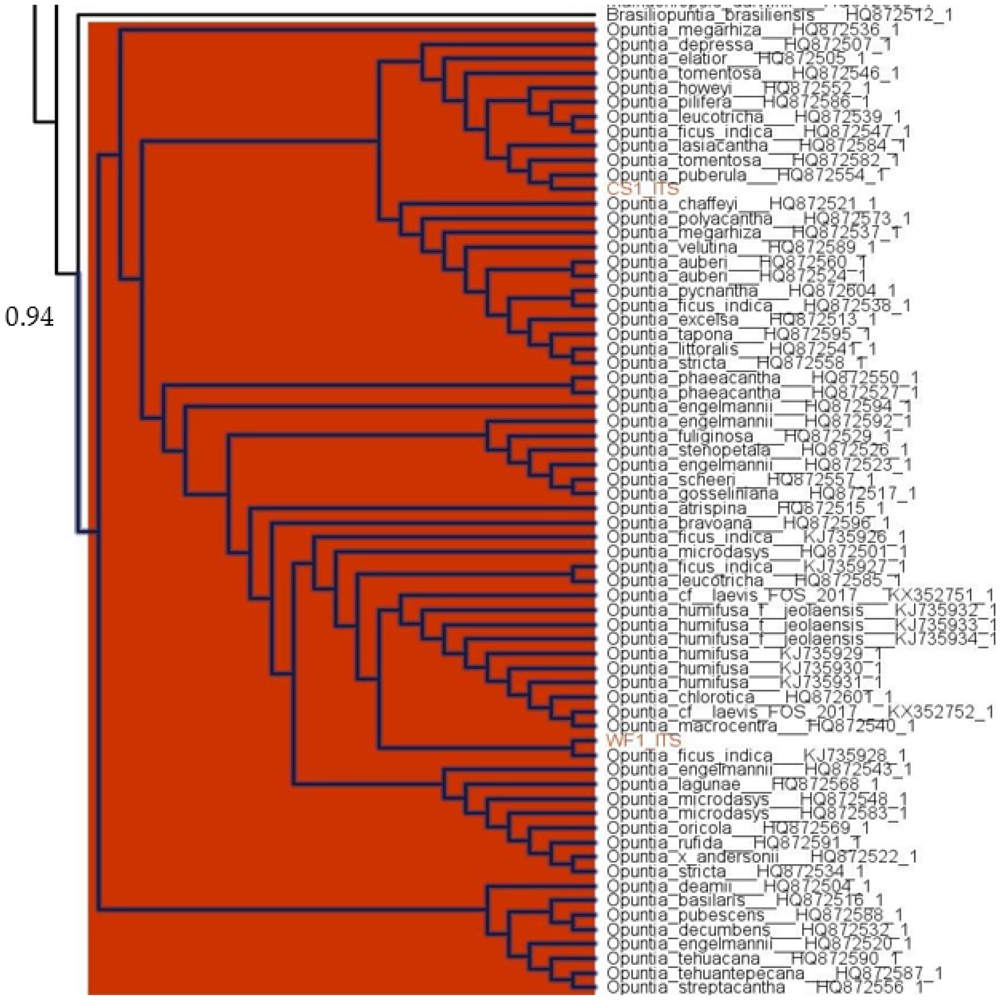
The phylogenetic analysis showed that both rcbL and ITS gene regions can be used to identify the genus of our samples (Opuntia). The obtained branch support value of 0.84 for the Opuntia genus branch of the rcbL gene tree, which included both the commercial and wild type sequences. The gene tree of ITS presented a support value of 0.94 for the branch that included the wild type samples of Opuntia and other species of the genus Opuntia (Figure 2). The sequences included in the obtained ITS gene tree branch were very similar, which did not allow for the identification of the species. The identification of the genus of the sequences for the gene regions COX-3, 18S and matk was not possible using the gene tree, which was mainly derived from the low diversity observed between Opuntia phylogenetic closest genera. The COX-3, 18S and matk alignments showed, respectively, pairwise identity values of 99.6%, 83.0%, 84.7%, which is in accordance with the low diversity in these genes region for the Cactaceae family [19,43]. Taking into consideration that the analysis of all Opuntia gene regions was performed using the intra and inter-specific variation of the DNA sequences through the Cactaceae family, our methodology can correctly identify the Opuntia genus when using the rcbL and ITS regions, even if the phylogenetic closest genera are included.
The BOLD identification system allowed the identification of the genus Opuntia for the matk gene region, as shown in Table 3, with a similarity value of 100% (E-value = 0). The rcbL analysis retrieved a similar result with the identification of the sample as belonging to the genus Opuntia (E-value = 0, similarity = 99.85). The results of the Geneious BLAST search for the COX-3 showed that the samples presented no differences in the nucleotides sequence when compared to all species of the genus Opuntia identified in the results with a higher bit-score (E-value = 0 and 100% identical sites). Although our previous phylogenetic analysis identified our sequenced sample in a branch with other closely related genera, the BLAST result allowed the assignment of the COX-3 sequence to the Opuntia genus. A similar result was also obtained for the ITS gene region with identical sites values higher than 97% for the first 200 hits (E-value = 0), which allowed us to confirm that the samples belonged to the Opuntia genus. The ITS gene region result analysis also showed that a match to the species Opuntia ficus-indica is present in the top three bit-scores. The 18S gene region was the only region with BLAST results that did not allow for the identification of the sequenced sample genus, producing several hits from different genera.
The combination of the global results considering the five different gene regions allowed an assignment of our sequences to the genus Opuntia. Nevertheless, a cautions analysis of each result should be performed taking into consideration the interspecific variation detected in our phylogenetic analysis considering the family Cactaceae. This is of major relevance considering that the Opuntia genus branch of the phylogenetic tree for the COX-3, 18S rRNA, and matk, included other closely related genera inside the Cactaceae family.

3.2. Total Polyphenols and Total Flavonoids Content

Fruits and vegetables are rich sources of many phytochemicals, particularly polyphenols, which represent a group of metabolites generally found in high amount, with a wide variety of structures, and that are able to protect humans against several diseases [44,45]. Many reports have mentioned the importance of flavonoids as the most common and widely distributed group of plant phenolic compounds; moreover, they point out their implication in a variety of health-promoting effects, and their indispensable role in a variety of nutraceutical, medicinal, cosmetic, and pharmaceutical applications [46,47].
Polyphenols are the main contributor to plants’ antioxidant capacity [48]; thus, it is highly rational to determine their total amount in plant extracts. The statistical analyses confirmed a significant difference in the TPC between both species (cultivated and wild) of fruits and their fractions (Figure 3a). The highest phenolic content was found in the wild Opuntia sp.; which fruit (WF) presented 700.54 ± 13.07 mg GAE/100 g DM, significantly higher than the value of the cultivated variety (CF), with 453.27 ± 6.80 mg GAE/100 g DM (p ≤ 0.05). Regarding the fruit fractions, for both wild and cultivated varieties, the pulp was richer in total phenols than the seeds (Figure 3a). For the wild pulp (WP), TPC was 1140.86 ± 11.85 mg GAE/100 g DM, while for the cultivated variety (CP) a significantly lower content of 429.39 ± 18.95 mg GAE/100 g DM was found (p ≤ 0.05). It is interesting to note that the seeds of both species contained a low amount of phenols (around 285 mg GAE/100 g DM) when compared with the pulp, but did not significantly differ among species.
Within the vast group of polyphenols, the flavonoids family represents the most important due to its high antioxidant capacity, which mainly results from the variable number and position of hydroxyl groups in the molecules [49]. The TFC was significantly different (p ≤ 0.05) between the two varieties and their fractions (Figure 3b). As for TPC, the highest value was recorded in the wild Opuntia sp., with 90.09 ± 4.96 mg QE/100 g DM, face to the 61.30 ± 5.44 mg QE/100 g DM of the cultivated variety (p ≤ 0.05). Regarding the fruit fractions, for both wild and cultivated varieties, the pulp was richer in total flavonoids than the seeds (Figure 3b). The pulp from the wild variety (WP) presented the highest TFC (155.62 ± 6.47 mg QE/100 g DM), significantly higher than the value obtained with the cultivated variety (CF), where the TFC did not surpass 42 mg QE/100 g DM. Contrary to the observation of the TPC, the seeds of both varieties presented significant differences in terms of total flavonoids (p ≤ 0.05), with the cultivated variety being richer (48.40 ± 3.24 face to 35.49 ± 3.04 mg QE/100 g for the wild Opuntia sp., respectively).
Several studies have reported the presence of different classes and amounts of phenolic compounds according to the Opuntia species and their tissues. Through the available reports undertaken with the Opuntia genus, the species OFI appear to be the most widespread, with many scientific investigations undertaken on it, in comparison to the other cacti. Moreover, from the studies found in the literature, different classes of phenols with different concentrations can be found in Opuntia sp. depending on the species, vegetative part (seeds, flower, pulp, peel, and cladodes), ripening stage, and other abiotic and geographic factors such as temperature, soil and altitude [5]. For some authors, ferulic acid was the most common phenolic acid found in seeds of Opuntia microdasys (Lehm.) N.E. Pfeiffer and Opuntia macrorhiza Engelm [50], while Amrane-abider et al. [51] found chlorogenic acid as the most prevalent in OFI seeds. Zenteno-Ramírez et al. [15] found a highest abundance of gallic acid and of the flavonoid epicatechin in the pulps of different Opuntia sp., while in different varieties of OFI peels, piscidic acid and isorhamnetin derivates were the most representative [52]. The most common phenols in Opuntia dillenii (Ker Gawl.) Haw. cladodes were quinic acid and myricetin, while piscidic acid and isorhamnetin derivates were found to be the most common in OFI [53,54,55]. Regarding the flowers, Ammar et al. [56] reported quinic acid and quercetin derivates as the most abundant in OFI, while for the same species, Ouerghemmi et al. [57] found a higher abundance of ferulic acid and quercetin.
In the quantitative profile, Andreu et al. [11] reported an average of 780.00 mg GAE/100 g in the pulp of six Spanish OFI cultivars, the values of which followed the same order of magnitude as those obtained herein. Saravanakumar et al. [37] reported a high content in TPC and TFC, with 300.88 mg GAE/100 g DM and 21.24 mg QE/100 g DM, respectively, in a methanolic extract of OFI fruits collected in the summer season. These values are significantly lower than those obtained in our study; despite the geographic variation and differences among abiotic factors to which species are exposed, these differences may be explained by the differences in the solvent used to prepare the extracts. Moreover, Chaalal et al. [58] reported that TPC ranged between 286.29 and 316.46 mg GAE/100 g DM, and TFC varied from 19.19 to 27.20 QE/100 DM, in seeds of three Algerian varieties of OFI. These differences may be attributed to many factors, such as the species, cultivar, geographical location, cultivation practices, and extraction conditions, which could highly influence bioactive compounds recovery, more particularly the extraction solvent chosen, the polarity of which had probably improved the recovery of polyphenols and flavonoid compounds in the present study.

3.3. Antioxidant Activity Evaluation

Antioxidant activity is one of the best known and important biological activities that all plant extracts are endowed with. This property is directly related to the high countenance and variability of plants secondary metabolites, including alkaloids, vitamins, carotenoids, betalains, flavonoids, and polyphenols. The antioxidant potential of plant extracts may have positive implications on humans’ health through the direct scavenging of deleterious free radicals or the modulation of their production, thus delaying the establishment and improving the recovering of several oxidative stress-associated diseases, such as atherosclerosis, cancer, neurodegenerative disorders and cardiovascular diseases [11,59].
The antioxidant activity of extracts may be evaluated by several methods; since both hydrophilic and lipophilic compounds are implicated, many antioxidant mechanisms are available, and a different reactivity of antioxidant compounds could be evaluated against each reactive species [60]. Thus, seven in vitro methods were used to access the antioxidant potential of both Opuntia sp. and their different fractions.
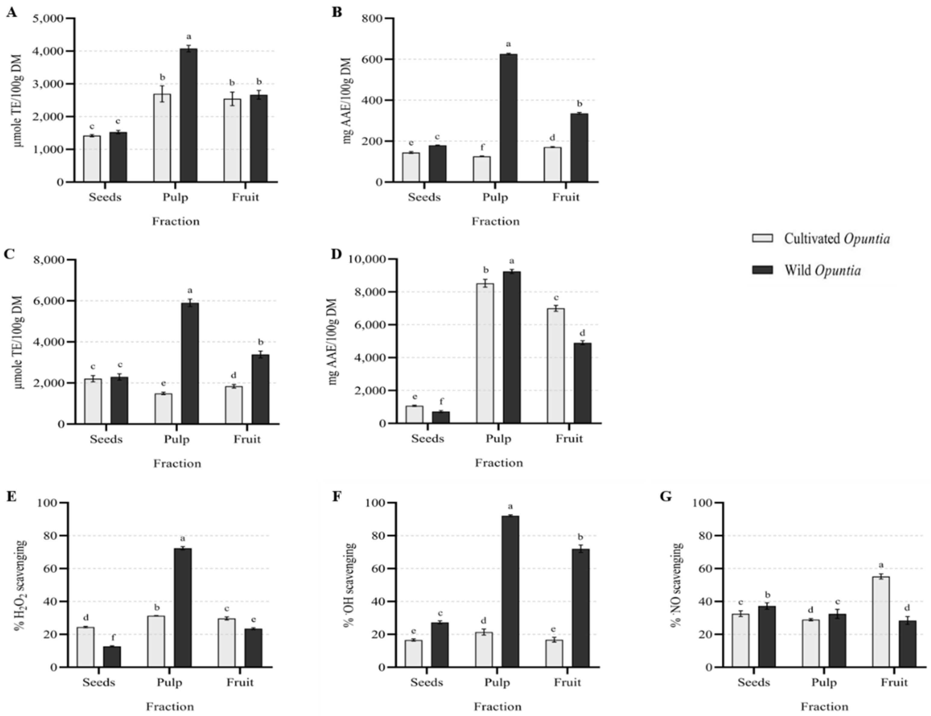
The TEAC is one of the most popular spectrophotometric assays to measure the antioxidant capacity of foods, and it is based in the reduction ability of antioxidant compounds found in extracts towards the synthetic blue-green chromophore ABTS [61]. This assay’s main advantage is the ability to measure both lipophilic and hydrophilic antioxidant components, making it more appropriate in comparison to the well-known anti-radical scavenging method of DPPH [62]. The results for TEAC (Figure 4A) showed that both Opuntias’ fruits presented the same antioxidant potential, with almost 2600 µmole TE/100 g DM. Regarding the fractions, it was found that the pulps of both species had a better potential (4076.95 ± 97.94 and 2694.11 ± 246.81 µmole TE/100 g DM for WP and CP, respectively) when compared to seeds that did not present significant differences among the species, with almost 1500 µmole TE/100 g DM for both cultivated and wild Opuntia sp. A significant positive correlation (p ≤ 0.001) was observed between TEAC and both TPC and TFC (0.92 and 0.85, respectively), suggesting that these compounds contributed to the antioxidant potential displayed by Opuntia sp. extracts. The ABTS assay undertaken by Andreu et al. [11] with the pulp of six OFI cultivars reported a range between 640.00 and 3060.00 µmole TE/100 g DM; the authors also found that peels possessed a higher antioxidant potential (1470.00–3690.00 µmole TE/100 g DM) than those recorded for cladodes (1580–2840 µmole TE/100 g DM). The TEAC values reported herein for both Opuntia sp. seeds were higher than others found in the literature, such as those found by Chaalal et al. [58], with 1067.00 to 1129.00 µmole TE/100 g DM for OFI seeds, and by Kolniak-Ostek et al. [63] for seeds of different sorts of the same species, with an ABTS•+ scavenging power ranging between 708.00 to 1149.00 µmole TE/100 g DM. The values obtained herein were more promising than those reported in the literature, especially for the wild pulp and seeds, highlighting the importance of exploring the wild Opuntia sp. from the biological and economic point of view.
The RP is an assay developed to measure the compounds able to reduce the ferric (Fe3+) ions to the reduced ferrous (Fe2+) forms; more precisely, this assay allows us to measure the reductant compounds that can react with potassium ferricyanide to form potassium ferrocyanide [64]. The results for RP (Figure 4B) showed a significantly higher potential for the wild Opuntia sp. extracts than for the cultivated one, with WF demonstrating 335.91 ± 4.25 mg AAE/100 g DM and CF having 170.94 ± 2.81 mg AAE/100 g DM. Regarding the fractions, it was found that WP and WS had the best reduction capacity (625.54 ± 3.54 and 179.28 ± 1.43 mg AAE/100 g DM for WP and WS, respectively); for the cultivated fraction, a higher activity was found for the seeds, with 144.65 ± 4.42 mg AAE/100 g DM, followed by the pulp with 125.86 ± 1.87 mg AAE/100 g DM. As previously observed for TEAC, a significant positive correlation (p ≤ 0.001) between the RP and TPC and TFC was observed herein (0.96 and 0.94, respectively). The study undertaken by Chougui et al. [65] in seeds of four OFI cultivars reported RP values ranging from 32.30 to 51.30 mg AAE/100 g DM. Elsewhere, Arró-Díaz et al. [60] recorded an excellent reducing potential for ellagic acid and the ethanol extract of Gymnanthes lucida Sw leaves when compared to the ascorbic acid solution. The authors consequently deduced that a higher amount of free hydroxyl groups in the evaluated compounds could be directly related to a higher reducing power of sample extracts. As for TEAC, the values reported herein are also more promising than the ones found in the literature.
FRAP is a widely used method to measure the total antioxidant potential of several foods and plant extracts; it is principally based on the reduction of ferric 2,4,6-tripyridyl-s-triazine complex ([Fe3+-(TPTZ)2]3+) to the intensely blue-colored ferrous complex [Fe2+-(TPTZ)2]2+ at low pH [14]. The results from the FRAP assay, confirmed by ANOVA analysis (Figure 4C), showed higher values for the wild fruit, with 3388.80 ± 169.76 µmole TE/100 g DM, than for the cultivated fruit (1844.40 ± 88.00 µmole TE/100 g DM). Concerning the fractions, WP displayed an antioxidant capacity almost six times higher than CP (5905.46 ± 181.05 µmole TE/100 g DM for WP vs. 1491.75 ± 62.95 µmole TE/100 g DM for CP), while the fraction of the seeds of both Opuntia sp. had a similar antioxidant potential, with less than 2300 µmole TE/100 g DM. As before, both total phenols (0.92, p ≤ 0.001) and flavonoids (0.95, p ≤ 0.001), in particular, seem to contribute to this biological activity. It was interesting to note that there were no significant differences between seeds of both Opuntia sp. fruits, which showed an even higher antioxidant capacity than the pulp of cultivated species. This may be due to the difference in bioactive compounds present in both fractions. In addition, CF had slightly higher potential than CP when compared to the WF; these results were in line with the polyphenols and flavonoids content, which were found to be higher in CF than in CP. The FRAP results obtained for WP were better when compared to the results obtained by Andreu et al. [11]. The authors reported a reducing capacity ranging between 1500 and 3230 µmole TE/100 g DM for the pulp of six OFI cultivars; this difference may also be explained by the differences between the Opuntia sp. studied. Moreover, Kolniak-Ostek et al. [63] reported a range between 367 and 889 µmole TE/100 g DM in seeds of six OFI varieties.
PMA is another assay developed to determine the total antioxidant activity of extracts, and is based in the evaluation of the reduction in molybdate ions from Mo6+ to Mo5+ in acidic conditions [61]. The statistical analysis of PMA showed a higher potential for cultivated Opuntia sp. (7000.84 ± 178.09 mg AAE/100 g DM) than for the wild one (4904.81 ± 121.15 mg AAE/100 g DM) (Figure 4D). The pulp’s fraction in both species revealed a higher activity than that recorded for seeds (9242.24 ± 124.50 and 8524.81 ± 235.86 mg AAE/100 g DM for WP and CP, respectively, vs. 1071.93 ± 41.07 and 720.25 ± 56.99 mg AAE/100 g DM for CS and WS, respectively). As before, both total phenols (0.67, p ≤ 0.001) and flavonoids (0.57, p ≤ 0.001) seemed to contribute to PMA. To our knowledge, there is only one report on PMA with Opuntia sp., developed by Shan et al. [66], who reported that PMA varied between the different fruits studied. The value found by the authors for O. dillenii fruits was almost 26.42 mg AAE/100 g of fresh weight. Once the values were presented in different units, comparisons with the present study were not possible to establish.

Effect on Physiological Reactive Species

Oxidative stress describes a condition of oxidative damage occurring in cells, and resulting from an imbalance between the pro-oxidant system and antioxidant defenses. The pro-oxidant system is reinforced by the excess of some molecular species, which can be classified into radical and non-radical reactive species. Free radicals are defined as species containing an unpaired electron in their outer shell, such as: OH, superoxide (O2•–), NO, and nitrogen dioxide (NO2) radicals. This makes these species unstable, hence they tend to trap an electron from adjacent molecules to compensate for their electron deficiency. The non-radical reactive derivatives, also called oxidants, encompass molecules such as H2O2, ozone (O3), singlet oxygen (1O2), hypochlorous acid (HOCl), and nitrous acid (HNO2). These species are more stable than free radicals but can easily lead to free radical reactions in living organisms. Both radical and non-radical reactive species are produced from cell metabolism or generated from external sources, such as pollution, cigarette smoke, radiation, and medication. Although free radicals can have an important role in biologic processes, when present in low amounts, their uncontrolled production and accumulation is at the root of oxidative stress and harmful effects [67,68,69].
H2O2 may occur in the body through the environment, by inhalation of vapor, or mist, and through eye or skin contact; otherwise, it can also be generated inside the body by several enzymes, such as superoxide dismutase (SOD) and amino-acid oxidases. H2O2 is a nonradical reactive species of oxygen (ROS). Even if it is considered to be poorly reactive due to its weak oxidation capacity, H2O2 can cross the cellular membrane easily, and once inside the cell, it can generate the OH, which is considered to be the most reactive ROS for the body [14,61]. The results of H2O2 inhibition (Figure 4E) showed a difference between both Opuntia sp. fruits, with 29.69 ± 0.95 and 23.46 ± 0.58% of inhibition for CF and WF crude extracts, respectively, for the highest concentration tested of 3.33 mg of DM/mL. As for the pulps, which showed a better efficiency than seeds, WP demonstrated two-times more effectiveness than that recorded for CP (72.34 ± 0.98 and 31.26 ± 0.16% for WP and CP, respectively), while seeds of cultivated Opuntia sp. had 24.42 ± 0.42% of scavenging potential, better than that found for WS (12.71 ± 0.36%). Through the literature, Chougui et al. [70] reported that H2O2 inhibition was between 76 and 96% for crude ethanolic extracts of seeds of OFI cultivars. The authors also found that this antioxidant effect was positively correlated with phenolic compounds, well-known to be good electron donors, allowing to accelerate the conversion of H2O2 into H2O. Saravanakumar et al. [37] found that the ripening stage and extraction solvent influenced the antioxidant activity of OFI fruit, with the highest H2O2 scavenging reaching 70% for the methanolic extract of fruit collected in summer, at 100 µg of dry extract/mL, while the water extracts of fruits presented approximately 43% of inhibition at the same concentration. The results obtained for the seeds and fruits fractions evaluated herein were lower when compared with the cited authors, which may be related to the bioactive compounds found in the extracts. Moreover, it should be noticed that for Saravanakumar et al.’s (2015) study, the aqueous extracts of OFI fruit gave much lower inhibition potential for H2O2 when comparing to the methanolic ones.
OH is one of the most potent ROS in biological systems [71], and is well known for its extreme reactivity, attacking most cellular compounds, such as amino acids, carbohydrates, lipids and nucleic acids [72]. The results for OH scavenging (Figure 4F) revealed more activity for the wild Opuntia sp. than for the cultivated one, with higher activity observed for WF, with 72.07 ± 2.28% than for CF (16.74 ± 1.44%), for the highest concentration tested of 3.88 mg of DM/mL. Concerning the fractions, Opuntia sp. pulps’ were better, with 92.12 ± 0.50 and 21.41 ± 1.82% for the WP and CP, respectively, while WS exhibited 27.30 ± 0.92% and CS 16.55 ± 0.71% of inhibition. The results obtained with the wild fruits were more promising than those obtained by Saravanakumar et al. [37], who found a OH scavenging capacity of 60% for OFI fruit methanolic extracts (100 µg/mL of dry extract). In addition to the concentration tested, the difference found can also be attributed to the chemical composition of Opuntia species, which can highly influence their antioxidant potential. As expected, a significant positive correlation was observed between H2O2 and OH scavenging and TPC (0.89 and 0.93, p ≤ 0.001, respectively) and TFC (0.88 and 0.91, p ≤ 0.001, respectively).
NO is a pleiotropic mediator in the body, mainly generated from an enzymatic pathway involving the nitric oxide synthase (NOS) and its isoforms. This free radical is implicated in many physiological processes, such as neurons signalization, smooth muscle relaxation, regulation of the immune system, and so forth [30,73]. However, when it is found in large excess, NO, with its brief second lifetime, is highly reactive, and can react with oxygen and other molecules to generate reactive nitrogen species (RNS), which can interact with many molecular targets within the cell and cause multiple deleterious effects [74]. The results of NO scavenging are displayed in Figure 4G. The cultivated fruit presented a significantly better ability to scavenge NO (55.22 ± 1.56%), almost two times higher than that found for the wild one (28.41 ± 2.36%), for the highest concentration tested of 1.50 mg of DM/mL. Regarding the fractions, seeds of both Opuntia sp. exhibited a better NO scavenging potential than their pulps, with 37.27 ± 1.94 and 32.58 ± 1.77% for WS and CS, respectively, in comparison to the 32.44 ± 2.74 and 28.92 ± 0.69% found for WP and CP, respectively. It is worth mentioning that there was no significant difference between WP and CS fractions. Contrary to what was observed for the previous assays, a statistical correlation between NO scavenging and the phenolic compounds analyzed herein was not established, possibly suggesting that Opuntia sp. phenols are more prone to scavenge free radicals of oxygen than free radicals of nitrogen. The involvement of other bioactive compounds present in the extract of NO scavenging should also not be disregarded. The results obtained for the seeds and fruit extracts were similar to some previous studies, which recorded a NO scavenging around 56% for methanolic extract of OFI fruits (100 µg of dry extract/mL), and from 21.51% to 34.67% for crude acetonic seeds extract of OFI, respectively [37,58]. Otherwise, when comparing the results obtained between both species studied herein, it can be hypothesized that the fruit of cultivated species possessed more compounds that were able to scavenge NO than the fruit of the wild species.
In order to better scrutinize the correlation between the studied extracts and the biological activities displayed by them, a principal component analysis (PCA) was applied. The results obtained from the measured phytochemical compounds and antioxidant activities were reduced from the multivariate data to a two-dimensional presentation, as shown in Figure 5.
As can be observed, the cumulative variance was about 89.41% of the variability, explained by the first principal component (PC1) accounting for 73.79% of the variance, and the second (PC2) with 15.62%. According to the PCA, it can be concluded that, with the exception of NO scavenging, the other activities are positively correlated, being positioned in the positive axis of PC1. TPC and TFC appear superimposed, as expected, once flavonoids contained in phenolic compounds are also determined in the TPC assay. These compounds do not explain the activity measured for NO, appearing in opposite quadrants of the PCA, but are positively correlated with the remaining assays. These observations are in accordance with the literature, with several studies reporting a positive correlation between polyphenols and the antioxidant capacity of plants [58,75,76,77,78]; moreover, Dong et al. [79] found that polyphenols from oat improved a range of metabolic syndromes in high-fat-diet-fed mice, including an improvement of oxidative stress in liver cells, by reducing lipid peroxidation and increasing the activity of some antioxidant enzymes (catalase, superoxide dismutase, and glutathione-peroxidase), which contributed to enhancing the liver protection from oxidative damage. Regarding Opuntia sp. cultivars and extracts, both wild and cultivated seeds appear very close, demonstrating that, in a general way, they are very similar, both in terms of chemical composition and biological activities. Regarding the other tissues, there is not a clear correlation between them, although, those from wild Opuntia sp. were more active than those from the cultivated group, and the wild pulp presented the strongest correlation with the radicals scavenging evaluated in this study.
In summary, the differences obtained between both species and their tissues should not be limited to the amount of phenolic compounds present in the extracts, but also extended to their profile, as well as to other secondary metabolites endowed with antioxidant potential. Regarding phenols, various studies have demonstrated positive correlations between antioxidant activity and the enrichment in phenolic acids, especially those which were highly associated with radical scavenging, such as ferulic acid, caffeic acid, apigenin and luteolin, also reporting the prebiotic effect of these compounds [80]. In relation to other compounds, Koss-Mikołajczyk et al. [81] reported that vitamin C and betalains were among the main contributors to the total antioxidant capacity of prickly pears; they also reported a better antioxidant efficiency for betacyanins, rather than betaxanthins. Furthermore, according to Kalinowska et al. [82], the differences in the results could also be related to the mechanism of action, as well as to the reactionary environment specific to each method, such as the pH and the type of solvent, which may influence the ionization state of biomolecules present in the extract and, therefore, the rate of their antioxidant mechanism.

4. Conclusions

This study was conducted for the first time in a wild Opuntia species grown in Bejaia, Algeria. The results showed that phenolic compounds and flavonoids were present in all fractions of both Opuntia species, with a significantly higher amount in the pulp of the wild species (1140 mg GAE/100 g vs. 429 mg GAE/100 g for the cultivated Opuntia). The different antioxidant assays realized prove the potential of both species to prevent oxidative stress. However, the wild species demonstrated a more significant antioxidant potential, and particularly its pulp, with the phenolic compounds highly correlated with the radical scavenging (p ≤ 0.001). Overall, the results clearly provide evidence that both Opuntia species can be of great benefit for human health, with the potential to prevent consumers from oxidative stress-related diseases. The wild Opuntia sp. stood out, emphasizing its potential application in the areas of nutraceuticals, cosmetics, and as a non-pharmacological approach, with application in different kinds of states where oxidative stress has a prominent role. Moreover, the exploitation of wild Opuntia sp. can have huge importance for the economy of Algeria, where this undervalued species is very abundant.

Author Contributions

Conceptualization, F.B.; methodology, F.B., W.Z., R.S. and J.M.; Data analysis: G.L. and J.C.; validation, F.B. and G.L.; investigation, F.B. and W.Z.; writing—original draft preparation, F.B. and W.Z.; writing—review and editing, F.B., W.Z., J.C., R.S., J.M., V.V. and G.L.; supervision, F.B and G.L. Funding: F.B. and V.V. All authors have read and agreed to the published version of the manuscript.

Funding

This research was partially funded by the strategic funding from UIDB/04423/2020 and UIDP/04423/2020 of CIIMAR.

Institutional Review Board Statement

Not applicable.

Data Availability Statement

Anyone can access the data by sending an email to [email protected].

Acknowledgments

The authors are grateful to the Algerian Ministry of Higher Education and Scientific Research for the financial support. João Carneiro acknowledges the Portuguese Foundation for Science and Technology (FCT) funding for his research contract at CIIMAR, established under the transitional rule of Decree Law 57/2016, amended by Law 57/2017. This work was produced with the support of INCD funded by FCT under the project 2021.09782.CPCA. Graciliana Lopes thanks FCT for the financial support for her work contract through the Scientific Employment Stimulus-Individual Call (CEECIND/01768/2021).

Conflicts of Interest

The authors declare no conflict of interest.

References

  1. Valero-Cases, E.; Cerdá-Bernad, D.; Pastor, J.-J.; Frutos, M.-J. Non-Dairy Fermented Beverages as Potential Carriers to Ensure Probiotics, Prebiotics, and Bioactive Compounds Arrival to the Gut and Their Health Benefits. Nutrients 2020, 12, 1666. [Google Scholar] [CrossRef] [PubMed]
  2. Osuna-Martínez, L.; Reyes Esparza, J.; Rodríguez-Fragoso, L. Cactus (Opuntia ficus-indica): A Review on Its Antioxidants Properties and Potential Pharmacological Use in Chronic Diseases. Nat. Prod. Chem. Res. 2014, 2, 153–160. [Google Scholar] [CrossRef] [Green Version]
  3. Zeghbib, W.; Boudjouan, F.; Vasconcelos, V.; Lopes, G. Phenolic Compounds’ Occurrence in Opuntia Species and Their Role in the Inflammatory Process: A Review. Molecules 2022, 27, 4763. [Google Scholar] [CrossRef] [PubMed]
  4. Cota-Sánchez, J.H. Chapter 28—Nutritional Composition of the Prickly Pear (Opuntia ficus-indica) Fruit. In Nutritional Composition of Fruit Cultivars; Simmonds, M.S.J., Preedy, V.R., Eds.; Academic Press: San Diego, CA, USA, 2016; pp. 691–712. ISBN 978-0-12-408117-8. [Google Scholar]
  5. El-Mostafa, K.; El-Kharrassi, Y.; Badreddine, A.; Andreoletti, P.; Vamecq, J.; El-Kebbaj, M.S.; Latruffe, N.; Lizard, G.; Nasser, B.; Cherkaoui-Malki, M. Nopal Cactus (Opuntia ficus-indica) as a Source of Bioactive Compounds for Nutrition, Health and Disease. Molecules 2014, 19, 14879–14901. [Google Scholar] [CrossRef] [Green Version]
  6. Leem, K.-H.; Kim, M.-G.; Hahm, Y.-T.; Kim, H.K. Hypoglycemic Effect of Opuntia ficus-indica Var. Saboten Is Due to Enhanced Peripheral Glucose Uptake through Activation of AMPK/P38 MAPK Pathway. Nutrients 2016, 8, 800. [Google Scholar] [CrossRef] [Green Version]
  7. Mazari, A.; Yahiaoui, K.; Fedjer, Z.; Mahdeb, A. Physical Characteristics, Phytochemical Content and Antioxidant Activity of Cactus Pear Fruits Growing in Northeast Algeria. J. Prof. Assoc. Cactus Dev. 2018, 20, 178–188. [Google Scholar] [CrossRef]
  8. Bouzoubaâ, Z.; Essoukrati, Y.; Tahrouch, S.; Hatimi, A.; Gharby, S.; Harhar, H. Phytochemical Study of Prickly Pear from Southern Morocco. J. Saudi Soc. Agric. Sci. 2016, 15, 155–161. [Google Scholar] [CrossRef] [Green Version]
  9. García, F.H.; Coll, L.A.; Cano-Lamadrid, M.; Lluch, D.L.; Barrachina, Á.A.C.; Murcia, P.L. Valorization of Prickly Pear [Opuntia ficus-indica (L.) Mill]: Nutritional Composition, Functional Properties and Economic Aspects; IntechOpen: London, UK, 2020; ISBN 978-1-78985-850-1. [Google Scholar]
  10. Ćavar Zeljković, S.; Šišková, J.; Komzáková, K.; De Diego, N.; Kaffková, K.; Tarkowski, P. Phenolic Compounds and Biological Activity of Selected Mentha Species. Plants 2021, 10, 550. [Google Scholar] [CrossRef]
  11. Andreu, L.; Nuncio-Jáuregui, N.; Carbonell-Barrachina, Á.A.; Legua, P.; Hernández, F. Antioxidant Properties and Chemical Characterization of Spanish Opuntia ficus-indica Mill. Cladodes and Fruits. J. Sci. Food Agric. 2018, 98, 1566–1573. [Google Scholar] [CrossRef]
  12. Wang, J.; Xu, J.; Gong, X.; Yang, M.; Zhang, C.; Li, M. Biosynthesis, Chemistry, and Pharmacology of Polyphenols from Chinese Salvia Species: A Review. Molecules 2019, 24, 155. [Google Scholar] [CrossRef]
  13. Alagumanivasagam, G.; Pasupathy, R.; Kottaimuthu, A.; Manavalan, R. A Review on In-Vitro Antioxidant Methods. Int. J. Pharm. Chem. Sci. 2012, 1, 662–674. [Google Scholar]
  14. Gulcin, İ. Antioxidants and Antioxidant Methods: An Updated Overview. Arch. Toxicol. 2020, 94, 651–715. [Google Scholar] [CrossRef] [PubMed] [Green Version]
  15. Zenteno-Ramírez, G.; Juárez-Flores, B.I.; Aguirre-Rivera, J.R.; Monreal-Montes, M.; García, J.M.; Serratosa, M.P.; Santos, M.Á.V.; Pérez, M.D.O.; Rendón-Huerta, J.A. Juices of Prickly Pear Fruits (Opuntia spp.) As Functional Foods. Ital. J. Food Sci. 2018, 30, 614–627. [Google Scholar] [CrossRef]
  16. Folgado, A.; Pires, A.S.; Figueiredo, A.C.; Pimentel, C.; Abranches, R. Toward Alternative Sources of Milk Coagulants for Cheese Manufacturing: Establishment of Hairy Roots Culture and Protease Characterization from Cynara cardunculus L. Plant Cell Rep. 2020, 39, 89–100. [Google Scholar] [CrossRef]
  17. Moon-van der Staay, S.Y.; van der Staay, G.W.M.; Guillou, L.; Vaulot, D.; Claustre, H.; Medlin, L.K. Abundance and Diversity of Prymnesiophytes in the Picoplankton Coμmunity from the Equatorial Pacific Ocean Inferred from 18S RDNA Sequences. Limnol. Oceanogr. 2000, 45, 98–109. [Google Scholar] [CrossRef] [Green Version]
  18. Katana, A.; Kwiatowski, J.; Spalik, K.; Zakryś, B.; Szalacha, E.; Szymańska, H. Phylogenetic Position of Koliella (Chlorophyta) as Inferred from Nuclear and Chloroplast Small Subunit RDNA. J. Phycol. 2001, 37, 443–451. [Google Scholar] [CrossRef]
  19. Martínez-González, C.R.; Ramírez-Mendoza, R.; Jiménez-Ramírez, J.; Gallegos-Vázquez, C.; Luna-Vega, I. Improved Method for Genomic DNA Extraction for Opuntia Mill. (Cactaceae). Plant Methods 2017, 13, 82. [Google Scholar] [CrossRef] [Green Version]
  20. Edwards, E.J.; Nyffeler, R.; Donoghue, M.J. Basal Cactus Phylogeny: Implications of Pereskia (Cactaceae) Paraphyly for the Transition to the Cactus Life Form. Am. J. Bot. 2005, 92, 1177–1188. [Google Scholar] [CrossRef] [Green Version]
  21. Kearse, M.; Moir, R.; Wilson, A.; Stones-Havas, S.; Cheung, M.; Sturrock, S.; Buxton, S.; Cooper, A.; Markowitz, S.; Duran, C.; et al. Geneious Basic: An Integrated and Extendable Desktop Software Platform for the Organization and Analysis of Sequence Data. Bioinformatics 2012, 28, 1647. [Google Scholar] [CrossRef] [PubMed] [Green Version]
  22. Santos, C.; Carneiro, J.; Pereira, F. A Web-Based Platform of Nucleotide Sequence Alignments of Plants. bioRxiv 2019, bioRxiv:617035. [Google Scholar] [CrossRef] [Green Version]
  23. Lemoine, F.; Correia, D.; Lefort, V.; Doppelt-Azeroual, O.; Mareuil, F.; Cohen-Boulakia, S.; Gascuel, O. NGPhylogeny.Fr: New Generation Phylogenetic Services for Non-Specialists. Nucleic Acids Res. 2019, 47, W260–W265. [Google Scholar] [CrossRef]
  24. Katoh, K.; Rozewicki, J.; Yamada, K.D. MAFFT Online Service: Multiple Sequence Alignment, Interactive Sequence Choice and Visualization. Brief. Bioinform. 2019, 20, 1160–1166. [Google Scholar] [CrossRef] [Green Version]
  25. Edgar, R.C. MUSCLE: Multiple Sequence Alignment with High Accuracy and High Throughput. Nucleic Acids Res. 2004, 32, 1792–1797. [Google Scholar] [CrossRef] [Green Version]
  26. Criscuolo, A.; Gribaldo, S. BMGE (Block Mapping and Gathering with Entropy): A New Software for Selection of Phylogenetic Informative Regions from Multiple Sequence Alignments. BMC Evol. Biol. 2010, 10, 210. [Google Scholar] [CrossRef] [Green Version]
  27. Lemoine, F.; Domelevo Entfellner, J.-B.; Wilkinson, E.; Correia, D.; Dávila Felipe, M.; De Oliveira, T.; Gascuel, O. Renewing Felsenstein’s Phylogenetic Bootstrap in the Era of Big Data. Nature 2018, 556, 452–456. [Google Scholar] [CrossRef]
  28. Guindon, S.; Dufayard, J.-F.; Lefort, V.; Anisimova, M.; Hordijk, W.; Gascuel, O. New Algorithms and Methods to Estimate Maximum-Likelihood Phylogenies: Assessing the Performance of PhyML 3.0. Syst. Biol. 2010, 59, 307–321. [Google Scholar] [CrossRef] [PubMed] [Green Version]
  29. Zeghbib, W.; Boudjouan, F.; Bachir-bey, M. Optimization of Phenolic Compounds Recovery and Antioxidant Activity Evaluation from Opuntia Ficus Indica Using Response Surface Methodology. J. Food Meas. Charact. 2022, 16, 1354–1366. [Google Scholar] [CrossRef]
  30. Surana, A.; Kumbhare, M.; Wagh, R. Estimation of Total Phenolic and Total Flavonoid Content and Assessment of in Vitro Antioxidant Activity of Extracts of Hamelia Patens Jacq. Stems. Res. J. Phytochem. 2016, 10, 67–74. [Google Scholar] [CrossRef] [Green Version]
  31. Re, R.; Pellegrini, N.; Proteggente, A.; Pannala, A.; Yang, M.; Rice-Evans, C. Antioxidant Activity Applying an Improved ABTS Radical Cation Decolorization Assay. Free Radic. Biol. Med. 1999, 26, 1231–1237. [Google Scholar] [CrossRef]
  32. Oyaizu, M. Studies on products of browning reaction--antioxidative activities of products of browning reaction prepared from glucosamine. Jpn. J. Nutr. Diet. 1986, 44, 307–315. [Google Scholar] [CrossRef] [Green Version]
  33. Prieto, P.; Pineda, M.; Aguilar, M. Spectrophotometric Quantitation of Antioxidant Capacity through the Formation of a Phosphomolybdenum Complex: Specific Application to the Determination of Vitamin E. Anal. Biochem. 1999, 269, 337–341. [Google Scholar] [CrossRef] [PubMed]
  34. Benzie, I.F.; Strain, J.J. Ferric Reducing/Antioxidant Power Assay: Direct Measure of Total Antioxidant Activity of Biological Fluids and Modified Version for Simultaneous Measurement of Total Antioxidant Power and Ascorbic Acid Concentration. Methods Enzymol. 1999, 299, 15–27. [Google Scholar] [CrossRef] [PubMed]
  35. Mukhopadhyay, D.; Dasgupta, P.; Roy, D.S.; Palchoudhuri, S.; Chatterjee, I.; Ali, S.; Dastidar, S.G. A Sensitive In Vitro Spectrophotometric Hydrogen Peroxide Scavenging Assay Using 1,10-Phenanthroline. Free Radic. Antioxid. 2016, 6, 124–132. [Google Scholar] [CrossRef]
  36. Smirnoff, N.; Cumbes, Q.J. Hydroxyl Radical Scavenging Activity of Compatible Solutes. Phytochemistry 1989, 28, 1057–1060. [Google Scholar] [CrossRef]
  37. Saravanakumar, A.; Ganesh, M.; Peng, M.M.; Aziz, A.S.; Jang, H.T. Comparative Antioxidant and Antimycobacterial Activities of Opuntia ficus-indica Fruit Extracts from Summer and Rainy Seasons. Front. Life Sci. 2015, 8, 182–191. [Google Scholar] [CrossRef] [Green Version]
  38. Dehbi, F.; Hasib, A.; Ouatmane, A.; Elbatal, H.; Jaouad, A. Physicochemical Characteristics of Moroccan Prickly Pear Juice (Opuntia ficus-indica L.). Int. J. Emerg. Technol. Adv. Eng. 2014, 4, 300–306. [Google Scholar]
  39. De Wit, M.; Nel, P.; Osthoff, G.; Labuschagne, M.T. The Effect of Variety and Location on Cactus Pear (Opuntia ficus-indica) Fruit Quality. Plant Foods Hum. Nutr. Dordr. Neth. 2010, 65, 136–145. [Google Scholar] [CrossRef]
  40. Felker, P.; Soulier, C.; Leguizamon, G.; Ochoa, J. A Comparison of the Fruit Parameters of 12 Opuntia Clones Grown in Argentina and the United States. J. Arid Environ. 2002, 52, 361–370. [Google Scholar] [CrossRef]
  41. Karababa, E.; Coşkuner, Y.; Aksay, S. Some Physical Fruit Properties of Cactus Pear (Opuntia spp.) That Grow Wild in the Eastern Mediterranean Region of Turkey. J. Prof. Assoc. Cactus Dev. 2004, 6, 1–8. [Google Scholar] [CrossRef]
  42. Bekir, E.A. Cactus Pear (Opuntia ficus-indica Mill.) In Turkey: Growing Regions and Pomological Traits of Cactus Pear Fruits. Acta Hortic. 2006, 728, 51–54. [Google Scholar] [CrossRef]
  43. Nyffeler, R. Phylogenetic Relationships in the Cactus Family (Cactaceae) Based on Evidence from TrnK/MatK and TrnL-TrnF Sequences. Am. J. Bot. 2002, 89, 312–326. [Google Scholar] [CrossRef] [PubMed] [Green Version]
  44. Zamora-Ros, R.; Touillaud, M.; Rothwell, J.A.; Romieu, I.; Scalbert, A. Measuring Exposure to the Polyphenol Metabolome in Observational Epidemiologic Studies: Current Tools and Applications and Their Limits123. Am. J. Clin. Nutr. 2014, 100, 11–26. [Google Scholar] [CrossRef] [PubMed]
  45. Guo, X.; Tresserra-Rimbau, A.; Estruch, R.; Martínez-González, M.A.; Medina-Remón, A.; Castañer, O.; Corella, D.; Salas-Salvadó, J.; Lamuela-Raventós, R.M. Effects of Polyphenol, Measured by a Biomarker of Total Polyphenols in Urine, on Cardiovascular Risk Factors After a Long-Term Follow-Up in the PREDIMED Study. Oxid. Med. Cell. Longev. 2016, 2016, 2572606. [Google Scholar] [CrossRef] [PubMed] [Green Version]
  46. Kumar, S.; Pandey, A.K. Chemistry and Biological Activities of Flavonoids: An Overview. Sci. World J. 2013, 2013, e162750. [Google Scholar] [CrossRef] [Green Version]
  47. Rufino, A.T.; Costa, V.M.; Carvalho, F.; Fernandes, E. Flavonoids as Antiobesity Agents: A Review. Med. Res. Rev. 2021, 41, 556–585. [Google Scholar] [CrossRef] [PubMed]
  48. Madoui, S.; Charef, N.; Arrar, L.; Baghianni, A.; Khennouf, S. In Vitro Antioxidant Activities of Various Extracts from Flowers-Leaves Mixture of Algerian Cytisus Triflorus. Annu. Res. Rev. Biol. 2018, 26, 1–13. [Google Scholar] [CrossRef]
  49. Pabón-Baquero, L.C.; Otálvaro-Álvarez, Á.M.; Fernández, M.R.R.; Chaparro-González, M.P. Plant Extracts as Antioxidant Additives for Food Industry. In Antioxidants in Foods and Its Applications; Shalaby, E., Azzam, G.M., Eds.; IntechOpen: London, UK, 2018; pp. 87–116. ISBN 978-1-78923-379-7. [Google Scholar]
  50. Chahdoura, H.; Barreira, J.C.M.; Barros, L.; Santos-Buelga, C.; Ferreira, I.C.F.R.; Achour, L. Seeds of Opuntia Spp. as a Novel High Potential by-Product: Phytochemical Characterization and Antioxidant Activity. Ind. Crops Prod. 2015, 65, 383–389. [Google Scholar] [CrossRef] [Green Version]
  51. Amrane-Abider, M.; Nerin, C.; Canellas, E.; Benkerrou, F.; Louaileche, H. Modeling and Optimization of Phenolic Compounds Extraction from Prickly Pear (Opuntia ficus-indica) Seeds via Ultrasound-Assisted Technique. Ann. Univ. Dunarea Jos Galati Fascicle VI-Food Technol. 2018, 42, 109–121. [Google Scholar]
  52. García-Cayuela, T.; Gómez-Maqueo, A.; Guajardo-Flores, D.; Welti-Chanes, J.; Cano, M.P. Characterization and Quantification of Individual Betalain and Phenolic Compounds in Mexican and Spanish Prickly Pear (Opuntia ficus-indica L. Mill) Tissues: A Comparative Study. J. Food Compos. Anal. 2019, 76, 1–13. [Google Scholar] [CrossRef]
  53. Mena, P.; Tassotti, M.; Andreu, L.; Nuncio-Jáuregui, N.; Legua, P.; Del Rio, D.; Hernández, F. Phytochemical Characterization of Different Prickly Pear (Opuntia ficus-indica (L.) Mill.) Cultivars and Botanical Parts: UHPLC-ESI-MSn Metabolomics Profiles and Their Chemometric Analysis. Food Res. Int. 2018, 108, 301–308. [Google Scholar] [CrossRef]
  54. Missaoui, M.; D’Antuono, I.; D’Imperio, M.; Linsalata, V.; Boukhchina, S.; Logrieco, A.F.; Cardinali, A. Characterization of Micronutrients, Bioaccessibility and Antioxidant Activity of Prickly Pear Cladodes as Functional Ingredient. Molecules 2020, 25, 2176. [Google Scholar] [CrossRef] [PubMed]
  55. Ben Lataief, S.; Zourgui, M.-N.; Rahmani, R.; Najjaa, H.; Gharsallah, N.; Zourgui, L. Chemical Composition, Antioxidant, Antimicrobial and Cytotoxic Activities of Bioactive Compounds Extracted from Opuntia Dillenii Cladodes. J. Food Meas. Charact. 2021, 15, 782–794. [Google Scholar] [CrossRef]
  56. Ammar, I.; Ben Salem, M.; Harrabi, B.; Mzid, M.; Bardaa, S.; Sahnoun, Z.; Attia, H.; Ennouri, M. Anti-Inflammatory Activity and Phenolic Composition of Prickly Pear (Opuntia ficus-indica) Flowers. Ind. Crops Prod. 2018, 112, 313–319. [Google Scholar] [CrossRef]
  57. Ouerghemmi, I.; Harbeoui, H.; Aidi Wannes, W.; Bettaieb Rebey, I.; Hammami, M.; Marzouk, B.; Saidani Tounsi, M. Phytochemical Composition and Antioxidant Activity of Tunisian Cactus Pear (Opuntia ficus-indica L.) Flower. J. Food Biochem. 2017, 41, e12390. [Google Scholar] [CrossRef]
  58. Chaalal, M.; Louaileche, H.; Touati, N.; Bachir Bey, M. Phytochemicals, in Vitro Antioxidant Capacity and Antiradical Potential of Whole and Ground Seeds of Three Prickly Pear Varieties: A Comparative Study. Ind. Crops Prod. 2013, 49, 386–391. [Google Scholar] [CrossRef]
  59. Tupec, M.; Hýsková, V.; Bělonožníková, K.; Hraníček, J.; Červený, V.; Ryšlavá, H. Characterization of Some Potential Medicinal Plants from Central Europe by Their Antioxidant Capacity and the Presence of Metal Elements. Food Biosci. 2017, 20, 43–50. [Google Scholar] [CrossRef]
  60. Arró-Díaz, D.J.; Castelnaux-Ochoa, N.; Ochoa-Pacheco, A.; Do-Nascimento, Y.M. Antioxidant Activity of Bioactive Compounds Isolated from Leaves and Bark of Gymnanthes Lucida Sw. Rev. Cuba. Quím. 2021, 33, 22–39. [Google Scholar]
  61. Alam, M.N.; Bristi, N.J.; Rafiquzzaman, M. Review on in Vivo and in Vitro Methods Evaluation of Antioxidant Activity. Saudi Pharm. J. 2013, 21, 143–152. [Google Scholar] [CrossRef] [Green Version]
  62. Floegel, A.; Kim, D.-O.; Chung, S.-J.; Koo, S.I.; Chun, O.K. Comparison of ABTS/DPPH Assays to Measure Antioxidant Capacity in Popular Antioxidant-Rich US Foods. J. Food Compos. Anal. 2011, 24, 1043–1048. [Google Scholar] [CrossRef]
  63. Kolniak-Ostek, J.; Kita, A.; Miedzianka, J.; Andreu-Coll, L.; Legua, P.; Hernandez, F. Characterization of Bioactive Compounds of Opuntia ficus-indica (L.) Mill. Seeds from Spanish Cultivars. Molecules 2020, 25, 5734. [Google Scholar] [CrossRef]
  64. Dontha, S. A Review on Antioxidant Methods. Asian J. Pharm. Clin. Res. 2016, 9, 14–32. [Google Scholar] [CrossRef]
  65. Chougui, N.; Tamendjari, A.; Hamidj, W.; Hallal, S.; Barras, A.; Richard, T.; Larbat, R. Oil Composition and Characterisation of Phenolic Compounds of Opuntia ficus-indica Seeds. Food Chem. 2013, 139, 796–803. [Google Scholar] [CrossRef] [PubMed]
  66. Shan, S.; Huang, X.; Shah, M.H.; Abbasi, A.M. Evaluation of Polyphenolics Content and Antioxidant Activity in Edible Wild Fruits. BioMed Res. Int. 2019, 2019, e1381989. [Google Scholar] [CrossRef] [Green Version]
  67. Pham-Huy, L.A.; He, H.; Pham-Huy, C. Free Radicals, Antioxidants in Disease and Health. Int. J. Biomed. Sci. 2008, 4, 89–96. [Google Scholar]
  68. Lobo, V.; Patil, A.; Phatak, A.; Chandra, N. Free Radicals, Antioxidants and Functional Foods: Impact on Human Health. Pharmacogn. Rev. 2010, 4, 118–126. [Google Scholar] [CrossRef]
  69. Santos-Sánchez, N.F.; Salas-Coronado, R.; Villanueva-Cañongo, C.; Hernández-Carlos, B. Antioxidant Compounds and Their Antioxidant Mechanism. In Antioxidants; Shalaby, E., Ed.; IntechOpen: London, UK, 2019; ISBN 978-1-78923-920-1. [Google Scholar]
  70. Chougui, N.; Louaileche, H.; Mohedeb, S.; Mouloudj, Y.; Hammoui, Y.; Tamendjari, A. Physico-Chemical Characterisation and Antioxidant Activity of Some Opuntia ficus-indica Varieties Grown in North Algeria. Afr. J. Biotechnol. 2013, 12, 299–307. [Google Scholar] [CrossRef] [Green Version]
  71. Treml, J.; Šmejkal, K. Flavonoids as Potent Scavengers of Hydroxyl Radicals. Compr. Rev. Food Sci. Food Saf. 2016, 15, 720–738. [Google Scholar] [CrossRef]
  72. Lubos, E.; Handy, D.E.; Loscalzo, J. Role of Oxidative Stress and Nitric Oxide in Atherothrombosis. Front. Biosci. J. Virtual Libr. 2008, 13, 5323–5344. [Google Scholar] [CrossRef] [Green Version]
  73. Luiking, Y.C.; Engelen, M.P.K.J.; Deutz, N.E.P. Regulation of Nitric Oxide Production in Health and Disease. Curr. Opin. Clin. Nutr. Metab. Care 2010, 13, 97–104. [Google Scholar] [CrossRef] [Green Version]
  74. Beevi, S.S.S.; Rasheed, A.M.H.; Geetha, A. Evaluation of Oxidative Stress and Nitric Oxide Levels in Patients with Oral Cavity Cancer. Jpn. J. Clin. Oncol. 2004, 34, 379–385. [Google Scholar] [CrossRef] [Green Version]
  75. Kumar, S.; Sandhir, R.; Ojha, S. Evaluation of Antioxidant Activity and Total Phenol in Different Varieties of Lantana Camara Leaves. BMC Res. Notes 2014, 7, 560. [Google Scholar] [CrossRef] [Green Version]
  76. Davies, C.V.; Gerard, L.M.; Ferreyra, M.M.; Schvab, M.D.C.; Solda, C.A. Bioactive Compounds and Antioxidant Activity Analysis during Orange Vinegar Production. Food Sci. Technol. 2017, 37, 449–455. [Google Scholar] [CrossRef] [Green Version]
  77. Fu, H.; Qiao, Y.; Wang, P.; Mu, X.; Zhang, J.; Fu, B.; Du, J. Changes of Bioactive Components and Antioxidant Potential during Fruit Development of Prunus Humilis Bunge. PLoS ONE 2021, 16, e0251300. [Google Scholar] [CrossRef]
  78. Boudjouan, F.; Zeghbib, W.; Bachir Bey, M.; Djaoudene, O.; Elothmani, D.; Bououdina, M.; Louaileche, H. Extraction Methods Comparison for Polyphenols Recovery and Antioxidant Activity from Opuntia Ficus Indica’s Seeds. Stud. Univ. Vasile Goldis Arad Ser. Stiintele Vietii 2021, 31, 164–171. [Google Scholar]
  79. Dong, L.; Qin, C.; Li, Y.; Wu, Z.; Liu, L. Oat Phenolic Compounds Regulate Metabolic Syndrome in High Fat Diet-Fed Mice via Gut Microbiota. Food Biosci. 2022, 50, 101946. [Google Scholar] [CrossRef]
  80. Zhang, Y.; Li, Y.; Ren, X.; Zhang, X.; Wu, Z.; Liu, L. The Positive Correlation of Antioxidant Activity and Prebiotic Effect about Oat Phenolic Compounds. Food Chem. 2023, 402, 134231. [Google Scholar] [CrossRef]
  81. Koss-Mikołajczyk, I.; Kusznierewicz, B.; Wiczkowski, W.; Sawicki, T.; Bartoszek, A. The Comparison of Betalain Composition and Chosen Biological Activities for Differently Pigmented Prickly Pear (Opuntia ficus-indica) and Beetroot (Beta Vulgaris) Varieties. Int. J. Food Sci. Nutr. 2019, 70, 442–452. [Google Scholar] [CrossRef]
  82. Kalinowska, M.; Gołębiewska, E.; Świderski, G.; Męczyńska-Wielgosz, S.; Lewandowska, H.; Pietryczuk, A.; Cudowski, A.; Astel, A.; Świsłocka, R.; Samsonowicz, M.; et al. Plant-Derived and Dietary Hydroxybenzoic Acids—A Comprehensive Study of Structural, Anti-/Pro-Oxidant, Lipophilic, Antimicrobial, and Cytotoxic Activity in MDA-MB-231 and MCF-7 Cell Lines. Nutrients 2021, 13, 3107. [Google Scholar] [CrossRef]
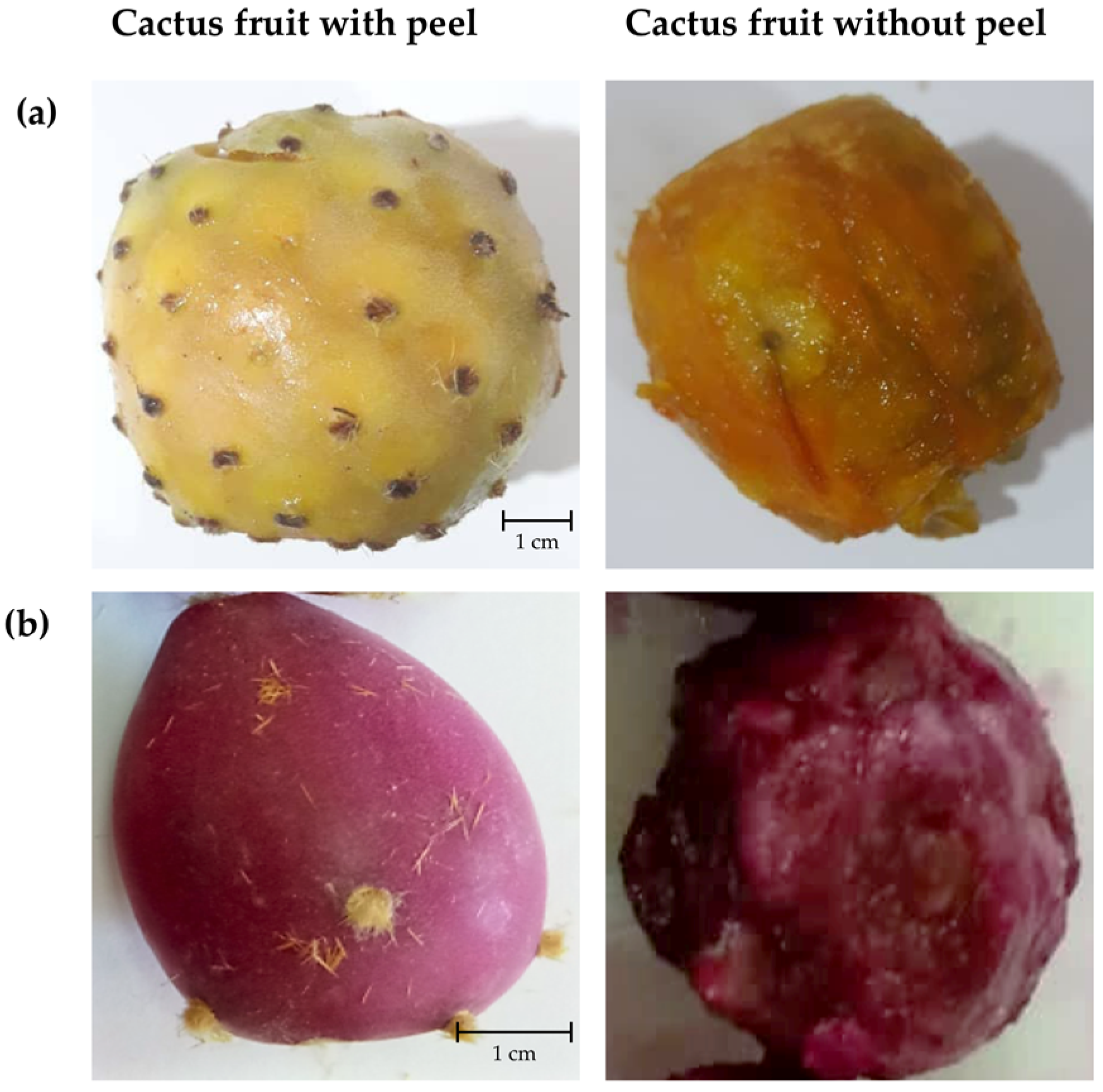
Figure 1. Photographs of Opuntia sp. fruits. (a) Cultivated Opuntia sp.; (b) Wild Opuntia sp. [photographs of the author (W.Z.)].
Agriculture 12 01755 g001
Figure 2. PHYML phylogenetic tree section of the ITS gene region, considering one of the Opuntia sp. branches (red) with a support value of 0.94 and both the comercial (CS1_ITS) and wild (WF1_ITS) sequences.
Agriculture 12 01755 g002
Figure 3. Phytochemical composition of different Opuntia sp. fractions. (a) Total Phenolic Content; (b) Total Flavonoid Content. Results are expressed as mean ± SD of at least five independent assays. TPC and TFC are expressed as mg of Gallic Acid Equivalents (GAE) and Quercetin Equivalents (QE) per 100 g of dry matter (DM). Significant differences between the samples were designed with different letters from the most significant (letter a) to the lowest significant (letter f) (p ≤ 0.05, ANOVA).
Agriculture 12 01755 g003
Figure 4. Antioxidant potential of different Opuntia sp. fractions. (A) Trolox equivalent antioxidant capacity; (B) Reducing power; (C) Ferric reducing antioxidant power; (D) Phosphomolybdenum antioxidant activity; (E) Hydrogen peroxide scavenging activity; (F) Hydroxyl radical inhibition assay; (G) Nitric oxide radical scavenging assay. TE, Trolox Equivalents; AAE, Ascorbic Acid Equivalents; H2O2, Hydrogen Peroxide; OH, Hydroxyl Radical; NO, Nitric Oxide Radical; DM, Dry Matter. Results are expressed as mean ± SD corresponding to the maximum percentage of inhibition of Opuntia sp. extracts, determined by at least five independent assays. Significant differences between the samples were designed with different letters from the most significant (letter a) to the lowest significant (letter f) (p ≤ 0.05, ANOVA).
Agriculture 12 01755 g004
Figure 5. Projection of Opuntia sp. extracts (A) [variables: Wild seeds (WS), Cultivated seeds (CS), Wild pulp (WP), Cultivated pulp (CP), Wild fruit (WF), Cultivated fruit (CF)] and loadings (B) by chemical composition and bioactivities [variables: total phenolic content (TPC), total flavonoid content (TFC), Trolox equivalent antioxidant capacity (TEAC), reducing power (RP), Ferric reducing antioxidant power (FRAP), Phosphomolybdenum antioxidant activity (PMA), Hydrogen peroxide scavenging activity (H2O2), Nitric oxide scavenging (NO), Hydroxyl radical scavenging (OH)] into the plane composed by the principal components PC1 and PC2 containing 89.41% of the total variance.
Agriculture 12 01755 g005
Table 1. List of specific primers used for PCR amplification.
RegionSequencesAnnealing TemperatureReference
COX-3COX-3f: CCGTAGGAGGTGTGATGT
COX-3r: CTCCCCACCAATAGATAGAG
51 °C[19]
18S18S-f: ACCTGGTTGATCCTGCCAG
18S-r: TGATCCTTCYGCAGGTTCAC
18S402-f: GCTACCACATCCAAGGAAGGCA
18S895-f: GTCAGAGGTGAAATTCTTGGAT
18S919-r: TAAATCCAAGAATTTCACCTCT
18S1339-r: CTCGTTCGTTAACGGAATTAACC
55 °C[17]
rbcL1f: ATGTCACCACAAACAGAAAC
724r: TCGCATGTACCTGCAGTAGC
48 °C[20]
matkmatkx: TAATTTACGATCAATTCATTC
matk5: GTTCTAGCACCAGAAAGTCG
48 °C[19]
ITSITS5: GGAAGTAAAAGTCGTAACAAGG
ITS4: TCCTCCGCTTATTGATATGC
57 °C[19]
Table 2. Physico-chemical characteristics of cultivated and wild Opuntia fruits.
Opuntia SpeciesWeight (g)Moisture (%)°BrixAsh (%)
Cultivated fruit65.62 ± 18.09 a86.48 ± 0.10 a11.48 ± 0.03 a0.32 ± 0.05 b
Wild fruit21.93 ± 1.93 b83.14 ± 0.14 b10.10 ± 0.10 b0.53 ± 0.09 a
Differences between the samples were designed with different letters from the most significant (letter a) to the lowest significant (letter b) (p ≤ 0.05, ANOVA).
Table 3. The BOLD identification system results table for the matk gene region.
Match RankPhylumClassOrderFamilyGenusSpeciesSubspeciesScoreSimilarityE-Value
1MagnoliophytaMagnoliopsidaCaryophyllalesCactaceaeOpuntiaficusindica 8741000
2MagnoliophytaMagnoliopsidaCaryophyllalesCactaceaeOpuntiaellisiana 8741000
3MagnoliophytaMagnoliopsidaCaryophyllalesCactaceaeOpuntiaengelmannii var. lindheimeri 8741000
4MagnoliophytaMagnoliopsidaCaryophyllalesCactaceaeOpuntiaengelmannii var. linguiformis 8741000
5MagnoliophytaMagnoliopsidaCaryophyllalesCactaceaeOpuntiaorbiculata 8741000
6MagnoliophytaMagnoliopsidaCaryophyllalesCactaceaeOpuntiadillenii 8741000
7MagnoliophytaMagnoliopsidaCaryophyllalesCactaceaeOpuntiaficusindica 8741000
8MagnoliophytaMagnoliopsidaCaryophyllalesCactaceaeOpuntiamagnifica 8741000
9MagnoliophytaMagnoliopsidaCaryophyllalesCactaceaeOpuntiaphaeacantha 8741000
10MagnoliophytaMagnoliopsidaCaryophyllalesCactaceaeOpuntiasetispina 8741000
11MagnoliophytaMagnoliopsidaCaryophyllalesCactaceaeOpuntiastricta 8741000
12MagnoliophytaMagnoliopsidaCaryophyllalesCactaceaeOpuntiadillenii 8741000
13MagnoliophytaMagnoliopsidaCaryophyllalesCactaceaeOpuntiaficusindica 8741000
14MagnoliophytaMagnoliopsidaCaryophyllalesCactaceaeOpuntiaengelmanniivar. engelmannii8741000
15MagnoliophytaMagnoliopsidaCaryophyllalesCactaceaeOpuntiamartiniana 8741000
16MagnoliophytaMagnoliopsidaCaryophyllalesCactaceaeOpuntiapailana 8741000
17MagnoliophytaMagnoliopsidaCaryophyllalesCactaceaeOpuntiaellisiana 8731000
18MagnoliophytaMagnoliopsidaCaryophyllalesCactaceaeOpuntiaengelmanniivar. engelmannii8731000
Publisher’s Note: MDPI stays neutral with regard to jurisdictional claims in published maps and institutional affiliations.