Article

Volatile Organic Compounds from Rice Rhizosphere Bacteria Inhibit Growth of the Pathogen Rhizoctonia solani

1
Key Laboratory of Plant Nutrition and Fertilizer, Ministry of Agriculture and Rural Affairs, Institute of Agricultural Resources and Regional Planning, Chinese Academy of Agricultural Sciences, Beijing 100081, China
2
Key Laboratory of Biometallurgy of Ministry of Education, School of Minerals Processing and Bioengineering, Central South University, Changsha 410083, China
3
Department of Entomology, Kansas State University, Manhattan, KS 66506, USA
*
Author to whom correspondence should be addressed.
Agriculture 2021, 11(4), 368; https://doi.org/10.3390/agriculture11040368
Submission received: 8 March 2021 / Revised: 12 April 2021 / Accepted: 12 April 2021 / Published: 19 April 2021
(This article belongs to the Special Issue Detection, Identification, and Control of Plant Pathogens)

Abstract

:
Rice sheath blight, a fungal disease caused by Rhizoctonia solani, seriously threatens rice production. Some of the volatile organic compounds (VOCs) produced by microbes are inhibitory to the growth of the plant pathogen, and hence may have the potential as environmentally friendly antifungal substances. However, information on the inhibitory effect of VOCs released by rice rhizosphere bacteria on R. solani is scarce. In this study, bacteria from the rice rhizosphere capable of inhibiting the growth of R. solani via releasing VOCs were screened using a double Petri dish assay. Headspace solid phase microextraction and gas chromatography mass spectrometry (GC/MS) were used to identify and quantify the VOCs. The contributions of VOCs to the inhibition of the growth of R. solani were estimated by constructing a random forest model, and were verified using pure compounds. Nine strains (i.e., Pseudomonas sp. No. 3, Enterobacter sp. No. 26, Enterobacter sp. No. 34, Pseudomonas sp. No. 35, Ralstonia sp. No. 50, Bacillus sp. No. 62, Arthrobacter sp. No. 146, Brevibacillus sp. No. 2–18, and Paenisporosarcina sp. No. 2–60) showed various inhibition on R. solani growth via VOCs. The inhibitory effect ranged from 7.84% to 100%, with Ralstonia sp. No. 50 completely inhibiting the growth of R. solani. Five VOCs (i.e., benzoic acid ethyl ester, 3-methyl-butanoic acid, 2-ethyl-1-hexanol, 3-methyl-1-butanol, and 6-methyl-5-hepten-2-one) identified by random forest model were confirmed to be toxic to R. solani when applied as a pure chemical compound. In particular, benzoic acid ethyl ester, 3-methyl-butanoic acid, and 2-ethyl-1-hexanol were lethal to R. solani. In summary, the rice rhizosphere bacteria (Ralstonia sp. No. 50) and VOCs (benzoic acid ethyl ester, 3-methyl-butanoic acid, and 2-ethyl-1-hexanol) showed potential to be used as new resources for biological control of rice sheath blight.

1. Introduction

Rice is an important staple food crop globally. More than 3.5 billion people rely on it to provide calorific energy every day. Rice sheath blight caused by Rhizoctonia solani is one of the most economically important rice diseases worldwide, causing devastating crop losses and representing a serious threat to global food security [1,2]. Currently, rice sheath blight is mainly controlled by fungicides [3]. For instance, Jingangmycin has been used widely to control sheath blight in China for the past 30 years [4]. However, the extensive and continuous use of a single fungicide increases the risk of resistance development in the pathogen [5]. Cultivating resistant rice varieties would be the best option to cope with the attack of this pathogen, but no such variety is available to the growers at present [6]. A range of alternate control measures such as fertilization, soil amendments, and agricultural management have also been recommended. Unfortunately, these alternative measures are time-consuming and laborious, which is not particularly effective [7,8,9]. Therefore, it is imperative to search for new active agents against rice sheath blight.
Biological control is widely studied as an environmentally friendly control strategy. For example, Kanjanamaneesathian et al. found three bacterial isolates that provided maximum suppression of sheath blight lesions [10]. Bacillus subtilis B4 is also shown to be a highly effective inhibitor of R. solani mycelial growth [11]. During rice growth, the root system can release various signal molecules to shape a unique root microbial community [12]. This microbial community plays an important role in rice growth and development. By releasing various metabolites, such as phytohormones and volatile organic compounds (VOCs), they not only promote plant growth but also help plants resist various diseases [13,14]. So far, there are a few studies on rice-associated bacteria inhibiting the growth of R. solani [2], but no reports on whether rice rhizosphere bacteria can inhibit R. solani growth via VOCs.
Among the many biological control strategies, VOCs have been studied widely [15,16]. They are potentially effective being small organic molecules (<C20) with low water solubility and high vapor pressure (0.01 kPa at 20 °C) that can readily evaporate and diffuse through heterogeneous mixtures of solids, liquids, and gasses. There is growing evidence that VOCs could modulate various biotic and abiotic stresses in plants, and are very likely to become a substitute for environmentally harmful pesticides and fungicides [17]. For instance, the endophytic bacteria Pseudomonas stutzeri E25 and Stenotrophomonas maltophilia CR71 inhibited the growth of Botrytis cinerea by releasing VOCs, with dimethyl disulfide (DMDS) being the main component [18]. VOCs produced by Bacillus subtilis GB03 can directly inhibit the mycelial growth of Arabidopsis gray mold and interfere with the attachment of pathogenic fungi on hydrophobic foliage [19]. A small number of VOCs were found to inhibit growth of R. solani specifically, such as sulfide, 1-methyl-4-(1-methylethenyl)-cyclohexene, methyl-2-methylvalerate, and 1,3,5-trichloro-2-methoxybenzene [14,19,20]. Overall, information on the identity and inhibitory effect of VOCs released by rice rhizosphere bacteria is fragmented.
In the present study, we searched for novel VOCs from rice rhizosphere bacteria with efficacy against rice sheath blight. To achieve this objective, bacteria from rice rhizosphere suppressive to R. solani were screened via a double Petri dish assay, VOCs were identified and quantified with GC-MS, and relative contribution of each VOC was estimated by constructing a random forest model and was further verified using pure compounds.

2. Materials and Methods

2.1. Test Strains, Culture Medium, and Culture Conditions

The test strains were isolated from the rice rhizosphere. All strains were sequenced at the Institute of Crop Science, Chinese Academy of Agricultural Sciences, and then the bacterial species were identified by BLAST search of 16S rDNA sequences using NCBI database. Freeze-dried bacterial strains were activated by culturing on a beef extract peptone medium (beef extract 3 g, peptone 10 g, sodium chloride 5 g, agar 20 g, distilled water 1 L, pH 7.2–7.4) in the dark at 30 °C for 48 h. Bacteria were amplified via culturing them in a liquid beef medium as described above except without containing an agar. Bacterial culturing was carried out under shaking at 180 rpm at 30 °C for 48 h.
Rhizoctonia solani was purchased from Agricultural Culture Collection of China (Strain number 36246). R. solani was activated on the potato dextrose agar (PDA) medium (potato 200 g, glucose 20 g, agar 20 g, water 1 L), and cultured in the dark at 30 °C for 48 h. The activated strain was inoculated into the potato dextrose broth (PDB) medium (the same formula as PDA, without agar). The R. solani fermentation broth was obtained after shaking cultures at 180 rpm and 30 °C for 48 h.

2.2. Inhibition of R. Solani by VOCs

The bacterial fermentation broth (100 μL) of the tested strains was spread by a sterile coating rod on the Petri dish containing beef extract peptone medium, and cultured at 30 °C for 24 h. R. solani fermentation broth (10 μL) was dropped in the center of a Petri dish containing PDA medium and placed on top of a Petri dish of PDA medium inoculated with R. solani. At the same time, a Petri dish without inoculation bacteria was set as the control. The two bottom dishes were sealed and cultured at 30 °C in the dark. The colony diameter of R. solani was recorded before the control R. solani reached the edge of the Petri dish. The treatment was repeated three times to assess the inhibitory effects of VOCs. The inhibition rate was measured as follows: Inhibition rate = [(colony diameter of control R. solani − colony diameter of treated R. solani)/colony diameter of control R. solani] × 100%.

2.3. Collection of VOCs

An aliquot of 5 mL of beef extract peptone culture medium and 0.1 g agar were added to a 20 mL headspace bottle, then sealed with kraft paper, and sterilized at 121 °C for 30 min. After sterilization, the bottles were tilted 30 °C to cool and solidify the medium. Then 100 μL of the prepared bacterial fermentation broth was added to each bottle, shaken to distribute the bacterial fermentation broth evenly on the agar slant, covered with kraft paper, and incubated at 30 °C in the dark. After 48 h, the kraft paper was replaced by a Polytetrafluoroethylene (PTFE) septum seal with a hollow screw cap. A headspace bottle containing the medium, but no bacteria, was used as the control. All treatments were repeated four times.
After culturing for 5 days, VOCs from the headspace of each vial were collected by a 50/30 μm DVB/CAR/PDMS extraction fiber (purchased from Supelco, Bellefonte, PA, USA). The extraction fiber was aged according to the manufacturer’s instructions before use and was equipped with a solid-phase microextraction (SPME) handle (Supelco, Bellefonte, PA, USA). The extraction was carried out at 30 °C for 12 h.

2.4. Analysis of VOCs

VOCs were analyzed by a 7890A-5975C GC-MS System (Agilent Technologies, Palo Alto, CA, USA) immediately after extraction. The chromatographic conditions were injection port temperature 250 °C, injection time 2.7 min, split-less mode, carrier gas 99.999% high purity helium, and column flow rate 1 mL min−1. The temperature program of a column oven was initial temperature 50 °C for 2 min, an increase to 180 °C at 8 °C min−1, and then further to 240 °C at 10 °C min−1, this was then held at 240 °C for 6 min. The mass spectrometer (MS) conditions were electron ionization (EI) mode, 70 eV, ion source temperature 230 °C, quadrupole temperature 150 °C, transmission line temperature 250 °C, full scan mode, and the scanning range 35–450 amu. The MS of VOCs detected were identified by comparison with the NIST/EPA/NIH database.

2.5. In Vitro Verification Using Pure Compounds

The PDA medium dish edge fragment was placed in a sterilized 200 μL container. R. solani fermentation broth (10 μL) was added to the center of the medium. Pure chemical compounds (50, 100, and 200 μL) benzoic acid ethyl ester (Sangon Biotech, Shanghai, China), 3-methyl-butanoic acid (Adamas, Basel, Switzerland), 3-methyl-1-butanol (Sigma-Aldrich, St. Louis, MO, USA ), 6-methyl-5-hepten-2-one (Sigma-Aldrich, St. Louis, MO, USA), 2-ethyl-1-hexanol (Sigma-Aldrich, St. Louis, MO, USA ), dimethyl disulfide (Macklin, Shanghai, China), and dimethyl trisulfide (Macklin, Shanghai, China) were added to the container separately, with sterile water added if the volume was less than 200 μL (the concentrations were 25%, 50%, and 100%, 25%: 50 μL pure compound and 150 μL sterile water/plate, 50%: 100 μL pure compound and 100 μL sterile water/plate, 100%: 200 μL of pure compound/plate). Their concentrations were greater than 98%. The same amount of sterile water (200 μL) was added to the control container, and was cultivated in the dark at 32 °C to observe growth. All treatments had four replicates.

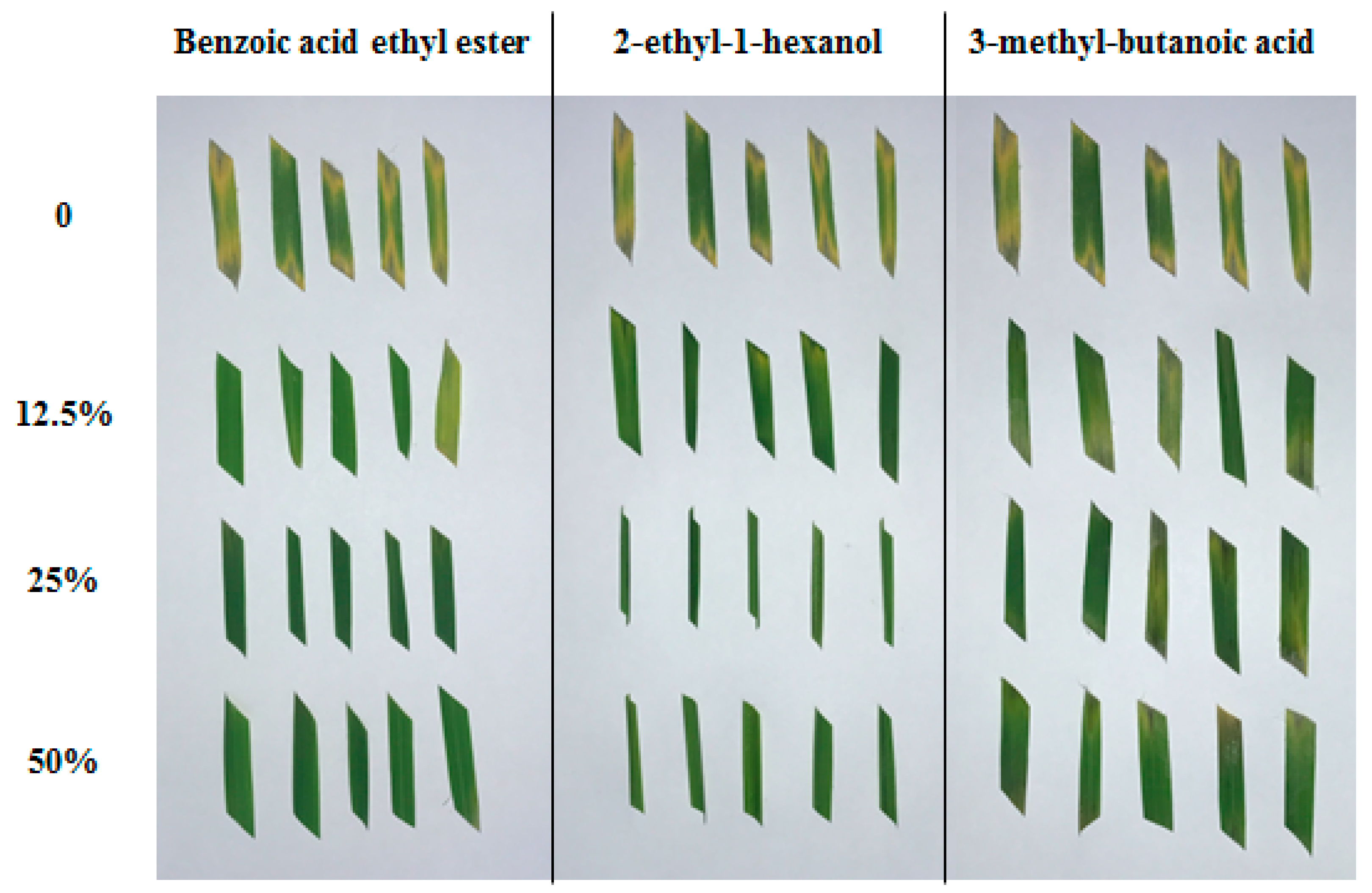
2.6. Evaluation of the Inhibitory Activity of Pure Compounds on the Lesion Development by R. solani on Detached Rice Leaves

Rhizoctonia solani was inoculated on PDA medium and pre-cultured for 48 h. The rice leaves were cut into 5-cm-long fragments and then placed on the PDA medium. Different compounds (200 μL) were added to the 200 μL container containing the plate edge fragment. The concentrations were 0%, 12.5%, 25%, and 50% (0%: 200 μL sterile water/plate, 12.5%: 25 μL pure compound and 175 μL sterile water/plate; 25% and 50% were same as above), respectively. Each treatment had five replicates and observations were made every 24 h.

2.7. Statistical Analyses

Microsoft Excel 2010 (Microsoft Corporation, Redmond, WA, USA) and SPSS for Windows version 21 (SPSS Institute Inc., Armonk, NY, USA) were used for statistical analysis. The random forest variable importance plot and Venn diagram were made in R 4.0.2, and the packages used were “randomForest” and “UpSetR”.

3. Results

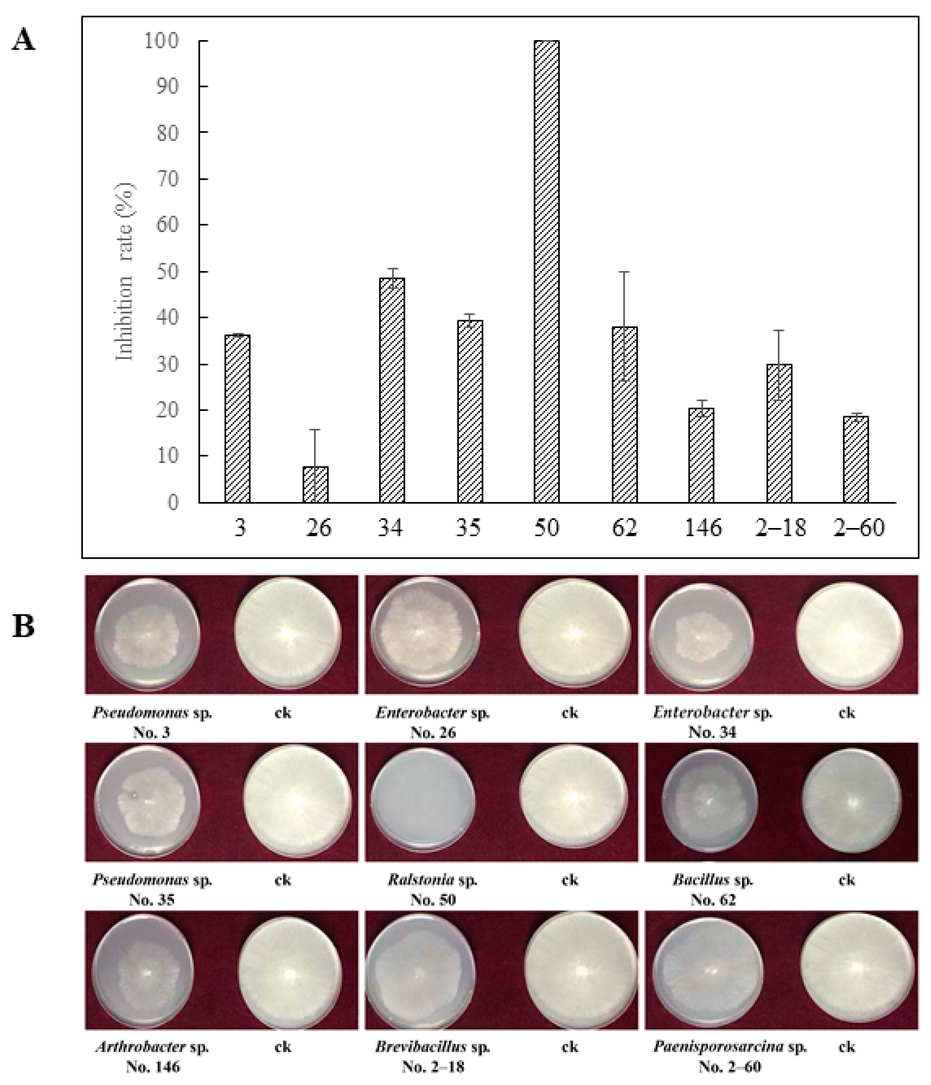
3.1. Inhibition of VOCs on R. solani

The tested strains were Pseudomonas sp. (No. 3), Enterobacter sp. (No. 26), Enterobacter sp. (No. 34), Pseudomonas sp. (No. 35), Ralstonia sp. (No. 50), Bacillus sp. (No. 62), Arthrobacter sp. (No. 146), Brevibacillus sp. (No. 2–18), and Paenisporosarcina sp. (No. 2–60), respectively. They all inhibited the growth of R. solani by releasing VOCs, with the inhibition ranging from 7.84% to 100%. The strain No. 50 of Ralstonia sp. had the strongest inhibitory effect (100%). The weakest inhibitory effect (7.84%) was recorded for the strain No. 26 of Enterobacter sp. The inhibitory effects of the strains No. 3, No. 34, No. 35, No. 62, No. 146, No. 2–18, and No. 2–60 were 36.08%, 48.63%, 39.22%, 38.04%, 20.39%, 29.80%, and 18.43%, respectively (Figure 1).

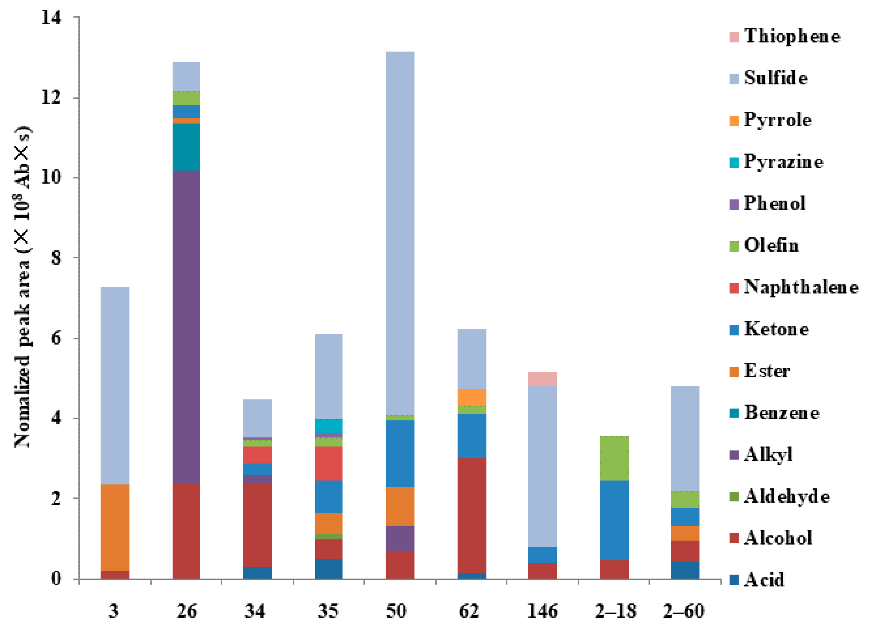
3.2. Composition of VOCs

A total of 89 compounds were detected across all tested strains, but different strains produced different types of VOCs. Some VOCs were unique to a specific strain, but others were produced by several strains. The number and abundance of VOCs produced by each strain were also different. The number of VOCs released by an individual strain ranged from 7 to 33. The number of VOCs released by strains No. 3, No. 26, No. 34, No. 35, No. 50, No. 62, No. 146, No. 2–18, and No. 2–60 were 7, 17, 21, 30, 10, 29, 10, 18, and 33, respectively (Table 1).
There were 15 compounds unique to strain No. 2–60, six unique to No. 35, seven to No. 62, six to No. 34, five to No. 2–18, eight to No. 26, six to No. 50, two to No. 146, and three unique to No. 3. One compound was released by eight strains, one compound was released by seven strains, five compounds were released by six strains, three compounds were released by five strains, three compounds were released by four strains, seven types of compounds were released by three strains, and 12 compounds were released by two strains (Figure 2).

3.3. Classification and Abundance of VOCs Emitted from Different Bacterial Strains

The VOCs released by all tested strains were mainly esters, olefins, alkanes, ketones, acids, thiophenes, aldehydes, naphthalenes, sulfides, phenols, alcohols, pyrazines, pyrroles, and benzenes. All strains released alcohols, but strains No. 34 and 62 released the most amounts. Strains No. 3, 35, 50, 146, and 2–60 released the largest amounts of sulfides, strain No. 26 released the largest amounts of alkanes, while strain No. 2–18 released the largest amounts of ketones (Figure 3).

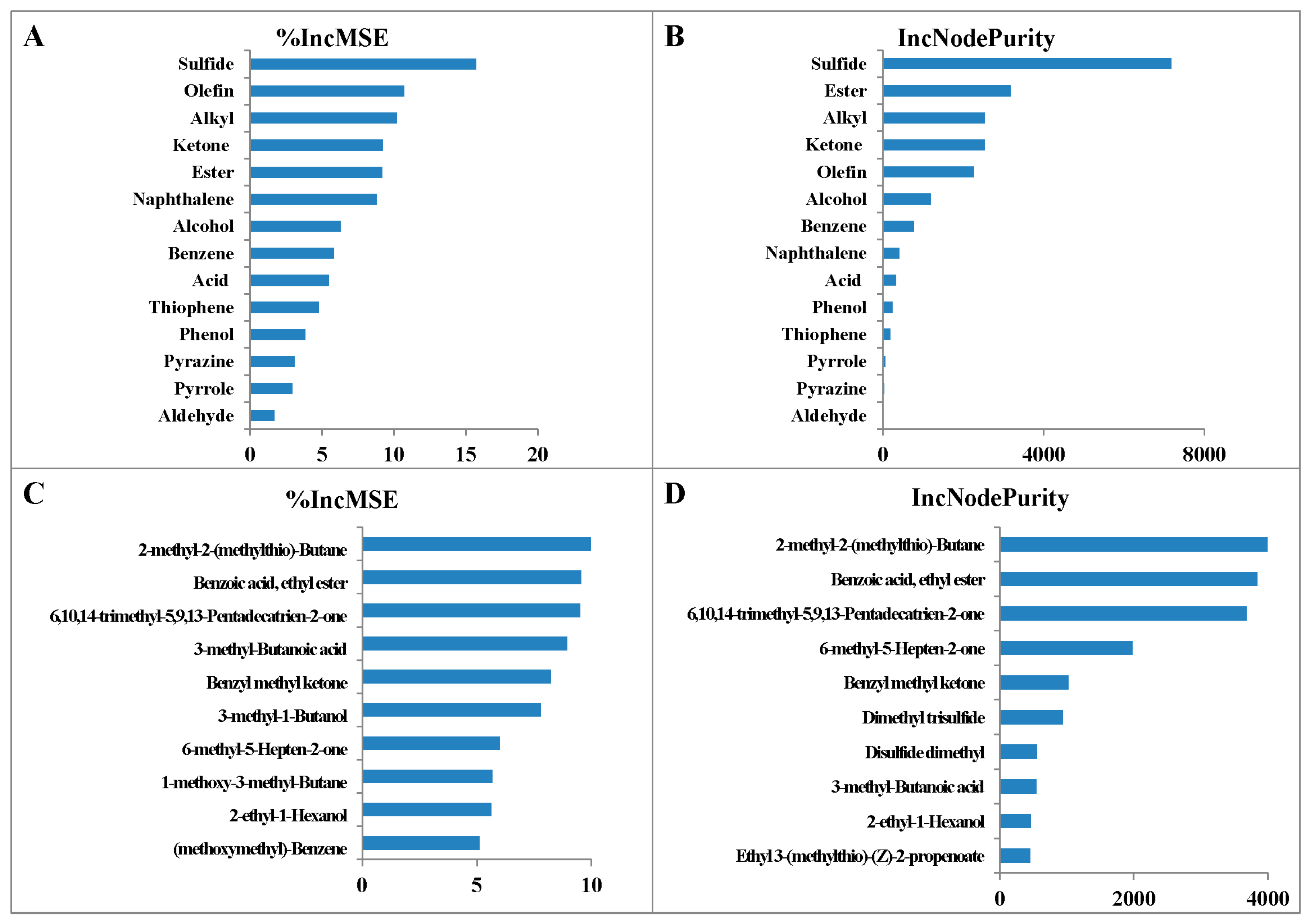
3.4. The Relative Importance of Different VOCs

The relative importance of different VOCs was estimated by constructing a random forest model. It was found that sulfides, olefins, alkyls, ketones, and esters were more important than the other types of VOCs. The ranking was based on two criteria: Mean Square Error and Node Purity. The proportion of variance explained by this model was 94.23% (Figure 4A,B). In addition, a random forest model of the importance of different individual VOCs was established. This model explained 95.24% of the variance. Using this model, the top 10 most important VOCs were ranked. These compounds with decreasing importance were 2-methyl-2-methylthio-butane, benzoic acid ethyl ester, 6,10,14-trimethyl-5,9,13-pentadecatrien-2-one, 3-methyl-butanoic acid, and benzyl methyl ketone (Figure 4C,D).

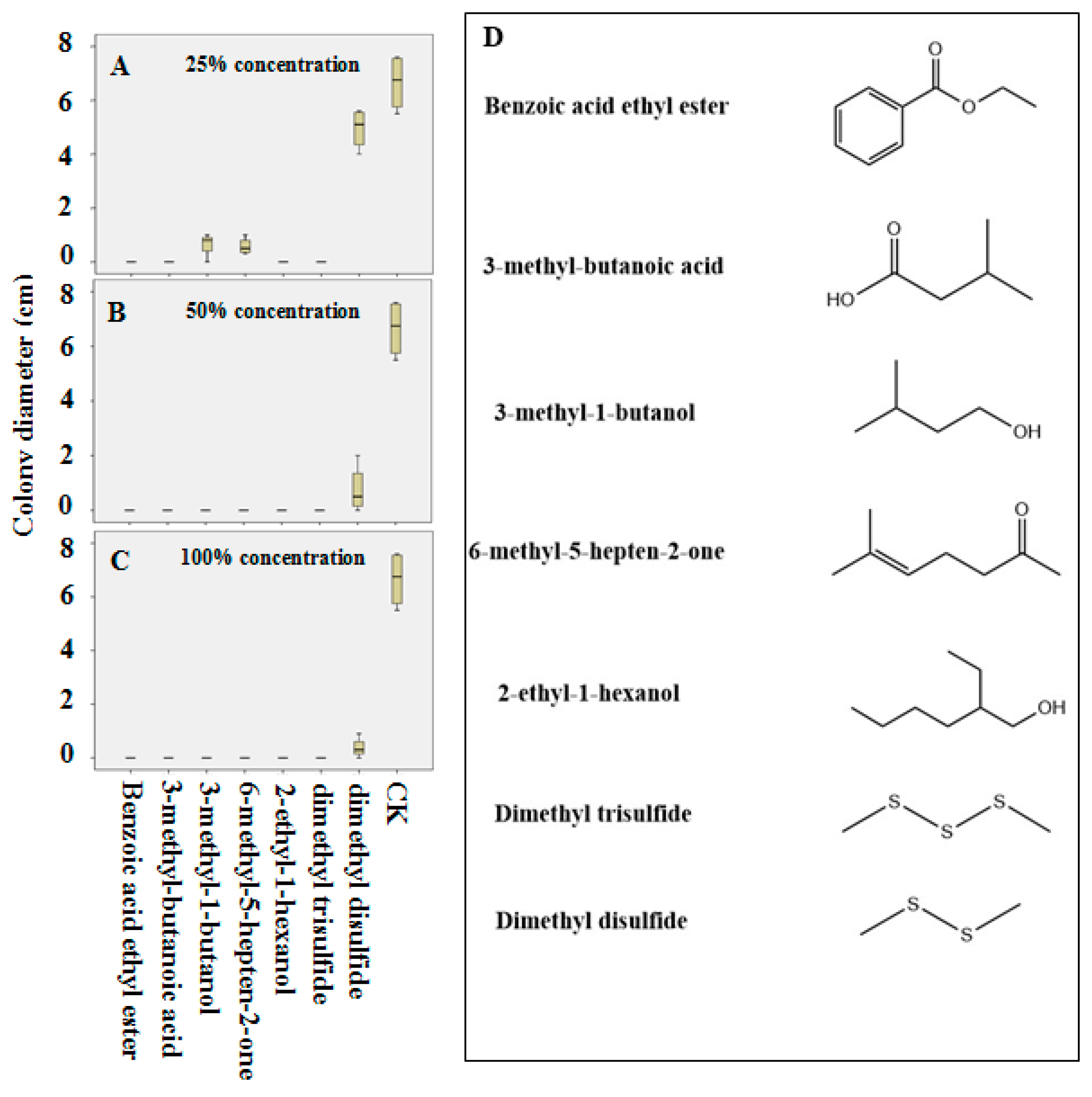
3.5. The Inhibition of R. solani by Pure Compounds

Benzoic acid ethyl ester, 3-methyl-butanoic acid, 2-ethyl-1-hexanol, and dimethyl trisulfide were lethal to R. solani, whereas 3-methyl-1-butanol and 6-methyl-5-hepten-2-one significantly inhibited the growth of R. solani at 25% concentration. At 50% and 100% concentrations, all compounds except dimethyl disulfide were lethal to R. solani. However, dimethyl disulfide also significantly inhibited the growth of R. solani (Figure 5).
Benzoic acid ethyl ester had the strongest suppression on R. Solani infection, with nearly complete inhibition under all tested concentrations greater than 12.5%. 2-Ethyl-1-hexanol inhibited the infection of detached leaves by R. solani at the concentration of 12.5%. The effect of benzoic acid ethyl ester became increasingly obvious as the concentration of benzoic acid ethyl ester increased. By contrast, 3-methyl-butanoic acid showed only a slightly inhibitory effect on R. solani at all tested concentrations, but inhibition did not reach very high levels. (Figure 6).

4. Discussion

In this study we found that nine bacterial strains isolated from the rice rhizosphere inhibited the growth of R. solani without direct contact (a double Petri dish assay), indicating that these bacteria released VOCs that were inhibitory to the growth of R. solani (Figure 1). Although there are many related reports in the literature on bacteria that could inhibit the growth of pathogens by releasing VOCs [21,22], no report can be found on the VOCs released by rice rhizosphere bacteria that can inhibit the growth of R. solani, specifically. Here we discovered nine bacterial strains that can release VOCs, which can then elicit inhibitory effects on R. solani. Based on sequencing and comparison in the NCBI database, we found these bacterial strains belong to Pseudomonas sp. (No. 3), Enterobacter sp. (No. 26), Enterobacter sp. (No. 34), Pseudomonas sp. (No. 35), Ralstonia sp. (No. 50), Bacillus sp. (No. 62), Arthrobacter sp. (No. 146), Brevibacillus sp. (No. 2–18), and Paenisporosarcina sp. (No. 2–60), respectively. As expected, different strains exhibited different inhibitory effects on R. solani, ranging from 7.84% to 100%. Surprisingly, strain No. 50 of Ralstonia sp. showed 100% inhibition of R. solani growth (Figure 1). In comparison, Mookherjee et al. found that the endophyte Geotrichum candidum utilized different carbon sources to varying extents [23], and the antifungal activity of its VOCs against phytopathogen R. solani changed with each carbon source, with the maximum mean growth inhibition of about 62% observed with glucose. Elkahoui et al. (2015) found 25 VOCs producing strains inhibited (by 27–50%) the growth of R. solani [24]. However, no previous study has found a strain that would kill R. solani. Hence, the present study is the first one to document complete (100%) inhibition of R. solani growth by bacterial VOCs produced by Ralstonia sp. No. 50.
The profiles of VOCs released by the nine bacterial strains were also analyzed in our study. We found that some VOCs were commonly released by different bacterial strains, whiles others were unique to each strain (Table 1, Figure 2). In addition to the differences of VOC profiles, the abundance of different VOCs was also different. These findings are consistent with those reported previously by Li et al. [25], who tested VOCs released by eight Bacillus strains, which released a variety of VOCs in different amounts. Hernández-León et al. also found that four Pseudomonas fluorescens UM16, UM240, UM256, and UM270 produced different VOCs in different concentrations [26]. The different bacterial strains characterized in the different studies are the likely reason for the differences in the type and content of VOCs observed.
In the random forest variable importance plot of different VOCs, benzoic acid ethyl ester and 3-methyl-butanoic acid contributed more to R. solani inhibition than 2-ethyl-1-hexanol, 3-methyl-1-butanol, and 6-methyl-5-hepten-2-one. Benzoic acid ethyl ester and 3-methyl-butanoic acid were found lethal to R. solani at 25% concentration. The VOCs 3-methyl-1-butanol, and 6-methyl-5-hepten-2-one were able to significantly inhibit the growth of R. solani at 25% concentration, but their contribution was less than that of benzoic acid ethyl ester and 3-methyl-butanoic acid. Therefore, it may be reasonable to speculate that 2-methyl-2-(methylthio)-butane and 6,10,14-trimethyl-5,9,13-pentadecatrien-2-one can also totally inhibit R. solani under different conditions. These two VOCs have not been reported hitherto to have antifungal activity previously and their antifungal activity remains to be investigated more extensively and vigorously.
2-Ethyl-1-hexanol was also lethal to R. solani at 25% concentration. Similarly, benzoic acid ethyl ester, 3-methyl-butanoic acid, 2-ethyl-1-hexanol, 3-methyl-1-butanol, and 6-methyl-5-hepten-2-one, which had similar contributions in the random forest model, all showed lethality or significant inhibition at 25% concentration, and were definitely lethal at 50% and 100% concentrations (Figure 4 and Figure 5). In the literature, some other VOCs, such as 2-phenylethanol, isopentyl acetate, naphthalene, methyl 2-methylpentanoate, 1,3,5-trichloro-2-methoxy benzene, 1-methyl-4-(1-methylethenyl)-cyclohexene, and 4-flavanone (4H-1-benzopyran-4-one, 2,3-dihydro-2-phenyl), were also reported to inhibit the growth of R. solani [19,23,27]. These VOCs were not detected in the present study, possibly because we isolated bacteria from the rhizosphere of rice, whereas the previous studies used other bacterial sources.
Several studies have described antifungal activity of bacterial VOCs, but few have identified a single VOC or a blends of VOCs responsible for the antifungal activity [28]. Fernando et al. used commercial VOCs for in vitro verification of their antifungal activity [29], and only a few of them were found effective. This approach is time-consuming, laborious, and costly [30,31]. In our study a random forest model was constructed to explore the relationship between VOCs and antifungal activity for the first time. This was an easy and effective way to compare the contribution of different VOCs to antifungal activity and identify the most effective VOCs. Tests of pure commercial compound showed that estimation based on the random forest model was reliable. This method saved time, labor, and money.
It is very interesting to note that some of the most potent VOCs were easily synthesized and are commercially available, such as benzoic acid ethyl ester, 3-methyl-butanoic acid, 2-ethyl-1-hexanol, 3-methyl-1-butanol, 6-methyl-5-hepten-2-one. In addition, we found that dimethyl trisulfide was lethal to R. solani at 25% concentration. Benzoic acid ethyl ester, 2-ethyl-1-hexanol, and 3-methyl-butanoic acid were also lethal to R. solani under certain conditions. These three compounds could actually inhibit R. solani infection on detached leaves (Figure 5 and Figure 6). However, the previous studies used pure compounds for verification, and no study has ever used smaller concentrations of compounds to verify the antifungal activity. Hence, the VOCs found in this study showed high antifungal activity. However, we assume that the VOCs produced by the rhizosphere bacteria when grown on a beef peptone medium would also be produced when they colonize roots of the rice plant. That may or may not be the case, as nutrition obtained from a complex medium may result in different products vs. those when grown in a natural system. Nonetheless, our findings are interesting. We also identified volatiles produced by the different bacterial strains as well as evaluated the impact those volatiles have on disease suppression. Again, this was done under “laboratory” conditions that do not necessarily reflect the real world, but there is enough information presented for us or others to pursue this line of investigation. For example, the potential interaction of production of the VOCs in a complex environment interacting with many potential feedback mechanisms. Therefore, the system presented here is not ready to release as a commercially ready biocontrol, but the data presented here are intriguing and sufficiently represent a starting point to have a decent system for further study.

5. Conclusions

Pseudomonas sp. No. 3, Enterobacter sp. No. 26, Enterobacter sp. No. 34, Pseudomonas sp. No. 35, Ralstonia sp. No. 50, Bacillus sp. No. 62, Arthrobacter sp. No. 146, Brevibacillus sp. No. 2–18, and Paenisporosarcina sp. No. 2–60 were nine strains of bacteria found for the first time to inhibit R. solani growth via VOCs. The inhibitory effect ranged from 7.84% to 100%, with Ralstonia sp. No. 50 inhibiting the growth of R. solani completely (100%). Benzoic acid ethyl ester, 3-methyl-butanoic acid, 2-ethyl-1-hexanol, 3-methyl-1-butanol, and 6-methyl-5-hepten-2-one were VOCs released by bacteria in the rice rhizosphere. These five VOCs were found to be toxic to R. solani for the first time. Among them, benzoic acid ethyl ester, 3-methyl-butanoic acid, and 2-ethyl-1-hexanol were lethal to R. solani. Overall, novel functions of bacterial strains from the rice rhizosphere and their VOCs have the potential to be used as resources for biological control of rice sheath blight in the future.

Author Contributions

Conceptualization, F.F., A.S. and E.W.; methodology, E.W., J.B., S.W. and J.Z.; formal analysis, E.W., Z.S., X.L. (Xu Li) and W.D.; writing—original draft preparation, E.W.; writing—review and editing, F.F., A.S., M.C., and X.L. (Xiongduo Liu). All authors have read and agreed to the published version of the manuscript.

Funding

The research was funded by the National Key Research and Development Program of China (2019YFD1002004) and Fundamental Research Funds for Central Non-profit Scientific Institution (No. 1610132019011, 1610132020012).

Institutional Review Board Statement

Not applicable.

Informed Consent Statement

Not applicable.

Data Availability Statement

The data presented in this study are available on request from the corresponding author.

Acknowledgments

We thank the reviewers for their valuable comments and recommendations. We also thank Timothy George for polishing the language.

Conflicts of Interest

The authors declare no conflict of interest.

References

  1. Spence, C.; Alff, E.; Johnson, C.; Ramos, C.; Donofrio, N.; Sundaresan, V.; Bais, H. Natural rice rhizospheric microbes suppress rice blast infections. BMC Plant Biol. 2014, 14, 1–17. [Google Scholar] [CrossRef] [Green Version]
  2. Shrestha, B.K.; Karki, H.S.; Groth, D.E.; Jungkhun, N.; Ham, J.H. Biological Control Activities of Rice-Associated Bacillus sp. Strains against Sheath Blight and Bacterial Panicle Blight of Rice. PLoS ONE 2016, 11, e0146764. [Google Scholar] [CrossRef] [Green Version]
  3. Boukaew, S.; Klinmanee, C.; Prasertsan, P. Potential for the integration of biological and chemical control of sheath blight disease caused by Rhizoctonia solani on rice. World J. Microb. Biotechnol. 2013, 29, 1885–1893. [Google Scholar] [CrossRef]
  4. Mew, T.W.; Cottyn, B.; Pamplona, R.; Barrios, H.; Li, X.M.; Chen, Z.Y.; Lu, F.; Nilpanit, N.; Arunyanart, P.; Kim, P.V.; et al. Applying rice seed-associated antagonistic bacteria to manage rice sheath blight in developing countries. Plant Dis. 2004, 88, 557–564. [Google Scholar] [CrossRef] [PubMed] [Green Version]
  5. Zhang, Z.J.; Jiang, Z.Y.; Zhu, Q.; Zhong, G.H. Discovery of beta-Carboline Oxadiazole Derivatives as Fungicidal Agents against Rice Sheath Blight. J. Agric. Food Chem. 2018, 66, 9598–9607. [Google Scholar] [CrossRef] [PubMed]
  6. Bag, M.; Yadav, M.; Mukherjee, A. Bioefficacy of Strobilurin Based Fungicides against Rice Sheath Blight Disease. Transcriptomics 2016, 4, 2. [Google Scholar]
  7. Postma, J.; Schilder, M.T. Enhancement of soil suppressiveness against Rhizoctonia solani in sugar beet by organic amendments. Appl. Soil Ecol. 2015, 94, 72–79. [Google Scholar] [CrossRef]
  8. Agtmaal, M.V.; Straathof, A.L.; Termorshuizen, A.; Lievens, B.; Hoffland, E.; Boer, W.D. Volatile-mediated suppression of plant pathogens is related to soil properties and microbial community composition. Soil Biol. Biochem. 2018, 117, 164–174. [Google Scholar] [CrossRef]
  9. Ahsan, T.; Chen, J.; Zhao, X.; Irfan, M.; Wu, Y. Extraction and identification of bioactive compounds (eicosane and dibutyl phthalate) produced by Streptomyces strain KX852460 for the biological control of Rhizoctonia solani AG-3 strain KX852461 to control target spot disease in tobacco leaf. AMB Express. 2017, 7, 54. [Google Scholar] [CrossRef] [Green Version]
  10. Kanjanamaneesathian, M.; Kusonwiriyawong, C.; Pengnoo, A.; Nilratana, L. Screening of potential bacterial antagonists for control of sheath blight in rice and development of suitable bacterial formulations for effective application. Australas. Plant Path. 1998, 27, 198–206. [Google Scholar] [CrossRef]
  11. Ahmad, A.-G.M.; Attia, A.-Z.G.; Mohamed, M.S.; Elsayed, H.E. Fermentation, formulation and evaluation of PGPR Bacillus subtilis isolate as a bioagent for reducing occurrence of peanut soil-borne diseases. J. Integr. Agric. 2019, 18, 2080–2092. [Google Scholar] [CrossRef]
  12. Goldford, J.E.; Lu, N.X.; Bajic, D.; Estrela, S.; Tikhonov, M.; Sanchez-Gorostiaga, A.; Segre, D.; Mehta, P.; Sanchez, A. Emergent simplicity in microbial community assembly. Science 2018, 361, 469–474. [Google Scholar] [CrossRef] [PubMed] [Green Version]
  13. Zhang, J.Y.; Liu, Y.X.; Zhang, N.; Hu, B.; Jin, T.; Xu, H.R.; Qin, Y.; Yan, P.X.; Zhang, X.N.; Guo, X.X.; et al. NRT1.1B is associated with root microbiota composition and nitrogen use in field-grown rice. Nat. Biotechnol. 2019, 37, 676. [Google Scholar] [CrossRef] [PubMed]
  14. Carrion, V.J.; Cordovez, V.; Tyc, O.; Etalo, D.W.; de Bruijn, I.; de Jager, V.C.L.; Medema, M.H.; Eberl, L.; Raaijmakers, J.M. Involvement of Burkholderiaceae and sulfurous volatiles in disease-suppressive soils. ISME J. 2018, 12, 2307–2321. [Google Scholar] [CrossRef] [PubMed] [Green Version]
  15. Audrain, B.; Farag, M.A.; Ryu, C.M.; Ghigo, J.M. Role of bacterial volatile compounds in bacterial biology. FEMS Microbiol. Rev. 2015, 39, 222–233. [Google Scholar] [CrossRef] [PubMed] [Green Version]
  16. Choudoir, M.; Rossabi, S.; Gebert, M.; Helmig, D.; Fierer, N. A Phylogenetic and Functional Perspective on Volatile Organic Compound Production by Actinobacteria. Msystems 2019, 4, e00295-18. [Google Scholar] [CrossRef] [Green Version]
  17. Cernava, T.; Aschenbrenner, I.A.; Grube, M.; Liebminger, S.; Berg, G. A novel assay for the detection of bioactive volatiles evaluated by screening of lichen-associated bacteria. Front. Microbiol. 2015, 6, 398. [Google Scholar] [CrossRef]
  18. Rojas-Solís, D.; Zetter-Salmón, E.; Contreras-Pérez, M.; Rocha-Granados, M.d.C.; Macías-Rodríguez, L.; Santoyo, G. Pseudomonas stutzeri E25 and Stenotrophomonas maltophilia CR71 endophytes produce antifungal volatile organic compounds and exhibit additive plant growth-promoting effects. Biocatal. Agric. Biotechnol. 2018, 13, 46–52. [Google Scholar] [CrossRef]
  19. Cordovez, V.; Carrion, V.J.; Etalo, D.W.; Mumm, R.; Zhu, H.; van Wezel, G.P.; Raaijmakers, J.M. Diversity and functions of volatile organic compounds produced by Streptomyces from a disease-suppressive soil. Front. Microbiol. 2015, 6, 1081. [Google Scholar] [CrossRef] [Green Version]
  20. Elshafie, H.S.; Camele, I.; Racioppi, R.; Scrano, L.; Iacobellis, N.S.; Bufo, S.A. In vitro antifungal activity of Burkholderia gladioli pv. agaricicola against some phytopathogenic fungi. Int. J. Mol. Sci. 2012, 13, 16291–16302. [Google Scholar] [CrossRef] [Green Version]
  21. Ossowicki, A.; Jafra, S.; Garbeva, P. The antimicrobial volatile power of the rhizospheric isolate Pseudomonas donghuensis P482. PLoS ONE 2017, 12, e0174362. [Google Scholar] [CrossRef] [Green Version]
  22. Raza, W.; Ling, N.; Yang, L.D.; Huang, Q.W.; Shen, Q.R. Response of tomato wilt pathogen Ralstonia solanacearum to the volatile organic compounds produced by a biocontrol strain Bacillus amyloliquefaciens SQR-9. Sci. Rep.-UK 2016, 6, 1–13. [Google Scholar] [CrossRef] [Green Version]
  23. Mookherjee, A.; Bera, P.; Mitra, A.; Maiti, M.K. Characterization and Synergistic Effect of Antifungal Volatile Organic Compounds Emitted by the Geotrichum candidum PF005, an Endophytic Fungus from the Eggplant. Microb. Ecol. 2018, 75, 647–661. [Google Scholar] [CrossRef] [PubMed]
  24. Elkahoui, S.; Djebali, N.; Yaich, N.; Azaiez, S.; Hammami, M.; Essid, R.; Limam, F. Antifungal activity of volatile compounds-producing Pseudomonas P2 strain against Rhizoctonia solani. World J. Microb. Biot. 2015, 31, 175–185. [Google Scholar] [CrossRef]
  25. Li, X.Y.; Mao, Z.C.; Wu, Y.X.; Ho, H.H.; He, Y.Q. Comprehensive volatile organic compounds profiling of Bacillus species with biocontrol properties by head space solid phase microextraction with gas chromatography-mass spectrometry. Biocontrol. Sci. Technol. 2015, 25, 132–143. [Google Scholar] [CrossRef]
  26. Hernandez-Leon, R.; Rojas-Solis, D.; Contreras-Perez, M.; Orozco-Mosqueda, M.D.; Macias-Rodriguez, L.I.; Reyes-de la Cruz, H.; Valencia-Cantero, E.; Santoyo, G. Characterization of the antifungal and plant growth-promoting effects of diffusible and volatile organic compounds produced by Pseudomonas fluorescens strains. Biol. Control 2015, 81, 83–92. [Google Scholar] [CrossRef]
  27. Elshafie, H.S.; Bufo, S.A.; Racioppi, R.; Camele, I. Biochemical characterization of volatile secondary matabolites produced by Burkholderia gladioli pv. agaricicola. Int. J. Drug Discov. 2013, 5, 181–184. [Google Scholar]
  28. Wang, Z.; Wang, C.; Li, F.; Li, Z.; Chen, M.; Wang, Y.; Qiao, X.; Zhang, H. Fumigant activity of volatiles from Streptomyces alboflavus TD-1 against Fusarium moniliforme Sheldon. J. Microbiol. 2013, 51, 477–483. [Google Scholar] [CrossRef] [PubMed]
  29. Fernando, W.G.D.; Ramarathnam, R.; Krishnamoorthy, A.S.; Savchuk, S.C. Identification and use of potential bacterial organic antifungal volatiles in biocontrol. Soil Biol. Biochem. 2005, 37, 955–964. [Google Scholar] [CrossRef]
  30. Yuan, J.; Raza, W.; Shen, Q.; Huang, Q. Antifungal activity of Bacillus amyloliquefaciens NJN-6 volatile compounds against Fusarium oxysporum f. sp. cubense. Appl. Environ. Microbiol. 2012, 78, 5942–5944. [Google Scholar] [CrossRef] [PubMed] [Green Version]
  31. Gao, Z.; Zhang, B.; Liu, H.; Han, J.; Zhang, Y. Identification of endophytic Bacillus velezensis ZSY-1 strain and antifungal activity of its volatile compounds against Alternaria solani and Botrytis cinerea. Biol. Control 2017, 105, 27–39. [Google Scholar] [CrossRef]
Figure 1. (A): Inhibition rate of different bacteria against Rhizoctonia solani. The inhibition rate was measured with the following formula: Inhibition rate = (The colony diameter of control R. solani − The colony diameter of treated R. solani)/The colony diameter of control R. solani × 100%. (B): Pictures of R. solani treated with volatile organic compounds (VOCs) from different bacteria. ck: R. solani treated with uninoculated beef extract peptone medium.
Agriculture 11 00368 g001
Figure 2. The venn diagram showing the amount of different and same VOCs released by different strains. The left side shows the total number of VOCs released by each strain, and the above shows the number of VOCs unique to each strain or shared with other strains.
Agriculture 11 00368 g002
Figure 3. Types and normalized peak area of VOCs emitted from different bacteria.
Agriculture 11 00368 g003
Figure 4. Random forests variable importance plot of different types VOCs ((A): % IncMSE, (B): IncNodePurity)) and random forests variable importance plot of different individual VOCs ((C): % IncMSE, (D): IncNodePurity)). % IncMSE: Relative importance.
Agriculture 11 00368 g004
Figure 5. The inhibitory effect of pure substances on R. solani. 25% concentration was 50 μL pure substances and 150 μL sterile water/plate (A), 50% concentration was 100 μL pure substances and 100 μL sterile water/plate (B), and 100% concentration was 200 μL of pure chemical substance/plate (C). CK was 200 μL sterile water/plate. (D): Chemical structures of purified compounds.
Agriculture 11 00368 g005
Figure 6. The inhibitory activities of pure compounds on the lesion development by R. solani on detached rice leaves.
Agriculture 11 00368 g006
Table 1. Composition of VOCs produced by different bacterium and their peak area on mass spectrum.
VOCs326343550621462–182–60
(×108 Ab × s)
(E)-1-Methyl-2-(prop-1-en-1-yl)disulfane 0.14
beta.-Phenylethyl butyrate 0.15
11-Dodecen-2-one 2.34
2-methyl-1-Butanol 1.93
3-methyl-1-Butanol 3.124.761.16 0.110.51 0.18
1-Butanol, 3-methyl-, acetate 0.170.12
1-Octadecene 0.22
1-Octanol 0.47
7-methyl-1-Octene 0.30
1-Pentadecene 0.26
2-Chloropropionic acid, hexadecyl ester 0.93
5-heptyldihydro-2(3H)-Furanone 0.13 0.42
2,4,6-Cycloheptatrien-1-one 1.23
2,4-Dithiapentane0.43
6-methyl-2,4-Heptanedione 0.26
3,7,11-trimethyl-2,6,10-Dodecatrien-1-ol1.82
2-Decanone 0.120.120.55 0.63 0.490.30
2-Dodecanone 0.25 1.45
2-ethyl-1-Hexanol0.20 0.180.680.140.300.680.97
2-Heptanone 0.180.251.99 1.67 0.270.38
3-methyl-2-Heptanone 0.24
5-methyl-2-Heptanone 3.84 7.120.50
6-methyl-2-Heptanone 1.12 2.75 1.361.48
2-Hexadecanone 0.12
2-Hexanone 0.12 0.41 0.18
3,4-dimethyl-2-Hexanone 0.13
5-methyl-2-Hexanone 3.86 0.520.92
2-Nonanone 0.480.400.71 0.87 0.40
2-Octanone 0.26 0.45
2-Pentadecanone 0.14
2-Pentanone 0.130.55
3-methyl-2-Pentanone 3.28
2-Tetradecanone 0.19 0.89 1.88
2-Tridecanone 0.26
2-Undecanone 0.620.551.18 0.63
3-Dodecanone 0.20
7-phenyl-3-Heptene 0.19
5-methyl-3-Hexanone 3.58 0.90 0.57
3-Pentadecanone 0.65
3-Pentanone 0.44
3-Tridecanone 0.520.12
5-methyl-4-Hexen-3-one 0.90
6,10,14-trimethyl-5,9,13-Pentadecatrien-2-one 3.20
6-methyl-5-Hepten-2-one 0.16 0.15
6-tert-Butyl-2,4-dimethylphenol 0.520.95
7-Methyloctane-2,4-dione, enol form 0.24
Acetic acid, 2-phenylethyl ester 0.31
Acetic acid, chloro-, hexadecyl ester 0.29
Acetic acid, non-3-enyl ester, cis- 0.22
(2-methoxyethyl)-Benzene 1.43
(methoxymethyl)-Benzene 1.35
Benzeneacetic acid, ethyl ester 0.41
Benzoic acid, ethyl ester 1.74
Benzyl alcohol 1.590.44
Benzyl methyl ketone 0.14 0.790.130.520.11
1-methoxy-3-methyl-Butane 9.96
2-methyl-2-(methylthio)-Butane 0.63
Butanethioic acid, S-methyl ester 0.74
Butanoic acid, 1-ethenylhexyl ester 0.12
3-methyl-Butanoic acid 0.451.87 0.16
Butanoic acid, 3-methyl-, ethyl ester 0.12 0.42
cis-Bicyclo [3.3.0]oct-2-ene 0.83
Cycloheptene 0.43
1-methyl-Cyclohexene 0.24
3-ethenyl-Cyclopentene 0.17
Dicyclopentadiene 0.160.240.140.20 0.290.18
Dimethyl trisulfide0.32 0.200.191.981.671.96 0.87
Disulfide dimethyl18.71.181.332.2316.13.139.98 3.21
Dodecanoic acid, ethyl ester 0.14
1-(2-aminophenyl)-Ethanone 0.130.16
Ethyl 13-methyl-tetradecanoate 0.23
Ethyl 3-(methylthio)-(E)-2-propenoate 0.23
Ethyl 3-(methylthio)-(Z)-2-propenoate 0.26
Ethyl tridecanoate 0.38
Methyl Isobutyl Ketone 1.29 0.42 0.42
Methyl isovalerate 0.58
Methyl thiolacetate2.62
decahydro-Naphthalene 0.440.84
Octadecanal 0.12
Phenylethyl Alcohol 1.691.630.15 0.57
Propanoic acid, 2-phenylethyl ester 0.15
Pyrazine, 2-ethyl-5-methyl- 0.24
trimethyl-Pyrazine 0.50
Pyrrole 0.41
S-Methyl 3-methylbutanethioate0.17 3.70 0.340.15 3.72
TATP 0.54
Tetradecanoic acid, ethyl ester 0.64
2-methoxy-5-methyl-Thiophene 0.35
Undecanoic acid, ethyl ester 0.24
Publisher’s Note: MDPI stays neutral with regard to jurisdictional claims in published maps and institutional affiliations.

