Article

Influence of Inorganic Metal (Ag, Cu) Nanoparticles on Biological Activity and Biochemical Properties of Brassica napus Rhizosphere Soil

by
Magdalena Kachel
1,
Artur Nowak
2,*,
Jolanta Jaroszuk-Ściseł
2,
Renata Tyśkiewicz
3,
Stanisław Parafiniuk
1,* and
Fabienne Rabier
4
1
Department of Machinery Exploitation and Management of Production Processes, Faculty of Production Engineering, University of Life Sciences in Lublin, Głęboka 28, 20-612 Lublin, Poland
2
Department of Industrial and Environmental Microbiology, Institute of Biological Sciences, Faculty of Biology and Biotechnology, Maria Curie–Skłodowska University, Akademicka 19, 20-033 Lublin, Poland
3
Analytical Laboratory, Łukasiewicz Research Network—New Chemical Syntheses Institute, Al. Tysiąclecia Państwa Polskiego 13a, 24-110 Puławy, Poland
4
Agricultural Research Centre of Wallonia, Production in Agriculture Department, 146, Chaussée de Namur, 5030 Gembloux, Belgium
*
Authors to whom correspondence should be addressed.
Agriculture 2021, 11(12), 1215; https://doi.org/10.3390/agriculture11121215
Submission received: 3 October 2021 / Revised: 22 November 2021 / Accepted: 29 November 2021 / Published: 2 December 2021

Abstract

:
Two separate forms of application of silver and copper nanoparticles (AgNP and CuNP in a nanocolloidal suspension) to winter oilseed rape seeds were used: (1) seed soaking (S) for 1 h at 20 °C in a NP suspension and (2) additional seed soaking and spraying plants 21-day-old seedlings (SP) with NP. The AgNP and CuNP colloidal suspensions in sterile distilled water were applied in three different NP concentrations (50, 100, 150 mg L−1). However, the changes in the biology and biochemistry of the Brassica napus rhizospheric soil after the application of CuNPs and AgNPs are not considerable, although mostly statistically significant, and the application of CuNPs is more beneficial for this activity than the application of AgNPs. The number of CFUs (colony–forming units) of the tested groups of culturable microorganisms (fungi and copiotrophic, oligotrophic, and siderophore-producing bacteria) indicates the following trend: the abundance of all the tested groups was slightly positively correlated with CuNPs and clearly negatively correlated with AgNPs in each version of application. The soil pH value and tested biochemical soil parameters (IAA: indole-3-acetic acid, PhC: phenolic compounds, FeCC: Fe–chelating compounds) were negatively correlated with AgNPs applied to the seeds (S) at all the tested concentrations and to the seeds and plants (SP) at the concentration of 50 mg L−1. In turn, these parameters were strongly positively correlated with CuNPs applied to the S and SP groups at the concentration of 50 mg L−1 as well as Ag applied to SP at 100 mg L−1. Decrease in dehydrogenase activity (DHA) was lower after the application of CuNPs and AgNPs in S than in the SP way, and the DHA activity was equal to the activity in the control sample after the CuNP application in 100 and 150 mg L−1 concentrations.

Graphical Abstract

1. Introduction

Nanotechnology has the potential to solve various problems of conventional agriculture and contribute to considerable modernization thereof [1]. Metal nanoparticles have been used in many areas of everyday life: cosmetics, clothing, packaging, food additives [2,3], textile industry, refrigeration, biology, medicine, and agriculture [4]. The growing interest in nanoparticles of gold, copper, palladium, zinc [5], magnesium [6], and silver [7] is associated with their antimicrobial properties and potential uses in environmental protection. Their optical, electrical, and catalytic properties derive from their physical characteristics, such as the form or size, which determine their chemical properties [8,9]. Products with silver (AgNP) and copper (CuNP) nanoparticles available on the market can potentially be used in agriculture due to their strong impact exceeding that of the classic bactericidal and antifungal agents [10]. Recently, they have received great interest due to their antiviral potential [11]. The wide use of nanomaterials and nanoparticles encourages the analysis of their impact on human health and the natural environment, including soil, plants, and accompanying microorganisms [12,13]. Nanoscale agrochemicals, such as nanofertilizers and nanopesticides, have changed traditional agropractices [14]. The application of these nanoproducts in real field situations requires a safety check of the dose size and exposure time/level evaluated through their positive influence on plant growth. The review paper by Singh et al. [15] presents collected data on the positive role of NPs of different metals on plant growth, indicating that NPs have a positive effect in very different, wide ranges of concentrations, expressed in different units: %, ppm, μg ml-1, mg L−1, mg kg−1. Fertilization preparations based on NPs are usually introduced directly into the soil, and the concentration range of NPs introduced into the soil is very wide, for example: TiO2 NPs were applied at concentrations of 2.5–40 μg g−1 (mg kg−1) soil [16], while ZnONPs, Cr2O3NPs, CuONPs, and NiNPs at a dose of 10–1000 μg g−1 (mg kg−1) of soil [17]. CuNPs are introduced on plants or soil in a wide range of concentration of 20–1500 mg L−1 or mg kg−1, and AgNPs in the range 418–4000 mg L−1 or mg kg−1 [18,19,20,21]. NPs are introduced into the soil at doses similar to the MIC values for pathogenic bacteria and the concentrations at which NPs exhibit cytotoxic properties for human cells. The studies of Seil and Webster [22] indicate that the MIC value of CuNPs (diameter = 10 nm) is approximately 200 µg mL−1 (mg L−1) for E. coli, and Ma et al. [16] showed that the MIC for methicillin-resistant Staphylococcus aureus and Staphylococcus epidermidis and vancomycin-resistant Enterococcus faecium are in the range 1.69–13.5 μg mL−1 (mg L−1). ZnO NPs at concentrations above 3 mmol L−1 (244.2 mg L−1) significantly inhibit growth of pathogenic postharvest fungi Botrytis cinerea and Penicillium expansum [23,24]. AgNPs tested at concentration 1–100 ppm were found to be the more effective antimicrobial agent against fungal pathogens Botrytis cinerea and Pilidium concavum in comparison to CuNPs tested at same concentrations [25]. Botrytis cinerea growth inhibition was not demonstrated in an in vitro well test with CuO NPs at concentrations 5, 50, 100, and 200 mg L−1 [26]. AuNPs at concentration of 160 µg mL−1 (mg L−1) show cytotoxic properties, as they cause an approximate 45% reduction in cell metabolic activity of two mammalian cell lines: mouse fibroblasts (NIH3T3) and human osteosarcoma cells (U2OS) [27].
For NPs introduced on plants, more than 95% end up in the soil, where they accumulate and undergo transformations depending on the properties of NPs and soil properties such as pH value, granulometric composition and content of organic matter and water bioavailability [28,29,30,31].
The effects of the introduction of NPs into the soil environment are difficult to pre-dict because NPs use a large number of oxidative and non-oxidative mechanisms of an-timicrobiological action: (1) disruption and penetration of the microbial cell membrane, (2) metal ion and cation release, (3) production of reactive oxygen species, (4) biomolecule damage, (5) ATP depletion, and (6) interaction with DNA and proteins [32,33,34]. The toxicity of NPs is affected by multiple parameters of NPs: the shape, size, composition, surface charge, stability, administration method, dose, and target tissue [32]. The use of metal NPs as nanofertilizers seems to be completely safe for the soil microbiome and higher organisms, as both these NPs can be produced by biosynthesis carried out by plants and such microorganisms as bacteria and, especially, fungi, e.g., Alternaria alternata with an ability to biosynthesize FeNPs [35]. NP biosynthesis is most likely a mechanism of protection against metal toxicity. It is also a cheap and environmentally friendly alternative to the chemical synthesis of NPs, which are of great importance as remediation, biocontrol, biofertilization, and therapeutic agents in environmental protection, agriculture, and medicine [35].
The environmental effects of using CuNPs are still poorly known, although several papers indicate their use as an agricultural biocide or an additive to anti-lichen fluids affecting the functioning of cell walls and membranes and agents that easily penetrate the interior of bacteria [36,37,38]. Research of the effects of the use of AgNPs on the soil microbiome and its abundance in microorganisms is limited to studies of the impact on plant growth and development [39,40], activity of soil enzymes [41], and numbers of metabolically active soil and rhizosphere microbial communities [42,43]. AgNPs in the concentration of 10 mg kg−1 of Haplic Chernozem caused a 13% decrease relative to the control in the index of biological state (IIBS), reflecting the overall condition of the soil [44]. The best determinants of microbiological activity are cultured microorganisms, for which the number of colony–forming units (CFUs) can be established using culture methods [45,46]. Viable but non-cultured microorganisms (VBNC) have minimal activity, or their activity is completely irrelevant to soil biological activity [47,48,49]. Determination of the abundance of VBNC microorganisms can be useful in the particular case of pathogenic microorganisms [50,51]. It is especially advisable to determine the growth rate of microorganisms and ecophysiological indicators [52,53].
Both plants and soil are essential elements in the natural ecosystem and play a key role in providing food sources for both animals and humans [54]. Soils are inhabited by various species of microorganisms at a level of 106–109 of bacterial and fungal cells per 1 g of soil [55]. Their number and activity are determined by many factors that may limit their development as well as the content of available organic matter. Microorganisms affect the efficiency of agricultural production, which depends on soil moisture, pH, presence of plant biomass, and availability of nitrogen or iron [56,57]. Microorganisms play an important role in the transformation of soil components that have a nutritional value for plants; therefore, knowledge of their distribution and activities in this environment has become indispensable to ensure optimal conditions for good development and yield of crops. The interaction of microorganisms and higher plants leads to balance in biocenotic systems in soil environments that can be disturbed by any chemical or physicochemical agent [58].
Any agricultural treatment and practice affects soil fertility and ecosystem productivity through its impact on biological activity, which consists of biochemical processes carried out by living organisms, mainly by soil-inhabiting microorganisms [59]. Groups of microorganisms differing in their requirements in relation to the amount of carbon and nitrogen compounds developing mainly at low (oligotrophs) and high (copiotrophs) concentrations of these elements can be distinguished. Most often, there is a clear dominance of bacterial oligotrophs [60]. There are also microorganisms capable of utilizing iron from the soil environment due to the synthesis of specific iron chelating/complexing compounds (FeCCs)—siderophores or microorganisms synthesizing phytohormones. Microorganisms that stimulate plant growth include both bacteria and fungi known as PGPR (plant–growth–promoting rhizobacteria) [61,62] and PGPF (plant–growth–promoting fungi) [63].
The basic indicator of soil biological activity is the activity of dehydrogenase (DHA)—an enzyme participating in the respiration process of all organisms. This indicator is commonly used to assess factors that exert an adverse effect on soil microorganisms [64,65]. The usefulness of measuring the activity of this enzyme as an ecological test has been confirmed by numerous studies of changes in biological activity in various cultivation and fertilization systems in the case of soil contamination with heavy metals, pesticides, or petroleum compounds [66]. The enzymatic activity of dehydrogenases as well as catalase was not as sensitive to the presence of NPs from Cu, Ni, and Zn as the number of bacteria or such growth parameters as seed germination or radish root elongation growth [67].
Compounds that play an important role in the development of plants and microorganisms and, at the same time, in the interaction between these organisms include phytohormones, such as auxi—IAA (indole-3-acetic acid), and phenolic compounds (PhCs) [68,69]. IAA is a phytohormone synthesized in natural conditions by both plants and microorganisms [61,70]. In addition, IAA can have an indirect effect on the growth and development of microorganisms, including plant pathogens, through participation in the regulation of plant resistance pathways [71]. As suggested by Enders and Strader [72], depending on the plant species, one can expect a different response to the contact of nanoparticles with IAA. The composition of soil was found to change upon application of NPs and affect the nutritional quality of bean seeds significantly [73,74]. In addition, measuring the enzyme activity in key biogeochemical cycles can improve our understanding of the impact of NPs (nanoparticles) on soil microorganisms [75,76]. Given the wide range of both positive and negative anticipated results of NP use, it seems highly justified to follow these interactions and determine the impact on the autochthonous state of the soil microbiome and its biochemical activity.
We hypothesize that the application of CuNPs and AgNPs in a colloidal suspension as a nanofertilizer in the form of rape seed dressing by soaking the seeds and application on leaves may have an impact. However, it does not produce substantial changes in the biological activity and biochemical parameters of the rhizosphere soil that could disturb its biological balance.
The aim of the conducted research was to determine the influence of the colloidal suspension of CuNPs and AgNPs (introduced in two forms: (1) rape seed dressing by soaking the seeds and (2) application on leaves) and concentrations of NPs suspension on biological activity and biochemical parameters of Brassica napus rhizospheric soil. It was assumed that the best reflection of the biological activity of the rhizosphere soil is the number of selected groups of metabolically active culturable microorganisms: fungi, oligotrophic and copiotrophic bacteria, microorganisms with an ability to synthesize metal complexing compounds. Additionally, the soil enzymatic (DHA) activity is both biochemical parameter and the basic indicator of soil biological activity, and the remaining biochemical parameters selected in this study (the concentration of PhCs, FeCCs, and IAA phytohormone) indicate the participation of rhizosphere microorganisms in metal binding and plant growth stimulation.

2. Materials and Methods

2.1. Plant and Soil Preparation

The test material consisted of spring oilseed rape cv. “Feliks” from the station “Hodowla Roślin Strzelce” Sp. z o.o. Soil samples originated from a farm with arable land located in cultivated areas in eastern Poland, Lublin province. Rapeseeds were sterilized in 1% (v/v) sodium hypochlorite for 10 min and then rinsed ten times with sterile distilled water (SDW). The seeds were then divided into five groups.
Soil samples were collected from the surface layer (0–20 cm depth) and then mixed to obtain representative samples (GPS: 51°18′12.8′ N 22°15′12.8′ E).
The properties of the soil in which rapeseed cultivation in phytotron was carried out were estimated by performing analyses of granulometric composition, pH value, and carbon and other elements’ (P, K, Mg) contents (Table 1 and Table 2).
The soil agronomic category was established as a carbonaceous mineral soil with alkaline pH (PN-ISO 10390-1997). The following devices were used for the soil analysis: granulometric composition—Mastersizer 2000 apparatus (MALVERN INSTRUMENTS LTD, type APA 2000, KQ/PB-34 version 05 from 01.07.2014), phosphorus—Genesis (Produced by Spectrolab Type 140, KO/PB-07-wew. 06 from 01.03.2013), potassium—Sherwood (Sherwood Scientific Ltd Type 410, KQ/PB-07-wew. 06 from 01.03.2013), and magnesium—AAS (Carl Zeiss Jena Type AAS III, KQ/PB-34 version 05 from 01.07.2014).
The soil agronomic category was defined as the mineral average of the dust group (sandy dust) (Table 1). The fraction with soil grain size <0.02 mm represented 32.77%. The largest sand was made up by very fine sand in the range 0.10–0.05 mm, which accounted for 20.89%; in the dust fraction, thick dust in the range 0.05–0.02 mm was dominant (39.45%).
The results showed contents of phosphorus, potassium, magnesium, and carbon in the soil of 69.07, 71.33, 11.47 mg/100 g soil and 1.03%, respectively, which indicates that the analyzed soil is characterized by very good parameters in terms of bioavailable components and does not require additional fertilization for experiments (Table 2).

2.2. Origin and Properties of Nanoparticles

Silver nanoparticles and copper nanoparticles at concentrations of ≥0.1% silver (ITP-1KAg PO) and ≥0.1% copper (ITP-1KCu PO) were produced by ITP-SYSTEM Sp. z o.o. in Dąbrowa Górnicza.
An atomic force microscope (AFM) from Veeco (USA) NanoScope V was used to map the solid surfaces in three dimensions in a magnification range from 2000 to 500,000 times. The reliability of the measurements is in accordance with the PN-EN ISO/IEC 17025 standard. Preparations for AFM microscopy were obtained by applying a drop of the suspension to a mica plate attached to a metal disc and evaporating the suspension. The measurements were carried out at room temperature. Records obtained using AFM were processed in the Nanoscope Analysis software (Bruker). This was carried out at the Central Laboratory of Electron Microscopy, UMCS, in Lublin.

2.3. Characteristic/Properties of Nanoparticles

The atomic force microscope (AFM) made it possible to detect the presence of silver and copper nanoparticles in the colloidal suspensions. In addition, using this microscope, individual particles and their groups can be visualized. Unlike other microscopic techniques, AFM allows 3D visualization of particles [77] as can be seen in Figure 1A,B.
The average diameter of AgNP was about 60 nm (Figure 1(Aa,Ab)). The minimum AgNP height in the nanopreparation was 0.918 nm, while the maximum height was about 20 nm (Figure 1(Ac,Ad)). The average diameter of CuNP was in the range of 30–50 nm (Figure 1(Ba,Bb)), and the CuNP height in the nanopreparation was mainly 5–10 nm (Figure 1(Bc,Bd)).

2.4. FTIR Spectroscopy Analysis

The aqueous suspensions of the nanoparticles were vacuum dried and reduced to uniform powder for FTIR measurement in the frequency range of 4000–500 cm−1 with the resolution of 4.0 cm−1 and co-addition of 320 scans. The FTIR spectrum of AgNPs (Figure 2A), and CuNPs (Figure 2B) was measured on an Agilent Cary 630 spectrometer.
The FTIR spectrum indicates clear differences between Cu and Ag nanoparticles resulting from differences in the number and type of bonds. In the spectrum of both NPs, there are 7 peaks, but 2 peaks (1260 and 626 cm−1) are different. The peaks at 3400 cm−1 shown for both AgNPs and CuNPs represent O-H bonds on the surface of NPs (alcoholic or phenolic) [78]. The peaks observed at 1562 cm−1 correspond to the aromatic ring from C=O [79]. The peaks at 1362 cm−1 correspond to C-OH bending and vibrations [78]. The peaks in the range of 1260–1096 cm−1 may come from C-O bonds on the surface of NPs as a residue of the solvents used during the synthesis. The peaks in the range of 898–842 cm−1 are related to Cu/Ag-O-H bonds, and their shift depends on the degree of hydrogen bonding. The peaks observed at 615 cm−1 correspond to metal–oxygen Cu-O/Ag-O binding and they are the remains of precursors [80].

2.5. Application of Ag and Cu Nanoparticles to Plants

The AgNPs and CuNPs colloidal suspension in a volume of 50, 100, and 150 mL were supplemented to 1 L with sterile distilled water (SDW) and stirred at room temperature for 15 min. The concentration of AgNPs and CuNPs in the colloidal suspension after preparation of the SDW suspension was 50, 100, and 150 mg L−1. Fifty seeds were placed on Petri dishes, flooded (in the case of seeds from the control object) with 2 mL of sterile distilled water, and left for 1 h at 20 °C. Then, the seeds were drained and plated in triplicate in pots with a size of 8 × 8 cm and a volume of 512 cm3 (6 pieces in three replications for each concentration) with 10 seeds in each. They were incubated in a two-chamber phytotron (Biogenet, P.U.H Warsaw, Poland) at a controlled temperature set at 18 °C and lighting (day/night) for 31 days, maintaining a constant soil moisture level and 60% relative humidity (RH).
Two separate forms of application of the AgNP and CuNP colloidal suspensions to rapeseeds were used:
  • Seeds soaked with the NP suspension—(S)AgNPs and (S)CuNPs
  • Seeds soaked with the NP suspension and plants (21-day-old seedlings) with additional foliar spraying with NP—(SP)AgNPs and (SP)CuNPs
The plants were harvested 31 days after sowing and the rhizosphere soil was collected and subjected to further analyses (stored at 4 °C).

2.6. Preparation of Soil Samples for Analysis

Samples for biochemical analysis: The soil (5 g) was suspended in SDW (1:1) and shaken (150 rpm) in an Innova chamber (New Brunswick Innova 43 Incubator Shaker) at a constant temperature of 20 °C for 30 min. Then, the obtained soil suspensions were centrifuged on an MPW-352 centrifuge (10,000 rpm, 4 °C, 15 min), and FeCCs, PhCs, and IAA were determined in the supernatant according to the procedures described below.
Samples for microbial analysis: The soil suspension was prepared by sterile weighing of a 10 g soil sample, sterile transfer to 90 mL of distilled H2O (in a 250 mL Erlenmeyer flask), and 30-min shaking at 150 rpm at 20 °C in the Innova chamber (New Brunswick, Innova 43 Incubator Shaker). Soil dilutions were sown on solid media specific to particular groups of microorganisms: microscopic fungi, oligotrophic and copiotrophic bacteria, and microorganisms synthesizing Fe-chelating compounds.

2.7. Determination of Soil pH Changes

The pH value of soil was measured in soil suspensions (1 g of soil: 2.5 mL of 1.0 M KCl) obtained after shaking in the Innova chamber at 150 rpm at 20 °C for 30 min. The measurement was made using a Beckman ɸ32 pH–meter. The average of 3 measurements made for 15 min was assumed as the real value.

2.8. Soil Biochemical Properties

2.8.1. Determination of the FeCC Concentration

The concentration of FeCCs was determined using the FeCl3 test proposed by Gibson and Magrath [81], modified by Atkin et al. [82], with desferrioxamine B methanesulfonate (DFOB) as a standard. 0.3 mL of 6% FeCl3·6H2O in 0.1 N HCl was introduced into 1.0 mL of the supernatant. The absorbance measurements were performed after 1 h at a wavelength λ = 520 nm using a Varian Cary 1E UV-VIS spectrophotometer.

2.8.2. Determination of the PhC Concentration

The concentration of total phenols was determined by reacting with Folin-Ciocalteau (F-C) reagent using ferulic acid (FA) as a standard. An aliquot of 0.25 mL of 50% (v/v) F-C reagent was mixed with 0.25 mL of the supernatant. After 3 min, 0.5 mL of 7% Na2CO3 was added, mixed, and incubated for 1 h at 25 °C. Absorbance was measured at a wavelength λ = 680 nm using a Varian Cary 1E UV-VIS spectrophotometer.

2.8.3. Determination of the Concentration of IAA

Determination of the IAA concentration was performed using the modified Pileta and Chollet method [83]. First, 0.1 mL of Salkowski reagent R1 (1.2% FeCl3 in 7.9M H2SO4) [84,85] was added to 0.1 mL of the supernatant and left for 30 min in the dark at room temperature. Spectrophotometric measurement of absorbance (Varian, Cary 1E UV-VIS spectrophotometer) was made at a wavelength λ = 530 nm.

2.9. Determination of DHA Activity

DHA activity was determined using Alef’s method [86] based on transformation by reduction of yellow-colored 2,3,5-triphenylphospholazole chloride (TTC) to reddish-colored formazan (TPF). Three-gram soil samples were weighed into 10 mL centrifuge tubes, then 0.5 mL of 3% TTC, 1.0 mL of 3% CaCO3 (CaCO3 suspension in H2O), and 1.0 mL of distilled water were added and incubated at 28 °C for 24 h. The formazan was extracted with 5 mL portions of methanol, shaking thoroughly for 1–2 min to obtain 25 mL of extract. The mixture was centrifuged at 10,000 rpm, and the supernatant was separated from the precipitate. Absorbance was measured at a wavelength λ = 485.

2.10. Determination of the Number of Microorganisms

The count expressed as log10 of colony forming units (CFU) g−1 dry weight (dw) of soil was determined by sowing of 0.1 mL of a series of decimal dilutions (in the range from 10−2 to 10−7) of the soil suspension in SDW onto solid medium (Section 2.5) using the grated plate method. The Petri dishes were incubated for 7 days at 20 °C, after which the colonies obtained in the subsequent dilutions were counted.

2.10.1. Medium for Cultures of Microscopic Fungi

The medium intended to determine the total number of microscopic fungi was prepared in accordance with Martin’s [87] guidelines. The Martin medium was composed of the following ingredients: glucose 10 g, peptone 5 g, KH2PO4 1g, MgSO4∙7·H2O 1.0 g agar 15 g, and H2O 1000 mL. After autoclaving, sterilized rose bengal (1.0 g/1000 mL) and streptomycin (1.0 g/1000 mL) were added separately.

2.10.2. Medium for Cultivation of Copiotrophic and Oligotrophic Bacteria

Medium with soil extract (PYS agar) intended to determine the total number of copiotrophic and oligotrophic bacteria was prepared in accordance with the recommendations proposed by Alef and Nannipieri [86]. The PYS medium consisted of glucose 1.0 g, peptone 0.2 g, yeast extract 0.1 g, K2HPO4 0.4 g, MgSO4·7H2O 0.05 g, 100 mL of horticulture soil extract, agar 15.0 g, and deionized water 900 mL. The pH value of PYS medium was 6.8–7.0.

2.10.3. Media for Growing and Counting Microorganisms Synthesizing Siderophores and Other Fe(III)–Chelating Compounds

The “blue” medium to determine the ability of microorganisms to produce FeCCs was prepared according to Schwyn and Neilands [88]. The medium was prepared in 850 mL of 0.1 M PIPES buffer by mixing the following components: glucose 4.0g, KH2PO4 3 g, NaCl 0.5 g, NH4Cl 1 g, MgSO4·7 H2O 0.2 g, and agar 15.0 g. Three solutions of 30 mL of 10% acidic casein hydrolysate, 10 mL of 0.01 M CaCl2, and a dark–blue solution obtained by combining the mixture of 50 mL of a solution containing 60.5 mg chromazurol S (CAS), 10 mL of 1 mM FeCl3·6 H2O in 10 mM HCl, and 40 mL of a solution containing 72.9 mg of detergent–hexadecyltrimethylammonium bromide (HDTMA) were sterilized separately.

2.11. Statistical Analysis

The analyses were carried out in three independent experiments and three replications. The data were expressed as means ± SD (shown as deviation bars) calculated from these experiments. p < 0.05 was used to denote significant differences between mean values determined by the analysis of variance, Pearson correlation, and Tukey’s post hoc test (ANOVA) with the use of Statistica 13.3 software [89]. The principal component analysis (PCA) was performed in the Statistica 13.3 and Microsoft Excel 2010 software. The PCA analyses were based on the average values of three independent replicates.

3. Results

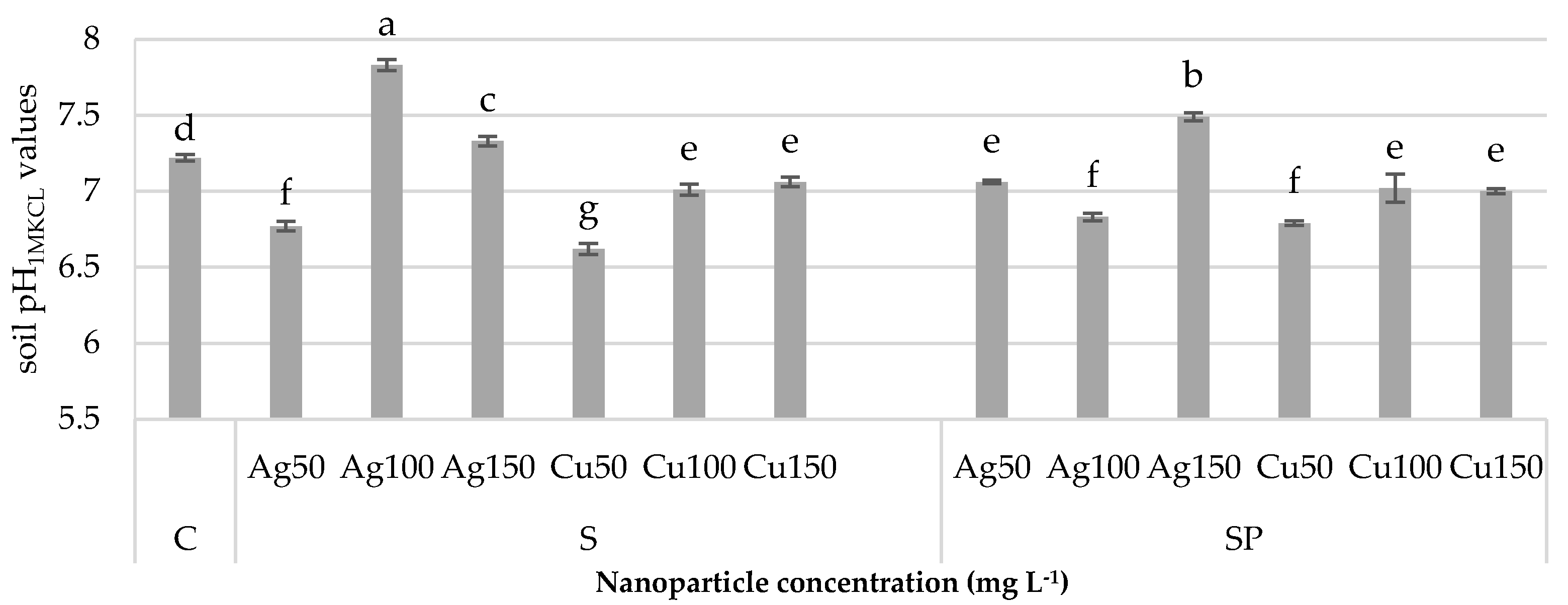
3.1. Influence of Silver and Copper Nanoparticles on Changes in Soil pH Values

The pH value of the soil, in which the soaked seeds were sown and the plants (seedlings) were sprayed with the silver and copper nanoparticles (AgNPs and CuNPs) in the nanocolloidal suspension (AgNP and CuNP) in the control object, was 7.22 (Figure 3). In the soil in which the seeds soaked in AgNPs were seeded, a significant reduction was observed compared to the pH value in the control soil. This value was significantly lower by 6.23%. At higher concentrations of the silver nanoparticles, the pH value increased significantly by 8.44% for a concentration of 100 mg L−1 and 1.5% for a concentration of 150 mg L−1.
It was found that, in the case of application of CuNPs at all the concentrations, the soil pH value decreased significantly compared to the soil pH in the control object. These values were lower by 8.31%, 2.91%, and 2.22%, respectively. The double application of NPs to the seeds and later to the plants (SP) caused lower soil pH values in most cases. In the case of the silver nanoparticles (SP)AgNPs at 50 and 100 mg L−1, these values were lower by 2.22% and 5.40%, respectively. At the highest concentration, an increase of 3.74% was observed compared to the control object. The copper nanoparticles at all the concentrations caused significant reduction in soil pH by 5.95%, 2.77%, and 3.05%, respectively, in comparison with the control.

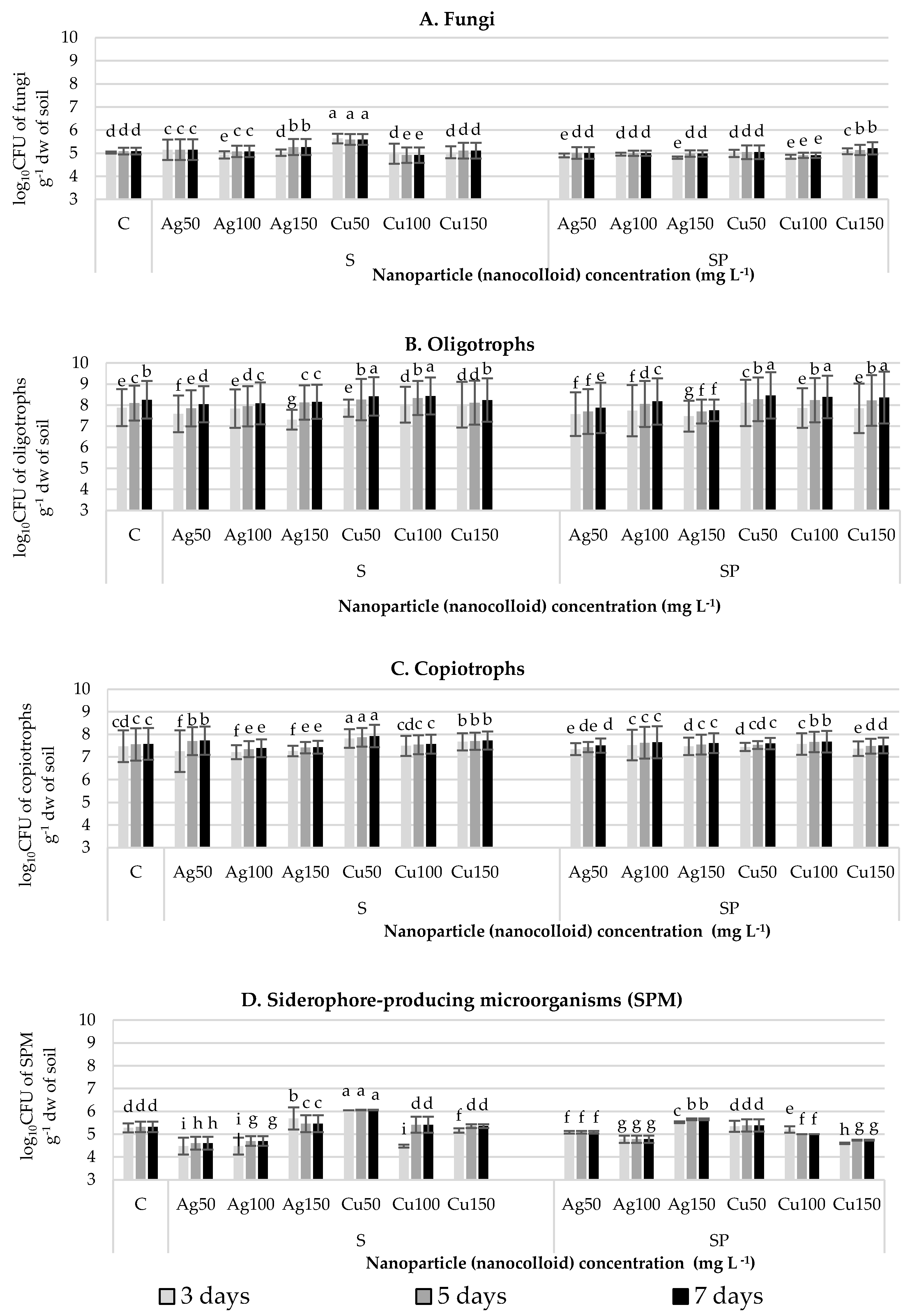
3.2. Influence of Silver and Copper Nanoparticles on Microbial Abundance and Diversity in Soil

The numbers of culturable fungi (CFU) (number of CFU g−1 dw of soil) on the subsequent incubation days (3, 5, 7) in the control object were very similar (about 1.0 × 10⁵ CFU g−1 dw of soil, log10CFU = 5.0 g−1 dw of soil) (Figure 4). After seeding the seeds soaked in the (50 mg L−1) AgNPs suspension, a statistically significant but small increase in the number of fungi (CFU) not exceeding 0.1 logarithmic unit was observed (0.1 log10CFU g−1 dw of soil), i.e., the CFU number increased by about 85% (day 3) and 62.66% (days 5 and 7) compared to the numbers in the control object (Figure 4A).
The log10CFU value for fungi increased by 0.5 units only in the case of the application of copper NPs in a dose of 50 mg L−1, i.e., there was a three- to four-fold increase in the number of fungal CFUs.
The concentration of CuNPs introduced to the seeds by soaking (S) was negatively (R = 0.67) correlated with the number of fungi in the soil on days 3 and 7 (R = 0.60) (Figure 4A).
Irrespective of the mode of the application of the nanoparticles (S—treatment of the seeds only, SP—treatment of the seeds and seedlings), the abundance of oligotrophs (Figure 4B) and copiotrophs (Figure 4C) in the soil on the consecutive days of incubation (days 3, 5, 7) only slightly differed from that in the control variant (no NP treatment).
The analysis of the number of culturable oligotrophs (Figure 4B) in the soil showed their presence in the control. On the subsequent days of incubation, the number of CFUs of oligotrophs in the control object was similar and did not exceed log10CFU 8.5 g−1 dw of soil. The seed soaking in the silver nanoparticles (S)AgNPs and in the copper nanoparticles (S)CuNPs and the subsequent plant spraying with these nanoparticles did not contribute to significant differences in the number of CFUs of the analyzed microorganisms.
The concentration of silver nanoparticles in the case of the soaked seeds on analysis days 5 and 7 was strongly positively (R = 0.67 and R = 0.98) correlated (p < 0.05) with the number of culturable oligotrophs in the analyzed soil. In the case of the copper nanoparticles, the concentrations applied for soaking the seeds showed a significant positive (R = 0.76) correlation (p < 0.05) with their content in soil on day 3 of the experiments and a negative (R = 0.62) correlation (p < 0.05) on analysis day 7. The double application of the copper nanoparticles to both seeds and plants was positively (R = 0.77) correlated (p < 0.05) on analysis day 3, negatively correlated (R = 0.95) on analysis day 5, and negatively correlated (R= 0.96) on analysis day 7 with the numbers of oligotrophs in the soil.
The second group of rhizospheric bacteria comprised culturable copiotrophs (Figure 4C). On subsequent days (3, 5, and 7) of incubation, the number of CFUs of copiotrophs in the control object was close to the value of approximately 1.0·10−5 CFU g−1 dw of soil. The application of the silver nanoparticles (S)AgNP and the copper nanoparticles (S)CuNP in the form of seed soaking and subsequent spraying of seedlings did not increase or decrease the soil copiotrophic count significantly.
The number of colonies of microorganisms capable of producing specific iron chelators–siderophores (Figure 4D) on Schwyn and Neilands [65] medium (containing a strong CAS-Fe-HDTMA iron complex) in the following days of the control soil incubation was about 2.0 × 10⁵ CFU g−1 dw of soil, i.e., about 5.3 log10 CFU g−1 dw of soil. After sowing seeds soaked in the silver nanoparticles at concentrations of 50 and 100 mg L−1, a decrease in the number of colonies (CFU) of culturable siderophore-synthesizing microorganisms was observed on all three days of analysis. Their number was significantly lower than in the control object, i.e., 84.21% (3 days) and 80.85% (5 and 7 days), respectively, and by 84.21% and 76.19%, respectively, at the higher concentration (100 mg L−1).
In the case of sowing the seeds soaked in the copper nanoparticles (S)CuNPs at the concentration of 50 mg L−1 to the soil, a significant increase in the number of CFUs of microorganisms able to synthesize siderophores not exceeding 0.8 logarithmic unit was observed (0.8 log10CFU g−1 dw of soil). This ultimately means an increase in the number of CFUs on the subsequent days by 68.4% (3 days) and 71.4% (days 5 and 7, respectively). The copper concentration of 100 mg L−1 on analysis day 3 contributed to a significant reduction in the number of CFUs of siderophore-producing microorganisms by 0.8 units, which constituted a reduction of 58% in relation to the control object.
The double application of the silver nanoparticles (SP)AgNPs to the seeds and rape seedlings contributed to a reduction in the number of colony-forming units (CFU) of siderophore-producing microorganisms in the soil at the concentrations of 50 and 100 mg L−1 of the nanoparticles. The significant reduction in the number of units did not exceed 0.25 for 50 mg L−1 and 5.5 log10CFU unit for 100 mg L−1. This indicates a reduction in the number of CFUs by 37% (on day 3) and 43% (on days 5 and 7) at a concentration of 50 mg L−1 and by 68% and 71% at a concentration of 100 mg L−1.
An opposite trend was observed at the highest concentration of 150 mg L−1. Here, the log10CFU value of the siderophore-producing microorganisms increased by 0.3 units, i.e., the number of CFUs increased on the subsequent days by 76.32% and 114.29% in comparison to their number in the control object.
The double application of the copper nanoparticles at the higher concentrations (100 and 150 mg L−1) contributed to the reduction of the number of CFUs of the siderophore-producing microorganisms not exceeding 0.3 and 0.5 logarithm units, respectively. They decreased on the subsequent days by 15.79% and 52.38% (in 100 mg L−1 NP concentrations) and 52.63% and 50% (150 mg L−1).
The concentration of the copper nanoparticles applied to the seeds as a dressing and at the later date of the plant growth on analysis days 3, 5, and 7 was strongly negatively (R = −0.69, R = −0.67, and R = −0.68) correlated (p = 0.05) with the content of siderophores in the soil.
The dependence between the number of copiotrophs, oligotrophs, fungi, SPM microorganisms producing siderophores, and the different concentrations of silver (Ag) and cooper (Cu) applied to seeds as well as seeds and plants in the form of nanoparticles (AgNPs, CuNPs) was determined via Principal Component Analysis (PCA) (Figure 5). The first two axes of the PCA based on the First Order Reaction (FOR) model explained 66.5% of data variability, with principal component 1 (PC1) accounting for 44.49%, and principal component 2 (PC2) accounting for 21.99% of the variance (Figure 5). The PCA analysis performed for the number of the tested microbial groups in the two different treatments (seeds—S and seeds and plants—SP) with two suspensions of metal colloidal particles (Ag and Cu) at the three different concentrations (50, 100, 150 mg L−1) indicated a clear trend. The abundance of all the tested groups was slightly positively correlated (green circle) with Cu regardless of the method of introducing its colloids and clearly negatively correlated (blue circle) with Ag in each application variant (Figure 5). As revealed by the PCA, no significant differences were found between the numbers of the studied groups of microorganisms and the application of the metal colloids in S or SP.

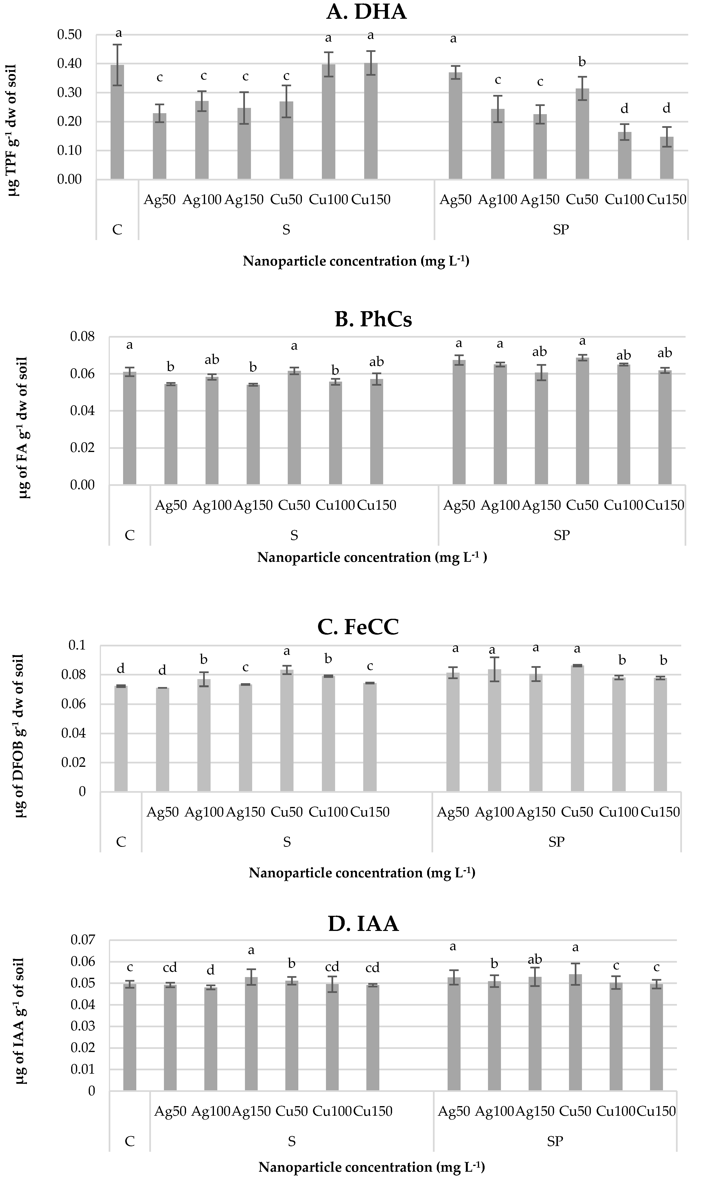
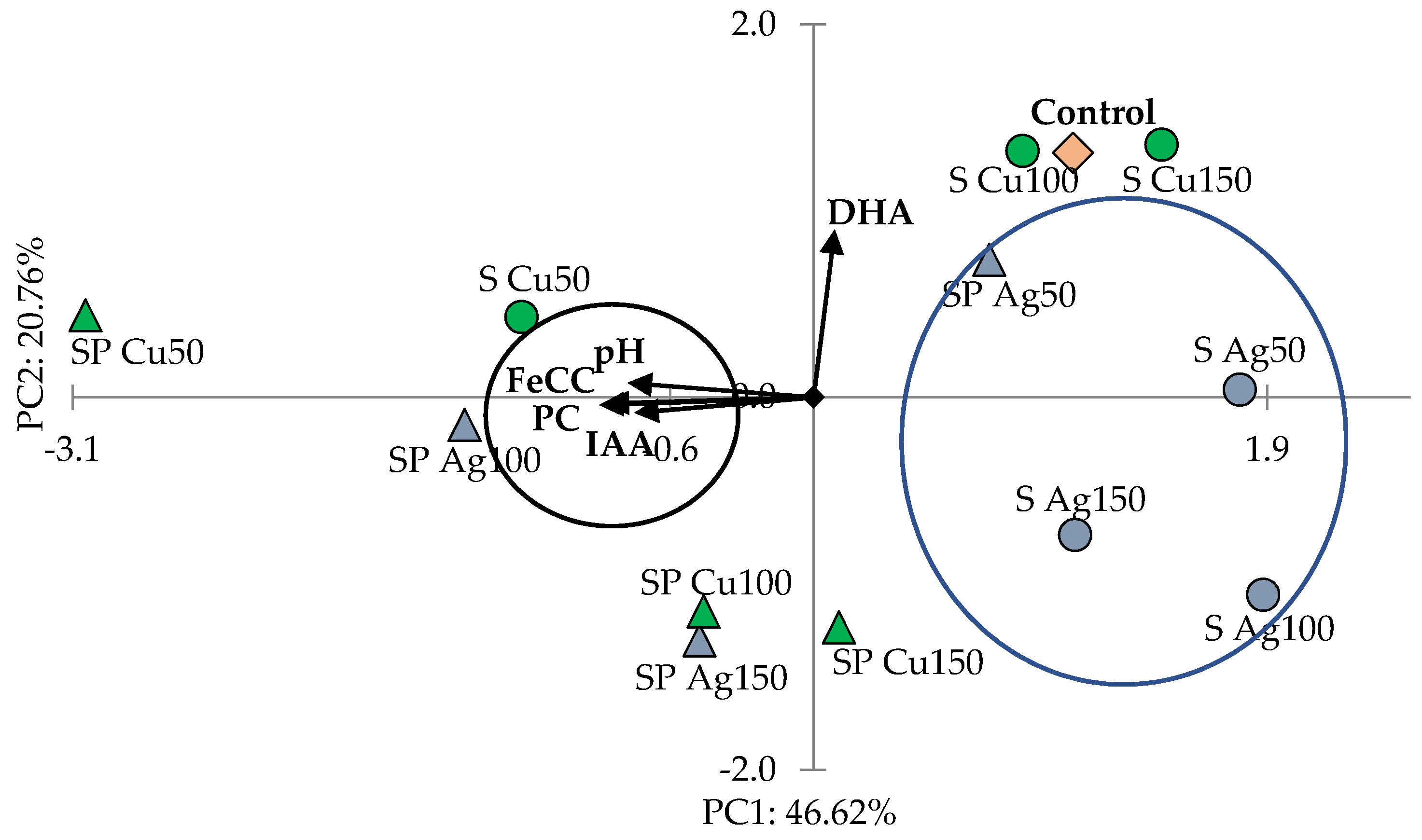
3.3. Influence of Silver and Copper NPs on Soil Biological Activity and Soil Biochemical Characteristics

Soil DHA activity was analyzed after the application of AgNPs and CuNPs (Figure 6A). DHA activity in the control soil (without added NPs) was 0.39 μg TPF g−1 dw of soil (Figure 6A). After using (S)CuNPs, a significant decrease in DHA activity was recorded only at the 50 mg L−1 concentration, which was a lower value (by 31.34%) than that obtained in the control object. The double application of the (SP)NPs of silver and copper contributed to a significant decrease in DHA activity in relation to the control object at all the doses in proportion to the increasing concentration of the nanopreparation: by 6.44%, 38.41%, and 43.09% for (SP)AgNPs and by 20.51%, 58.56%, and 62.76% for (SP)CuNPs.
The concentration of the applied silver and copper nanoparticles (SP) to the seeds and plants was strongly negatively (R = 0.744) correlated (p < 0.05) with the activity of DHA in the soil.
The concentration of phenolic compounds (PhCs) in the soil, in which seeds soaked in nanoparticles were sown and the seedlings were sprayed with AgNPs and CuNPs, was 0.062 μg g−1 dw in the control object (Figure 6B). The application of the nanoparticles via seed soaking contributed to reduction in their contents in the majority of the concentrations used. Significant reduction was observed after the application of (S)AgNPs at 50 mg L−1 and 150 mg L−1. The values obtained were lower than those in the control object, i.e., by 10.86%, 4.53%, and 11.41%, respectively. In the case of (S)CuNPs applied at the concentrations 100 mg L−1, significant reduction (8.84%) in PhCs was observed. The application of NPs for soaking seeds and spraying plant seedlings caused an increase in the concentration of PhCs practically at all the concentrations. The use of AgNPs at the lower concentrations was accompanied by a significant increase in their content successively by 10.31% and 6.44%. The application of (SP)CuNPs to the seeds and plants produced a significant increase in the content of PhCs at the concentrations of 50 and 100 mg L−1. These values were by 6.38% higher in both cases than those in the control object.
The concentration of the silver and copper NPs applied to the seeds and plants was strongly negatively correlated with the concentration of PhCs in the soil in the case of (SP)AgNP (R = 0.971) and (SP)CuNP (R = 0.997) (p < 0.05).
The content of Fe-chelating compounds (FeCCs) in the soil of the control object was 0.072 μg g−1 (Figure 6C). After the application of (S)AgNPs for soaking the seeds, a significant increase (by 8.08%) in their content was observed at the concentration of 100 mg L−1. After the application of (S)CuNPs, a significant increase was observed at the lowest concentration (50 mg L−1). This value was by 13.50% higher than in the control object. The double application of AgNPs and CuNPs (SP) to the plants contributed to the increase in the amount of the compounds produced by microorganisms at all the analyzed nanoparticle concentrations. A significant increase was observed in relation to the control object after the application of AgNPs. The obtained values were 12.52%, 15.77%, and 11.34%, respectively. After using CuNPs in the seeds and plant seedlings, the content of FeCCs in the soil increased by 19.30%, 8.02%, and 9.42%, respectively.
The concentration of the applied copper nanoparticles (SP)CuNPs was strongly positively (R = 0.772) correlated (at the significance level of p < 0.05) with the content of FeCCs in the soil after sowing the soaked seeds and seedlings sprayed with the copper nanopreparation.
In the control object, the concentration of IAA was 0.0495 μg g−1 (Figure 6D). For the use of AgNPs to soak the seeds at the concentration of 150 mg L−1, a slight increase (by 6.65%) in the content of the phytohormone was observed compared to the level in the control object. The application of (S) CuNPs to the seeds at the concentration of 50 mg L−1 induced a small increase of 3.23%. After applying the concentration of 150 mg L−1, a slight reduction was observed, which was by 0.94% lower than that in the control object.
The concentration of the applied copper nanoparticles (SP)CuNPs was strongly negatively (R = −0.866) correlated (p < 0.05) with the IAA concentration in the soil in the case of sowing the seeds soaked in the copper nanoparticles and after spraying the seedlings on day 21 of their growth.
The relationship between the evaluated parameters of soil, i.e., the pH values; IAA (auxin), PhC (phenolic compounds), and FeCC (Fe-chelating compounds) concentration, dehydrogenase (DHA) activity; and three different concentrations (50, 100, 150 mg L−1) of silver (Ag) and cooper (Cu) applied to the seeds or seeds and plants in the form of nanoparticles (AgNPs, CuNPs), was determined via Principal Component Analysis (PCA). The first two axes of the PCA based on the First Order Reaction (FOR) model explained 67.38% of data variability, with principal component 1 (PC1) accounting for 46.62% and principal component 2 (PC2) accounting for 20.76% of the variance (Figure 7). The PCA analysis showed that, except DHA, the main examined parameters were positively correlated with each other (black circle) and formed a clear cluster. The samples on the left side of the diagram were characterized by the highest pH, IAA, PhC, and FeCC values, which decreased in the case of the samples located on the right side of the diagram. It was observed that pH, IAA, PhC, and FeCC were negatively correlated with Ag applied to the seeds (S) at all the tested concentrations and to the seeds and plants applied (SP) at the concentration of 50 mg L−1 (blue circle—a clear cluster was noted). In turn, these parameters were strongly positively correlated with Cu applied to S and SP at the concentration of 50 mg L−1 as well as Ag applied to SP at 100 mg L−1. An opposite trend was shown by the DHA vector, which was positively correlated with the control variant and with the S variant treated with the colloidal Cu suspension at the concentrations of 150 and 100 mg L−1 (Figure 7). In general, as shown by the PCA diagram, the influence of the colloidal suspensions of Ag and Cu on the evaluated soil parameters is not clear.

4. Discussion

The results presented in this paper indicate that the application of CuNPs and AgNPs in three concentrations (50, 100, 150 mg L−1) by soaking the seeds and spraying rape plants has an impact on the biological activity and biochemical properties of the Brassica napus rhizospheric soil, but the changes that occur in the biology and biochemistry of the rhizospheric soil are not considerable, although mostly statistically significant, and the application of CuNPs is more beneficial for this activity than the application of AgNPs. NPs constantly enter the environment not only as nanofertilizers but also from numerous other sources due to their wide application in many fields. Hence, comprehensive investigations proving their beneficial potential or toxic effect are necessary to show the purposefulness of their use as nanofertilizers to improve the condition of plants and the lack of harmful effects on human health and the environment, which is dealt with by econanotoxicology [90].
The presence of metallic NPs may affect soil biodiversity, the abundance of the soil microbial community, and their metabolic activity in soil nutrient cycling, especially in C and N dynamics [91]. Few studies have shown a dependence between the type of NPs and soil characteristics [92,93]. Sánchez-López et al. [93] showed that TiO2 NPs had a limited effect on the bacterial community structure, as only members of Firmicutes and Acetobacter were enriched, while the relative abundance of Verrucomicrobia and Pedobacter decreased. The application of organic material reduced the effect of TiO2 NPs applied to soil.
The number, efficiency of agricultural production, and diversity of microorganisms in the soil are influenced by, e.g., factors related to soil moisture, pH, or the availability of iron [94,95]. In this paper, it was shown that the introduction of NPs on seeds or foliar can cause a decrease in soil pH, which was observed in most treatments except for the application of AgNPs to seeds (S) at concentrations of 100 and 150 mg L−1 and CuNPs on seeds and foliar (SP) at a concentration of 150 mg L−1.
When incorporated into the soil, seeds, or leaves, metal nanoparticles (NP) in nanocolloidal suspensions can have a major influence on these properties and a direct and indirect effect on plant growth. Some research results have reported positive effects of metallic NPs as fertilizers [90,96,97]. Medina-Pérez et al. [90] showed that such NPs as hematite, ferrihydrite, or magnetite can increase significantly such growth parameters of the common bean as the total N of roots or shoots, the number of pods, the dry weight of pods, and the number of seeds and yield. However, it has not been shown that the use of these NPs and two other (zinc oxide or titanium dioxide) compounds resulted in a significant modification of chlorophyll content in common bean plants. The analysis of the quality parameters of oils from seed harvested in a field experiment carried out in our previous research revealed that, regardless of the year of harvest or method of nanocolloid CuNPs application (seeds soaking and leaves spraying) at a concentration of 0.005% (w/v) = 50 mg L−1), the content of chlorophyll a and b as well as carotenoids was statistically significantly higher than in the control samples [98]. This is consistent with the results obtained in this work, i.e., the application of CuNPs seems to be more beneficial for the activity and their biochemical properties of rhizospheric soil than the application of AgNPs.
The effects of NP seed treatment depend on the type of plant, type and concentration of NPs used, conditions (e.g., temperature), and the duration of the experiment [99,100,101,102].
The results of the application of silver and copper NPs to seeds presented in many publications are contradictory. AuNPs and CuNPs had a positive effect on the germination of lettuce seeds [103], and AgNPs increased the germination capacity of oats and radishes [104]. CuO NPs cause an increase in Triticum aestivum biomass when used at a concentration 500 mg kg−1 [105], and AgNPs used in concentrations 10–30 µg mL−1 (mg L−1) cause an increase in seedling weight and seed germination of Boswellia ovalifoliolata [106]. The application of AgNC with a size of 15 nm and at a concentration of 2 ppm (mg L−1) had a positive effect on the growth of soybean grown in flood areas [107].
The effect of AgNPs and CuNPs depended on the method of introduction of the suspension: (1) only by soaking the seeds, (2) application to both seeds and plants. It should be noted that both positive and negative effects of NPs on plant growth and yielding have been reported [1]. AgNPs have been shown to not only strengthen but also weaken plant growth. For example, AgNPs enhanced the growth of Trigonella foenum-graecum and diosgenin synthesis by this plant but reduced shoot and root length in Triticum aestivum; reduced shoot length in Linum usitatissimum and Lolium perenne; reduced germination of Hordeum vulgare; decreased transpiration, biomass, and root growth in Cucurbita pepo; and inhibited seedling growth in Phaseolus radiatus. Copper nanoparticles have already been widely applied as fertilization with slow release of micronutrients and for enhancement of the bioavailability of Cu for soil microorganisms [14,108].
Silver NPs have a large surface area and fraction of surface atoms; as a result, they have high antimicrobial effects compared to bulk silver [109]. CuNP fungicides proved to be effective in protection of various crops against diseases caused by Fusarium spp. (F. solani and F. oxysporum) [14,110]. CuNPs and AgNPs have shown effectiveness in protecting trees Rhizoctonia solani and Phytophthora cactorum [14,111]. AgNPs are nanobactericides effectively combating bacterial infections caused by Pseudomonas syringae [14,112] and Xanthomonas axonopodis [14,113].
The use of nanoparticles in agriculture may increase the indirect possibility of contamination of agricultural areas by these particles. Possible disruptions of plant growth and bioremediation processes, in which different soil microorganisms play a key role, can have serious consequences for agriculture and other environmental processes. Nanoparticles contribute to changes in the social structure of microorganisms (population and microbial diversity) or microbial physiology in the soil. However, the NP impact on terrestrial organisms (soils) is still not clear [114,115]. There are studies on the interaction of NPs with microorganisms, especially with regard to bacteria [116]. They show that bacterial colonies in the soil are decimated or are subjected to modification after the application of Ag [117], CuO, ZnO [118], CeO2, or SnO2 nanoparticles [119]. In these studies, significant differences in the number of microbial groups were found. After the treatment of the seeds (S) and seeds and plants (SP) with Ag and Cu nanoparticles (NP), significant variation was shown depending on the type of NPs, their concentration, and type of application. Copiotrophs and oligotrophs were less sensitive to the application of metal NPs than siderophore-producing bacteria and fungi. As reported by Ge et al. [120], the application of TiO2 and ZnO nanoparticles reduces microbial biomass as well as their diversity. Research conducted by Baek et al. [121] confirms that the application of CuO, ZnO, NiO2, and Sb2O3 nanoparticles, in this order, inhibited the growth of B. subtilis. In turn, Dhas et al. [122] found that both AgNP and ZnONP inhibited the growth of B. subtilis, B. barbaricus, and P. aeruginosa. Over time, however, the strains were able to adapt to the toxic effects of the applied nanoparticles, presumably through NP agglomeration into less toxic microparticles. Shah and Belozerova [103] found no significant effect of Si, Pd, Au, and Cu nanoparticles on the number of colony–forming units (CFU) of culturable microorganisms in soil. Next to biochemical properties, the number of culturable microorganisms (determined as CFUs) is an excellent commonly accepted indicator of changes in the soil microbiome induced by introduction of the tested factor into the soil [45].
Plant–growth–promoting microorganisms (PGPRs) influence many processes in the soil ecosystem, particularly the rhizosphere, participate in the circulation of elements and provide nutrients to plants or produce substances that directly stimulate plant growth (phytohormones), and exert an indirect effect through induction of plant resistance to phytopathogens and abiotic factors [123]. These microorganisms competitively colonize the root system of plants and stimulate plant growth through various mechanisms, including dissolution of inaccessible salts (e.g., phosphate) [124], nitrogen binding [125], and production of indole-3-acetic acid (IAA) or siderophores [126].
Due to environmental restrictions, many anaerobic microorganisms synthesize and release specific molecules known as siderophores that can effectively intercept iron from the environment and make it available to other microorganisms [127]. Thus, the presence of siderophores in the soil affects the availability and mobility of Fe. The affinity system and conversion of Fe from hard-to-reach complex compounds into an easily absorbable form [127,128] are activated when sufficient abundance of iron is ensured in the environment, i.e., when its concentration is at least 1.0 × 10⁻⁵ mol dm−3. This mechanism is based on free diffusion of iron ions through cell membranes [128,129].
A statistically significant increase in dehydrogenase activity (DHA) was observed in these studies after the application of AgNPs as well as after seed treatment with CuNP at a concentration of 50 mg L−1 and after additional rape plant spraying with CuNP at a dose of 100 and 150 mg L−1.
DHA is often used as an indicator of both fertility and soil contamination with heavy metals or pesticides [130] and can also indicate the amount and activity of soil microorganisms [131,132]. To date, however, relatively few studies have focused on the effects of NPs on dehydrogenase activities, and the available results are contradictory. Most studies carried out to date analyze enzymatic activity upon ZnONP, CuONP, and NiNP applications [17,133,134]. Research conducted by Kim et al. [134] comparing the sensitivity of different enzymes to NPs showed that dehydrogenases were more sensitive to CuONPs than to ZnONPs. Analyses conducted by Jośko et al. [17], in which ZnONPs, Cr2O3NPs, CuONPs, and NiNPs (at doses of 10, 100, and 1000 μg g−1 of soil) were used, showed positive effects on the activity of dehydrogenases in the soil. However, Murata et al. [135] showed that CuNPs can inhibit DHA by fifty times and AgNPs by a hundredfold.
In the presented studies, it was shown that the treatment of rapeseed with NPs in a different way affected the concentration of phenolic compounds (PhC) in the soil. A decrease in PhC concentration was observed after the application of AgNPs to seeds (S) at concentrations of 50 and 150 mg L−1. In contrast, the application of CuNPs to seeds and in the form of spraying plants resulted in a significant increase in the concentration of PhCs in the soil. Phenolic compounds (PhCs) are the most important plant allelochemicals in soil ecosystems. The compounds (PhCs) are commonly found not only in plant tissues but also in soil, which they penetrate as root exudates. They exhibit strong biological activity against microorganisms colonizing plant tissues and soil and reduce their growth, development, and abundance [136]. These compounds may also affect the supply of soil nutrients directly and indirectly to plants and microorganisms [137,138]. Some of them may affect the microbial balance by modifying the rhizosphere structure [139] or protect plants against biotic and abiotic stress reactions [130].
The value of iron ions depends on soil acidity (pH), and their high concentration in soil is not synonymous with iron availability for plants, bacteria, and fungi [140]. It is assumed that iron is an element for which microorganisms, as well as those capable of producing strong iron chelating compounds, strongly compete [140].
The presented studies showed that the application of CuNPs and AgNPs, regardless of the method of introduction, caused in most cases an increase in the concentration of IAA in the soil, and the greatest increase was recorded in the soil after the cultivation of oilseed rape treated with the lowest dose (50 mg L−1) of both AgNPs and CuNPs. Dimpka et al. [141] showed that CuONPs can enhance and ZnONPs inhibit the production of IAA by PGPR strain Pseudomonas chlororaphis O6, which can affect the level of IAA concentration in soil. Indole-3-acetic acid (IAA) is a plant hormone from the auxin group affecting the development of plants, bacteria, and fungi [142,143]. This acid is the most prominent molecular signal used to build the root system. Auxin can stimulate the growth of not only plants but also roots by activating dormant cells and initiating their division. It can also regulate the response of the plant immune system [144]. Auxins are preferred for root development and proper synthesis [145].
The use of NPs introduced on plants in colloidal suspensions as nanofertilizer or na-nopesticide against phytopathogens is fully justified, effective, does not affect biodiversity and at the same time is cheap, easy to apply using traditionally used agricultural machinery and equipment technologies, and introduced during sowing or with other fertilizers or protection products.

5. Conclusions

The analysis of the number and activity of microorganisms in the soil after treatment of seeds (S) and seeds and plants (SP) with the colloidal suspensions of Ag and Cu nanoparticles (NP) at three different concentrations (50, 100, 150 mg L−1) showed significant variation depending on the NPs used, their concentration, and type of application. Copiotrophs and oligotrophs were less sensitive to the application of metal NPs than siderophore-producing bacteria and fungi.
The PCA analysis of the number of the tested microbial groups indicated the following trend: the abundance of all the tested groups was slightly positively correlated with Cu regardless of the method of introducing its colloids and clearly negatively correlated with Ag in each application variant.
The influence of the Ag and Cu NPs on the evaluated soil parameters (pH values and concentration of IAA – auxin, PhC – phenolic compounds, FeCC – Fe-chelating compounds) is not clear. These parameters were clearly negatively correlated with Ag applied to the seeds (S) at all the tested concentrations and to the seeds and plants (SP) at the concentration of 50 mg L−1. In turn, these parameters were strongly positively correlated with Cu applied to the S and SP at the concentration of 50 mg L−1 and with Ag applied to SP at 100 mg L−1. An opposite trend was shown by the DHA vector, which was positively correlated with the control variant and with the S variant treated with the colloidal Cu suspension at the concentrations of 150 and 100 mg L−1.
It seems that both methods of NP introduction and the three tested doses can be used without causing negative effects on microbial abundance in rape rhizospheric soil and the soil activity and quality, but the application of CuNPs seems to be more beneficial than the application of AgNPs. The application of the 50 mg L−1 dose and the introduction of NPs by coating seeds via soaking is more economically viable and creates a lower risk of accumulating NPs in excessive concentrations in the soil and, consequently, in plants.

Author Contributions

Conceptualization, M.K., J.J.-Ś. and A.N.; methodology, J.J.-Ś., A.N. and M.K.; formal analysis, S.P. and F.R.; writing—original draft preparation, J.J.-Ś.; AN and M.K.; writing—review and editing, J.J.-Ś., AN and M.K. and visualization, J.J.-Ś., R.T. and A.N.; supervision, S.P., F.R. and J.J.-Ś.; statistical analysis, R.T. All authors have read and agreed to the published version of the manuscript.

Funding

The APC was funded from the ’Excellent science’ program of the Ministry of Education and Science as a part of the contract no. DNK/SP/465641/2020 "The role of the agricultural engineering and environmental engineering in the sustainable agriculture development". Research supported by the Ministry of Science and Higher Education of Poland as part of the statutory activities of the Department of Industrial and Environmental Microbiology, Maria Curie–Skłodowska University.

Conflicts of Interest

No potential conflict of interest was reported by the authors.

References

  1. Duhan, J.S.; Kumar, R.; Kumar, N.; Kaur, P.; Nehra, K.; Duhan, S. Nanotechnology: The new perspective in precision agriculture. Biotechnol Rep. 2017, 15, 11–23. [Google Scholar] [CrossRef]
  2. Philbrook, N.A.; Winn, L.M.; Afrooz, A.R.M.N.; Saleh, N.B.; Walker, V.K. The effect of TiO2 and Ag nanoparticles on reproduction and development of Drosophila melanogaster and CD-1 mice. Toxicol. Appl. Pharmacol. 2011, 257, 429–436. [Google Scholar] [CrossRef]
  3. Kim, S.W.; An, Y.J. Effect of ZnO and TiO2 nanoparticles preilluminated with UVA and UVB light on Escherichia coli and Bacillus subtilis. Appl. Microbiol. Biotechnol. 2012, 95, 243–253. [Google Scholar] [CrossRef] [PubMed]
  4. Yang, F.; Jin, C.; Subedi, S.; Lee, C.L.; Wang, Q.; Jiang, Y.; Li, J.; Di, Y.; Fu, D. Emerging inorganic nanomaterials for pancreatic cancer diagnosis and treatment. Cancer Treat. Rev. 2012, 38, 566–579. [Google Scholar] [CrossRef]
  5. Retchkiman-Schabes, P.S.; Canizal, G.; Becerra-Herrera, R.; Zorrilla, C.; Liu, H.B.; Ascencio, J.A. Biosynthesis and characterization of Ti/Ni bimetallic nanoparticles. Opt. Mater. 2006, 29, 95–99. [Google Scholar] [CrossRef]
  6. Gu, H.; Ho, P.L.; Tong, E.; Wang, L.; Xu, B. Presenting vancomycin on nanoparticles to enhance antimicrobial activities. Nano Lett. 2003, 3, 1261–1263. [Google Scholar] [CrossRef]
  7. Gong, P.; Li, H.; He, X.; Wang, K.; Hu, J.; Tan, W.; Zhang, S.; Yang, X. Preparation and antibacterial activity of Fe3O4@Ag nanoparticles. Nanotechnol 2007, 18, 604–611. [Google Scholar] [CrossRef]
  8. Rahim, E.H.; Kamounah, F.S.; Frederiksen, J.; Christensen, J.B. Heck reactions catalyzed by PAMAM-dendrimer encapsulated Pd(0) nanoparticles. Nano Lett. 2001, 1, 499–501. [Google Scholar] [CrossRef]
  9. Kim, Y.G.; Oh, S.K.; Crools, R.M. Preparation and characterization of 1–2 nmdendrimer-encapsulated gold nanoparticles having very narrow size distributions. Chem. Mater. 2004, 16, 167. [Google Scholar] [CrossRef]
  10. Mirzajani, F.; Askari, H.; Hamzelou, S.; Schober, Y.; Römpp, A.; Ghassempour, A.; Spengler, B. Proteomics study of silver nanoparticles toxicity on Oryza sativa L. Ecotoxic. Environ. Safety 2014, 108, 335–339. [Google Scholar] [CrossRef] [PubMed]
  11. Zhou, J.; Hu, Z.; Zabihi, F.; Chen, Z.; Zhu, M. Progress and perspective of antiviral protectoive material. Adv. Fiber Mater. 2020, 2, 123–139. [Google Scholar] [CrossRef]
  12. Navarro, E.; Baun, A.; Behra, R.; Hartmann, N.B.; Filser, J.; Miao, A.J.; Quigg, A.; Santschi, P.H.; Sigg, L. Environmental behavior and ecotoxicity of engineered nanoparticles to algae, plants, and fungi. Ecotoxicology 2008, 17, 372–386. [Google Scholar] [CrossRef] [PubMed] [Green Version]
  13. Gu, M.B.; Niazi, J.H.; Sang, B.I.; Kim, Y.S. Global gene response in Saccharomyces cerevisiae exposed to silver nanoparticles. Appl. Biochem. Biotechnol. 2011, 164, 1278–1291. [Google Scholar] [CrossRef]
  14. Singh, H.; Sharma, A.; Bhardwaj, S.K.; Arya, S.K.; Bhardwaj, N.; Khatri, M. Recent advances in the applications of nano-agrochemicals for sustainable agricultural development. Environ. Sci. Process. Impacts 2021, 23, 213–239. [Google Scholar] [CrossRef]
  15. Singh, A.; Singh, N.B.; Hussain, I.; Singh, H.; Singh, S.C. Plant-nanoparticle interaction: An approach to improve agricultural practices and plant productivity. Int. J. Pharm. Sci. Invent. 2015, 4, 25–40. [Google Scholar]
  16. Ma, X.; Geisler-Lee, J.; Deng, Y.; Kolmakov, A. Interactions between engineered nanoparticles (ENPs) and plants: Phytotoxicity, uptake and accumulation. Sci Total Environ 2010, 408, 3053–3061. [Google Scholar] [CrossRef]
  17. Jośko, I.; Oleszczuk, P.; Futa, B. The effect of inorganic nanoparticles (ZnO, Cr2O3,CuO and Ni) and their bulk counterparts on enzyme activities in different soils. Geoderma. 2014, 232, 528–537. [Google Scholar] [CrossRef]
  18. Rao, S.; Shekhawat, G.S. Phytotoxicity and oxidative stress perspective of two selected nanoparticles in Brassica juncea. Biotechnology 2016, 3, 244. [Google Scholar] [CrossRef] [Green Version]
  19. Hong, J.; Rico, C.M.; Zhao, L.; Adeleye, A.S.; Keller, A.A.; Peralta-Videa, J.R.; Gardea-Torresdey, J.L. Toxic effects of copper-based nanoparticles or compounds to lettuce (Lactuca sativa) and alfalfa (Medicago sativa). Environ Sci Process Impacts. 2015, 17, 177–185. [Google Scholar] [CrossRef] [Green Version]
  20. Doolette, C.L.; McLaughlin, M.J.; Kirby, J.K.; Navarro, D.A. Bioavailability of silver and silver sulfide nanoparticles to lettuce (Lactuca sativa): Effect of agricultural amendments on plant uptake. J. Hazard. Mater. 2015, 300, 788–795. [Google Scholar] [CrossRef]
  21. Hernandez-Viezcas, J.A.; Castillo-Michel, H.; Andrews, J.C.; Cotte, M.; Rico, C.; Peralta-Videa, J.R.; Ge, Y.; Priester, J.H.; Holden, P.A.; Gardea-Torresdey, J.L. In situ synchrotron X-ray fluorescence mapping and speciation of CeO2 and ZnO nanoparticles in soil cultivated soybean (Glycine max). ACS Nano 2013, 7, 1415–1423. [Google Scholar] [CrossRef] [PubMed]
  22. Seil, J.T.; Webster, T.J. Antimicrobial applications of nanotechnology: Methods and literature. Int. J. Nanomedicine 2012, 7, 2767–2781. [Google Scholar] [CrossRef] [PubMed] [Green Version]
  23. Arciniegas-Grijalba, P.A.; Patiño-Portela, M.C.; Mosquera-Sánchez, L.P.; Guerrero-Vargas, J.A.; Rodríguez-Páez, J.E. ZnO nanoparticles (ZnO-NPs) and their antifungal activity against coffee fungus Erythricium salmonicolor. Appl Nanosci. 2017, 7, 225–241. [Google Scholar] [CrossRef] [Green Version]
  24. He, L.; Liu, Y.; Mustapha, A.; Lin, M. Antifungal activity of zinc oxide nanoparticles against Botrytis cinerea and Penicillium expansum. Microbiol. Res. 2011, 166, 207–215. [Google Scholar] [CrossRef]
  25. Bayat, M.; Zargar, M.; Chudinova, E.; Astarkhanova, T.; Pakina, E. In vitro evaluation of antibacterial and antifungal activity of biogenic silver and copper nanoparticles: The first report of applying biogenic nanoparticles against Pilidium concavum and Pestalotia sp. fungi. Molecules 2021, 26, 5402. [Google Scholar] [CrossRef] [PubMed]
  26. Hao, Y.; Cao, X.; Ma, C.; Zhang, Z.; Zhao, N.; Ali, A.; Hou, T.; Xiang, Z.; Zhuang, J.; Wu, S.; et al. Potential applications and antifungal activities of engineered nanomaterials against gray mold disease agent Botrytis cinerea on rose petals. Front. Plant. Sci. 2017, 8, 1332. [Google Scholar] [CrossRef] [Green Version]
  27. Ben Tahar, I.; Fickers, P.; Dziedzic, A.; Płoch, D.; Skóra, B.; Kus-Liśkiewicz, M. Green pyomelanin-mediated synthesis of gold nanoparticles: Modelling and design, physico-chemical and biological characteristics. Microb. Cell Fact 2019, 18, 210. [Google Scholar] [CrossRef]
  28. Keller, A.A.; Adeleye, A.S.; Conway, J.R.; Garner, K.L.; Zhao, L.; Cherr, G.N.; Hong, J.; Gardea-Torresdey, J.L.; Godwin, H.A.; Hanna, S.; et al. Comparative environmental fate and toxicity of copper nanomaterials. NanoImpact 2017, 7, 28–40. [Google Scholar] [CrossRef] [Green Version]
  29. Feng, X.; Yan, Y.; Wan, B.; Li, W.; Jaisi, D.P.; Zheng, L.; Zhang, J.; Liu, F. Enhanced dissolution and transformation of ZnO nanoparticles: The role of inositol hexakisphosphate. Environ. Sci. Technol. 2016, 50, 5651–5660. [Google Scholar] [CrossRef]
  30. Benoit, R.; Wilkinson, K.J.; Sauvé, S. Partitioning of silver and chemical speciation of free Ag in soils amended with nanoparticles. Chem. Cent. J. 2013, 7, 75. [Google Scholar] [CrossRef] [Green Version]
  31. Cornelis, G.; Doolette, M.T.C.; McLaughlin, M.J.; Kirby, J.K.; Beak, D.G.; Chittleborough, D. Retention and dissolution of engineered silver nanoparticles in natural soils. Soil Sci. Soc. Am. J. 2012, 76, 891–902. [Google Scholar] [CrossRef]
  32. Fernando, S.S.N.; Gunasekara, T.D.C.P.; Holton, J. Antimicrobial nanoparticles: Applications and mechanisms of action. Sri Lankan. J. Infect. Dis. 2018, 8, 2–11. [Google Scholar] [CrossRef]
  33. Slavin, Y.N.; Asnis, J.; Häfeli, U.O.; Bach, H. Metal nanoparticles: Understanding the mechanisms behind antibacterial activity. J. Nanobiotechnol. 2017, 15. [Google Scholar] [CrossRef] [PubMed]
  34. Wang, L.; Hu, C.; Shao, L. The antimicrobial activity of nanoparticles: Present situation and prospects for the future. Int. J. Nanomedicine. 2017, 12, 1227–1249. [Google Scholar] [CrossRef] [PubMed] [Green Version]
  35. Alamilla-Martínez, D.G.; Rojas-Avelizapa, N.G.; Domínguez-López, I.; Pool, H.; Gómez-Ramírez, M. Biosynthesis of iron nanoparticles (FeNPs) by Alternaria alternata MVSS-AH-5. Mex. J. Biotechnol. 2019, 4, 1–14. [Google Scholar] [CrossRef]
  36. Gomes, T.; Pinheiro, J.P.; Cancio, I.; Pereira, C.G.; Cardoso, C.; Bebianno, M.J. Effects of copper nanoparticles expo-sure in the mussel Mytilus galloprovincialis. Environ. Scien. Technol. 2011, 45, 9356–9362. [Google Scholar] [CrossRef] [PubMed]
  37. Llorens, A.; Lloret, E.; Picouet, P.A.; Trbojevich, R.; Fernandez, A. Metallic-based micro and nanocomposites in food contact materials and active food packaging. Trends Food Sci. Technol. 2012, 24, 19–29. [Google Scholar] [CrossRef]
  38. Orabi, A.S.; Abou El-Nour, K.M.; Youssef, M.F.; Salem, H.A. Novel and highly effective composites of silver and zinc oxide nanoparticles with some transition metalcomplexes against different microorganisms. Arabian J. Chem. 2018, 13, 262–2648. [Google Scholar] [CrossRef]
  39. Barrena, R.; Casals, E.; Colón, J.; Font, X.; Sánchez, A.; Puntes, V. Evaluation of the ecotoxicity of model nanoparticles. Chemosphere 2009, 75, 850–857. [Google Scholar] [CrossRef] [Green Version]
  40. Yin, F.; Peng, S.; Sun, P.; Shi, Z. Effects of low salinity on antioxidant enzymes activities in kidney and muscle of juvenile silver pomfret Pampus argenteus. Acta Ecologica Sinica. 2011, 31, 55–60. [Google Scholar] [CrossRef]
  41. Hänsch, M.; Emmerling, C. Effects of silver nanoparticles on the microbiota and enzyme activity in soil. J. Plant Nutr. Soil Sci. 2010, 173, 554–558. [Google Scholar] [CrossRef]
  42. Kong, C.H.; Wang, P.; Gu, Y.; Xu, X.H.; Wang, M.L. Fate and impact on microorganisms of rice allelochemicals in paddy soil. J. Agric. Food Chem. 2008, 56, 5043–5049. [Google Scholar] [CrossRef]
  43. Xu, C.; Peng, C.; Sun, L.; Zhang, S.; Huang, H.; Chen, Y.; Shi, J. Distinctive effects of TiO2 and CuO nanoparticles on soil microbes and their community structures inflooded paddy soi. Soil Biol Biochem. 2015, 86, 24–33. [Google Scholar] [CrossRef]
  44. Kolesnikov, S.; Timoshenko, A.; Minnikova, T.; Tsepina, N.; Kazeev, K.; Akimenko, Y.; Zhadobin, A.; Shuvaeva, V.; Rajput, V.D.; Mandzhieva, S.; et al. Impact of metal-based nanoparticles on cambisol microbial functionality, enzyme activity, and plant growth. Plants 2021, 10, 2080. [Google Scholar] [CrossRef]
  45. Schottroff, F.; Fröhling, A.; Zunabovic-Pichler, M.; Krottenthaler, A.; Schlüter, O.; Jäger, H. Sublethal injury and viable but non-culturable (vbnc) state in microorganisms during preservation of food and biological materials by non-thermal processes. Front. Microbiol. 2018, 9, 2773. [Google Scholar] [CrossRef] [PubMed]
  46. Coelho, E.; Teixeira, J.A.; Domingues, L.; Tavares, T.; Oliveira, J.M. Factors affecting extraction of adsorbed wine volatile compounds and wood extractives from used oak wood. Food Chem. 2019, 295, 156–164. [Google Scholar] [CrossRef] [Green Version]
  47. Epstein, S.S. The phenomenon of microbial uncultivability. Curr. Opin. Microbiol. 2013, 16, 636–642. [Google Scholar] [CrossRef]
  48. Ali, W.A.; Al-Jaff, B.M.A.; Al-Saeedi, S.S.S. Cultivation and detection of viable but non-culturable fungi in soil using yoghurt whey infusion agar. ICPAS 2018, 1–6. [Google Scholar] [CrossRef]
  49. Bamford, R.A.; Smith, A.; Metz, J.; Glover, G.; Titball, R.W.; Pagliara, S. Investigating the physiology of viable but non-culturable bacteria by microfluidics and time-lapse microscopy. BMC Biol. 2017, 15, 1–12. [Google Scholar] [CrossRef] [PubMed]
  50. Fakruddin, M.; Mannan, K.S.B.; Andrews, S. Viable but nonculturable bacteria: Food safety and public health perspective. Int. Sch. Res. Notices. 2013, 2013, 121. [Google Scholar] [CrossRef]
  51. Highmore, C.J.; Rothwell, S.D.; Keevil, C.W. Improved sample preparation for direct quantitative detection of Escherichia coli O157 in soil using qPCR without pre-enrichment. Microb. Biotechnol. 2017, 10, 969–976. [Google Scholar] [CrossRef]
  52. Koch, E.W. Beyond light: Physical, geological, and geochemical parameters as possible submersed aquatic vegetation habitat requirements. Estuaries 2001, 24, 1–17. [Google Scholar] [CrossRef]
  53. Ho, A.; Di Lonardo, D.P.; Bodelier, P.L.E. Revisiting life strategy concepts in environmental microbial ecology. FEMS Microbiol. Ecol. 2017, 93, 1–14. [Google Scholar] [CrossRef] [Green Version]
  54. Judy, J.D.; Unrine, J.M.; Bertsch, P.M. Evidence for biomagnification of gold nanoparticles within a terrestrial food chain. Environ. Sci. Technol. 2011, 45, 776–781. [Google Scholar] [CrossRef] [PubMed]
  55. Dąbrowska, G.; Zdziechowska, E. The role of rhizobacteria in the stimulation of the growth and development processes and protection of plants against environmental factors. Prog. Plant Prot. 2015, 55, 498–506. [Google Scholar] [CrossRef] [Green Version]
  56. Van Vliet, P.C.J.; Bloem, J.; de Goede, R.G.M. Microbial diversity, nitrogen loss and grass production after addition of Effective Microorganisms (EM) to slurry manure. Appl. Soil Ecol. 2006, 32, 188–198. [Google Scholar] [CrossRef]
  57. Bhattacharyya, P.N.; Jha, D.K. Plant growth-promoting rhizobacteria (PGPR): Emergence in agriculture. World J. Microbiol. Biotechnol. 2012, 28, 1327–1350. [Google Scholar] [CrossRef] [PubMed]
  58. Natywa, M.K.; Ambroży, K.; Sawicka, A. Changes in the number of selected groups of soil microorganisms under the cultivation of maize, depending on the development phase of the plant and the use of sprinkler irrigation. Scien. Nature Technician 2010, 4, 1–9. [Google Scholar]
  59. Parelho, C.; Rodrigues, A.S.; Cruz, J.V.; Garcia, P. Linking trace metals and agricultural land use in volcanic soils—a multivariate approach. Sci. Total Environ. 2014, 496, 241–247. [Google Scholar] [CrossRef]
  60. Gao, Y.; Wu, M. Free-living bacterial communities are mostly dominated by oligotrophs. bioRxiv 2018. [Google Scholar]
  61. Pawlik, M.; Płociniczak, T.; Piotrowska-Seget, Z. Endophytic bacteria and their importance in environmental microbiology, medicine and industry. Post. Mikrobiol. 2015, 54, 115–122. [Google Scholar]
  62. Vejan, P.; Abdullah, R.; Khadiran, T.; Ismail, S.; Boyce, A.N. Role of plant growth promoting rhizobacteria in agricultural sustainability—a review. Molecules. 2016, 21, 537. [Google Scholar] [CrossRef] [PubMed]
  63. Podile, A.R.; Kishore, G.K. Plant growth-promoting rhizobacteria. In Gnanamanickam SS (ed) Plant-Associated Bacteria; Springer: Berlin/Heidelberg, Germany, 2006; pp. 195–230. [Google Scholar]
  64. Burns, R.G. Enzyme activity in soil: Location and a possible role in microbial ecology. Soil Biol. Biochem. 1982, 14, 423–427. [Google Scholar] [CrossRef]
  65. Praveen-Kumar, J.; Tarafdar, C. 2,3,5-Tripheny ltetrazolium chloride (TTC) as electron acceptor of culturable soil bacteria, fungi and actimomycetes. Biol. Fertil. Soils. 2003, 38, 186–189. [Google Scholar] [CrossRef]
  66. Gajda, A.M.; Przewłoka, B.; Gawryjołek, K. Determination of the effect of used tillage system on changes of soil environm ent measured with parameters of soil microbiological activity. Nauka Przyr. Technol. 2010, 4, 1–11. [Google Scholar]
  67. Kolesnikov, S.; Tsepina, N.; Minnikova, T.; Kazeev, K.; Mandzhieva, S.; Sushkova, S.; Minkina, T.; Mazarji, M.; Singh, R.K.; Rajput, V.D. Influence of silver nanoparticles on the biological indicators of haplic chernozem. Plants 2021, 10, 1022. [Google Scholar] [CrossRef] [PubMed]
  68. Patten, D.M. The Relation Between Environmental Performance and Environmental Disclosure: A Research Note. Account. Organ. Soc. 2002, 27, 763–773. [Google Scholar] [CrossRef]
  69. Simon, S.; Petrášek, J. Why plants need more than one type of auxin. Plant Sci. 2011, 180, 454–460. [Google Scholar] [CrossRef] [Green Version]
  70. Jaroszuk-Ściseł, J.; Kurek, E.; Trytek, M. Efficiency of indoleacetic acid, gibberellic acid and ethylene synthesized in vitro by Fusarium culmorum strains with different effects on cereal growth. Biology 2014, 69, 281–292. [Google Scholar] [CrossRef] [Green Version]
  71. Spaepen, S.; Vanderleyden, J.; Remans, R. Indole-3-acetic acid in microbial and microorganism-plant signaling. FEMS Microbiol. 2007, 31, 425–448. [Google Scholar] [CrossRef] [PubMed] [Green Version]
  72. Enders, T.A.; Strader, L.C. Auxin activity: Past, present, and future. Am. J. Botany. 2015, 102, 180–196. [Google Scholar] [CrossRef] [PubMed] [Green Version]
  73. Majumdar, S.; Peralta-Videa, J.R.; Trujillo-Reyes, J.; Sun, Y.; Barrios, A.C.; Niu, G.; Flores-Margez, J.P.; Gardea-Torresdey, J.L. Soil organic matter influences cerium translocation and physiological processes in kidney bean plants exposed to cerium oxide nanoparticles. Sci. Total Environ. 2016, 569–570, 201–211. [Google Scholar] [CrossRef] [PubMed] [Green Version]
  74. Medina-Velo, I.A.; Dominguez, O.E.; Ochoa, L.; Barrios, A.C.; Hernández-Viezcas, J.A.; White, J.C.; Peralta-Videa, J.R.; Gardea-Torresdey, J.L. Nutritional quality of bean seeds harvested from plants grown in different soils amended with coated and uncoated zinc oxide nanomaterials. Environ. Sci. Nano. 2017, 4, 2336–2347. [Google Scholar] [CrossRef]
  75. Savi, T.; Casolo, V.; Luglio, J.; Bertuzzi, S.; Trifilo, P.; Lo Gullo, M.A.; Nardini, A. Species-specific reversal of stem xylem embolism after a prolonged drought correlates to endpoint concentration of soluble sugars. Plant Physiol. Biochem. 2016, 106, 198–207. [Google Scholar] [CrossRef] [PubMed]
  76. Peyrot, C.; Evin, K.; Wilkinson, J.; Élanie, M.; Esrosiers, D.; Sauvé, S. Effects of silver nanoparticles on soil enzyme activities with and without added organic matter. Environmental Toxico. Chem. 2014, 33, 115–125. [Google Scholar] [CrossRef]
  77. Ebenstein, Y.; Nahum, E.; Banin, U. Tapping mode atomic force microscopy for nanoparticle sizing: Tip−sample interaction effects. Nano Letters. 2002, 2, 945–950. [Google Scholar] [CrossRef]
  78. Mohamed, E.A. Green synthesis of copper & copper oxide nanoparticles using the extract of seedless dates. Heliyon 2020, 6, e03123. [Google Scholar] [CrossRef] [Green Version]
  79. Wan Mat Khalir, W.K.A.; Shameli, K.; Jazayeri, S.D.; Othman, N.A.; Che Jusoh, N.W.; Hassan, N.M. Biosynthesized silver nanoparticles by aqueous stem extract of entada spiralis and screening of their biomedical activity. Front Chem. 2020, 8, 620. [Google Scholar] [CrossRef] [PubMed]
  80. Moniri, S.; Ghoranneviss, M.; Hantehzadeh, M.R.; Asadabad, M.A. Synthesis and optical characterization of copper nanoparticles prepared by laser ablation. Bull. Mater. Sci. 2017, 40, 37–43. [Google Scholar] [CrossRef]
  81. Gibson, F.; Magrath, D.I. The isolation and characterization of hydroxamic acid (aerobactin) formed by Aerobacter aerogenes 62-I. Biochimica Biophysica Acta 1969, 192, 175–184. [Google Scholar] [CrossRef]
  82. Atkin, C.L.; Neilands, J.B.; Phaff, H. Rhodotorulic acid from species of Rhodospirillum, Rhodotorula, Sporidiobolus and Sporobolomyces. J. Bacteriology. 1970, 103, 722–733. [Google Scholar] [CrossRef] [Green Version]
  83. Pilet, P.E.; Chollet, R. Colorimetric determination of indole-3-acetic acid, C. R. Acad. Sci. Serv. 1970, 271, 1675–1678. [Google Scholar]
  84. Glickmann, E.; Dessaux, Y. A critical examination of the specificity of the Salkowski reagent for indolic compounds produced by phytopathogenic bacteria. Applied Environ. Microbio. 1995, 61, 793–796. [Google Scholar] [CrossRef] [Green Version]
  85. Salkowski, E. Ueber des Verhalten der Skatolcarbonsaure im Organismus. Physiol. Chem. 1885, 9, 23–33. [Google Scholar] [CrossRef]
  86. Alef, K. Dehydrogenase activity. In Methods in Applied Soil Microbiology and Biochemistry; Alef, K., Nannipieri, P., Eds.; Academic Press: London, UK, 1995; pp. 228–231. [Google Scholar]
  87. Martin, J.P. Use of acid rose bengal and streptomycin in the plate methods for estimating soil fungi. Soil Scien. 1950, 38, 215–220. [Google Scholar] [CrossRef]
  88. Schwyn, B.; Neilands, J.B. Universal chemical assay for the detection and determination of siderophores. Anal. Biochem. 1987, 160, 47–56. [Google Scholar] [CrossRef]
  89. Carlton, M.A.; Brandt, S. Data analysis: Statistical and computational methods for scientists and engineers. Am. Stat. 2000, 54, 155. [Google Scholar] [CrossRef]
  90. Medina-Pérez, G.; Fernández-Luqueño, F.; Trejo-Téllez, L.I.; López-Valdez, F.; Pampillón-González, L. Growth and development of common bean (Phaseolus vulgaris l.) var. pinto saltillo exposed to iron, titanium, and zinc oxide nanoparticles in an agricultural soil. App.l Ecol. Environ. Res. 2018, 16, 1883–1897. [Google Scholar] [CrossRef]
  91. Nogueira, V.; Lopes, I.; Rocha-Santos, T.; Santos, A.L.; Rasteiro, G.M.; Antunes, F.; Gonçalves, F.; Soares, A.M.; Cunha, A.; Almeida, A.; et al. Impact of organic and inorganic nanomaterials in the soil microbial community structure. Sci. Total. Environ. 2012, 424, 344–350. [Google Scholar] [CrossRef]
  92. Frenk, S.; Ben-Moshe, T.; Dror, I.; Berkowitz, B.; Minz, D. Effect of metal oxide nanoparticles on microbial community structure and function in two different soil types. PLoS ONE. 2013, 8, e84441. [Google Scholar] [CrossRef] [Green Version]
  93. Sánchez-López, K.B.; De Los Santos-Ramos, F.J.; Gómez-Acata, E.S.; Luna-Guido, M.; Navarro-Noya, Y.E.; Fernández-Luqueño, F.; Dendooven, L. TiO2 nanoparticles affect the bacterial community structure and Eisenia fetida (Savigny, 1826) in an arable soil. PeerJ. 2019, 7, e6939. [Google Scholar] [CrossRef] [PubMed]
  94. Gajendiran, J.; Rajendran, V. Synthesis and characterization of coupled semiconductor metal oxide (ZnO/CuO) nanocomposite. Mater. Lett. 2014, 116, 311–313. [Google Scholar] [CrossRef]
  95. Mimmo, T.; Del Buono, D.; Terzano, R.; Tomasi, N.; Vigani, G.; Crecchio, C.; Pinton, R.; Zocchi, G.; Cesco, S. Rhizospheric organic compounds in the soil-microorganism-plant system: Their role in iron availability. Eur. J. Soil Sci. 2014, 65, 629–642. [Google Scholar] [CrossRef]
  96. Burke, D.J.; Pietrasiak, N.; Situ, S.F.; Abenojar, E.C.; Porche, M.; Kraj, P.; Lakliang, Y.; Samia, A.C.S. Iron oxide and titanium dioxide nanoparticle effects on plant performance and root associated microbes. Int. J. Mol. Sci. 2015, 16, 23630–23650. [Google Scholar] [CrossRef]
  97. Rico, C.M.; Majumdar, S.; Duarte-Gardea, M.; Peralta-Videa, J.R.; Gardea-Torresdey, J.L. Interaction of nanoparticles with edible plants and their possible implications in the food chain. J. Agric. Food Chem. 2011, 59, 3485–3498. [Google Scholar] [CrossRef] [Green Version]
  98. Kachel, M.; Matwijczuk, A.; Sujak, A.; Czernel, G.; Niemczynowicz, A.; Nowicka, A. The influence of copper and silver nanocolloids on the quality of pressed spring rapeseed oil. Agronomy 2019, 9, 643. [Google Scholar] [CrossRef] [Green Version]
  99. Coutris, C.; Joner, E.J.; Oughton, D.H. Aging and soil organic matter content affect the fate of silver nanoparticles in soil. Sci. Total Environ. 2012, 420, 327–333. [Google Scholar] [CrossRef]
  100. Joshi, N.; Ngwenya, B.T.; French, C.E. Enhanced resistance to nanoparticle toxicity is conferred by overproduction of extracellular polymeric substances. J. Hazard. Mater. 2012, 241−242, 363–370. [Google Scholar] [CrossRef] [PubMed]
  101. Singh, D.; Kumar, A. Effects of nano silver oxide and silver ions on growth of vigna radiate. Bull. Environ. Contam. Toxicol. 2015, 95, 379–384. [Google Scholar] [CrossRef]
  102. Judy, J.D.; Kirby, J.K.; Creamer, C.; McLaughlin, M.J.; Fiebiger, C.; Wright, C.; Cavagnaro, T.R.; Bertsch, P.M. Effects of silver sulfide nanomaterials on mycorrhizal colonization of tomato plants and soil microbial communities in biosolid-amended soil. Environ. Pollut. 2015, 206, 256–263. [Google Scholar] [CrossRef]
  103. Shah, V.; Belozerova, I. Influence of metal nanoparticles on the soil microbial community and germination of lettuce seeds. Water Air Soil Pollut. 2009, 197, 143–148. [Google Scholar] [CrossRef]
  104. Tomacheski, D.; Pittol, M.; Simoes, D.N.; Ribeiro, V.F. Effect of natural ageing on surface of silver loaded TPE and its influence in antimicrobial efficacy. Appl. Surf. Sci. 2017, 405, 137–145. [Google Scholar] [CrossRef]
  105. Dimkpa, C.O.; McLean, J.E.; Latta, D.E.; Manangón, E.; Britt, D.W.; Johnson, W.P.; Boyanov, M.I.; Anderson, A.J. CuO and ZnO nanoparticles: Phytotoxicity, metal speciation, and induction of oxidative stress in sand-grown wheat. J Nanopart Res 2012, 14, 1125. [Google Scholar] [CrossRef]
  106. Savithramma, N.; Ankanna, S.; Bhumi, G. Effect of nanoparticles on seed germination and seedling growth of Boswellia ovalifoliolata an endemic and endangered medicinal tree taxon. Nano Vision 2012, 2, 61–68. [Google Scholar]
  107. Mustafa, G.; Sakata, K.; Hossain, Z.; Komatsu, S. Proteomic study on the effects of silver nanoparticles on soybean. J. Proteom. 2015, 122, 100–118. [Google Scholar] [CrossRef]
  108. VandeVoort, A.R.; Arai, Y. Macroscopic observation of soil nitrification kinetics impacted by copper nanoparticles: Implications for micronutrient nanofertilizer. Nanomaterials 2018, 8, 927. [Google Scholar] [CrossRef] [Green Version]
  109. Cho, K.H.; Park, J.E.; Osaka, T.; Park, S.G. Study of antimicrobial activity and preservative effects of nanosilver ingredient. Electrochim. Acta. 2005, 51, 956–960. [Google Scholar] [CrossRef]
  110. Pariona, N.; Mtz-Enriquez, A.I.; Sanchez-Rangel, D.; Carrion, G. Paraguay-Delgado, F.; Rosas-Saito, G. Green-synthesized copper nanoparticles as a potential antifungal against plant pathogens. RSC Adv. 2019, 9, 18835–18843. [Google Scholar] [CrossRef] [Green Version]
  111. Aleksandrowicz-Trzcińska, M.; Szaniawski, A.; Olchowik, J.; Drozdowski, S. Effects of copper and silver nanoparticles on growth of selected species of pathogenic and wood-decay fungi in vitro. For. Chron. 2018, 94, 109–116. [Google Scholar] [CrossRef] [Green Version]
  112. Shahryari, F.; Rabiei, Z.; Sadighian, S. Antibacterial activity of synthesized silver nanoparticles by sumac aqueous extract and silver-chitosan nanocomposite against Pseudomonas syringae pv. syringae. J. Plant Pathol. 2020, 102, 469–475. [Google Scholar] [CrossRef]
  113. Sherkhane, A.S.; Suryawanshi, H.H.; Mundada, P.S.; Shinde, B.P. Control of bacterial blight disease of pomegranate using silver nanoparticles. J. Nanomed. Nanotechnol. 2018, 9, 500. [Google Scholar] [CrossRef]
  114. Dinesh, R.; Anandaraj, M.; Srinivasan, V.; Hamza, S. Engineered nanoparticles in the soil and their potential implications to microbial activity. Geoderma. 2012, 173, 19–27. [Google Scholar] [CrossRef]
  115. Thul, S.T.; Sarangi, B.K.; Pandey, R.A. Nanotechnology in agroecosystem: Implications on plant productivity and its soil environment. Expert Opin. Environ. Biol. 2013, 2, 1–7. [Google Scholar] [CrossRef]
  116. Ingle, A.P.; Duran, N.; Rai, M. Bioactivity, mechanism of action, and cytotoxicity of copper-based nanoparticles: A review. Appl. Microbiol. Biotechnol. 2014, 98, 1001–1009. [Google Scholar] [CrossRef]
  117. Mirzajani, F.; Askari, H.; Hamzelou, S.; Farzaneh, M.; Ghassempour, A. Effect of silver nanoparticles on Oryza sativa L. and its rhizosphere bacteria. Ecotoxicol. Env. Safety 2013, 88, 48–54. [Google Scholar] [CrossRef]
  118. Rousk, J.; Ackermann, K.; Curling, S.F.; Jones, D.L. Comparative toxicity of nanoparticulate CuO and ZnO to soil bacterial communities. PLoS ONE 2012, 7, 34197. [Google Scholar] [CrossRef]
  119. Antisari, L.V.; Carbone, S.; Gatti, A.; Vianello, G.; Nannipieri, P. Toxicity of metal oxide (CeO2, Fe3O4, SnO2) engineered nanoparticles on soil microbial biomass and their distribution in soil. Soil Biol. Biochem. 2013, 60, 87–94. [Google Scholar] [CrossRef]
  120. Ge, Y.; Schimel, J.P.; Holden, P.A. Evidence for negative effects of TiO2 and ZnO nanoparticles on soil bacterial communities. Environ. Sci. Tech. 2011, 45, 1659–1664. [Google Scholar] [CrossRef]
  121. Baek, Y.W.; An, Y.J. Microbial toxicity of metal oxide nanoparticles (CuO, NiO, ZnO, and Sb2O3) to Escherichia coli, Bacillus subtilis, and Streptococcus aureus. Sci. Total Environ. 2011, 409, 1603–1608. [Google Scholar] [CrossRef]
  122. Dhas, S.P.; Shiny, P.J.; Khan, S.S.; Mukherjee, A.; Chandrasekaran, N. Toxic behavior of silver and zinc oxide nanoparticles on environmental microorganisms. J. Basic Microbiol. 2013, 54, 916–927. [Google Scholar] [CrossRef]
  123. Gupta, G.; Parihar, S.S.; Ahirwar, N.K.; Snehi, S.K.; Singh, V. Plant growth promoting rhizobacteria (PGPR): Current and future prospects for development of sustainable agriculture. J. Microb. Biochem. Technol. 2015, 7, 96–102. [Google Scholar] [CrossRef]
  124. Ahemad, M.; Khan, M.S. Evaluation of plant-growth-promoting activities of rhizobacterium Pseudomonas putida under herbicide stress. Ann. Microbiol. 2012, 62, 1531–1540. [Google Scholar] [CrossRef]
  125. Glick, B.R. Plant Growth-Promoting Bacteria: Mechanisms and Applications. Hindawi Publishing Corporation Scien. 2012, 1–15. [Google Scholar] [CrossRef] [Green Version]
  126. Jahanian, A.; Chaichi, M.R.; Rezaei, K.; Rezayazdi, K.; Khavazi, K. The effect of plant growth promoting rhizobacteria (PGPR) on germination and primary growth of artichoke (Cynara scolymus). Int J Agri Crop Sci. 2012, 4, 923–929. [Google Scholar]
  127. Chincholkar, S.B.; Chaudhari, B.L.; Rane, M.R. Microbial siderophore: A state of art. In A, Varma & S.B. Chincholkar (Eds.). Soil Biology 2007, 12, 233–242. [Google Scholar]
  128. Jankiewicz, U.; Kudelska, J. Cross-reacting of siderophores synthesized by Pseudomonas. Water Resour. Rural. Dev. 2010, 10, 93–102. [Google Scholar]
  129. Saha, R.; Saha, N.; Donofrio Lorelle, R.S.; Bestervelt, L. Microbial siderophores: A mini review. J. Basic Microbi. Environ. Health Techn. 2013, 303–317. [Google Scholar] [CrossRef]
  130. Becerra-Moreno, A.; Benavides, J.; Cisneros-Zevallos, L.; Jacobo-Velázquez, D.A. Plants as biofactories: Glyphosate-induced production of shikimic acid and phenolic antioxidants in wounded carrot tissue. J. Agric. Food Chem. 2012, 60, 11378–11386. [Google Scholar] [CrossRef]
  131. Bolton, H., Jr.; Smith, J.L.; Link, S.O. Soil microbial biomass and activity of a disturbed and undisturbed shrub-steppe ecosystem. Soil Biol. Biochem. 1993, 25, 545–552. [Google Scholar] [CrossRef]
  132. Gil-Sotres, F.; Trasar-Cepeda, C.; Leirós, M.C.; Seoane, S. Different approaches to evaluating soil quality using biochemical properties. Soil Biol. Biochem. 2005, 37, 877–887. [Google Scholar] [CrossRef]
  133. Heinlaan, M.; Ivask, A.; Blinova, I.; Dubourguier, H.; Kahru, A. Toxicity of nanosized and bulk ZnO, CuO and TiO2 to bacteria Vilbrio fischeri and crustaceans Daphnia magna and Thamnocephalus platyurus. Chemosphere. 2008, 71, 1308–1316. [Google Scholar] [CrossRef]
  134. Kim, S.; Sin, H.; Lee, S.; Lee, I. Influence of metal oxide particles on soil enzyme activity and bioaccumulation of two plants. J. Micro. Biotech. 2013, 23, 1279–1286. [Google Scholar] [CrossRef] [Green Version]
  135. Murata, T.; Kanao-Koshikawa, M.; Takamatsu, T. Effects of Pb, Cu, Sb, In and Ag contamination on the proliferation of soil bacterial colonies, soil dehydrogenase activity, and phospholipid fatty acid profiles of soil microbial communities. Water Air Soil Pollut. 2005, 164, 103–118. [Google Scholar] [CrossRef]
  136. Schmidt, H.; Günther, C.; Weber, M. Metabolome analysis of Arabidopsis thaliana roots identifies a key metabolic pathway for iron acquisition. PLoS ONE. 2014, 9, 102444. [Google Scholar] [CrossRef] [Green Version]
  137. Zibilske, L.M.; Bradford, J.M. Soil aggregation, aggregate carbon and nitrogen, and moisture retention induced by conservation tillage. Soil Sci. Soc. Am. J. 2007, 71, 793–802. [Google Scholar] [CrossRef] [Green Version]
  138. Qu, X.H.; Wang, J.G. Effect of amendments with different phenolic acids on soil microbial biomass, activity, and community diversity. App. Soil Ecol. 2008, 39, 172–179. [Google Scholar] [CrossRef]
  139. Zhou, B.; Kong, C.; Li, Y.; Wang, P.; Xu, X. Crabgrass (Digitaria sanguinalis) allelochemicals that interfere with crop growth and the soil microbial community. J. Agricul. Food Chem. 2013, 61, 5310–5317. [Google Scholar] [CrossRef]
  140. Masalha, J.; Kosegarten, H.; Elmaci, Ö.; Mengal, K. The central role of microbial activity for iron acquisition in maize and sunflower. Biol. Fertil. Soils. 2000, 30, 433–439. [Google Scholar] [CrossRef]
  141. Dimpka, C.O.; Zeng, J.; McLean, J.E.; Breet, D.W.; Zhan, J.; Anderson, A.J. Production of indole-3-acetic acid via the indole-3-acetamide pathway in the plant-beneficial bacterium Pseudomonas chlororaphis O6 is inhibited by ZnO nanoparticles but enhanced by CuO nanoparticles. Appl. Environ. Microbiol. 2012, 78, 1404–1410. [Google Scholar] [CrossRef] [Green Version]
  142. Pokojska-Burdiej, A. The effect of microorganisms, microbial metabolites and plaant growth regulators (IAA and GA) on the growth of pine seedlings (Pinus sylvestries L.). Pol. J. Soil Sci. 1982, 15, 137–143. [Google Scholar]
  143. Szajdak, L.; Maryganowa, V. Occurrence of IAA auxin in some organic soil. Agron. Res. 2007, 5, 175–187. [Google Scholar]
  144. Ivanchenko, M.G.; Napsucialy-Mendivil, S.; Dubrovsky, J.G. Auxin-induced inhibition of lateral root initiation contributes to root system shaping in Arabidopsis thaliana. Plant. J. 2010, 64, 740–752. [Google Scholar] [CrossRef]
  145. Peret, B.; De Rybel, B.; Casimiro, I.; Benkova, E.; Swarup, R.; Laplaze, L.; Beeckman, T.; Bennett, M.J. Arabidopsis lateral root development: An emerging story. Trends Plant. Sci. 2009, 14, 399–408. [Google Scholar] [CrossRef]
Figure 1. AFM analysis results for AgNP (A) and CuNP (B) shape and size diversity. (a) Image in the 2D format according to the particle diameter and (b) histogram of the particle diameter; (c) image in the 3D format and (d) histogram of the nanoparticle height.
Agriculture 11 01215 g001
Figure 2. FTIR spectrum of (A) AgNPs and (B) CuNPs.
Agriculture 11 01215 g002
Figure 3. Soil pH1MKCl after sowing the soaked seeds (S) and the soaked seeds and plants sprayed (SP) with AgNPs and CuNPs. C–control soil; mean values ± SD for (n = 9); a–g means—bars with the different letter are statistically significantly different from each other and control object (p > 0.05 ANOVA).
Agriculture 11 01215 g003
Figure 4. Number of culturable microorganisms after sowing of soaked seeds (S) and soaked seeds and plants sprayed (SP) with AgNPs and CuNPs, C—control soil, mean values ± SD for (n = 9). (A) fungi; (B) oligotrophic; (C) copiotrophic and (D) siderophore-producing SPM microorganisms after 3, 5, and 7 days of incubation. Mean values ± SD for (n = 9); a–i means—bars with the different letter are statistically significantly different from each other and the control object (p > 0.05 ANOVA).
Agriculture 11 01215 g004
Figure 5. Ordination diagram showing the result of the principal component analysis (PCA) for the in vivo study of the relationship between the number of copiotrophs, oligotrophs, fungi, and SPM microorganisms producing siderophores, and different concentrations of Cu and Ag after sowing of soaked seeds (S) and soaked seeds and plants sprayed (SP) with AgNPs and CuNPs.
Agriculture 11 01215 g005
Figure 6. Biological activity and biochemical characteristics of soil after sowing of soaked seeds (S) and soaked seeds and plants sprayed (SP) with AgNPs and CuNPs. (A) activity of soil dehydrogenase (DHA); (B) concentration of phenolic compounds (PhCs); (C) concentration of Fe-chelating compounds (FeCC). (D) concentration of auxin indole-3-acetic acid (IAA); mean values ± SD for (n = 9); a–d means—bars with the different letter are statistically significantly different from each other and control object (p > 0.05 ANOVA).
Agriculture 11 01215 g006
Figure 7. Ordination diagram showing the result of the principal component analysis (PCA) for the in vivo study of the relationship between such parameters of soil as the pH values, IAA—auxin, PC—phenolic compounds, FeCC concentration, DHA activity, and different concentrations of Cu and Ag after sowing of soaked seeds (S) and soaked seeds and plants sprayed (SP) with AgNPs and CuNPs.
Agriculture 11 01215 g007
Table 1. Granulometric composition of the analyzed soil.
Content of Fraction <0.02 mm (%)Particle Size Distribution of Fractions in (%)
Sand (mm)Dust (mm)Loam (mm)
2.0–1.01.0–0.50.5–0.250.25–0.100.10–0.050.05–0.020.02–0.0050.005–0.002<0.002
32.767 ± 0.891.250 ± 1.541.603 ± 1.090.960 ± 0.342.793 ± 0.8320.887 ± 0.9839.447 ± 0.8821.833 ± 1.887.740 ± 0.954.457 ± 0.26
Table 2. Content of bioavailable components in the analyzed soil.
Soil Agronomic CategoryAcidityNeed for LimingContent of
C-Org (%)
Contents of Bioavailable Components (mg/100 g soil)
pH in KClReactionP2O2RatingK2ORatingMgRating
Mineral average7.23alkalineNo need1.0369.0
±6.77
Very high71.3
±0.65
Very
high
11.4
±0.55
Very
high
Publisher’s Note: MDPI stays neutral with regard to jurisdictional claims in published maps and institutional affiliations.

