Article

Nosema Ceranae Interactions with Nosema apis and Black Queen Cell Virus

by
Anna Maria Gajda
1,*,
Ewa Danuta Mazur
1,
Andrzej Marcin Bober
2 and
Michał Czopowicz
1
1
Institute of Veterinary Medicine, Warsaw University of Life Sciences, Nowoursynowska St. 166, 02-787 Warsaw, Poland
2
Department of Honeybee Diseases, National Veterinary Research Institute, 57 Partyzantow Avenue, 24-100 Pulawy, Poland
*
Author to whom correspondence should be addressed.
Agriculture 2021, 11(10), 963; https://doi.org/10.3390/agriculture11100963
Submission received: 3 September 2021 / Revised: 28 September 2021 / Accepted: 30 September 2021 / Published: 3 October 2021
(This article belongs to the Special Issue Emerging Problems of Modern Beekeeping)

Abstract

:
Nosema ceranae is a relatively new pathogen of the honeybee (Apis mellifera) and the course of type C nosemosis (the disease that it causes) is not entirely known. In order to better understand the course and the consequences of this disease, laboratory experiments were performed. They aimed to compare the course of N. ceranae infection with the course of Nosema apis infection, taking its influence on the black queen cell virus (BQCV) into account. Determination of the quantity of N. ceranae and BQCV genetic material in laboratory tests was performed using real-time PCR. In mixed Nosema infections, N. ceranae “wins” the competition and manages to outnumber N. apis significantly. BQCV exacerbates the course of both A and C nosemoses, but the data shows that in the case of nosemosis C and this viral infection, the mortality rate was the highest from all examined groups. Obtained results show that N. ceranae is more pathogenic for A. mellifera than N. apis, and the course of type C nosemosis is much heavier, which results in the shortened life spans of bees, and in connection with BQCV it becomes even more dangerous to bees.

1. Introduction

Nosema ceranae, originating from the Asian honeybee, Apis cerana [1], and quite recently in European honeybees [2], is an emergent pathogen of the honeybee Apis mellifera, found almost everywhere in the world [3]. In Poland, its presence was confirmed in 2007 [4].
It is classified as Microsporidium, an intracellular parasitic fungus that multiplies in epithelial cells and regeneration crypts in the midgut of the honeybee, and (in consequence) damages them [5]. This parasite is known to cause extensive cell degeneration and lysis (as a consequence of an oxidative stress produced by the honeybees’ immune system) [6]. The bee thus suffers from an energetic stress, which alters its basic physiological functions [7].
As a highly virulent pathogen, N. ceranae seems to be ousting the long present N. apis from colonies in some European countries [8,9], but the mechanism here is yet unclear. In warmer parts of Europe, it is associated with colony depopulation and collapse [10]. For example, in Spain [11], and Greece [12], it is correlated with high summer and winter colony mortality. It was also thought to be one of the top causes of winter colony mortality in Poland [13,14]. In Italy, beekeepers reported that the spread of N. ceranae was favored by high temperatures and high humidity during the summer, leading to a reduction in colony development and wintering of smaller colonies [15] Northern Europe does not seem to have this problem (but the pathogen is present). This is likewise in both North and South America [16,17,18,19]. A study from the United States also shows that N. ceranae in American bees may not be as virulent and dangerous as described in other studies [20].
However, in Spain (subtropical climate) it is considered as the main factor causing major colony losses [2], whereas in Sweden (polar climate), even though the parasite is seen, it is not a major threat to bees [21]. As Poland is located in a temperate climatic zone, it is “in-between” those two extremes, which may have an influence on N. ceranae pathogenicity and host-pathogen, as well as pathogen–pathogen interactions. To date, there is not much comprehensive research on N. ceranae in Poland.
There are three viral infections associated with nosemosis, among which black queen cell virus (BQCV) seems to pose the biggest threat to bees [22]. It is thought that Nosema opens the infection route for BQCV by damaging the epithelial cells of the midgut [23]. BQCV and N. ceranae have been shown to interact synergistically in honeybee workers, which significantly decreased their survival. It is classified as a Dicistrovirus (Dicistroviridae) and was originally found in dead queen larvae and prepupae [24], but more recent studies have also revealed its presence in pollen and honey [25], and it is known to aggravate the course of Nosema infection without showing any distinct symptoms in adult bees [22,26].
BQCV is a very common infection found in Europe [27,28,29,30,31,32], however it is unevenly distributed. For instance, in Denmark this virus is scarce [33], whereas in France it is found very frequently [28].
In this study, we try to unravel interactions of N. ceranae with N. apis and BQCV in Poland, as well as investigate differences in both Nosema species on bee mortality.

2. Material and Methods

All experiments were conducted in wooden hoarding cages (Figure 1), kept in incubators in 34 °C and 80% RH, and 60% sucrose solution, distilled water and protein supplement (FeedBee™) were available for bees ad libitum.
Each cage was given a commercially available strip with artificial queen pheromone, to ensure conditions were as close to natural as possible.

2.1. The Origin of Test Subjects

Bees, spores and the virus were all obtained from Polish apiaries.

2.2. The Bees

Bees used in experiments were obtained at emergence from three colonies with no Nosema, BQCV, ABPV, DWV, SBV, and CBPV infections in August. They were hand-picked immediately after emergence from the cell and evenly distributed in cages.
During both experiments, the cages were inspected daily, and dead bees were removed and frozen for further examination.

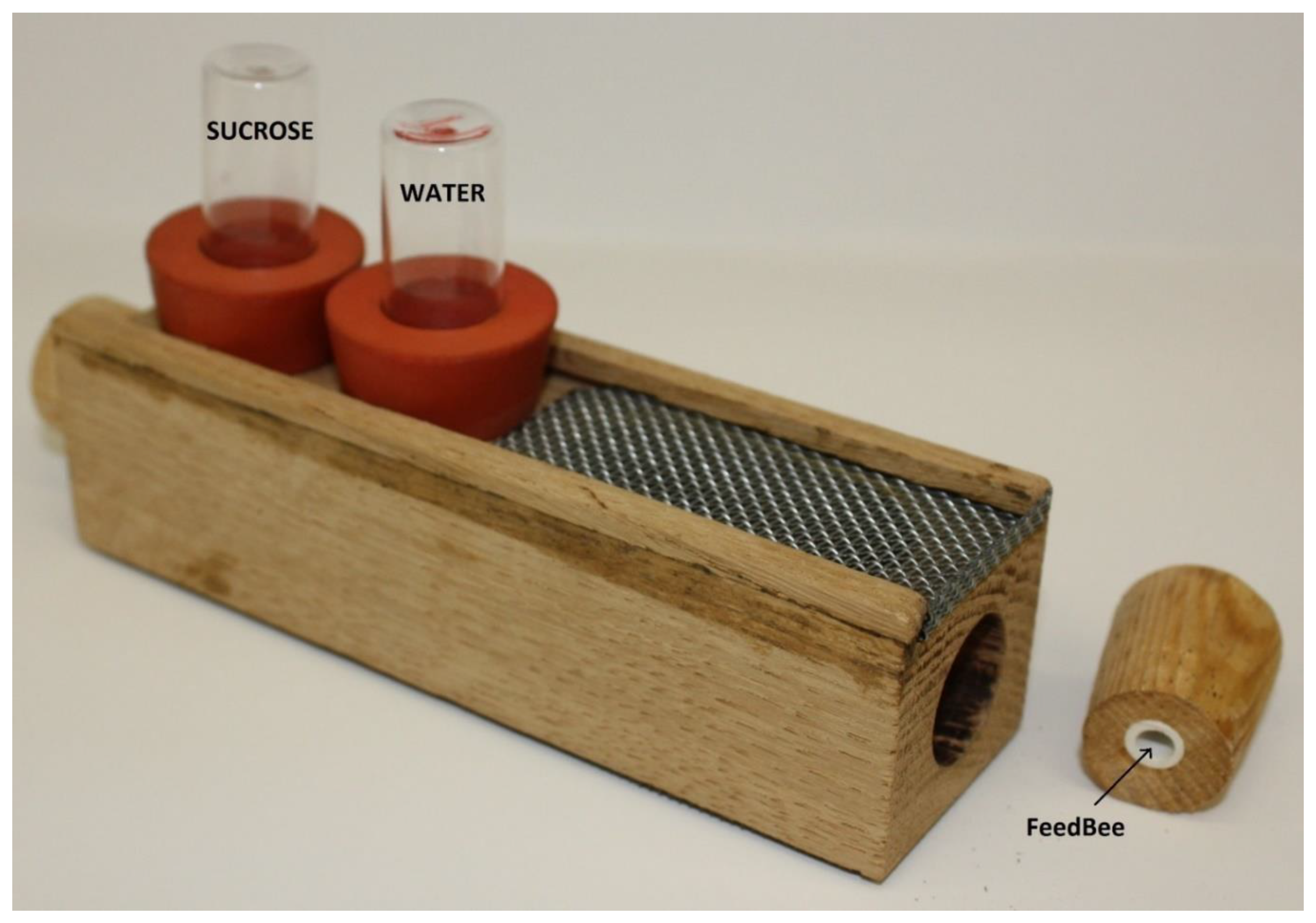
2.3. Material for Infection

N. ceranae spores were obtained and purified from foragers from heavily infected colonies which tested positive for pure N. ceranae infection.
N. apis spores were obtained from field samples of frozen dead bees and then cultivated in pathogen free bees in hoarding cages to ensure viability and freshness of spores for infections in both experiments.
Spore suspensions were prepared at the day of infection from the digestive tracts of infected bees after grinding them up in distilled water, filtering and centrifuging five times in 8000 g to ensure pureness of the spores. The resulting pure spore pellet was suspended in distilled water and the number of spores was calculated using the haemocytometric method, using Fuchs-Rosenthal chamber.
To each 10 mL of spore solution, 1 mL of an anti- BQCV serum (1:32 titre) was added in order to avoid unwanted BQCV infections, since this virus is very common in colonies with nosemosis.
BQCV was obtained pre-experiment from dead queen cells, using the high speed centrifugation method [22], and frozen (for 2 days) until the day of infection. The number of virus copies was determined (during that 2 day period) using a quantitative real-time RT-PCR (as described later, in Section 2.5).

2.4. Experiment 1: Competition between Nosema ceranae and Nosema apis in Mixed Infection

2.4.1. The Infection Procedure

At the 5th day after emergence bees were starved for 2 h and then individually fed with 10 µL of 60% sucrose solution containing spores (or clean sucrose solution in case of the control group). No sedation method was used, bees were placed under a Petri dish and picked one by one by both wings. Experimental groups were created as shown in Table 1. Each group consisted of three cages of 20 bees.
Dead bees were collected daily and kept at a temperature of −20 °C before further examination.

2.4.2. Nosema DNA Extraction and Quantification

Abdomens of individual bees were macerated in 1 ml of distilled water and filtered in extraction bags and then transferred to 1.5 mL Eppendorf tubes and centrifuged to obtain spore pellets. Pellets were crushed in liquid nitrogen and Nosema DNA was extracted according to OIE guidelines [34].
Quantitative real-time PCR to determine Nosema DNA copy numbers was carried out as described in [35], using MX 3005P thermal cycler (Stratagene).

2.5. Experiment 2: Influence of BQCV on the Course of Nosema Infection

2.5.1. The Infection Procedure

The first infection step (infections with Nosema spp.) was performed as described above in Experiment 1.
The second step was the infection with BQCV; 10 µL of 60% sucrose solution containing the virus particles was fed to respective groups on the 8th day post emergence (3rd day after Nosema infection). All control groups were individually fed sucrose solution two times, as the infected groups received spores and the virus.
Each group consisted of three cages of 20 bees. Experimental groups were created as shown in Table 2.
Dead bees were collected daily and kept at a temperature of −20 °C before further examination.

2.5.2. BQCV RNA Extraction and Nosema Spore Count Assessment

Abdomens of individual bees infected with each Nosema spp. and the virus were pulverized in liquid nitrogen and further submitted to RNA isolation using Total RNA Mini Kit (A & A Biotechnology, Gdańsk, Poland). According to manufacturer’s protocol, the powder was suspended in phenosol, but 20 µL of the suspension was transferred to a new tube, and the phenosol was evaporated. Next, the resulting spore pellet was resuspended in 20 µL of distilled water and the spores were counted in Fuchs-Rosenthal haemocytometer. The RNA extraction from the rest of the sample was carried out normally, right after transferring the 20 µL to a new tube.

2.5.3. RT qPCR for BQCV Detection and Quantification

The reverse transcription of obtained RNA was carried out using RevertAid First Strand cDNA Synthesis Kit (Thermo Scientific) according to manufacturer’s protocol.
Resulting cDNA was quantified using a quantitative real-time PCR. The template BQCV fragments for creating standard curves were obtained from SLU University Uppsala. Standard curve was prepared by using serial dilutions of target cDNA fragments, ranging from 102 to1012 as quantification standards in every run.
The quantitative real-time PCR reactions (all in duplicates) were performed in 20 µL volumes using 10 µL of iQTM SYBR® Green Super mix (Bio-Rad Laboratories, Warsaw, Poland), 0.4 µM of each primer (BQCV-qF7893, BQCV-qBB8150), 2 µL of template cDNA and the final reaction volume adjusted to 20 µL with nuclease-free water. A reaction containing water instead of the DNA template was set up as a negative control. In a positive control reaction, a cDNA previously tested positive for BQCV was used. The amplification and data collection were performed using MX 3005P thermal cycler (Stratagene) under the following conditions: initial activation step in 95 °C for 5 min, and PCR cycling consisting of 40 cycles of 94 °C for 30 s, 63 °C for 30 s, 72 °C for 30 s, including data collection. The amplified product was confirmed using melting curve analysis. To create the melting curve the reaction was incubated by raising the incubation temperature from 55 to 95 °C in 0.5 °C increments with a hold of 1 s at each increment. The specificity of amplicons was determined based on the melting temperature of amplified products. Reported results are an average of the duplicate reactions (standards, unknowns, controls).

2.5.4. Statistical Analysis

All statistical calculations were performed using Statistica 10 (StatSoft Inc., Tulsa, OK, USA).
Survival in both experiments was analyzed using Kaplan–Meier test.
A multivariate scatter Graph was used to determine the nature of change in N. apis and N. ceranae copy numbers. Single variation scatter Graphs were also used to compare the overtime change between bees with single N. ceranae and N. apis infections, but also to compare the overtime differences in cDNA copy number changes of BQCV in bees with N. ceranae and N. apis.
A Mann–Whitney U Test was applied to determine the differences in DNA copy numbers of N. ceranae and N. apis spores in bees with and without BQCV.
To determine differences between the number of DNA copies of N. ceranae and N. apis in mixed infection, a T-student test was used, and to determine statistical differences between DNA copy numbers in groups with N. ceranae, N. apis and a group with both species, a Kruskal–Wallis test was used.
To determine the differences in cDNA copy numbers of BQCV between bees with N. apis and N. ceranae a Brown–Forsyth test was used.
In experiments on influence of BQCV on the course of Nosema infection, a post hoc analysis (Gehan–Wilcoxon test) Bonferroni corrected was used to determine the differences in survival rates in bees infected and not infected with BQCV.

3. Results

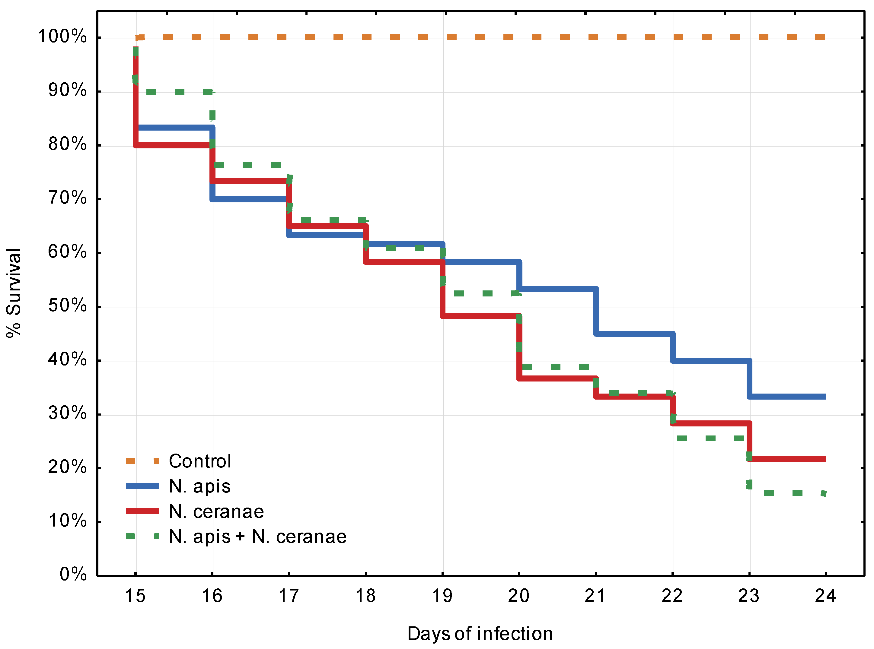
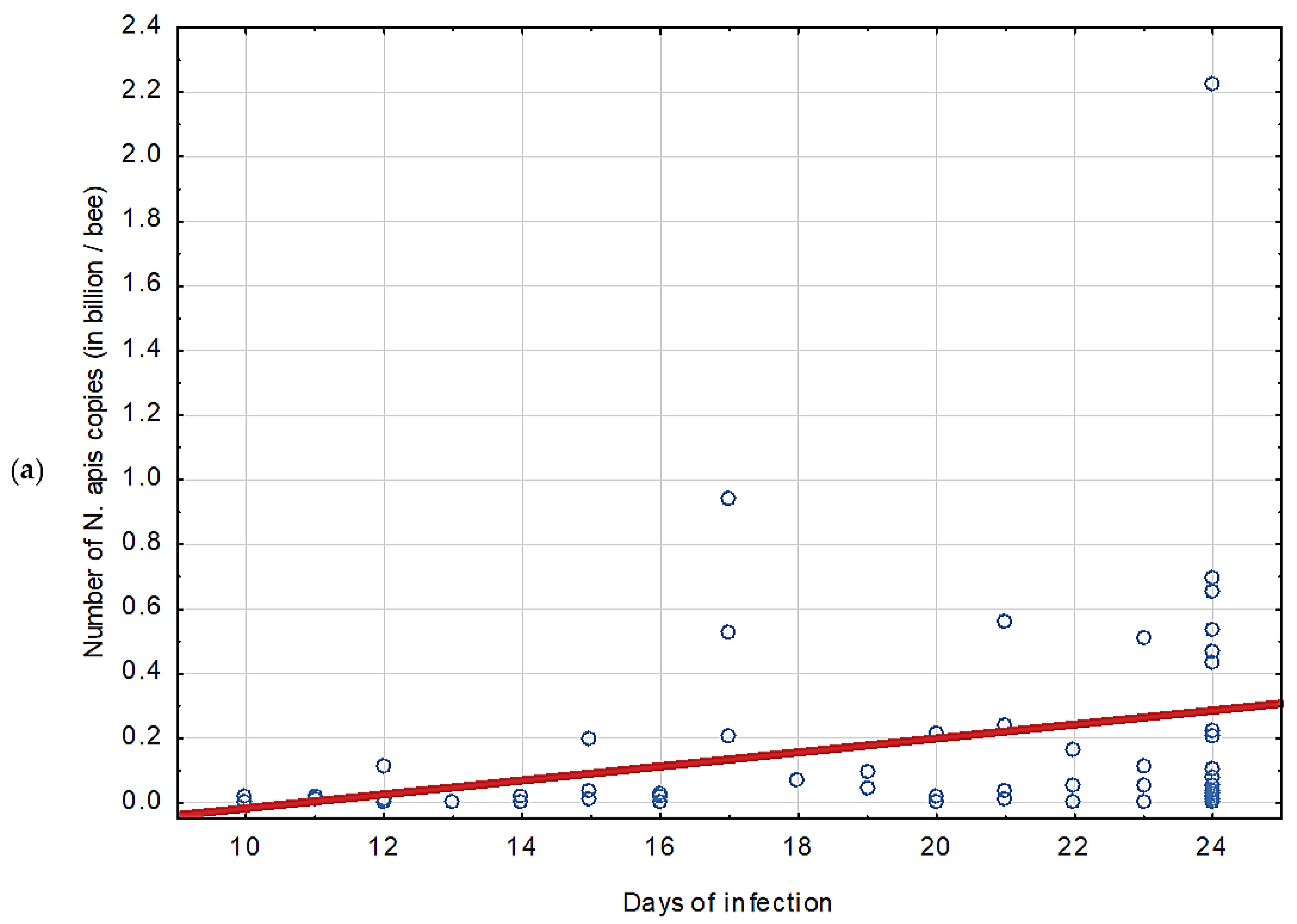
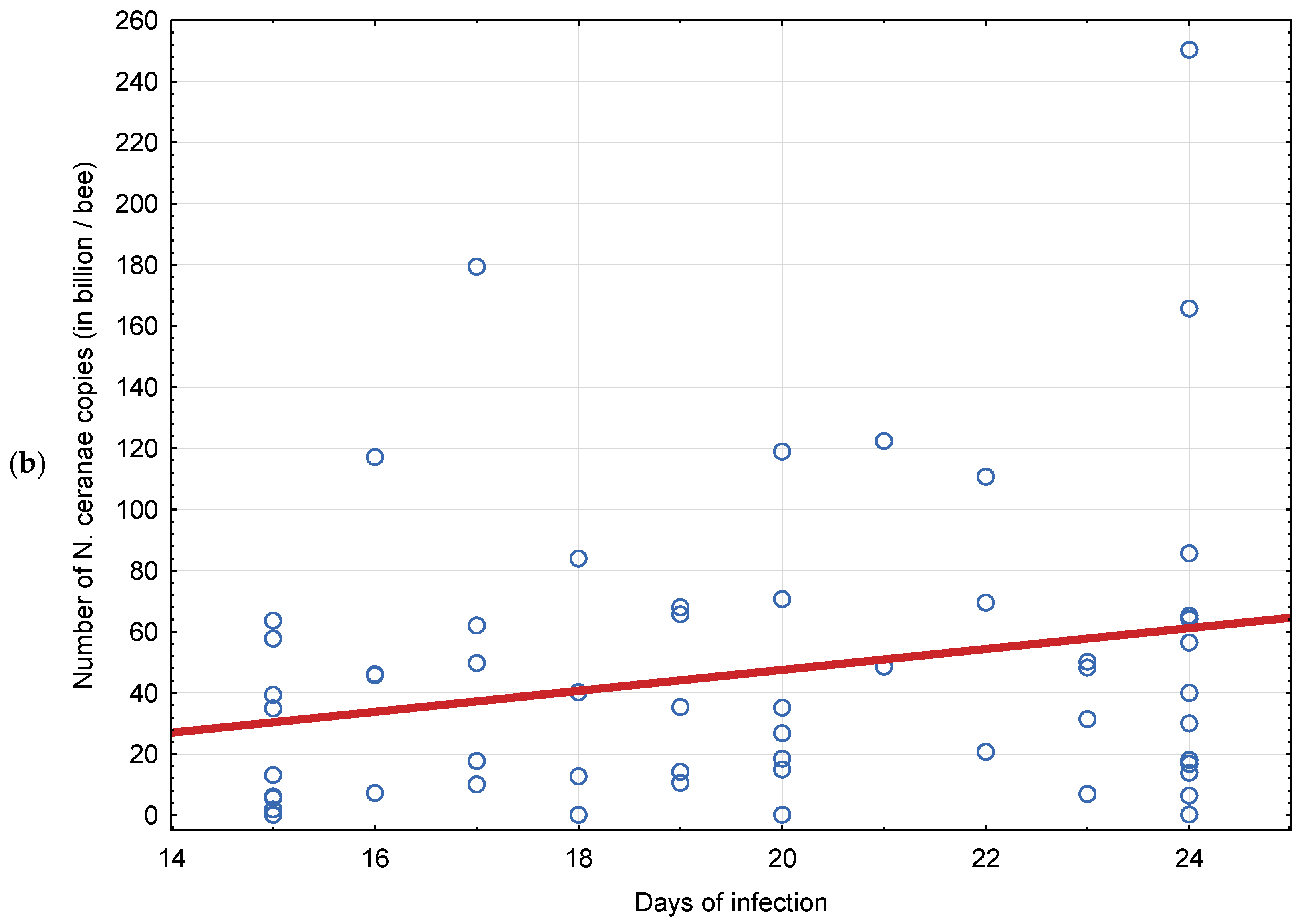
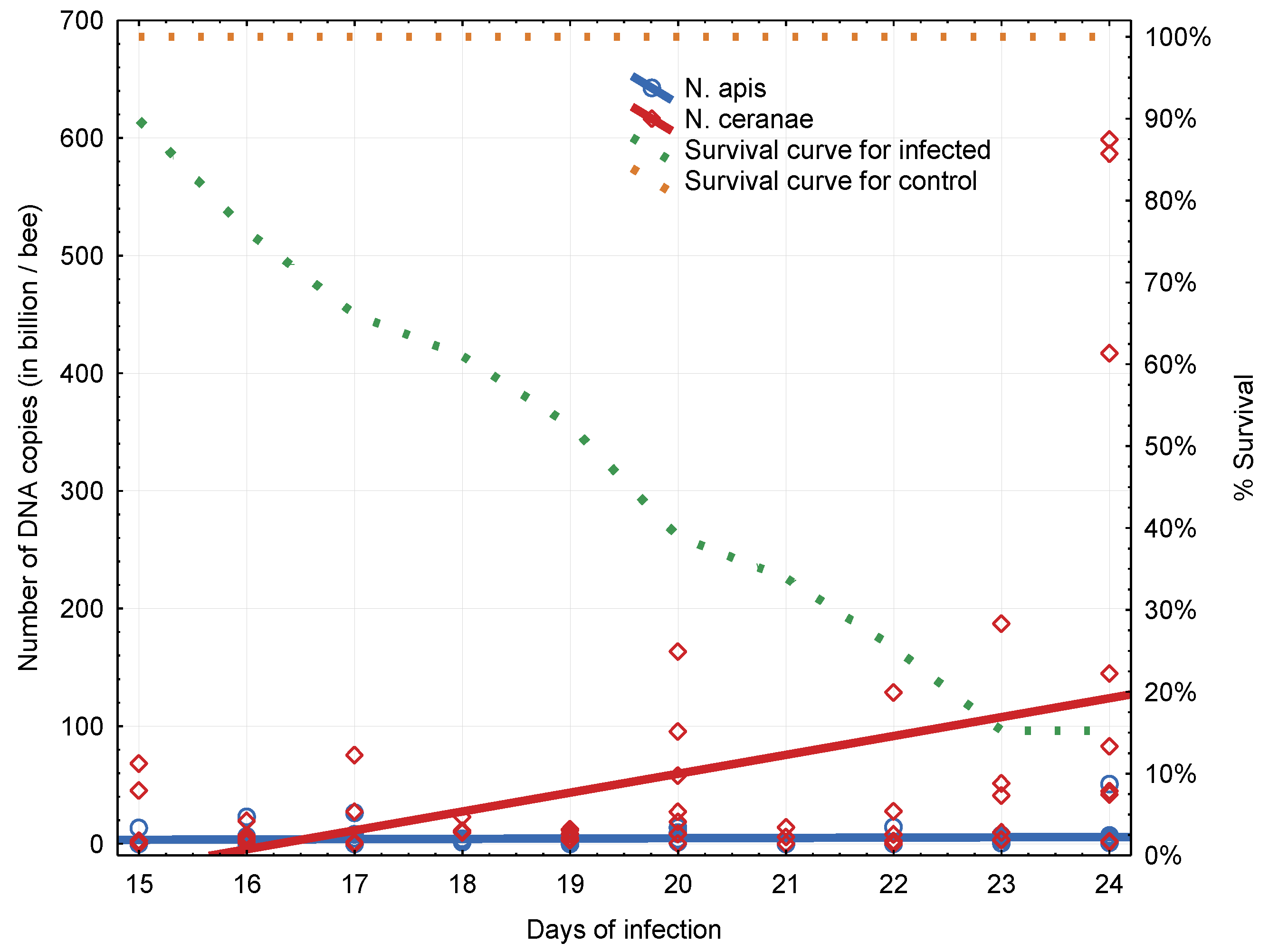
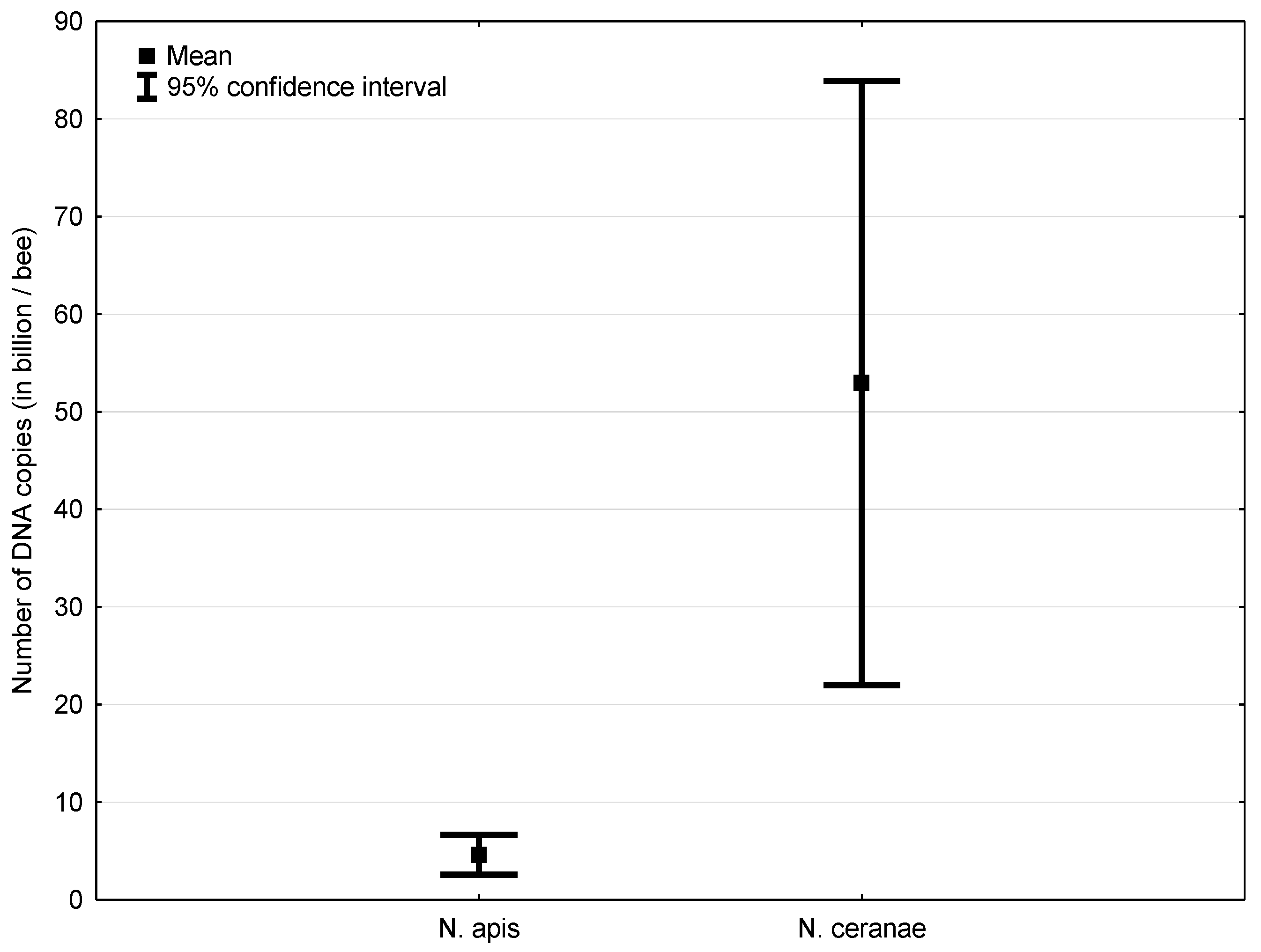
3.1. Experiment 1: Competition between Nosema ceranae and Nosema apis in Mixed Infection

Bees in all infected groups started dying after the 14th day post infection (Figure 2). All bees from the control group remained alive through the entire experiment, so until the 24th day when all the bees that survived were frozen until further examination. There was no statistically significant difference (Kaplan–Meier test) between the survival rate of bees infected with N. apis, N. ceranae and both N. apis and N. ceranae (in equal amounts). However, the Kaplan–Meier test showed a clear tendency for the bees infected with N. apis to survive longer than bees infected with N. ceranae and bees infected with both species.
In both groups of bees infected with only one Nosema species, the DNA copy numbers of both species were growing overtime, but in the case of N. apis, the numbers were significantly lower (Figure 3).
In bees infected with both Nosema species, DNA copy numbers of N. apis were growing minimally, whereas DNA copy numbers of N. ceranae were growing significantly more over time (Figure 4).
In general, in bees infected with both species in a mixed infection, DNA copy numbers of N. apis were significantly lower (Kruskal-Wallis test, p < 0.0001) than DNA copy numbers of N. ceranae in all bees independently from the day of death (Figure 5).

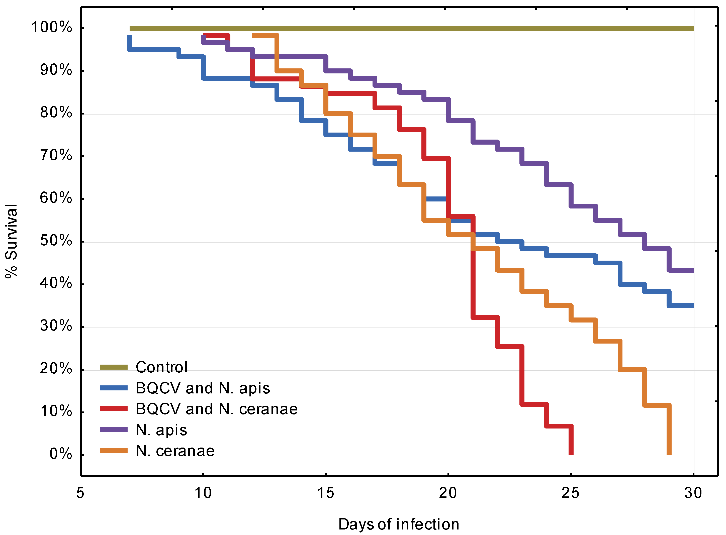
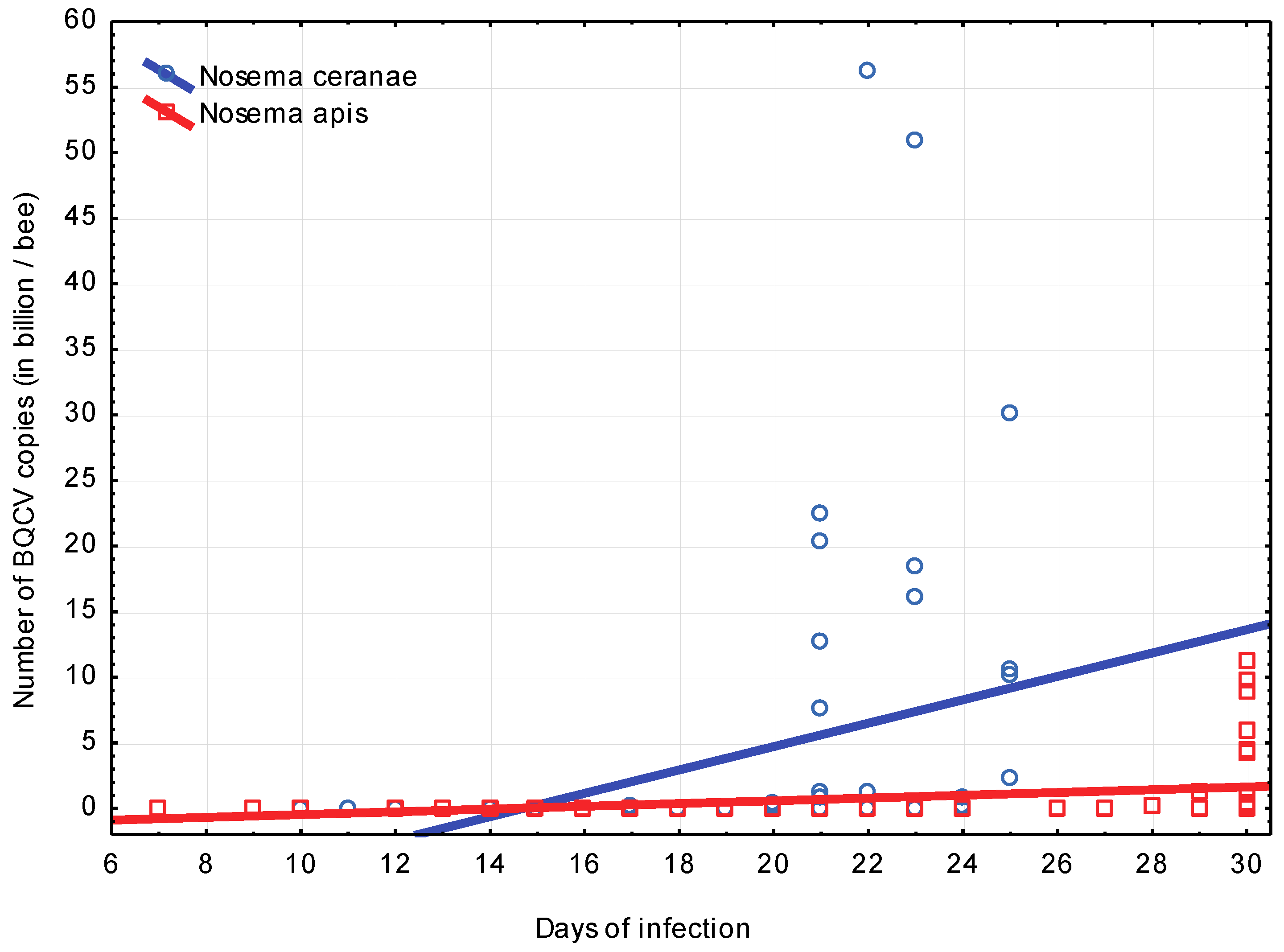
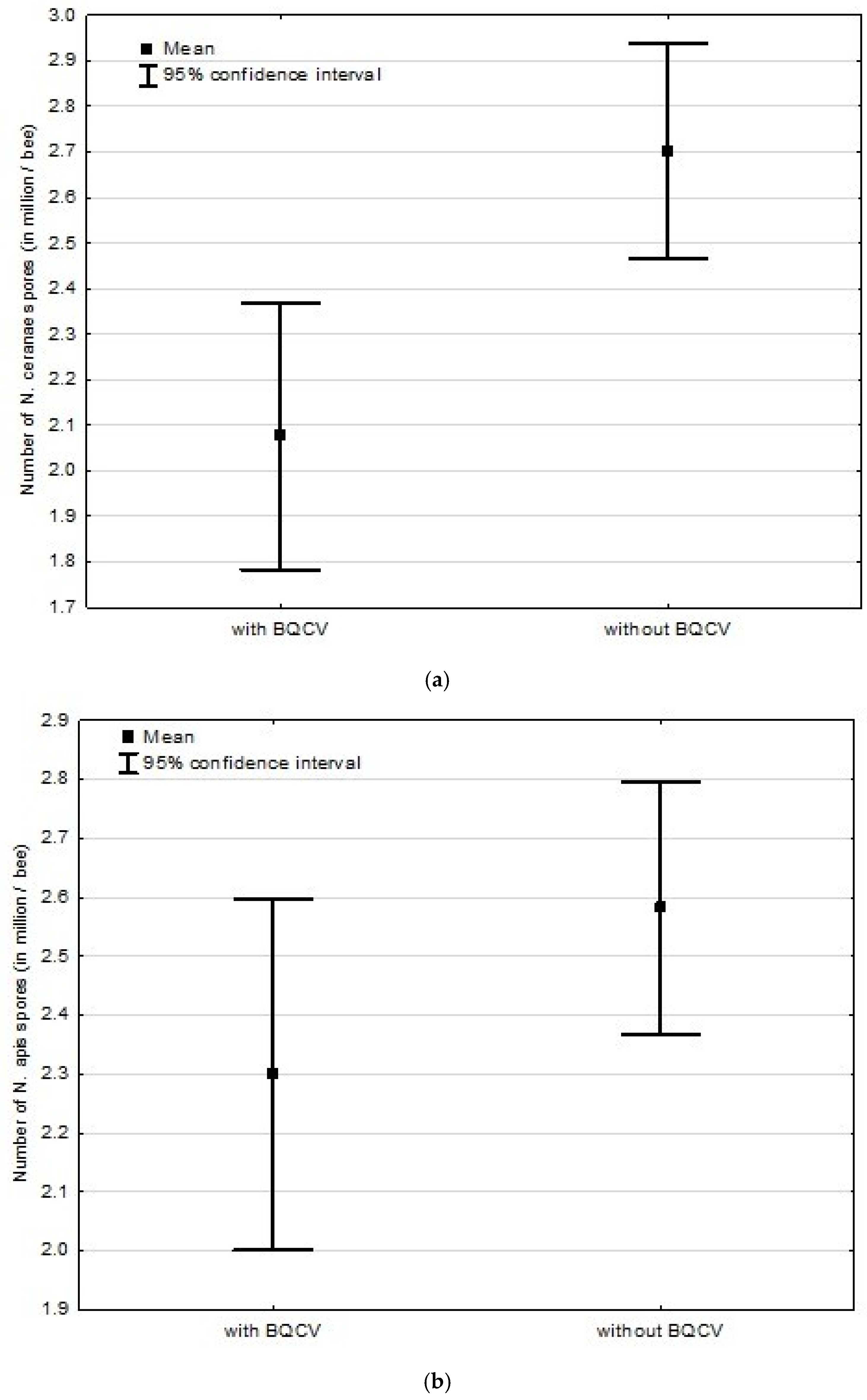
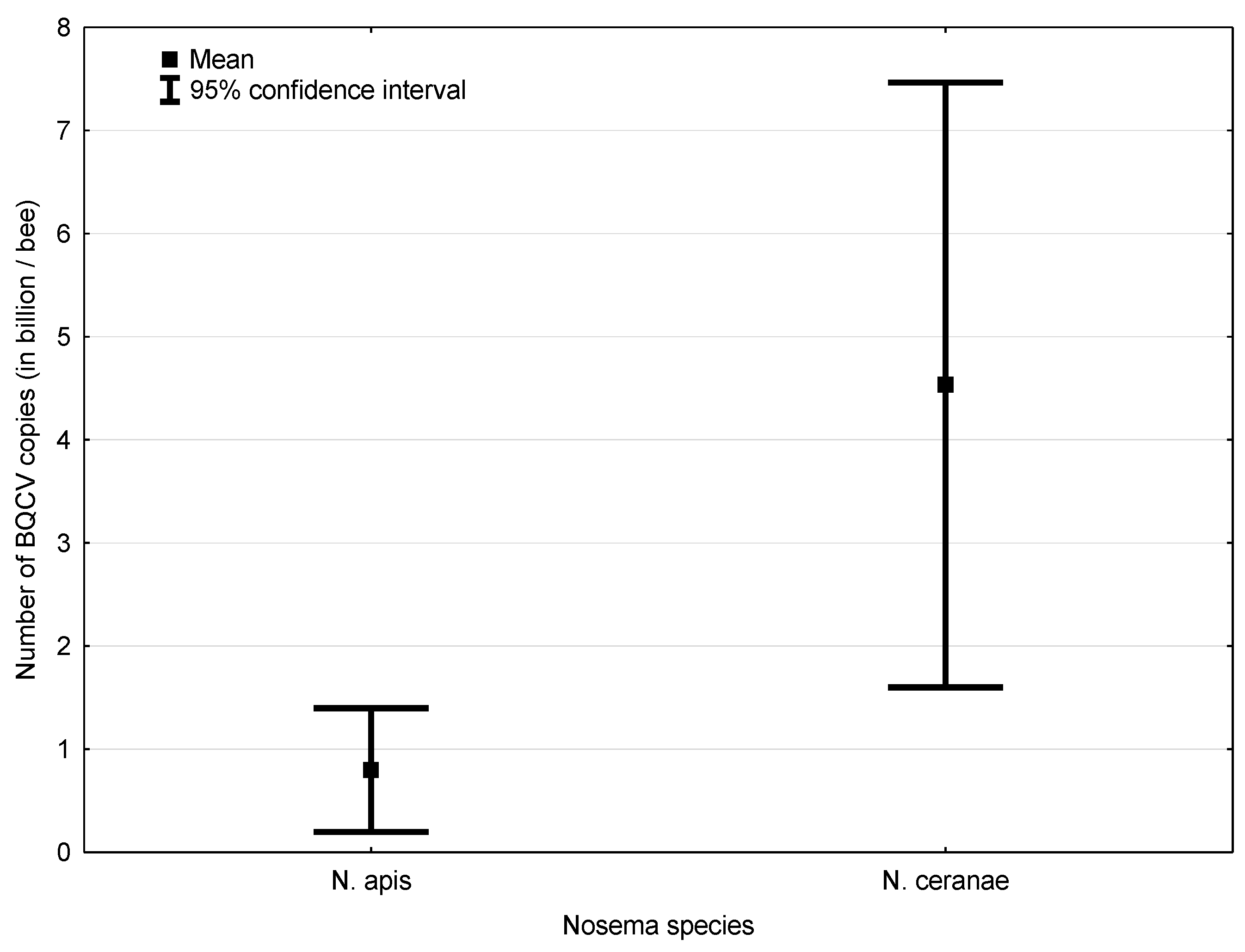
3.2. Experiment 2: Influence of BQCV on the Course of Nosema Infection

All bees from (non-infected) control group survived to the end of experiment, that is to the 30th day after infection with Nosema spores. The experiment ended on day 30, when all bees that were still alive were frozen. This was due to the fact that the group infected with the virus had no remaining living bee. The lowest survival rate was in bees infected with N. ceranae and BQCV. Survival of bees infected with only N. ceranae was also significantly lower (Kaplan–Meyer Test) than of those infected only with N. apis and even with N. apis and BQCV (Table 3 and Figure 6). The highest survival rate was observed in the group infected only with N. apis. The last bees infected with N. ceranae and BQCV died five days before the end of the experiment, while the last bees infected with only N. ceranae died one day before the end of the experiment. In general, bees infected with Nosema spp. and BQCV had a significantly lower survival rate than bees infected only with Nosema spp. (Figure 6).
BQCV cDNA copy numbers in bees infected with N. ceranae and BQCV grew significantly more overtime than cDNA copy numbers in bees infected with N. apis and BQCV (Figure 7).
Interestingly, spore counts in bees infected with N. ceranae and BQCV were significantly lower than in case of bees not infected with BQCV. Additionally, in the case of bees infected with N. apis and BQCV, the same trend was observed, although it was not as clear as in the group infected with N. ceranae and BQCV (Figure 8). In general, BQCV cDNA copy numbers were significantly higher in bees infected with N. ceranae and BQCV in comparison to bees infected with N. apis and BQCV (Figure 9).

4. Discussion and Conclusions

4.1. Experiment 1: Competition between Nosema ceranae and Nosema apis in Mixed Infection

Our results suggest that N. ceranae has a negative effect on the longevity of infected bees. The effect seems to be more clearly visible than in case of N. apis, although in the first experiment (when the bees that survived were frozen at 24th day post infection) only the tendency (a clear one nevertheless) was observed. However, during the second experiment the difference was significant, which confirms the findings of a Canadian study [36]. However, this contradicts with findings from Sweden and Germany [35,37], where such a difference was not found.
This may suggest a geographical/climatic component of the virulence level of N. ceranae compared to N. apis. As mentioned before, in the warmer parts of Europe it causes high colony losses, and in the colder ones it seems to be quite a mild infection.
There is also the experimental design component, which may significantly impact the results. We infected five-day old bees and mentioned authors who infected, respectively, two-week old bees, 48 h old bees and three-day old bees. Younger bees may be more susceptible to N. ceranae infection since their perytrophic membrane is slightly thinner and may be more easily penetrated by the spores and the shorter life cycle of N. ceranae than N. apis (three days, and five days respectively) [5] enabling faster multiplication. It would also explain why we found higher N. ceranae DNA copy numbers in mixed infection (N. apis and N. ceranae), but also singular infections with either N. apis or N. ceranae. Young bees may also be more prone to energetic stress [7], which is more evident during the infection caused by N. ceranae than N. apis. It was proven [38] that five-day old bees (or slightly older) are the most susceptible to infection. However, the two-week old bees may just be too old already, and their peritrophic membrane too thick to accommodate Nosema as well as in the case of younger bees. On the other hand, the newly emerged bees are known to be more resistant to Nosema infection (the reasons of this phenomenon are still unclear), therefore newly emerged bees are always free from infection, even after they chew on infected wax or eat infected food [38]. Our findings confirm that bees infected with N. ceranae had higher cumulative mortality than those infected with N. apis [39]. The results from our experiment also correspond with the research showing higher virulence of N. ceranae than N. apis [16]. Discrepancies between results may be also due to different temperatures in which bees were kept, namely bees in the Swedish experiment [35], which were kept in 30 °C, when bees in our experiment were kept in 34 °C, quite similarly to a 2009 study [40] with which we received comparable results.
We think that infecting young bees and keeping them at a temperature of 34 °C more accurately mimics natural conditions, where bees can be infected during the first days after emergence. During the following days, they mainly stay in warmer parts of the hive because of the tasks performed in the colony, allowing the spores time to multiply. This is why, in field conditions (apart from constant ingestion of spores with food overtime), the numbers of spores in older bees are higher.
In our experiment, infection of individual bees at the 5th day post emergence with N. ceranae resulted in the first deaths after the 14th day post infection, similar to the case of bees infected with N. apis (in the second experiment it was 12 and 10 days, respectively), and with the mixture of both species. These findings are similar to the results of another study researching the virulence of both Nosema species in individual bees [35]. But another study [31] found that the first deaths were observed at the 6th day post infection, and all infected bees were dead at the 8th day post infection. The difference between results might have been caused by using bees from different times of the season, namely, in the contradicting paper, late spring bees were used, and in our experiment we used August bees, which are practically winter bees already and have different body constitutions and, in general, live much longer than spring bees. We also did not use CO2 when handling the bees, since it was proven to shorten their life significantly [41,42]. In addition, more recent studies clearly show, that CO2 treatment influence was so considerable, that the influence of N. ceranae on the longevity of bees could no longer be observed [43].
It is also highly possible that bees (either used for the experiment or used to extract spores from) in experiments of most of the contradicting studies, might have been infected with BQCV (partly or entirely), which was not investigated. The results might be due to presence of this virus in the group of bees infected with N. apis and absence in the group infected with N. ceranae [38] and its presence in the N. apis spore solution [20]. Adding the BQCV antiserum to Nosema spore solutions eliminated this possibility in our experiments.
Our finding, that N. ceranae is highly competitive against N. apis in mixed infections and reaches much higher spore counts contradicts other results [20], in which N. apis was the one with higher (or similar to N. ceranae) spore counts. This might suggest that there may be several N. ceranae strains exhibiting different virulence rates, but this was proven untrue [44], so, in this case, a conclusion that different bee subspecies undergo nosemosis differently seems logical, which is supported by the Danish case [45] and other studies [39,46,47]. In one of the studies [38], A. m. ligustica was used, whereas in others it was Buckfast bees [36] and A. m. carnica [37].

4.2. Experiment 2: Influence of BQCV on the Course of Nosema Infection

In the 1980s [22], it was proven that bees infected with N. apis and BQCV lived shorter than bees infected with N. apis alone. In our study, in the case of N. ceranae, this effect was expressed even better. This can also be correlated to the higher number of virus copies in bees with N. ceranae, which, because of a very quick multiplication and causing much more damage to epithelial cells than N. apis, opened more entrance sites for BQCV. What is more, greater (than with N. apis) energetic stress caused by N. ceranae can explain why BQCV would multiply better in N. ceranae infected bees.
The fact that Nosema and BQCV act synergistically in adult honeybees and induce high and rapid mortality is in agreement with several other findings [36,48] and seems to be universally true.
Occurrence and mean peptide count of viral pathogens and Nosema in honeybee colonies sampled from widespread locations in USA CCD colonies showed, instead, a high frequency of iridescent virus and a low frequency of BQCV [49].
Bees infected with either species of Nosema and BQCV showed lower spore counts than the ones infected with only Nosema. Since dead bees were investigated, this result was probably due to the synergistic effect of the virus and Microsporidium, which caused faster death of bees, hence the spores could not multiply further. As the effect was much stronger in the case of N. ceranae, as expected, this dissonance is much clearer between bees with N. ceranae, together with BQCV, and bees with only N. ceranae, than in respective groups with N. apis.
Since this experiment was conducted in 2013, we based the design on available publications on this topic [22,50], which state that there is no virus propagation, or it is very minor, without the presence of Nosema sp. However, a more recent study [48] proved that the virus can multiply on its own, so a design with a control group with only BQCV infected bees is needed.
Further experiments are needed since it would also be desirable to compare the above results with spore and virus counts from live bees, as well as through the experiment. Studies on temperature influence on the course of virus/Nosema infection are also needed.
It has also become more apparent that a thorough comparison of Nosema infection course in different bee subspecies and, simultaneously, different climatic conditions, is needed. Those two factors together may have a greater influence on the course of this infection than previously thought.
This study shows the effects of interaction between N. ceranae, N. apis and black queen cell virus, as well as the effect of both Nosema species on the longevity of bees. All organisms (bees, Nosema), as well as the virus, were of Polish origin and show only the interaction of actors of said origin. As shown above, further multi-direction research is needed to say with certainty what influences the pathogenicity/virulence of N. ceranae, which, in time, may open new paths to controlling Nosema infections and preventing related colony losses.

Author Contributions

A.M.G.: conceptualization, laboratory testing, data acquisition, data interpretation, manuscript writing; E.D.M.: data interpretation, manuscript writing and proofreading; A.M.B.: data Interpretation, manuscript writing and proofreading, M.C.: statistical analyses, data interpretation, proofreading. All authors have read and agreed to the published version of the manuscript.

Funding

This research received no external funding.

Institutional Review Board Statement

Not applicable.

Informed Consent Statement

Informed consent was obtained from all subjects involved in the study.

Data Availability Statement

In section “MDPI Research Data Policies” at https://www.mdpi.com/ethics (accessed on 1 September 2021).

Conflicts of Interest

The authors declare no conflict of interest.

Appendix A. Pre-Testing Colonies for the Presence of Viruses and Nosema spp.

To test the presence of abovementioned viruses at least 20 emerged bees from each comb were pulverized in liquid nitrogen. RNA extraction was carried out using Total RNA Mini (A&A Biotechnology) according to manufacturer’s protocol.
RNA was reverse-transcribed to cDNA using RevertAid First Strand cDNA Synthesis Kit (Thermo Scientific) also according to protocol enclosed by the manufacturer.
To detect viruses specific primer pairs were used as shown in Table A1.
Table A1. Primers used to detect specific virus sequences.
VirusPrimer PairsSource
BQCVBQCV76f, BQCV299r(Bakonyi, 2002) [51]
ABPVABPV1f, ABPV2r(Bakonyi, 2002) [51]
CBPVCBPV1f, CBPV2r(Ribiere et al., 2002) [52]
SBVSBV1f, SBV2r(Ghosh et al., 1997) [53]
DWVDWV2345f, DWV2779r(Lanzi et al., 2006) [54]
All PCRs were performed in 50 µL reactions, each containing 0.3 µM of each (forward and reverse) primer, 0.2 mM of each dNTP, 1.5 mM MgCl2, 1.5 U of Taq Polymerase and 2 µL of query cDNA. A positive and negative (no template) controls were included for every virus.
All reactions were carried out in MiniCycler 25 (MJ Research). Thermal protocol for BQCV, ABPV, CBPV and SBV consisted of a single initial activation step in 94 °C for 3 min followed by 40 PCR cycles (denaturation: 94 °C for 1 min, annealing: see Table A2, elongation: 72 °C for 1 min). The reaction was closed with a final elongation step in 72 °C for 2 min.
Table A2. Annealing temperatures for specific primer pairs.
Primer PairsAnnealing Temperatures and Times
BQCV76f, BQCV299r50 °C for 1 min
ABPV1f, ABPV2r55 °C for 1 min
CBPV1f, CBPV2r50 °C for 1 min
SBV1f, SBV2r50 °C for 1 min
Thermal protocol for DWV consisted of an initial activation step in 94 °C for 3 min followed by 35 PCR cycles (denaturation: 94 °C for 30 s, annealing: 53 °C for 1 min, elongation: 72 °C for 30 min). The reaction was closed with a final elongation step in 72 °C for 10 min.
The size of PCR product for each virus is presented in Table A3.
Table A3. The size of PCR product for each virus.
VirusPCR Product Size (bp)
BQCV224
ABPV398
CBPV455
SBV646
DWV435
Products were separated electrophoretically in 1.5% agarose gel. Visualization under UV light and documentation of obtained results was carried out using GelDoc-It Imaging System (UVP).
The absence of Nosema spores was first assessed microscopically, and confirmed with a PCR according to the OIE protocol (2008) [34].

References

  1. Fries, I.; Feng, F.; da Silva, A.; Slemenda, S.B.; Pieniazek, N.J. Nosema ceranae n. sp. (Microspora, Nosematidae), morphological and molecular characterization of a microsporidian parasite of the Asian honey bee Apis cerana (Hymenoptera, Apidae). Eur. J. Protistol. 1996, 32, 356–365. [Google Scholar] [CrossRef]
  2. Higes, M.; Martín, R.; Meana, A. Nosema ceranae, a new microsporidian parasite in honeybees in Europe. J. Invertebr. Pathol. 2006, 92, 93–95. [Google Scholar] [CrossRef]
  3. Klee, J.; Besana, A.M.; Genersch, E.; Gisder, S.; Nanetti, A.; Tam, D.Q.; Chinh, T.X.; Puerta, F.; Ruz, J.M.; Kryger, P.; et al. Widespread dispersal of the microsporidian Nosema ceranae, an emergent pathogen of the western honey bee, Apis mellifera. J. Invertebr. Pathol. 2007, 96, 1–10. [Google Scholar] [CrossRef] [PubMed]
  4. Topolska, G.; Kasprzak, S. Pierwsze przypadki zarażenia pszczół w Polsce przez Nosema ceranae. Med. Wet. 2007, 63, 1504–1506. [Google Scholar]
  5. Higes, M.; Esperón, F.; Sánchez-Vizcaino, J.M. Short communication. First report of black queen-cell virus detection in honey bees (Apis mellifera) in Spain. Span. J. Agric. Res. 2007, 5, 322–325. [Google Scholar] [CrossRef]
  6. Vidau, C.; Panek, J.; Texier, C.; Biron, D.G.; Belzunces, L.P.; Le Gall, M.; Broussard, C.; Delbac, F.; El Alaoui, H. Differential proteomic analysis of midguts from Nosema ceranae-infected honeybees reveals manipulation of key host functions. J. Invertebr. Pathol. 2014, 121, 89–96. [Google Scholar] [CrossRef]
  7. Mayack, C.; Naug, D. Energetic stress in the honeybee Apis mellifera from Nosema ceranae infection. J. Invertebr. Pathol. 2009, 100, 185–188. [Google Scholar] [CrossRef]
  8. Gajda, A.M.; Grzęda, U.; Topolska, G.; Wilde, J.; Bieńkowska, M.; Gerula, D.; Panasiuk, B. Nosema ceranae has been present in honey bee colonies in Poland at least since 1994 and appears to have ousted Nosema apis. In Proceedings of the 9th COLOSS Conference, Kyiv, Ukraine, 27–29 September 2013; p. 38. [Google Scholar]
  9. Meixner, M.D.; Francis, R.M.; Gajda, A.; Kryger, P.; Andonov, S.; Uzunov, A.; Topolska, G.; Costa, C.; Amiri, E.; Berg, S.; et al. Occurrence of parasites and pathogens in honey bee colonies used in a European genotype-environment interactions experiment. J. Apic. Res. 2014, 53, 215–219. [Google Scholar] [CrossRef]
  10. Higes, M.; Martin-Hernández, R.; Botias, C.; Garrido-Bailón, E.; González-Porto, A.V.; Barrios, L.; Del Nozal, M.J.; Bernal, J.L.; Jiménez, J.J.; Palencia, P.G.; et al. How natural infection by Nosema ceranae causes honeybee colony collapse. Environ. Microbiol. 2008, 10, 2659–2669. [Google Scholar] [CrossRef]
  11. Higes, M.; Martín-Hernández, R.; Martínez-Salvador, A.; Garrido-Bailón, E.; González-Porto, A.V.; Meana, A.; Bernal, J.L.; Del Nozal, M.J.; Bernal, J. A preliminary study of the epidemiological factors related to honey bee colony loss in Spain. Environ. Microbiol. Rep. 2010, 2, 243–250. [Google Scholar] [CrossRef] [PubMed]
  12. Hatjina, F.; Tsoktouridis, G.; Bouga, M.; Charistos, L.; Evangelou, V.; Avtzis, D.; Meeus, I.; Brunain, M.; Smagghe, G.; de Graaf, D.C. Polar tube protein gene diversity among Nosema ceranae strains derived from a Greek honey bee health study. J. Invertebr. Pathol. 2011, 108, 131–134. [Google Scholar] [CrossRef]
  13. Topolska, G.; Gajda, A. Polish Honey Bee Colony-Loss During the Winter of 2007/2008. J. Apic. Sci. 2008, 52, 95–104. [Google Scholar]
  14. Topolska, G.; Gajda, A.; Pohorecka, K.; Bober, A.; Kasprzak, S.; Semkiw, P.; Skubida, M. Winter colony losses in Poland. J. Apic. Res. 2010, 49, 126–128. [Google Scholar] [CrossRef]
  15. Vercelli, M.; Novelli, S.; Ferrazzi, P.; Lentini, G.; Ferracini, C. A Qualitative Analysis of Beekeepers’ Perceptions and Farm Management Adaptations to the Impact of Climate Change on Honey Bees. Insects 2021, 12, 228. [Google Scholar] [CrossRef] [PubMed]
  16. Gisder, S.; Hedtke, K.; Möckel, N.; Frielitz, M.-C.; Linde, A.; Genersch, E. Five-year cohort study of Nosema spp. in Germany: Does climate shape virulence and assertiveness of Nosema ceranae? Appl. Environ. Microbiol. 2010, 76, 3032–3038. [Google Scholar] [CrossRef] [PubMed] [Green Version]
  17. Guzman-Novoa, E.; Eccles, L.; Calvete, Y.; Mcgowan, J.; Kelly, P.G.; Correa-Benitez, A. Varroa destructor is the main culprit for the death and reduced populations of overwintered honey bee (Apis mellifera) colonies in Ontario, Canada. Apidologie 2021, 41, 443–450. [Google Scholar] [CrossRef] [Green Version]
  18. Williams, G.R.; Shutler, D.; Rogers, R.E.L. Effect at Nearctic north-temperate latitudes of indoor versus outdoor overwintering on the microsporidium Nosema ceranae and western honey bees (Apis mellifera). J. Invertebr. Pathol. 2010, 104, 4–7. [Google Scholar] [CrossRef]
  19. Invernizzi, C.; Abud, C.; Tomasco, I.H.; Harriet, J.; Ramallo, G.; Campa, J.; Katz, H.; Gardiol, G.; Mendoza, Y. Presence of Nosema ceranae in honeybees (Apis mellifera) in Uruguay. J.Invertebr. Pathol. 2009, 101, 150–153. [Google Scholar] [CrossRef]
  20. Milbrath, M.O.; van Tran, T.; Huang, W.-F.; Solter, L.F.; Tarpy, D.R.; Lawrence, F.; Huang, Z.Y. Comparative virulence and competition between Nosema apis and Nosema ceranae in honey bees (Apis mellifera). J. Invertebr. Pathol. 2015, 125, 9–15. [Google Scholar] [CrossRef]
  21. Forsgren, E.; Fries, I. Temporal study of Nosema spp. in a cold climate. Environ. Microbiol. Rep. 2013, 5, 78–82. [Google Scholar] [CrossRef]
  22. Bailey, L.; Ball, V.B.; Perry, J.N. Association of viruses with two protozoal pathogens of the honey bee. Ann. Appl. Biol. 1983, 103, 13–20. [Google Scholar] [CrossRef]
  23. Al Naggar, Y.; Paxton, R.J. Mode of Transmission Determines the Virulence of Black Queen Cell Virus in Adult Honey Bees, Posing a Future Threat to Bees and Apiculture. Viruses 2020, 12, 535. [Google Scholar] [CrossRef]
  24. Bailey, L.; Woods, R.D. Two more small RNA viruses from honey bees and further observations on sacbrood and acute bee-paralysis viruses. J. Gen. Virol. 1977, 37, 175–182. [Google Scholar] [CrossRef]
  25. Chen, Y.; Evans, J.; Feldlaufer, M. Horizontal and vertical transmission of viruses in the honey bee, Apis mellifera. J. Invertebr. Pathol. 2006, 92, 152–159. [Google Scholar] [CrossRef]
  26. Bailey, E. Biochemistry of Insect Flight. In Insect Biochemistry and Function; Candy, D.J., Kilby, B.A., Eds.; Chapman and Hall: London, UK, 1975. [Google Scholar] [CrossRef]
  27. Lodesani, M.; Costa, C.; Besana, A.; Dall’Olio, R.; Franceschetti, S.; Tesoriero, D.; Giacomo, D. Impact of control strategies for Varroa destructor on colony survival and health in northern and central regions of Italy. J. Apic. Res. 2014, 53, 155–164. [Google Scholar] [CrossRef]
  28. Tentcheva, D.; Gauthier, L.; Zappulla, N.; Dainat, B.; Cousserans, F.; Colin, M.E.; Bergoin, M. Prevalence and Seasonal Variations of Six Bee Viruses in Apis mellifera L. and Varroa destructor Mite Populations in France. Appl. Environ. Microbiol. 2004, 70, 7185–7191. [Google Scholar] [CrossRef] [PubMed] [Green Version]
  29. Ravoet, J.; Maharramov, J.; Meeus, I.; De Smet, L.; Wenseleers, T.; Smagghe, G.; de Graaf, D.C. Comprehensive Bee Pathogen Screening in Belgium Reveals Crithidia mellificae as a New Contributory Factor to Winter Mortality. PLoS ONE 2013, 8, e72443. [Google Scholar] [CrossRef] [PubMed] [Green Version]
  30. Topolska, G.; Krzyżańska, K.; Hartwig, A.; Gajda, A. The investigation of bee virus infections in Poland. Ann. Wars. Univ. Life Sci. SGGW 2009, 46, 125–133. [Google Scholar]
  31. Higes, M.; García-Palencia, P.; Martín-Hernández, R.; Meana, A. Experimental infection of Apis mellifera honeybees with Nosema ceranae (Microsporidia). J. Invertebr. Pathol. 2007, 94, 211–217. [Google Scholar] [CrossRef]
  32. Siede, R.; Büchler, R. Symptomatic Black Queen Cell Virus infection of drone brood in Hessian apiaries. Berl. Munch. Tierarztl. Wochensch. 2003, 116, 130–133. [Google Scholar]
  33. Nielsen, S.L.; Nicolaisen, M.; Kryger, P. Incidence of acute bee paralysis virus, black queen cell virus, chronic bee paralysis virus, deformed wing virus, Kashmir bee virus and sacbrood virus in honey bees (Apis mellifera) in Denmark. Apidologie. 2008, 39, 310–314. [Google Scholar] [CrossRef] [Green Version]
  34. Nosemosis of Honey Bees. OIE Terrestrial Manual. 2018. Available online: https://www.oie.int/fileadmin/Home/eng/Health_standards/tahm/3.02.04_NOSEMOSIS_FINAL.pdf (accessed on 13 July 2021).
  35. Forsgren, E.; Fries, I. Comparative virulence of Nosema ceranae and Nosema apis in individual European honey bees. Vet. Parasitol. 2010, 170, 212–217. [Google Scholar] [CrossRef]
  36. Williams, G.R.; Shutler, D.; Burgher-MacLellan, K.L.; Rogers, R.E.L. Infra-population and -community dynamics of the parasites Nosema apis and Nosema ceranae, and consequences for honey bee (Apis mellifera) hosts. PLoS ONE 2014, 9, e99465. [Google Scholar] [CrossRef] [Green Version]
  37. Natsopoulou, M.E.; Doublet, V.; Paxton, R.J. European isolates of the Microsporidia Nosema apis and Nosema ceranae have similar virulence in laboratory tests on European worker honey bees. Apidologie 2016, 47, 57–65. [Google Scholar] [CrossRef] [Green Version]
  38. Huang, W.-F.; Solter, L.; Aronstein, K.; Huang, Z. Infectivity and virulence of Nosema ceranae and Nosema apis in commercially available North American honey bees. J. Invertebr. Pathol. 2015, 124, 107–113. [Google Scholar] [CrossRef] [PubMed]
  39. Paxton, R.J.; Klee, J.; Korpela, S.; Fries, I. Nosema ceranae has infected Apis mellifera in Europe since at least 1998 and may be more virulent than Nosema apis. Apidologie 2007, 38, 558–565. [Google Scholar] [CrossRef]
  40. Martín-Hernández, R.; Meana, A.; García-Palencia, P.; Marín, P.; Botías, C.; Garrido-Bailón, E.; Barrios, L.; Higes, M. Effect of temperature on the biotic potential of honeybee microsporidia. Appl. Environ. Microbiol. 2009, 75, 2554–2557. [Google Scholar] [CrossRef] [Green Version]
  41. Czekońska, K. The effect of different concentrations of carbon dioxide (CO2) in a mixture with air or nitrogen upon the survival of the honey bee (Apis mellifera). J. Apic. Res. 2009, 48, 67–71. [Google Scholar] [CrossRef]
  42. Tustain, R.C.R.; Faulke, J. Effect of carbon dioxide anaesthesia on the longevity of honey bees in the laboratory. N. Z. J. Exp. Agric. 1979, 7, 327–329. [Google Scholar] [CrossRef]
  43. Milbrath, M.O.; Xie, X.; Huang, Z.Y. Nosema ceranae induced mortality in honey bees (Apis mellifera) depends on infection methods. J. Invertebr. Pathol. 2013, 114, 42–44. [Google Scholar] [CrossRef]
  44. Dussaubat, C.; Sagastume, S.; Gómez-Moracho, T.; Botías, C.; García-Palencia, P.; Martín-Hernández, R.; Le Conte, Y.; Higes, M. Comparative study of Nosema ceranae (Microsporidia) isolates from two different geographic origins. Vet. Microbiol. 2013, 162, 670–678. [Google Scholar] [CrossRef]
  45. Huang, Q.; Kryger, P.; Le Conte, Y.; Moritz, R.F.F. Survival and immune response of drones of a Nosemosis tolerant honey bee strain towards N. ceranae infections. J. Invertebr. Pathol. 2012, 109, 297–302. [Google Scholar] [CrossRef]
  46. Martín-Hernández, R.; Botías, C.; Barrios, L.; Martinez-Salvador, A.; Meana, A.; Mayack, C.; Higes, M. Comparison of the energetic stress associated with experimental Nosema ceranae and Nosema apis infection of honeybees (Apis mellifera). Parasitol. Res. 2011, 109, 605–612. [Google Scholar] [CrossRef]
  47. Branchiccela, B.; Arredondo, D.; Higes, M.; Invernizzi, C.; Martín-Hernández, R.; Tomasco, I.; Zunino, P.; Antúnez, K. Characterization of Nosema ceranae genetic variants from different geographic origins. Microb. Ecol. 2017, 73, 978–987. [Google Scholar] [CrossRef]
  48. Doublet, V.; Labarussias, M.; de Miranda, J.R.; Moritz, R.F.A.; Paxton, R.J. Bees under stress: Sublethal doses of a neonicotinoid pesticide and pathogens interact to elevate honey bee mortality across the life cycle. Environ. Microbiol. 2015, 17, 969–983. [Google Scholar] [CrossRef]
  49. Bromenshenk, J.J.; Henderson, C.B.; Wick, C.H.; Stanford, M.F.; Zulich, A.W.; Jabbour, R.E.; Deshpande, S.V.; McCubbin, P.E.; Seccomb, R.A.; Welch, P.M.; et al. Iridovirus and microsporidian linked to honey bee colony decline. PLoS ONE. 2010, 5, e13181. [Google Scholar] [CrossRef] [PubMed]
  50. Virology and the Honey Bee. European Commission, Directorate-General for Research. 2008. Available online: http://edepot.wur.nl/1862 (accessed on 13 July 2021).
  51. Bakonyi, T. Occurrence of the Honey Bee Viruses in Hungary, Investigation of Molecular Structure of Certain Viruses. Ph.D. Thesis, Szent Istvan University, Budapest, Hungary, 2002. [Google Scholar]
  52. Ribiere, M.; Triboulot, C.; Mathieu, L.; Aurieres, C.; Faucon, J.-P.; Pepin, M. Molecular diagnosis of chronic bee paralysis virus infection. Apidologie 2002, 33, 339–351. [Google Scholar] [CrossRef]
  53. Ghosh, R.C.; Ball, B.V.; Willcocks, M.M.; Carter, M.J. The nucleotide sequence of sacbrood virus of the honey bee: An insect picorna-like virus. J. Gen. Virol. 1999, 80, 1541–1549. [Google Scholar] [CrossRef] [PubMed]
  54. Lanzi, G.; de Miranda, J.R.; Boniotti, M.B.; Cameron, C.E.; Lavazza, A.; Capucci, L.; Camazine, S.M.; Rossi, C. Molecular and biological characterization of deformed wing virus of honeybees (Apis mellifera L.). J. Virol. 2006, 80, 4998–5009. [Google Scholar] [CrossRef] [PubMed] [Green Version]
Figure 1. Hoarding cage used in both experiments.
Agriculture 11 00963 g001
Figure 2. Survival rate of bees infected with N. apis, between N. ceranae and both species at the same time.
Agriculture 11 00963 g002
Figure 3. Overtime change in DNA copy numbers in bees infected with one species of Nosema (a) of N. apis (b) of N. ceranae.
Agriculture 11 00963 g003aAgriculture 11 00963 g003b
Figure 4. Change in N. apis and N. ceranae DNA copy numbers overtime in bees infected with both species (N. ceranae and N. apis) (showing survival rate curves for bees infected and non-infected bees).
Agriculture 11 00963 g004
Figure 5. Differences in N. apis and N. ceranae DNA copy numbers in bees infected with both species (N. ceranae and N. apis). Paired Student’s t-test p = 0.003.
Agriculture 11 00963 g005
Figure 6. Survival rate in bees infected with N. apis, N. ceranae, BQCV + N. apis and BQCV + N. ceranae.
Agriculture 11 00963 g006
Figure 7. Overtime BQCV cDNA copy numbers in bees infected with N. ceranae and BQCV (shown as a blue line) and N. apis and BQCV (shown as a red line) Pearson’s product-moment correlation coefficient (r). For N. ceranae r = 0.41, p = 0.001. For N. apis r = 0.10, p = 0.443.
Agriculture 11 00963 g007
Figure 8. Nosema spp. spore counts in bees infected and not infected with BQCV and (a) N. ceranae, Mann-Whitney U test p = 0.001 (b) N. apis Unpaired Student’s t-test p = 0.135.
Agriculture 11 00963 g008
Figure 9. BQCV cDNA copy numbers in group of bees infected with N. ceranae and BQCV and N. apis and BQCV Unpaired Student’s t-test p = 0.017.
Agriculture 11 00963 g009
Table 1. The setup of experimental groups for experiment on competition between Nosema ceranae and Nosema apis in mixed infection.
GroupSpores Per Bee
N. apis106
N. ceranae106
N. apis + N. ceranae106 + 106
Control0 (sucrose solution)
Table 2. The setup of experimental groups for experiment on influence of BQCV on the course of Nosema infection.
Nosema sp.Spores Per BeeVirus Copies Per Bee
N. apis + BQCV1061010
N. ceranae + BQCV1061010
N. apis1060 (sucrose solution)
N. ceranae1060 (sucrose solution)
Control0 (sucrose solution)0 (sucrose solution)
Table 3. Post hoc analysis (Gehan-Wilcoxon test) Bonferroni corrected of differences in survival rates in bees infected with BQCV and N. apis, BQCV and N. ceranae, N. apis and N. ceranae.
GroupN. apis + BQCV N. ceranae + BQCV N. apisN. ceranae
N. apis + BCQV XXXX
N. ceranae + BCQV 0.08XXXX
N. apis0.04<0.0001XXXX
N. ceranae0.150.22<0.0001XXXX
Publisher’s Note: MDPI stays neutral with regard to jurisdictional claims in published maps and institutional affiliations.

