Article

Impact of Ag Nanoparticles on Seed Germination and Seedling Growth of Green Beans in Normal and Chill Temperatures

1
Institute of Plant Genetics, Breeding and Biotechnology, University of Life Sciences in Lublin, St. Akademicka 15, 20-950 Lublin, Poland
2
Department of Environmental Microbiology, University of Life Sciences in Lublin, St. Leszczyńskiego 7, 20-069 Lublin, Poland
3
Faculty of Food Sciences and Biotechnology, University of Life Sciences in Lublin, St. Skromna 8, 20-704 Lublin, Poland
4
Department of Botany and Plant Physiology, Faculty of Environmental Biology, University of Life Sciences in Lublin, St. Akademicka 15, 20-950 Lublin, Poland
5
Department of Biophysics, Faculty of Environmental Biology, University of Life Sciences in Lublin, 20-950 Lublin, Poland
*
Author to whom correspondence should be addressed.
Agriculture 2020, 10(8), 312; https://doi.org/10.3390/agriculture10080312
Submission received: 17 June 2020 / Revised: 15 July 2020 / Accepted: 23 July 2020 / Published: 27 July 2020
(This article belongs to the Special Issue Innovative Agronomic Practices for Maximizing Crop Growth and Yield)

Abstract

:
The study analysed the effect of silver nanoparticles (AgNPs) on seed germination, field emergence and the physiological parameters of seedlings of two bean cultivars, ‘Bali’ and ‘Delfina’, under normal and chill temperatures. AgNPs solutions (0.25, 1.25 and 2.5 mg dm−3) were applied together with the microbial preparation Nitragina (containing Rhizobium leguminosarum bv. phaseoli) on seeds as a short-term pre-sowing treatment. Low concentrations of AgNPs (0.25, 1.25 mg dm−3) had an immediate beneficial effect, resulting in fast and uniform germination in laboratory and field conditions, as well as a positive effect in the later stages of seedling development, manifested as an increase in the average seedling height, fresh and dry weight and net photosynthesis. Particularly, favourable effects were noted in suboptimal temperature conditions, suggesting that AgNPs activate plant mechanisms of tolerance to environmental stress. The highest concentration tested of AgNPs was not particularly effective for the plants but had a strong antimicrobial effect, which was beneficial in period of seed germination, but at the later stage of plant development was unfavourable probably due to disruption of symbiosis between the bean seedlings and rhizobia.

1. Introduction

Green beans (Phaseolus vulgaris L.) are an important nutritional component of the human diet. They have a high nutritional value (e.g., high content in vitamins, minerals, protein, starch and fibber) and high-quality parameters (such as texture, colour, aroma and flavour). Its share in crop production is steadily increasing in Poland and many other countries. It is a plant, sensitive to low temperatures and water deficiencies, especially in the early stages of development. In the temperate climate zone, chilling temperatures often occur during seed germination and seedling emergence, limiting the growth, development and yield of this plant [1,2]. Therefore, various techniques are used in agricultural practice to improve seed vigour and seedling tolerance to cold stress and other environmental stressors. These include the treatment of seeds before sowing with various bioactive compounds, among which nanomaterials are a promising alternative to standard substances [3].
Nanomaterials used in agriculture exhibit properties of bioregulators, fertilizers and pesticides, and are used as components of sensors that monitor environmental quality [4]. According to the European Commission Recommendation, a ‘nanomaterial’ is a material produced or formed naturally or accidentally that contains at least 50% particles in the range of 1–100 nm [5]. Nanoparticles of elements and chemical compounds are neutral, have one dimension <100 nm, and have a different atomic structure and properties from the material they are derived from. Owing to their large surface area in relation to their weight, they exhibit greater chemical and biological reactivity [6]. They are obtained as a result of reduction of the ionic form by various chemical and biological methods, but only biological methods eliminate contamination of the final product with toxic chemical reagents. Nanoparticles of carbon, iron oxides, copper, zinc, manganese, caesium, and precious metals—gold and silver—are currently the best known.
Research results to date have shown that silver nanoparticles (AgNPs) exert both beneficial and harmful effects on plants and other organisms. The effect of nanomaterials depends on many factors, such as the genotype of test organism, the method of application, the concentration and size of the nanoparticles, their physicochemical properties, the degree of dispersion in the dispersion medium, or the type of coating substance.
The best known and documented properties of AgNPs are their antimicrobial effects [7], and thus in agricultural practice they are used to protect plants against fungal diseases [8,9]. Their suitability as anti-phytopathogenic agents is further supported by research results indicating that they can have a positive effect on the growth and development of crop plants. Almutairi and Alharbi [10] have observed an increase in seed germination parameters and improved plant growth in the presence of AgNPs in maize (Zea mays L.), watermelon (Citrullus lanatus Thunb.) and zucchini (Cucurbita pepo L.), although maize root elongation was inhibited under these conditions. Beneficial effects of Ag nanomaterials have also been reported in the case of growth of Brassica juncea, Panicum virgatum, Phytolacca americana, Phaseolus vulgaris and Zea mays [11,12,13]. Seed germination was positively affected by treatment with AgNPs in Boswellia ovalifoliolata plants and in Pennisetum glaucum [14,15].
Another aspect of the beneficial effect of AgNPs that predisposes them to agricultural use is their ability to induce tolerance to abiotic stress conditions in plants. Abou-Zeid and Ismail [16], have noted that biosynthesized AgNPs induce salt stress tolerance in Triticum aestivum L. Similarly, Bhati-Kushwaha et al. [17] and Almutairi [18] have reported that AgNPs appeared to mitigate the detrimental effect of chilling and salinity in wheat (Triticum aestivum L.) and tomato (Solanum lycopersicum L.), respectively. Application of AgNPs may protect wheat plants against heat stress [19].
There are also many examples in the literature indicating the adverse effects of nanomaterials, including AgNPs, on plants and beneficial soil microbes. Genotoxic effects and harmful changes at the biochemical and physiological level have been observed in plants in the presence of AgNPs [20,21,22,23]. These findings raise doubts as to the safety of their use in agriculture.
This issue becomes even more important when we consider that AgNPs enter the soil environment not only as a result of their deliberate use as agrochemicals but also indirectly via the use of consumer and industrial products. Global nanoparticle production is expected to grow to over half a million tons by 2020, so the release of nanoparticles into the environment will increase as well. Data on environmental pollution with nanoparticles are incomplete. However, the scale of this phenomenon is illustrated by estimates that the release of AgNPs into the aquatic environment from personal care products in Singapore amounts to 26.7–27.5 t/year [24]. Predicted environmental concentrations of AgNPs range from 0.088–10.000 ng dm−3 in surface water to 1.29–39 mg kg−1 in sludge from wastewater treatment plants [25]. Therefore, it seems advisable to search for methods of agricultural application of AgNPs that will be effective for plants and microorganisms while minimizing the risk of contaminating the agricultural environment. This requires monitoring of the impact of nanomaterials on the vital parameters of both plants and the microorganisms in their surroundings. Our research deals with this issue. The aim of the study was to assess the impact of AgNPs on seed germination and parameters of seedling growth in green beans in normal (optimal for the species according to ISTA (International Seed Testing Association) rules and chilling temperatures. To reduce the amount of nanomaterials emitted into the soil, AgNPs were used in our research in the form of a solution for pre-sowing treatment of green beans seeds. The treatment was applied immediately before sowing. AgNPs taken up by seeds during imbibition have the ability to move to embryonic cells and can cause long-term biological effects. In addition, the use of the biopreparation Nitragina, which contains active microorganisms (Rhizobium leguminosarum bv. phaseoli), during this procedure made it possible to assess their effect on the symbiotic plant-bacteria system. Laboratory tests were performed to assess the germination capacity of seeds, and a field experiment was conducted to study the impact of AgNPs on the emergence, growth and physiological condition of seedlings of two cultivars of green beans, ‘Delfina’ and ‘Bali’.

2. Materials and Methods

2.1. Plant Material and Seed Treatment with AgNPs

The seeds of two cultivars of green beans (Phaseolus vulgaris L.), ‘Delfina’ and ‘Bali’, were used for the research. Seeds were purchased from the PNOS (Przedsiębiorstwo Nasiennictwa Ogrodniczego i Szkółkarstwa) gardening centre, located in Lublin (Poland). These are early, high-yielding dwarf cultivars. The ‘Delfina’ cultivar is resistant to halo blight and bean anthracnose.
The nanosilver solution containing 25 mg AgNPs dm−3 with a particle diameter of ~10 nm was a kind gift from prof. Wojciech Grudziński, Institute of Physics, UMCS in Lublin, Poland. It was synthesized in a trisodium citrate reduction reaction according to Pyatenko et al. [26]. The company Vet-Agro Sp. z o.o. in Lublin used this solution to develop a commercial preparation called Viflo-chitosol-silver for use as a disinfectant in fruit farming.
Aqueous solutions prepared from the stock solution contained 0.25, 1.25 and 2.5 mg AgNPs dm−3. The biopreparation Nitragina (BIOFOOD S.C., Wałcz, Poland), containing a strain of Rhizobium leguminosarum bv. phaseoli, was added to these solutions. According to the manufacturer of the biopreparation, bean seeds can be treated with Nitragina together with chemical plant protection agents, provided that the treatment is carried out a few hours before sowing. In the control, the seeds were treated with Nitragina water solution alone. Seeds of the tested green beans cultivars were soaked in these solutions (AgNPs treatment, control) for 1.5 h and stirred every 10 min. The volume of the solutions was adjusted to the seed sample. Treatments were carried out at 20 °C.
Following the pre-sowing treatment, the seeds were laid out on plastic sieves to filter solutions and dray them. Next, the seeds were plated in Petri dishes (laboratory experiment) or sown in soil (field experiment) 4 h after treatment.

2.2. Laboratory Experiment

An electrical conductivity (EC) test and a germination test were carried out under laboratory conditions. The EC test is a specific, commonly used test for assessing the vigour of seeds of legume plants, except broad bean [27]. The EC test was carried out 2 and 12 h after the seeds had been treated with AgNPs + Nitragina or only Nitragina solution. Seed samples (10 g) were placed in glass flasks with 300 cm3 deionized water and shaken for 2 and 12 h. Then the solutions were poured into chemically clean beakers, and electrolyte leakage was measured with an CC-551 conductivity meter (Elmetron, Zabrze, Poland).
The germination test was performed under controlled laboratory conditions specified for Phaseolus vulgaris L. by ISTA [27]. Samples of 20 seeds were placed in 20 cm Petri dishes lined with Whatman filter paper with 20 cm3 deionized water. Then, the Petri dishes with the seeds were placed in two phytotrons. In one of them, in accordance with the ISTA recommendations for this species [27], the temperature was 25/20 °C day/night (normal temperature), while in the other, the temperature was reduced to 15/10 °C (chill temperature). The light intensity in both phytotrons was 100 µmol m−2 s−1 with a 14 h photoperiod. In accordance with ISTA recommendations, germination rate (energy) was determined after 5 days, and germination capacity after 9 days [27]. The number of seeds and sprouts infected with fungus pathogens was also determined in the control and test samples.
Both control and treated samples were analysed in germination and EC tests in ten replicates (five technical replicates in two independent experiments).

2.3. Field Experiments

Field experiments were carried out in 2016 and 2017 on a horticultural farm in south-eastern Poland (Lublin Province; 50°70′93.8″ N, 23°22′31.1″ E). The experiment was set up as a randomized block design with three replications. The soil at the site of the experiment had been anthropogenically modified into a typical hortisol. It was a neutral soil with a humus level of approximately 35–40 cm and average organic carbon content of 2.3% (Table 1).
Basic soil pre-plant fertilization was carried out in mid-April. Polifoska compound fertilizer (Grupa Azoty S.A., Police, Poland) was applied at 20 kg ha1. This fertilizer contained 2% N in the form of ammonium (starter rate of nitrogen for beans), 8.73% available P, 24.91% available K and 2.80% water-soluble S. Weather conditions were different in 2016 and 2017. In May 2017, the average temperature was more than 3 °C lower than in 2016 (Table 2). Furthermore, in 2017 there were cold periods lasting several days in which the temperature at night was ≤10 °C. Common bean, as a thermophilic plant, is sensitive to cold, and its seeds will not germinate if the temperature drops to 8 °C.
In both years of research, seeds were sown on 10 May, at 30 cm row spacing, in the amount of 75 plants per m2. Seedling emergence was scored on a daily basis, and the number of emerged seedlings and mean time of emergence were calculated in Excel (2016). The emergence rate (Maguire’s index) was calculated as the sum of the quotients of the number of emerged seedlings and the number of days since the sowing day [28,29]. A high value of this index indicates fast and uniform emergence.
The biological quality of the bean seedlings was evaluated by measuring their biometric, physiological and structural parameters. These measurements were made on 20 randomly selected 28-day-old plants from each plot. Stomatal conductance and net photosynthesis were measured on fully grown leaves that were assimilate donors. Stomatal conductance and net photosynthesis were measured using an LCA-4 apparatus (ADC BioSientific Ltd., Hoddesdon, UK) equipped with an IRGA (Infrared Gas Analyser). Measurements were made in the morning at 1200–1300 µmol m2 s1 light intensity and a 23–25 °C leaf chamber temperature. Then the leaves in which the measurements had been taken were collected and their chlorophyll content (a + b) was determined according to Lichtenthalar and Wellbourn [30]. First the efficiency of the photosynthetic apparatus was measured, and then the plants were collected and the shoots were separated from the roots. The fresh weight of shoots was determined by the gravimetric method and the dry weight by the oven-drying method. The number of root nodules on the separated roots was determined and their morphological features were photographed. Next, nodules characteristic of a given sample were isolated from the roots, after which sections were prefixed w 2.5% buffered glutaraldehyde (0.1 M phosphate buffer, pH 7.2) for 8 h at room temperature. After dehydration through a graded ethanol series, samples were dried with a CPD (CO2 Critical-Point Drying) system, sputter-coated with gold (Jeol JFC-1100 E ion-sputtering system, Jeol Ltd., Tokyo, Japan) and observed with a scanning electron microscope Tesla BS-300 (Tesla, Brno, Czech Republic).

2.4. Statistical Analysis

The figures presenting photos of root nodules and bacteroids represent typical image for analysed seedlings, unless stated otherwise. All data were analysed by ANOVA using Statistica 13.1 (StatSoft, Krakow, Poland). Means were compared by Tukey’s test, and statistical significance was determined at a level of 5%.

3. Results and Discussion

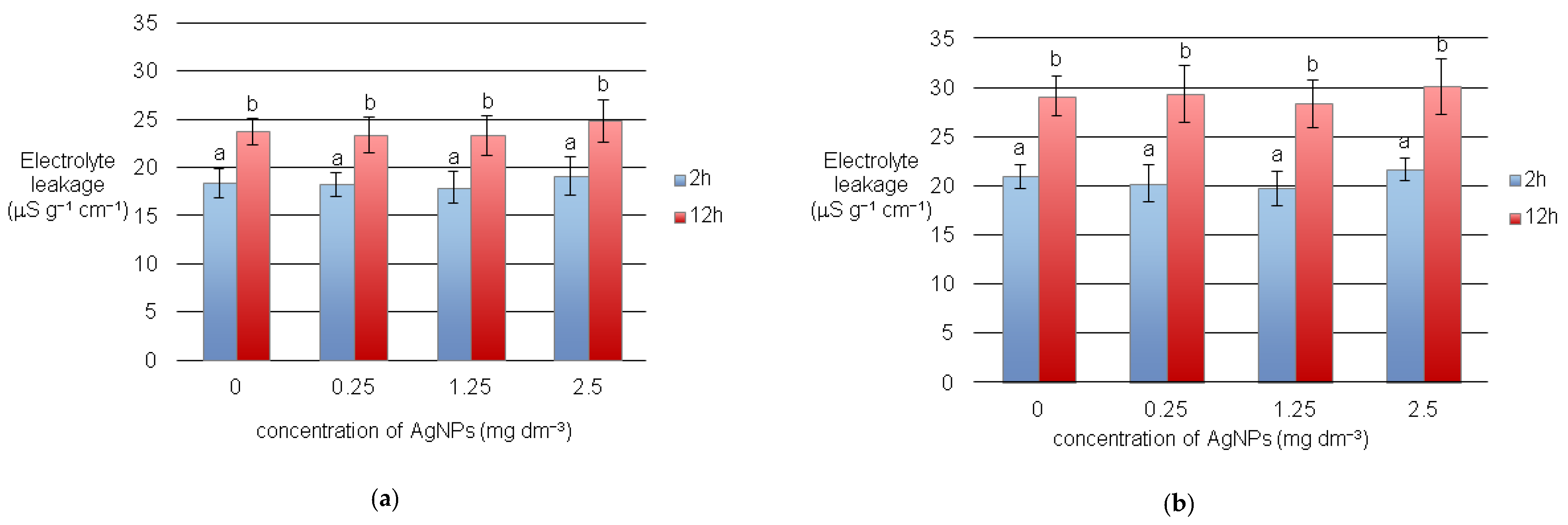
3.1. Electrolyte Leakages and Seed Germination

The EC of the ‘Bali’ seed effusate ranged from 19.75 to 30.05 µS cm1 g1, while that of the ‘Delfina’ cultivar was 17.9–24.8 µS cm1 g1. According to the EC test assessment criteria adopted in the European Union for legume plants and the criteria for peas developed by Kolasińska et al. [31], a result <25 µS cm1 g1 indicates that the seeds are suitable for sowing even in adverse environmental conditions. Seeds whose effusates exhibit EC in the range of 25–29 µS cm1 g1 are also suitable for sowing, but in adverse environmental conditions, there is a risk of poor growth. An EC test result of 30–43 µS cm1 g1 indicates that the seeds are not suitable for sowing in adverse environmental conditions, and at >43 µS cm1 g1 seeds are not suitable for sowing at all. According to this scale, the seeds of both bean cultivars subjected to pre-sowing hydration in Nitragina solution and treated with AgNPs were suitable for sowing, although ‘Bali’ seeds were more sensitive to adverse environmental conditions than ‘Delfina’ seeds. The electrolytes leakage from the seeds of the ‘Bali’ cultivar was greater than from the ‘Delfina’ seeds, both after 2 and 12 h of testing (Figure 1).
The results of the EC test showed that the AgNPs treatment did not significantly affect the biological quality of seeds of either bean cultivar. Electrolytes leakage from seeds treated with AgNPs did not differ significantly from control seeds (Figure 1). The efflux of electrolytes from plant cells increases with the permeability of cell membranes, e.g., due to lipid peroxidation taking place during oxidative stress [32]. Previous research indicates that AgNPs in sufficiently high concentrations induce oxidative stress in plant cells [33,34].
In the present study, however, the EC test results clearly show that AgNPs used at concentrations of 0.25, 1.25 and 2.5 mg dm3 for pre-sowing seed treatment do not affect the permeability of cell membranes for electrolytes in either of the bean cultivars. It can therefore be suggested that AgNPs at these concentrations do not induce oxidative stress in the cells of the seed embryos of this plant. On the other hand, the EC test results suggest that AgNPs at low concentrations (0.25 and 1.25 mg dm−3) slightly improve the vigour of bean seeds. Electrolyte leakage from seeds treated with AgNPs at these concentrations was somewhat lower than from the control seeds, although the differences were not statistically significant (Figure 1).
The germination test results showed that AgNPs treatment had a positive effect on the germination rate, particularly at chill temperature (Table 3).
The germination rate of the ‘Bali’ and ‘Delfina’ bean cultivars at normal temperature, i.e., optimal temperature for germination of seeds of this species according to ISTA [27], was higher than in the control by 7.5–11.4% and 4.8–12.4%, respectively. At chill temperature, the effectiveness of AgNPs was greater than at normal temperature. The germination rate of seeds treated with AgNPs was 9.0–15.7% (for ‘Bali’) and 6.8–16.0% (for ‘Delfina’): greater than that of control seeds. At normal temperature, there were no significant differences in seed capacity between the AgNPs treatments and the control (Table 3). However, significant differences were noted at chill temperature, except for the seed sample treated with AgNPs at a concentration of 2.5 mg dm3. The germination capacity of seeds treated with AgNPs at 0.25 and 1.25 mg dm3 was greater than that of the control seeds by 9.3% and 9.8%, respectively, for the ‘Bali’ cultivar and by 10.2% and 11.5% for ‘Delfina’.
The rate of seed infection with fungal pathogens was much lower at chill temperature than at normal temperature (Table 3). This was probably an expression of the universal response of microorganisms to suboptimal temperatures. However, pre-treatment of seeds with AgNPs significantly reduced the frequency of mould infection at both normal and chill temperatures. Moulds, which are natural epiphytic microflora of seeds but can also originate in storage, may pose a threat to the growing plant organism during seed germination [35]. Thus, in this context, the disinfectant effect observed is a positive and expected phenomenon due to the well-known and well-researched antimicrobial properties of AgNPs.
Previous studies have shown that the effect of AgNPs on seed germination and seedling growth can be positive or negative, depending on the size, properties and concentration of the nanoparticles, the test plant and the application method [4]. The stimulating effect of pre-sowing treatment of seeds with AgNPs at concentrations of 20 and 50 mg dm3 for 2 h on the germination rate of Pennisetum glaucum seeds has been demonstrated by Parveen and Rao [15]. These results are consistent with the results presented here, although the concentration of AgNPs used to treat the bean seeds was more than 10 times lower. Parveen and Rao [15], however, noted an adverse effect of this treatment on the growth rate of Pennisetum glaucum seedlings, whereas its effect on seedling growth in both tested bean cultivars was favourable. On the other hand, the results of the seed germination test were consistent with the findings of Hojjat et al. [36] and Michalek et al. [7], according to which AgNPs at very low concentrations (≤10 mg dm3) significantly enhance seed germination potential, mean germination time, seed germination index, seed vigour index, seedling fresh weight and dry weight of fenugreek and maize. A positive effect of low concentrations of AgNPs on seed germination under optimal and stress conditions for this process has also been reported by Rajjou et al. [37], Chen and Arora [38], Panyuta et al. [39] and Mahakham et al. [34]. The mechanism of the positive effect of AgNPs on seed germination has not been adequately explained. The results of previous studies indicate at least three mechanisms. The first of these probably occurs at the level of gene expression regulation. Ag nanoparticles activate genes coding for aquaporins, transmembrane proteins that facilitate water and gas transport and the flow of reactive oxygen species (ROS) through biological membranes [40]. The second occurs at the level of activation of gibberellin biosynthesis and activation of hydrolytic enzymes, i.e., α-amylase, proteases and lipases, by gibberellins [4]. These enzymes catalyse hydrolysis of high-molecular-weight organic compounds to low-molecular-weight compounds, which are respiratory substrates for embryo cells. In addition, the increase in the concentration of monosaccharides and other low-molecular-weight compounds causes an increase in the osmotic potential of cells, which further accelerates the imbibition of storage materials and other seed structures. Activation of α-amylase and increased water uptake by germinating rice seeds primed in an AgNPs solution has been noted by Mahakham et al. [34]. The third mechanism of action of AgNPs may involve a change in the relationship between the level of oxidative stress and the antioxidant system activity of the embryo cells [34,41]. AgNPs stimulate the generation of reactive oxygen species (ROS) (hydrogen peroxide and hydroxyl radical), which leads to the activation of antioxidant enzymes (catalase and superoxide dismutase) in cells. These changes condition the appropriate level of ROS in embryo cells, constituting the so called ‘Oxidative window’ needed to trigger signal pathways that initiate seed germination. As a result of the direct action of AgNPs and/or physical and biochemical activation of the seed germination phase by AgNPs, the physiological phase of this process, i.e., seedling differentiation and growth, is accelerated as well. Wang et al. [22], Mahakham et al. [34] and Michałek et al. [7] note a stimulatory effect of AgNPs on radicle elongation and an increase in the biomass of seedlings of Arabidopsis, poplar, rice and maize.

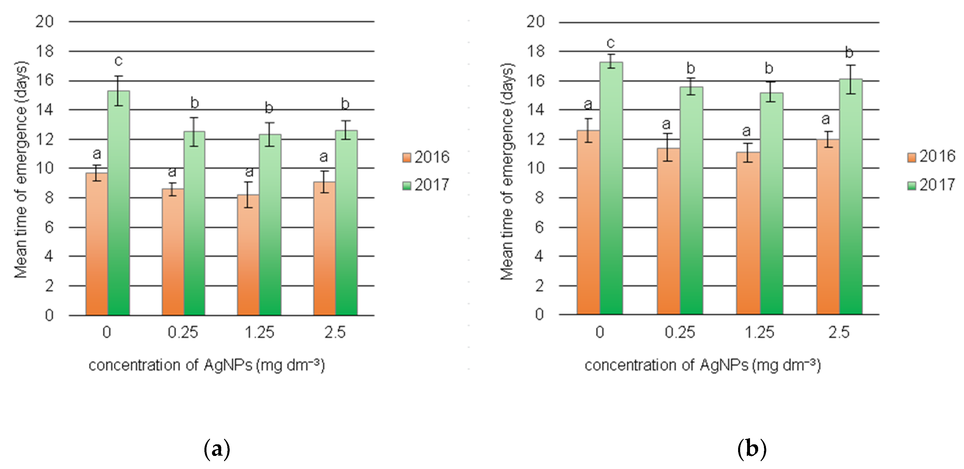
3.2. Dynamics of Field Emergence of Seedlings

Previous research has shown a positive correlation between seed vigour and emergence rate in both laboratory and field conditions [31].
A beneficial effect of treatment of green beans seeds with AgNPs on the field emergence rate was noted in our experiment (Figure 2).
The average time of emergence of seedlings from control seeds of the ‘Bali’ cultivar was about 2–3 days longer than for the ‘Delfina’ cultivar. In 2017, when the average temperature was lower than in 2016 and there were cold periods lasting several days, emergence of both bean varieties was prolonged by about 4–5 days. After seed treatment with AgNPs, emergence was faster than in the case of control seeds in both years of the study. In 2016, the emergence period of seedlings of both bean cultivars was reduced by an average of one day, and in 2017 by about 3 days (‘Delfina’) and about 2 days (‘Bali’). The results indicate that pre-sowing treatment of bean seeds with AgNPs is more effective in the long term than hydration in Nitragina solutions, especially in cold stress conditions. These results are of great practical importance, as fast and uniform emergence ensures uniform plant growth and development, which in turn ensures greater yield and enables mechanical harvesting without losses.

3.3. Biometric and Physiological Parameters of Seedlings

Pre-sowing seed treatment with AgNPs positively affected the growth and development of seedlings in field conditions (Table 4) and their photosynthetic activity (Table 5).
The height and the fresh and dry weight of the shoots of seedlings of both bean cultivars obtained from seeds treated with AgNPs were usually significantly greater than in the control seedlings, except for samples treated with 2.5 mg dm−3 AgNPs (Table 4). This stimulating effect of AgNPs on the biometric parameters of bean seedlings (height, fresh and dry weight of shoots) was noted in both 2016 and 2017. In 2017, when there were cold periods and the average temperature in May was about 3 °C lower than in May 2016 (Table 2), the growth rate of seedlings of both cultivars was lower, irrespective of the pre-sowing seed treatment. However, at chill temperatures (2017) the effect of AgNPs on seedling growth was stronger than at normal temperatures (2016), with a particularly positive effect noted for AgNPs at a concentration of 1.25 mg dm−3 (Table 4). The differences noted at normal and chill temperature were slightly greater in the ‘Delfina’ cultivar than in ‘Bali’, which confirms that the plant genotype plays a key role in the response to environmental factors.
The highest concentration of AgNPs (2.5 mg dm−3) used in our study had little effect on the seedling height of both bean cultivars, and the differences observed in relation to the control plants were not statistically significant in most cases. In contrast, the fresh and dry weight of seedlings in the sample treated with AgNPs at this concentration was usually lower compared to the control seedlings.
Similar relationships between temperature conditions during growth and AgNPs levels were found for the physiological parameters of the bean seedlings. Pre-sowing treatment of both cultivars with low concentrations of AgNPs (0.25, 1.25 mg dm−3) had a positive long-term effect on leaf chlorophyll content, stomatal conductance and net photosynthesis rate (Table 5). In both bean varieties, the net photosynthesis rate and stomatal conductance were lower at chill temperatures (2017) than at normal temperatures (2016), and AgNPs treatment generally improved these parameters to a greater extent at chill temperatures. Seed treatment with 2.5 mg dm−3 AgNPs led to a reduction in photosynthetic pigments in both bean cultivars in both years of the field experiment and in the photosynthesis rate at normal temperature (Table 5).
The physiological parameters of the bean seedlings correspond to their biometric indices, such as height and fresh weight of shoots (Table 4 and Table 5).
A stimulating effect of application of AgNPs at low concentrations (0.01–1 mg dm−3) on the growth, fresh weight and physiological activity of plants has been found in Arabidopsis and poplar, in cucumber, in Eruca sativa, and in rice [22,23,34,42]. A field experiment by Ashrafi et al. [43] showed a positive effect of nanosilver application on such agronomic features of soybean as chlorophyll content, nitrogen content in seeds, and seed yield. In contrast, a 10-fold higher level of AgNPs (10 mg dm−3) inhibited the growth of alfalfa plants (both without a bacterial symbiont and in conditions of symbiosis with Sinorhizobium meliloti) [44]. Comparable effects, even higher concentrations (12.5–100 mg dm−3), were induced by ZnONPs nanoparticles.
Even higher concentrations of AgNPs (1 and 3 M) have led to a drastic decrease in the growth parameters of pea (Pisum sativum) seedlings, content of photosynthetic pigments and chlorophyll fluorescence intensity, which was correlated with Ag accumulation in the roots and stems [45]. The examples cited, as well as the results of this work, support the thesis that the effects of AgNPs depend primarily on their concentration, the test plant and the application method, which determines the scope of this factor’s effect on the plant.
The study showed that AgNPs exerted a stronger growth-promoting effect in cold conditions than at the optimum temperature for bean growth, suggesting that metallic nanoparticles may modulate the plant’s tolerance to adverse abiotic factors. This type of report is not isolated; for example, application of silver nanoparticles has been found quite effective in improving resistance to salinity during germination of fennel and cumin [46,47]. Exposure to AgNPs has been reported to mitigate the adverse effects of salt stress and improve the germination, root length and fresh and dry seedling weight of tomato under NaCl stress [18]. Despite numerous studies on the promotion of plant growth and tolerance induced by nanomaterials, the underlying mechanism is still little understood. There are indications that a key role in this phenomenon is played by increased activity of enzymes, including antioxidants, which protect the interior of the cell from the effects of oxidative stress [48]. In addition, nanomaterials can regulate the expression of genes involved in response to environmental stress. In a microarray analysis, a number of genes were up-regulated or down-regulated by the application of AgNPs in Arabidopsis [21]. A major portion of up-regulated genes are associated with the response to metals and oxidative stress, whereas down-regulated genes are associated with the response to pathogens and hormonal stimuli. Interestingly, some of these genes overlap with genes induced by Ag ions, suggesting that the response to AgNPs involves two metabolic programmes, i.e., the AgNP-specific and Ag+1-induced programme. The AgNP-specific response includes genes belonging to the thalianol biosynthetic pathway, suggesting that this type of response is directly involved in the plant’s defence mechanisms [21].

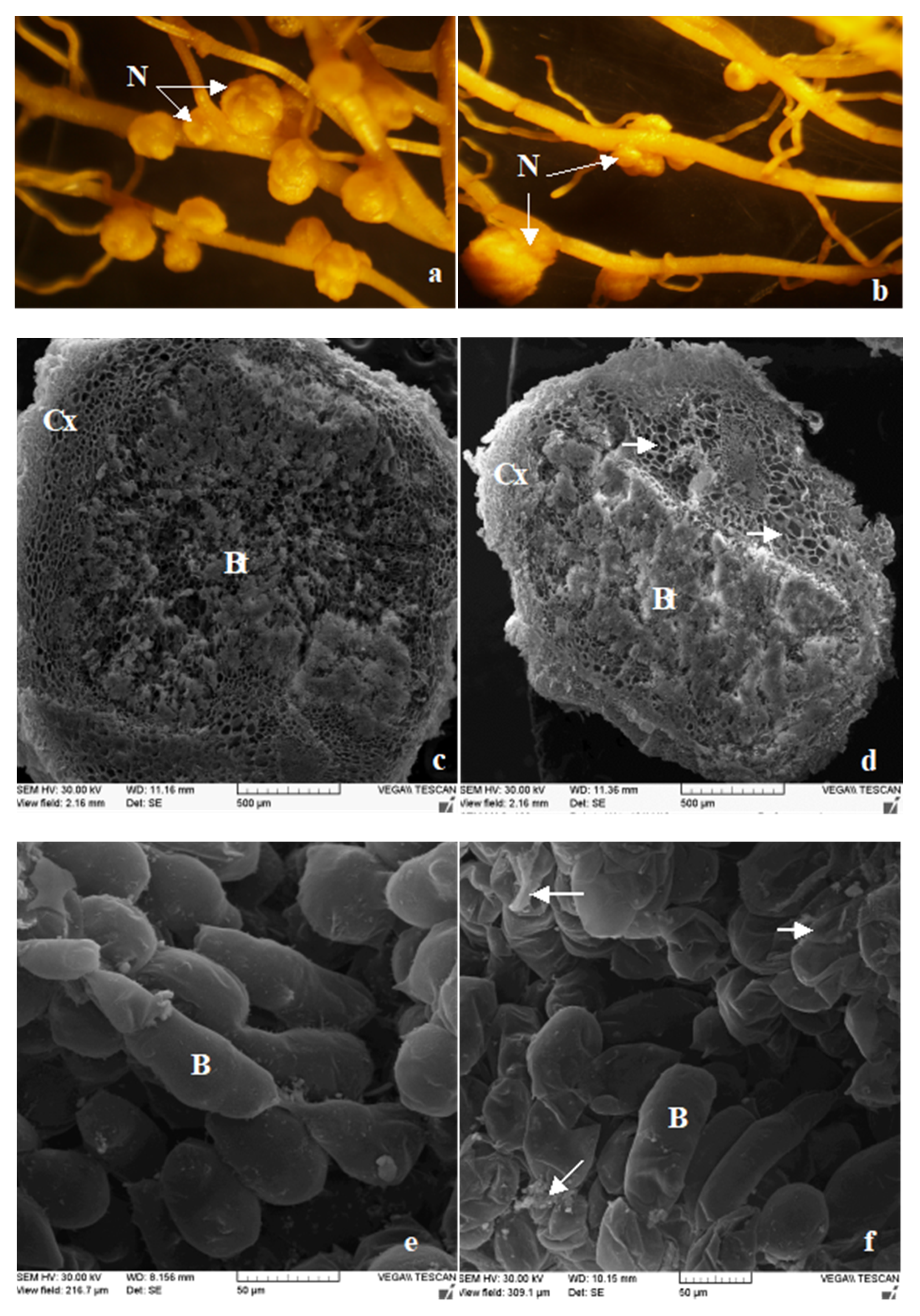
3.4. The Number and Structure of Root Nodules

Pre-sowing treatment of bean seeds with AgNPs also affected the number and structure of root nodules. Root nodules in bean plants begin to differentiate about 12 days after seedling emergence. These are determinate, spherical nodules whose meristem only functions for a few days [49]. Bacteroid tissue does not show differentiation into mature and immature tissue, and the vascular bundles form a closed system. In the anatomical structure of the nodules, we distinguish the outer tissue—the cortex (Cx), with the vascular system, and the inner tissue—the bacteroid tissue, which contains bacteroids surrounded by a peribacteroid membrane and uninfected plant cells. The central part of the nodule is the nitrogen-fixing (Nif) zone (Figure 3).
The results of the study showed that the number of root nodules in the bean cultivars depended on the weather conditions during the growing season, seed preparation for sowing and plant genotype (Table 4). Overall, the average number of root nodules in the ‘Delfina’ cultivar was greater than in ‘Bali’. In both cultivars, there were fewer root nodules in cold conditions, i.e., in 2017, than at normal temperature, i.e., in 2016. Irrespective of weather conditions, the number of root nodules was significantly higher in plants of both bean cultivars obtained from seeds treated with AgNPs before sowing, except for the concentration of 2.5 mg dm−3 (Table 4). The size of the nodules was varied in both the control and test samples. However, the smallest nodules were identified in seedlings obtained from seeds treated with 2.5 mg AgNPs dm−3 (Figure 3b). Observations of root nodules and bacteroids with scanning electron microscopy (SEM) showed no abnormalities or damage in the anatomical structure of the nodules of the control plants or those obtained from seeds treated with AgNPs at 0.25 and 1.25 mg dm−3 (data not shown). The bacteroids also showed no morphological abnormalities (Figure 3e). In contrast, in the small nodules of plants obtained from seeds treated with AgNPs at 2.5 mg·dm−3, smaller or larger areas of uninfected cells were identified by SEM in the bacteroid tissue (Figure 3b,d). In addition, the relatively numerous bacteroids found in these nodules showed morphological deformations (Figure 3f) reminiscent of changes occurring during nodule stress induced senescence [50]. The cells of these bacteroids shrank unevenly, and the peribacteroid membrane began to invaginate.
Thus, at a concentration of just 2.5 mg dm−3 AgNPs reduce infection of bean roots by Rhizobium phaseoli bacteria and adversely affect nodule formation and the bacteroid cells themselves. Similar effects have been observed by Moghaddam at al. [44] in the case of legume-Sinorhizobium symbiosis. The number of nodules in symbiotic plants in the presence of 5 and 10 mg dm−3 AgNPs was significantly decreased, and they were smaller than in the control plants.
In this study, the effectiveness of AgNPs in high concentrations in relation to the analysed microorganisms and test plant was varied. AgNPs were more toxic to epiphytic fungi and Rhizobium than to the bean seedlings. Similarly, high sensitivity of microorganisms to AgNPs has been noted by other researchers using a variety of research protocols. Even at low concentrations AgNPs cause a broad spectrum of antimicrobial effects. At a concentration of 1 mg dm−3, they have been found to reduce the activity of agricultural-important microorganisms, such as nitrifying bacteria and S. meliloti isolates [44,51]; at 2–22 mg dm−3 they inhibited symbiotic nitrogen fixers, phosphate solubilizers and biofilm producers [52]; and at 0.8 mg kg−1 soil, they reduced the activity of Rhizobium leguminosarum bacteria [53].
Electron microscopy study has shown that AgNPs are able to physically interact with the cell surface of various bacteria and cause disintegration of the cell wall and membrane [54,55]. This leads to leakage of intracellular substances and induction of cell death. AgNPs activity is strongly dependent on their concentration, size and a zeta potential value [56].
Smaller nanoparticles with a positive zeta potential seem better able to adhere to and interact with the cell surface and penetrate bacteria. As a consequence, this leads to damage of its intracellular structures. Ribosomes may be denatured with inhibition of protein synthesis, as well as translation and transcription can be blocked by the binding with the DNA of the bacterial cell [57].
Protein synthesis has been shown to be altered by treatment with AgNPs and proteomic data have shown an accumulation of immature precursors of membrane proteins resulting in destabilization of the composition of the outer membrane [58]. AgNPs can also interfere with the respiratory chain and reduce energy production [59]. The observed changes may result from the synergistic effects on cellular processes of both AgNPs and silver ions released from them. It is known that, silver ions interact with a number of electron donor groups such as thiols, phosphates, hydroxyls, imidazoles and indoles. AgNPs also damage membranes and cause the release of ROS, with powerful bactericidal activity [56].

4. Conclusions

This study was designed to evaluate the possibility of using AgNPs to improve bean growth and yield, especially in adverse climatic conditions. The research employed a strategy consistent with the principles of precision agriculture, involving the controlled application of AgNPs on bean seeds intended for sowing.
Pre-sowing treatment is believed to enable effective uptake of AgNPs and their accumulation in the seed, which can cause immediate and/or remote in time biological effects. The results of the study support this assumption. Application of AgNPs influenced not only seed germination in the laboratory but also the dynamics of emergence of bean seedlings and their growth and development in field conditions. The biological significance of the effects induced by AgNPs varied depending on the concentration of nanoparticles and on growth conditions. On the other hand, genetic variation between cultivars did not significantly affect the susceptibility of bean seeds and seedlings to AgNPs.
Low concentrations of AgNPs generally promoted the growth and development of bean seedlings. They were particularly effective in suboptimal growth conditions for this plant (chill temperature). On the other hand, the highest AgNPs concentration used in the study had little effect on bean growth, but exerted a strong antimicrobial effect on the microorganisms associated with this plant. The fungicidal properties of AgNPs proved to be beneficial during seed germination in both laboratory and field conditions, while its bactericidal properties reduced rhizobial infection and bacteroid activity in the root nodules, which could have caused the poorer growth and slower development of these seedlings. Thus, the harmful effects of AgNPs observed in this plant may be the combined result of their antimicrobial and phytotoxic effects. The presented research raises important issues associated with the environmental safety of using Ag nanoparticles. Despite promising results regarding the possibility of promoting bean growth, especially in adverse field conditions, it is important to consider the possible interactions of these nanoparticles with soil microorganisms, which, in the case of legumes, may have far-reaching negative economic consequences.

Author Contributions

Conceptualization, R.P. and S.M.; methodology, A.Ś. and A.K.; software, M.A.; validation, M.A. and A.K.; formal analysis, R.P., A.K. and S.M.; investigation, R.P., A.K., S.M. and A.Ś.; resources, M.A. and A.Ś.; data curation, S.M. and A.K.; writing, A.Ś.—original draft preparation; writing—review and editing, A.Ś., A.K. and R.P.; visualization, M.A. and A.K.; supervision, R.P. and A.Ś. All authors have read and agreed to the published version of the manuscript contributed substantially to the work reported.

Funding

This research received no external funding.

Conflicts of Interest

The authors declare no conflict of interest. The funders had no role in the design of the study; in the collection, analyses, or interpretation of data; in the writing of the manuscript, or in the decision to publish the results.

References

  1. El-Tohamy, W.A.; Singer, S.M.; El-Behairy, U.A.; Abou-Hadid, A.F. Effects of low tunnels, plastic much and mineral nutrient treatments on chilling tolerance of snap bean plants. Acta Hortic. 2001, 559, 127–134. [Google Scholar] [CrossRef]
  2. Głowacka, A.; Klikocka, H.; Onuch, J. Content of zinc and iron in common bean seeds (Phaseolus vulgaris L.) in different weed control methods. J. Elem. 2015, 20, 293–303. [Google Scholar]
  3. Abou-Zeid, H.M.; Moustafa, Y. Physiological and cytogenetic responses of wheat and barley to silver nanopriming treatment. Int. J. Appl. Biol. Pharm. Technol. 2014, 5, 265–278. [Google Scholar]
  4. Feregrino-Perez, A.A.; Magaña-López, E.; Guzmán, C.; Esquivel, K. A general overview of the benefits and possible negative effects of the nanotechnology in horticulture. Sci. Hort. 2018, 238, 126–137. [Google Scholar] [CrossRef]
  5. European Commission. Recommendation 2011/696/ EU of 18 October 2011 on the Definition of Nanomaterial; OJEU L 275/38; European Commission Recommendation: Brussel, Belgium, 2011; p. 3. [Google Scholar]
  6. Jo, D.H.; Lee, T.G.; Kim, J.H. Nanotechnology and nanotoxicology in retinopathy. Int. J. Mol. Sci. 2011, 12, 8288–8301. [Google Scholar] [CrossRef] [Green Version]
  7. Michałek, S.; Święciło, A.; Molas, J. Effect of silver nanoparticles and ions on seeds epiphytic microorganisms activity and early stages of sweetcorn development. Przem. Chem. 2018, 97, 1654–1658. [Google Scholar]
  8. Servin, A.; Elmer, W.; Mukherjee, A.; De La Torre Roche, R.; Hamdi, H.; White, J.; Bindraban, P.S.; Dimkpa, C. A review of the use of engineered nanomaterials to suppress plant disease and enhance crop yield. J. Nanopart. Res. 2015, 17, 92. [Google Scholar] [CrossRef]
  9. Zalewska, E.D.; Machowicz-Stefaniak, Z.; Król, E.D. Antifungal activity of nanoparticles against chosen species of the pathogenic fungi towards caraway. Acta Sci. Pol. Hortorum Cultus 2016, 15, 121–137. [Google Scholar]
  10. Almutairi, Z.; Alharbi, A. Effect of silver nanoparticles on seed germination of crop plants. J. Adv. Agric. 2015, 4, 280–285. [Google Scholar] [CrossRef]
  11. Sharma, P.; Bhatt, D.; Zaidi, M.G.; Saradhi, P.P.; Khanna, P.K.; Arora, S. Silver nanoparticle mediated enhancement in growth and antioxidant status of Brassica juncea. Appl. Biochem. Biotechnol. 2012, 167, 2225–2233. [Google Scholar] [CrossRef]
  12. Yin, L.; Colman, B.; McGill, B.; Wright, J.; Bernhardt, E. Effects of silver nanoparticle exposure on germination and early growth of eleven wetland plants. PLoS ONE 2012, 7, e47674. [Google Scholar] [CrossRef] [PubMed] [Green Version]
  13. Salama, H.M.H. Effects of silver nanoparticles in some crop plants, common bean (Phaseolus vulgaris L.) and corn (Zea mays L.). Int. Res. J. Biotech. 2012, 3, 190–197. [Google Scholar]
  14. Savithramma, N.; Ankanna, S.; Bhumi, G. Effect of nanoparticles on seed germination and seedling growth of Boswellia Ovalifoliolata—An endemic and endangered medicinal tree Taxon. Nano Vis. 2012, 2, 61–68. [Google Scholar]
  15. Parveen, A.; Rao, S.O. Effect of nanosilver on seed germination and seedling growth in Pennisetum glaucum. J. Clust. Sci. 2015, 26, 693–701. [Google Scholar] [CrossRef]
  16. Abou-Zeid, H.; Ismail, G. The role of priming with biosynthesized silver nanoparticles in the response of Triticum aestivum L. to salt stress. Egypt. J. Bot. 2018, 58, 73–85. [Google Scholar]
  17. Bhati-Kushwaha, H.; Kaur, A.; Malik, C.P. The synthesis and role of biogenic nanoparticles in overcoming chilling stresses. Ind. J. Plant Sci. 2013, 2, 54–62. [Google Scholar]
  18. Almutairi, Z.M. Influence of silver nano-particles on the salt resistance of tomato (Solanum lycopersicum) during germination. Int. J. Agric. Biol. 2016, 18, 449–457. [Google Scholar] [CrossRef] [Green Version]
  19. Iqbal, M.; Raja, N.; Mashwani, Z.R.; Hussain, M.; Ejaz, M.; Yasmeen, F. Effect of silver nanoparticles on growth of wheat under heat stress. Iran. J. Sci. Technol. Trans. A Sci. 2019, 43, 287. [Google Scholar] [CrossRef]
  20. Kumari, M.; Mukherjee, A.; Chandrasekaran, N. Genotoxicity of silver nanoparticles in Alium cepa. Sci. Total Environ. 2009, 407, 5243–5246. [Google Scholar] [CrossRef]
  21. Kaveh, R.; Li, Y.S.; Ranjbar, S.; Tehrani, R.; Brueck, C.L.; Van Aken, B. Changes in Arabidopsis thaliana gene expression in response to silver nanoparticles and silver ions. Environ. Sci. Technol. 2013, 47, 10637–10644. [Google Scholar] [CrossRef]
  22. Wang, J.; Koo, Y.; Alexander, A.; Yang, Y.; Westerhof, S.; Zhang, Q.B.; Shnoor, J.L.; Colvin, V.L.; Brasm, J.; Alvarez, P.J.J. Phytostimulation of poplars and Arabidopsis exposed to silver nanoparticles and Ag+ at sublethal concentrations. Environ. Sci. Technol. 2013, 47, 5442–5449. [Google Scholar] [CrossRef] [PubMed]
  23. Vannini, C.; Domingo, G.; Onelli, E.; Prinsi, B.; Marsoni, M.; Espen, L.; Bracale, M. Morphological and protreomic responses of Eruca sativa exposed to silver nanparticles or silver nitrate. PLoS ONE 2013, 8, e68752. [Google Scholar] [CrossRef] [Green Version]
  24. Zhang, Y.; Leu, Y.R.; Aitken, R.J.; Riediker, M. Inventory of engineered nanoparticle-containing consumer products available in the Singapore retail market and likelihood of release into the aquatic environment. Int. J. Environ. Res. Public Health 2015, 12, 8717–8743. [Google Scholar] [CrossRef] [PubMed] [Green Version]
  25. Maurer-Jones, M.; Gunsolus, I.; Murphy, C.; Haynes, C. Toxicity of engineered nanoparticles in the environment. Anal. Chem. 2013, 85, 3036–3049. [Google Scholar] [CrossRef] [Green Version]
  26. Pyatenko, A.; Yamaguchi, M.; Suzuki, M. Synthesis of spherical silver nanoparticles with controllable sizes in aqueous solutions. J. Phys. Chem. 2007, 111, 7910–7917. [Google Scholar] [CrossRef]
  27. ISTA International Seed Testing Association. International Rules for Seed Testing; International Seed Testing Association: Bassersdorf, Switzerland, 2014. [Google Scholar]
  28. Maguire, J.D. Speed of germination—Aid in selection and evaluation for seedling emergence and vigor. Crop Sci. 1962, 2, 176–177. [Google Scholar] [CrossRef]
  29. Piekutowska, M.; Szparaga, A. Evaluation of germmination of Soy beans treated with natural environmental friendly extracts depending on the method of their appplication. Annu. Set Environ. Prot. 2018, 20, 375–391. [Google Scholar]
  30. Lichtenthaler, H.K.; Wellburn, A.R. Determinations of total carotenoids and chlorophylls a and b of leaf extracts in different solvents. Biochem. Soc. Trans. 1983, 11, 591–592. [Google Scholar] [CrossRef] [Green Version]
  31. Kolasinska, K.; Szyrmer, J.; Dul, S. Relationship between laboratory seed quality tests and field emergence of common bean seed. Crop Sci. 2000, 40, 470–475. [Google Scholar] [CrossRef]
  32. Campos, P.S.; Quartin, V.; Ramalho, J.C.; Nunes, M.A. Electrolyte leakage and lipid degradation account for cold sensitivity in leaves of Coffea sp. plants. J. Plant Physiol. 2003, 160, 283–292. [Google Scholar] [CrossRef] [Green Version]
  33. Jiang, H.S.; Qiu, X.N.; Li, G.B.; Li, W.; Yin, L.Y. Silver nanoparticles induced accumulation of reactive oxygen species and alteration of antioxidant systems in the aquatic plant Spirodela polyrhiza. Environ. Toxicol. Chem. 2014, 33, 1398–1405. [Google Scholar] [CrossRef] [PubMed]
  34. Mahakham, W.; Sarmah, A.K.; Maensiri, S.; Theerakulpisut, P. Nanopriming technology for enhancing germination and starch metabolism of aged rice seeds using phytosynthesized silver nanoparticles. Sci. Rep. 2017, 7, 1–21. [Google Scholar] [CrossRef] [PubMed]
  35. Yago, J.I.; Roh, J.H.; Bae, S.D.; Yoon, Y.N.; Kim, H.J.; Nam, M.H. The effect of seed-borne mycoflora from sorghum and foxtail millet seeds on germination and disease transmission. Mycobiology 2011, 39, 206–218. [Google Scholar] [CrossRef] [PubMed] [Green Version]
  36. Hojjat, S.S. Impact of silver nanoparticles on germinated fenugreek seed. Int. J. Agric. Crop. Sci. 2015, 8, 627–630. [Google Scholar]
  37. Rajjou, L.; Duval, M.; Gallardo, K.; Catusse, J.; Bally, J.; Job, C.; Job, D. Seed germination and vigor. Annu. Rev. Plant Biol. 2012, 63, 507–533. [Google Scholar] [CrossRef] [Green Version]
  38. Chen, K.; Arora, R. Priming memory invokes seed stress-tolerance. Environ. Exp. Bot. 2013, 94, 33–45. [Google Scholar] [CrossRef]
  39. Panyuta, O.; Belava, V.; Fomaidi, S.; Kalinichenko, O.; Volkogon, M.; Taran, N. The effect of pre-sowing seed treatment with metal nanoparticles on the formation of the defensive reaction of wheat seedlings infected with the eyespot causal agent. Nanoscale Res. Lett. 2016, 11, 92. [Google Scholar] [CrossRef] [Green Version]
  40. Maurel, C.; Boursiac, Y.; Luu, D.T.; Santoni, V.; Shahzad, Z.; Verdoucq, L. Aquaporins in plants. Physiol. Rev. 2015, 95, 1321–1358. [Google Scholar] [CrossRef]
  41. Kibinza, S.; Bazin, J.; Bailly, C.; Farrant, J.M.; Corbineau, F.; El-Maarouf-Bouteau, H. Catalase is a key enzyme in seed recovery from ageing during priming. Plant Sci. 2011, 181, 309–315. [Google Scholar] [CrossRef]
  42. Shams, G.; Ranjbar, M.; Amiri, A. Effect of silver nanoparticles on concentration of silver heavy element and growth indexes in cucumber (Cucumis sativus L. negeen). J. Nanopart. Res. 2013, 15, 1630–1642. [Google Scholar] [CrossRef]
  43. Ashrafi, M.; Sarajuoghi, M.; Mohammadi, K.; Zarei, S. Effect of nanosilver application on agronomic traits of soybean in relation to different fertilizers and weed density in field conditions. Environ. Exper. Biol. 2013, 11, 53–58. [Google Scholar]
  44. Moghaddam, M.N.; Sabzevar, A.H.; Mortazaei, Z. Impact of ZnO and silver nanoparticles on legume-Sinorhizobium symbiosis. Adv. Stud. Biol. 2017, 9, 83–90. [Google Scholar] [CrossRef]
  45. Tripathi, D.K.; Tripathi, A.; Singh, S.; Singh, Y.; Vishwakarma, K.; Yadav, G.; Sharma, S.; Singh, V.K.; Mishra, R.K.; Upadhyay, R.G.; et al. Uptake, accumulation and toxicity of silver nanoparticle in autotrophic plants, and heterotrophic microbes: A concentric review. Front. Microbiol. 2017, 8, 7. [Google Scholar] [CrossRef] [PubMed]
  46. Ekhtiyari, R.; Mohebbi, H.; Mansouri, M. The study of the effects of nano silver technology on salinity tolerance of (Foeniculum vulgare Mill.). Plant Ecosyst. 2011, 7, 55–62. [Google Scholar]
  47. Ekhtiyari, R.; Moraghebi, F. The study of the effects of nano silver technology on salinity tolerance of cumin seed (Cuminum cyminum L.). Plant Ecosyst. 2011, 25, 99–107. [Google Scholar]
  48. Shojaei, T.R.; Salleh, M.A.M.; Tabatabaei, M.; Mobli, H.; Aghbashlo, M.; Rashid, S.A.; Tan, T. Applications of nanotechnology and carbon nanoparticles in agriculture. In Synthesis, Technology and Applications of Carbon Nanomaterials; Suraya, A.R., Raja, N.I.R.O., Mohd, Z.H., Eds.; Elsevier: Amsterdam, The Netherlands, 2019; pp. 247–277. [Google Scholar]
  49. Vance, C.P. Legume symbiotic nitrogen fixation: Agronomic aspects. In The Rhizobiaceae; Spaink, H.P., Kondorosi, A., Hooykaas, P.J.J., Eds.; Kluwer Academic Publishers: Dordrecht, The Netherlands, 1998; pp. 509–530. [Google Scholar]
  50. Pérez Guerra, J.C.; Coussens, G.; De Keyser, A.; De Rycke, R.; De Bodt, S.; Van De Velde, W.; Goormachtig, S.; Holsters, M. Comparison of developmental and stress-induced nodule senescence in Medicago truncatula. Plant Physiol. 2010, 152, 1574–1584. [Google Scholar] [CrossRef] [Green Version]
  51. Liang, Z.H.; Das, A.; Hu, Z.Q. Bacterial response to a shock load of nanosilver in an activated sludge treatment system. Water Res. 2010, 44, 5432–5438. [Google Scholar]
  52. Chavan, S.; Nadanathangam, V. Effects of nanoparticles on plant growth-promoting bacteria in Indian agricultural soil. Agronomy 2019, 9, 140. [Google Scholar] [CrossRef] [Green Version]
  53. Abd-Alla, M.H.; Nafady, N.A.; Khalaf, D.M. Assessment of silver nanoparticles contamination on faba bean Rhizobium leguminosarum bv. ViciaeGlomus aggregatum symbiosis: Implications for induction of autophagy process in root nodule. Agric. Ecosyst. Environ. 2016, 218, 163–177. [Google Scholar] [CrossRef]
  54. Zhang, L.; Wu, L.; Si, Y.; Shu, K. Size-dependent cytotoxicity of silver nanoparticles to Azotobacter vinelandii: Growth inhibition, cell injury, oxidative stress and internalization. PLoS ONE 2018, 13, e0209020. [Google Scholar] [CrossRef] [Green Version]
  55. Michels, C.; Perazzoli, S.; Soares, H.M. Inhibition of an enriched culture of ammonia oxidizing bacteria by two different nanoparticles: Silver and magnetite. Sci. Total Environ. 2017, 586, 995–1002. [Google Scholar] [CrossRef] [PubMed]
  56. Wu, D.; Fan, W.; Kishen, A.; Gutmann, J.L.; Fan, B. Evaluation of the antibacterial efficacy of silver nanoparticles against Enterococcus faecalis biofilm. J. Endod. 2014, 40, 285–290. [Google Scholar] [CrossRef] [PubMed]
  57. Jung, W.K.; Koo, H.C.; Kim, K.W.; Shin, S.; Kim, S.H.; Park, Y.H. Antibacterial activity and mechanism of action of the silver ion in Staphylococcus aureus and Escherichia coli. Appl. Environ. Microbiol. 2008, 74, 2171–2178. [Google Scholar] [CrossRef] [PubMed] [Green Version]
  58. Mirzajani, F.; Ghassempour, A.; Aliahmadi, A.; Esmaeili, M.A. Antibacterial effect of silver nanoparticles on Staphylococcus aureus. Res. Microbiol. 2011, 162, 542–549. [Google Scholar] [CrossRef]
  59. Durán, N.; Durán, M.; de Jesus, M.B.; Seabra, A.B.; Fávaro, W.J.; Nakazato, G. Silver nanoparticles: A new view on mechanistic aspects on antimicrobial activity. Nanomedicine 2016, 12, 789–799. [Google Scholar] [CrossRef]
Figure 1. Electrolytes leakage from green bean seeds treated with AgNPs. Different letters in graphs denote significant differences between treatments (Tukey test, p < 0.05). The figure shows the average values ± SD (n = 10) for the electrolytes leakage for: (a) ‘Delfina’ seeds, (b) ‘Bali’ seeds.
Agriculture 10 00312 g001
Figure 2. The field emergence rate of green beans seedlings obtained from seeds treated with AgNPs. Different letters in graphs denote significant differences between treatments (Tukey test, p < 0.05). The figure shows the average values ± SD for the time of emergence for: (a) ‘Delfina’ seedlings, (b) ‘Bali’ seedlings.
Agriculture 10 00312 g002
Figure 3. Nodule roots (a,b), nodule cross sections (c,d) and bacteroids from Nif region of nodules (e,f) of green bean seedlings obtained from seeds treated with AgNPs in concentration of 1.25 mg dm−3 (a,c,e) and 2.5 mg dm−3 (b,d,f). Explanation: N—nodules, Cx—primary cortex, Nif—region Nif (N2-fixing region of nodule), B—bacteroid; arrows indices big and small nodules (a,b), the not infection cell layers (d) and morphologically deformed of bacteroids (f).
Agriculture 10 00312 g003
Table 1. Soil conditions prior to the experiment.
YearpHC-org%N-NH4N-NO3PKCaMg
mg kg−1 Air Dry Mass
20166.82.4159177164725
20177.12.21912216378429
Table 2. Mean of air temperature and sum rainfall in the early stages of green bean vegetation.
MonthYearTemperature (°C)Rainfall (mm)The Occurrence of Chill Period
May201615.632.7No
201712.446.2Yes *
June201617.657.3No
201718.212.4No
* There were periods of chill weather lasting several days; average temperature between 10 and 12 May was 10.5 °C and between 13 and 16 May was 12 °C. At night the temperatures were lower than during the day (data from the Institute of Meteorology and Water Management for the Zamość-Mokre meteorological station, the station closest to the site of the field experiment).
Table 3. The germination characteristics of seeds treated with AgNPs.
CultivarTemperatureTreatment
ControlAgNPs (mg dm−3)
0.251.252.5
Germination rate (%)
BaliNormal61.2 a68.7 b72.6 c69.4 b
Chill29.6 a40.1 b45.3 c38.6 b
DelfinaNormal67.8 a75.3 b80.2 c72.6 b
Chill33.5 a49.5 b45.9 b40.3 c
Germination capacity (%)
BaliNormal91.6 a90.9 a92.2 a90.5 a
Chill59.8 a69.1 b69.6 b62.4 a
DelfinaNormal95.2 a94.2 a95,5 a93.4 b
Chill61.2 a71.4 b72.7 b64.6 a
Percentage of fungus infections
BaliNormal10.4 a6.7 b6.1 b3.6 c
Chill3.6 a3.1 a2.5 b1.7 c
DelfinaNormal7.4 a5.7 b3.6 c2.3 d
Chill2.2 a1.6 b1.5 b0.5 c
Different letters in line denote significant differences between treatments (Tukey test, p < 0.05).
Table 4. Biometrical parameters of green beans seedlings.
CultivarYearTreatments
ControlAgNPs (mg dm−3)
0.251.252.5
Height of plants (cm)
Bali201612.6 a13.6 b14.4 c13.3 b
20179.7 a12.2 b11.8 b10.1 a
Delfina201615.2 a17.4 b18.1 c15.3 a
201713.6 a16.7 b16.0 b13.9 a
Fresh mass of shoots (g plant−1)
Bali20169.5 a9.9 a11.2 b9.35 a
20177.9 a8.85 b9.9 c8.1 a
Delfina201611.8 a13.5 b14.1 b9.2 d
20178.6 a11.3 b11.5 b8.6 a
Dry mass of shoots (g plant−1)
Bali20160.77 a
(8.1) *
0.81 a
(8.2)
0.92 b
(8.2)
0.67 c
(7.2)
20170.73 a
(9.2)
0.79 b
(8.9)
0.85 c
(8.6)
0.57 d
(7.0)
Delfina20161.04 a
(8.8)
1.17 b
(8.7)
1.21 b
(8.6)
0.78 c
(8.5)
20170.81 a
(9.4)
0.95 b
(8.4)
0.98 b
(8.5)
0.66 c
(7.7)
Number of root nodules per plant
Bali201620.4 a21.2 b21.8 b18.6 c
201716.3 a18.2 b17.5 c12.3 d
Delfina201624.7 a27.6 b28.2 b23.7 a
201719.5 a22.8 b22.9 b15.7 c
Different letters in lines denote significant differences between treatments (Tukey test, p < 0.05); * In parentheses the percentage of shoot fresh mass.
Table 5. Physiological parameters of green beans seedlings.
CultivarYearTreatments
ControlAgNPs (mg dm−3)
0.251.252.5
Leaf chlorophyll (a + b) content (mg g−1 f.w.)
Bali20161.99 a2.04 a2.01 a1.97 a
20172.02 a2.16 b2.19 b1.94 b
Delfina20162.21 a2.22 a2.25 a2.09 b
20172.07 a2.34 b2.39 b2.06 a
Stomata conductance (mol H2O m−2 s−1)
Bali20160.75 a0.76 a0.83 a0.84 a
20170.62 a0.73 b0.77 b0.71 b
Delfina20161.03 a1.02 a1.04 a0.98 a
20170.74 a0.86b0.86 b0.80 b
Net photosynthesis intensity (µmol CO2 m−2 s−1)
Bali201612.45 a13.51 a14.85 b12.06 a
201711.07 a12.96 b13.18 b11.22 a
Delfina201613.68 a14.38 b15.02 c13.56 a
201711.89 a13.14 b13.77 c12.17 a
Different letters in lines denote significant differences between treatments (Tukey test, p < 0.05).

