Article

Isolation, Identification and Characterization of Rhizobacteria Strains for Biological Control of Bacterial Wilt (Ralstonia solanacearum) of Eggplant in China

1
State Key Laboratory of Rice Biology, Ministry of Agriculture, Key Lab of Molecular Biology of Crop Pathogens and Insects, Institute of Biotechnology, Zhejiang University, Hangzhou 310058, China
2
Plant Protection, Agriculture Directorate, Maysan Province, AL-amarah 62001, Iraq
3
Plant Protection, College of Agriculture, University of Misan, AL-amarah 62001, Iraq
*
Author to whom correspondence should be addressed.
Agriculture 2020, 10(2), 37; https://doi.org/10.3390/agriculture10020037
Submission received: 9 December 2019 / Revised: 3 February 2020 / Accepted: 4 February 2020 / Published: 6 February 2020

Abstract

:
Bacterial wilt of eggplant is the most destructive disease caused by Ralstonia solanacearum throughout the world. Eleven bacterial strains with high antagonistic activity were obtained from 245 rhizobacteria. Based on analysis of morphology, 16S rRNA sequences, fatty acid profiles, gyrA and rpoB genes, they were identified as Pseudomonas putida (IMA3), Paenibacillus polymyxa (IMA5), Bacillus cereus (IMA4, IMA7 and IMA11) and the “operational group Bacillus amyloliquefaciens” (IMA1, IMA2, IMA6, IMA8, IMA9 and IMA10). The lipopeptide compounds produced by each strain also were determined. The biocontrol tests demonstrated that co-inoculation by strain IMA5 and the pathogen gave the greatest biocontrol efficiency of 87.0% and 69.2% 30 and 40 days after co-inoculation, respectively. Plant growth promotion tests revealed that IMA5 markedly promoted eggplant growth, enhancing aboveground seedling length and biomass by 60.8% and by 107.6% and underground root length and biomass by 33.0% and 69.2%, respectively. Hence, strain IMA5 could be considered for developing potential biocontrol agents and for promoting plant growth characteristics, to aid the management of the pathogen R. solanacearum in eggplants.

1. Introduction

Eggplant (Solanum melongena), also called aubergine or brinjal, is an economically important vegetable crop that is grown around the world and can provide significant nutritive benefits thanks to its abundance of vitamins, phenolics and antioxidants [1]. In addition, eggplant has potential pharmaceutical uses due to high antioxidant activity in removal of free radicals and can improve memory deficits caused by diabetes [2]. Zhejiang province is one of the principal centres of eggplant production in China and its cultivated area reached 26,666 ha in 2018 (data from the Zhejiang provincial department of agriculture). However, bacterial wilt of eggplant, caused by Ralstonia solanacearum, is one of the most severe diseases in eggplant, thus posing a substantial threat to its production and food security.
R. solanacearum, the causal agent of bacterial wilt disease, ranks among the most devastating pathogens in solanaceous crops [3]. This pathogen has a wide host range of 200 plant species belonging to more than 50 families [4,5], including the economically important eggplant crop. Due to the wide host range and highly diverse nature of the pathogen, no successful control method exists for bacterial wilt disease [6,7]. Numerous studies have been devoted to bacteria showing a capacity to control R. solanacearum either by producing antibiotics or bacteriocins, which inhibit the growth of the pathogen within the rhizosphere or by inducing host-plant resistance. These bacteria are distributed among different genera and species, including Pseudomonas spp., Paenibacillus spp., Bacillus spp., Erwinia spp. and the avirulent mutants 8224PC and 8103PC of R. solanacearum [8,9,10]. Antagonistic bacteria produce antimicrobial compounds, including a well-known class of lipopeptides, to control plant pathogens. In Bacillus spp., prominent among these are surfactins, iturins, polymyxins, fengycins, kurstakins and bacitracins [11], while in P. polymyxa, antibiotics are known to include two types. One type, consisting of polymyxin, polypeptin, gavaserin, saltavalin, and jolipeptin, is only active against bacteria, and a second type, made up of gatavalin and fusaricidin, against fungi, Gram-positive bacteria and actinomycetes [12]. However, due to the complex soil environment, biocontrol disease efficacy usually depends on local edaphic adaptation of the biocontrol agents, while poor competition ability and poor edaphic adaptation of the introduced strains lead to limited success only. Therefore, it is important to screen the antagonistic strains adapting to the local soil environment as biocontrol agents.
In addition, some studies showed that plant growth-promoting rhizobacteria (PGPR) have a suppressive effect on R. solanacearum [13,14]. PGPR reduces this disease by various modes of action, such as antagonism by producing antibiotics, chelating iron, competition for space and nutrients and induction of systemic resistance [15]. However, the variations in the composition of antagonistic compounds might be primarily due to differences among different species or strains, leading to diversely antagonistic activity against pathogens [16,17]. While in order to achieve effective disease control, it is especially necessary to screen biocontrol or PGPR strains antagonistic to specific plant pathogens.
Thus, the main purpose of this study was to isolate, identify and characterize the bacterial strains with strong biocontrol potentials against R. solanacearum from contiguous eggplant-growing fields, and to determine their biocontrol efficacy for controlling bacterial wilt of eggplant.

2. Materials and Methods

2.1. Sample Collection and Bacterial Isolation

The soil samples were collected from the rhizospheres of the eggplant plants in different fields in Hangzhou city, Zhejiang province, China, on June 20, 2017. In these fields, eggplant plants had been growing in fields that were heavily infected by R. solanacearum for many years. A total of ten samples collected from ten fields were taken to the laboratory. The bacterial strains were isolated from each serially diluted soil sample with sterile distilled water on Lysogeny broth (LB) agar (yeast extract 5 g, tryptone 10 g, NaCl 10 g, agar 15 g, pH 7.0–7.5). The different colonies were chosen on the bases of colony characteristics such as shape, colour, size, regular or irregular and convex or flat. Subsequently, each colony was purified at least three times and was stored in 20% glycerol. The bacterial isolates were deposited in the Culture Collection of Biotechnology Institute, Zhejiang University, Zhejiang Province, China.

2.2. In Vitro Assessment of Antagonistic Activity of Bacterial Strains against R. solanacearum

The antagonistic activity of all isolated bacterial strains was determined by using an agar diffusion method [17]. The LB Agar plates containing bacterial cells of R. solanacearum YY06 (a highly aggressive strain provided by Plant Pathology Department, Zhejiang University, China) cultured in a WY-211B rotary shaker at 180 rpm at 30 °C for 12 h were prepared. A sterile filter paper disc (5 mm) containing cell suspension (107 cells/mL) of a test strain cultured under the same condition above for 12 h was placed into the centre of a LB agar plate. Inhibition zone diameters around filter papers were measured after incubation at 30 °C for 48 h. A disc containing sterile water was used as control. Three replicate plates per treatment were inoculated for each strain. The strains with strong antagonistic activity were chosen for identification.

2.3. Identification of Bacterial Strains

To identify the bacterial strains chosen, they were cultured on LB at 30 °C for 20–24 h and characterized as described by [17].
To analyse the I6S ribosomal RNA (rRNA) genes, 16S rRNA genes of the strains were amplified in an automated thermal cycler (Eppendorf AG, Germany) with primer pairs 27F and 1492R [17]. The amplified products were observed on a 1% agarose gel under an ultraviolet transilluminator (GenoSens 1850, Clinx Science Instruments Co., Ltd., China) after the ethidium bromide staining [5,18], and sent to the Sangon Biotech Company Limited (Shanghai, China) using the Sanger method for sequencing of both strands on automated DNA sequencer (ABI 3730xl, Applied Biosystems, USA) The resulted sequence was edited with the Bioedit 7.19, aligned with the Clustalx1.83, and then compared with others in the GenBank database using BLAST for searching the most similar sequences. The sequences of the representative strains were submitted to the GenBank database and accession numbers were obtained. Phylogenetic trees were generated using the neighbour-joining method in the MEGA 7.0 program [19]. Bootstrap replication (1000) was used as a statistical analysis for the nodes in the phylogenetic trees.
All bacterial strains for whole-cell fatty acid methyl ester (FAME) analysis were grown on tryptic soy agar (TSA) plates at 28 °C for 20 h as described by [20]. Methanolic NaOH solutions were added to the bacterial cells for heat treatment. After cooling, fatty acids were extracted with hexane after adding hydrochloric acid. The fatty acid composition was analysed with the Sherlock system following the protocol of the Microbial Identification System with the TSBA 6 6.0 library. The similarity index for each strain was generated based on fatty acid data.

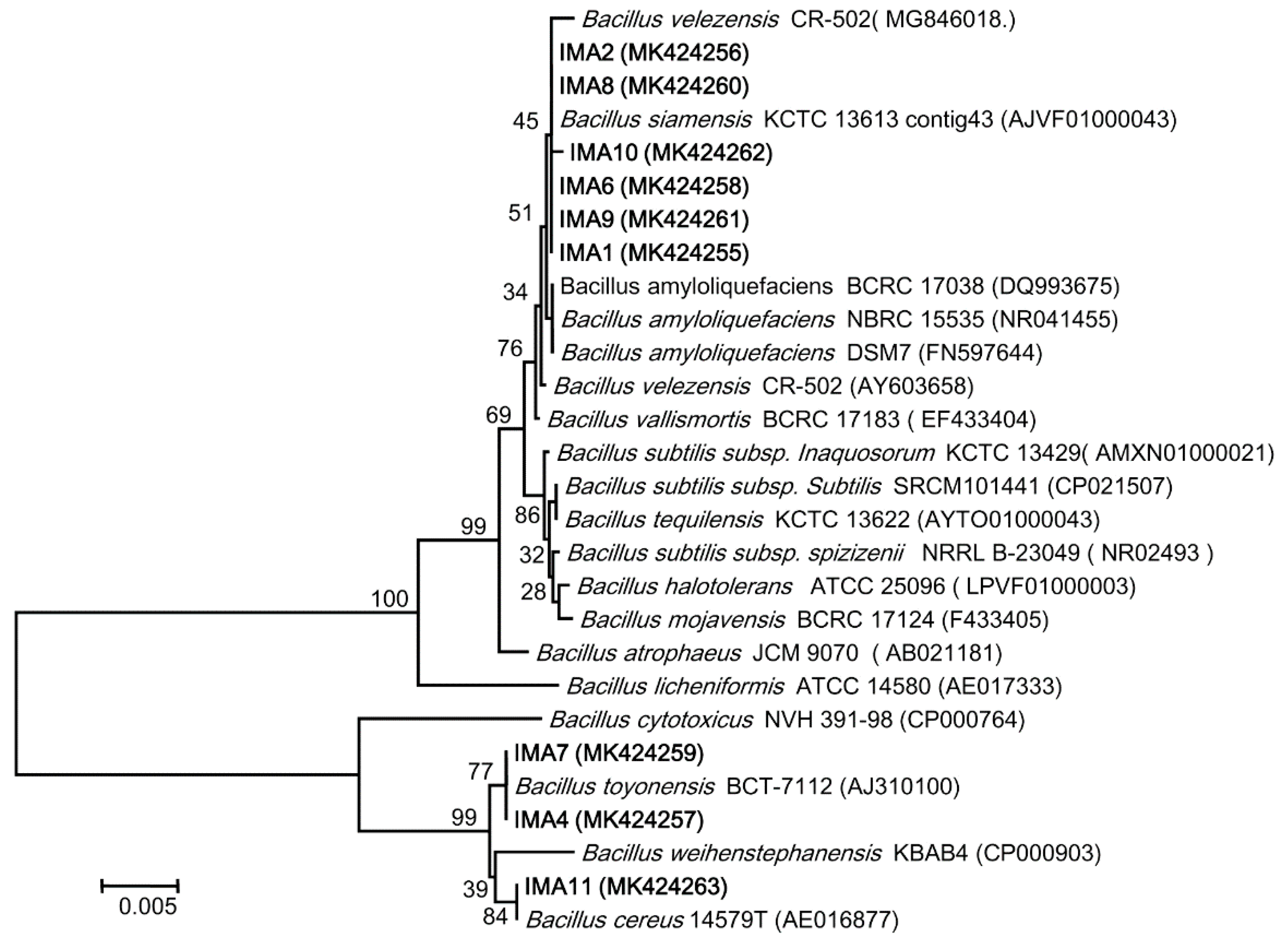
2.4. Phylogenetic Analyses of gyrA and rpoB Gene Sequences

To delineate the species boundaries among species being closely related in the same genus, phylogenetic analysis was conducted by using the gyrA and rpoB genes. The gyrA gene was amplified using the primer pair gyrA-f (5′-CAGTCAGGAAATGCGTACGTCCTT-3′) and gyrA-r (5′-CAAGGTAATGCTCCAGGCATTGCT-′3) [21], while the rpoB gene was amplified using the primer pair rpoB-f (5′-AGGTCAACTAGTTCAGTATGGAC-3′) and rpoB-r (5′-AAGAACCGTAACCGGCAACTT-3′) [22]. Phylogenetic trees were constructed as described above.

2.5. Lipopeptides Detection by MALDI-TOF MS Analysis

To identify the lipopeptides compounds produced by antagonistic strains, matrix-assisted laser desorption ionization-time of flight mass spectrometry (MALDI-TOF MS) was used to detect and characterise lipopeptides, as described by [23]. Individual colonies grown on LB agar plates at 30 °C for 72 h were carefully suspended in Eppendorf tubes containing a matrix solution (10 mg/mL α-cyano-4-hydroxycinnamic acid (Bruker Daltonik) in 70% water, 30% acetonitrile (Wako Pure Chemical Industries, Osaka, Japan) and 0.1% trifluoroacetic acid. The sample was homogenised and then centrifuged at 5000 rpm. For classical analysis, 1 µL of the sample solution was spotted onto a MALDI-TOF MTP 384 target plate (Bruker ultra flextreme instrument) and let dry before analysis.
MALDI-TOF mass spectra were recorded using an ultraflextreme instrument MALDI-TOF (Bruker, Bremen, Germany) equipped with a smart beam laser. Samples were analysed using an accelerating voltage of 25 kV and matrix suppression in deflex-ion mode at m/z 750. The laser power was set to just above the threshold of ionization (around 35%). Spectra were acquired in reflector positive mode in the range from 800 to 3000 Da. Each spectrum was the result of 1000 laser shots per m/z segment per sample delivered in 10 sets of 50 shots distributed in three different locations on the surface of the matrix spot. Spectral data were investigated for the presence of lipopeptides.

2.6. Greenhouse Experiment of Biocontrol Effect of Selected Bacteria Strain on Severity Index of Eggplant Bacterial Wilt and Plant Biomass

Based on the results of the bioactive species and lipopeptides identification, the representative strains were used for in vivo assessment for the suppression of eggplant wilt. The cells suspension (about 108 CFU/mL) of the test strains and R. solanacearum were prepared in LB media on a rotary shaker for 130 rpm at 30 °C for 24 h. A susceptible variety of eggplant plants (cv. Zheqie-3, a eggplant F1 hybrid) obtained from the Vegetable Institution, Hangzhou Academy of Agricultural Sciences were used for inoculation assay. The seeds were sterilized with 2% sodium hypochlorite solution for 2 min, rinsed three times with sterile water and submerged in the cell suspensions of the test strains for eight hours and then air-dried in a laminar flow hood. Subsequently, they were placed onto the surfaces of the wet sterilized filter papers and incubated at room temperature for five days. The germinated seeds were sowed into the pots containing non-sterilized mixing soil (peat:vermiculite:farmyard soil in a 2:1:1 ratio) and pots were moved to the greenhouse with a relative humidity level of 70%–80% and temperatures of 25–30 °C. When plants grew at the four-leaf stage, 10 mL of cell suspension of each test stain were poured into the soil around the plants. After growing for a week, eggplant seedlings were removed out from the pots, rinsed with tap water and then their roots were immersed in the cell suspension of R. solanacearum for one hour after their root tips were excised. Subsequently, they were transplanted to the same pots and incubated under the same condition above. The treatment for the roots immersed in sterile water was used as the control. Thirty plants (n = 30) were used for each treatment and three independent replicates were performed for each treatment. The seedlings were observed daily and symptoms and their changes were recorded. The disease incidence, disease severity and control efficacy were assessed 30 and 40 days after inoculation, respectively. The disease assessment was carried out for each plant using a scale of 0–5, where 0 = no symptoms, 1 = one leaf partially wilted, 2 = two or three leaves wilted, 3 = all leaves wilted except the top two or three leaves, 4 = all leaves wilted, and 5 = plant is dead [24]. Among them, the disease incidence (%) = (number of diseased plants/total number of plants investigated) × 100%; the disease severity (%) = Σ (disease ratings × number of diseased plants)/(maximum rating value × total number of plants) × 100; the biological control efficacy (%) = (wilt incidence of control − wilt incidence of treatment/wilt incidence of control) ×100%. In addition, to evaluate effect of different strains on eggplant plant biomass, plant lengths and fresh and dry weights were also determined 40 day after inoculation. To weight dry weights of plant organs, they were dried in an oven at 60 °C for three days. The growth promotion efficacy (GPE %) was assessed using the following formula: GPE % = ((treatment−control)/control) × 100%.

2.7. Plant Growth Promotion

To evaluate the plant growth promotion (PGP) ability of different strains, the PGP assay was conducted under greenhouse conditions. The seeds and seedlings were treated with each test strain, respectively, as described above. The inoculation with sterile water was used as the control. Thirty plants (n = 30) were used for each treatment and three independent replicates were performed for each treatment. In the same growing period (40 day after inoculation, as described above), seedling lengths and root length as well as their fresh and dry weights were determined. The growth promotion efficacy (GPE %) was assessed using the formula described above.

2.8. Statistical Analysis

Statistical analyses were done using SPSS software version 16 (SPSS, Chicago, IL, USA) and the level of significance for the LSD test was set at p < 0.05.

3. Results

3.1. Isolation, Screening and Assessment of Bacterial Strains

The bacterial strains were isolated from 10 soil samples (Table 1). After purification, 245 isolates were obtained. After being screened by antagonistic tests, 11 strains were obtained with the bigger zone of inhibition (diameter more than 15.0 mm) and they were designated as IMA1, IMA2, IMA3, IMA4, IMA5, IMA6, IMA7, IMA8, IMA9, IM10 and IMA11. Antagonistic tests also showed that the strains IMA2, IMA3, IMA5, IMA6, IMA8 and IMA10 produced more than 20.0 mm inhibition zone diameters, followed by IMA4, IMA11 and IMA7 (more than 17.0 mm) (Table 1).

3.2. Identification of Bacterial Strains

The 11 strains were grown on LB agar plates and characteristics of their colonies were observed. The colonies of strain IMA5 were round, translucent with smooth surfaces and entire margins; strain IMA3 was yellow green; strains IMA4. IMA7 and IMA11 were flat, creamy; while the strains IMA1, IMA2, IMA6, IMA8, IMA9 and IMA10 were wavy, flat, with a slightly creamy colour and undulate margins. Gram reaction showed that all strains except IMA3 were Gram-positive. The scanning electron microscopy micrographs demonstrated that the vegetative cells of all strains were rod-shaped (Figure S1), with polar flagella inserted at one end to form a tuft in the strain IMA3 and peritrichous flagella in other strains.
The BLAST search analysis of the 16S rRNA genes showed that the IMA3 had identities of 100% for Pseudomonas putida and P. monteilii (GenBank No. KC207085, and JN688162) and IMA5 100% for Paenibacillus polymyxa (GenBank No. MH794236). Other nine isolates had identities 99.2%–100%, being closely related to species of Bacillus. IMA1, IMA2, IMA3, IMA4, IMA5, IMA6, IMA7, IMA8, IMA9, IMA10 and IMA11 were submitted to GenBank under accession numbers MK424255, MK424256, MH794234, MK424257, MH794236, MK424258, MK424259, MK424260, MK424261, MK424262 and MK424263.
To delineate closely related species boundaries in Bacillus, a phylogenetic tree was constructed by using the 16S rRNA gene sequences (Figure 1). It clearly showed that the nine strains were grouped into two group (Figure 1): one group included the strains IMA1, IMA2, IMA6, IMA8, IMA9 and IMA10, as well as the B. siamensis, B. velezensis and B. amyloliquefaciens strains, whereas the other group contained the strains IMA4, IMA7 and IMA11, as well as the B. cereus, B. toyonensis and B. weihenstephanensis strains, being well-separated from the other species of Bacillus.
Analysis of cellular fatty acid profiles of the 11 stains are shown in Table 2. In strain IMA3, cellular fatty acid profiles revealed that 10:0 3OH (4.52), 12:0 (2.27), 12:0 2OH (4.71), 12:0 3OH (3.32), 16:0 (24.58) and 17:0 cyclo (1.35) were predominant, as described by [25], and it was identified as P. putida with a strong match with the MIDI database (with a similarity index of 0.86). In strain IMA5, C15:0 iso, C15:0 anteiso, C16:0, C16:0 iso and C17:0 anteiso were predominant, described by [17], and it was identified as P. polymyxa (with a similarity index of 0.867). In IMA4, IMA7 and IMA11, the major fatty acids detected were C15:0 iso, C17:0 iso C13:0 iso and 16:0 iso, and their fatty acid profiles were rather similar, being close to that of B. cereus, as already reported by [26,27]. So, they were identified as B. cereus (with a similarity index of 0.689 for IMA4, 0.639 for IMA7 and 0.676 for IMA11). For IMA1, IMA2, IMA6, IMA8, IMA9 and IMA10, 13-methyl tetradecanoic acid (15:0 iso), 12-methyl tetradecanoic acid (C15:0 anteiso), 14-methyl hexadecanoic acid (C17:0 anteiso) and hexadecanoic acid (16:0) were found to be the major fatty acids (but with a similarity index less than 5.0).
Due to six strains (IMA1, IMA2, IMA6, IMA8, IMA9 and IMA10) having a low similarity index in the FAMEs analysis, they were analysed further using the gyrA and rpoB gene sequences. The phylogenetic tree of gyrA generated by using the neighbour-joining method with 25 taxa, indicated that IMA1, IMA2, IMA6, IMA8, IMA9 and IMA10 clustered with both reference strain SQR9 (B. velezensis) and BPD1 (B. amyloliquefaciens) as a clad with a bootstrap value of 97 belonging to the operational group B. amyloliquefaciens with a bootstrap value of 100 (Figure S2). Similarly, the phylogenetic tree of rpoB showed that IMA1, IMA2, IMA6, IMA8, IMA9 and IMA10 clustered with the reference strain UMAF6639 (B. amyloliquefaciens) as a clad with a bootstrap value of 63. They formed the sister clades with the taxa including B. amyloliquefaciens, B. velezensis and B. siamensis of the operational group B. amyloliquefaciens with a bootstrap value of 99 (Figure S3).

3.3. Lipopeptides Detection by MALDI-TOF MS Analyses

MALDI-TOF mass spectrometry was used for the detection and identification of lipid molecules from whole cells of the 11 strains. In strain IMA3 (Figure 2a), the major peaks observed revealed the presence of syringfactin A (m/z 1105.602) [28]. While the peaks with low intensity exhibited the mass spectrum of nunamycin (m/z 1138.592) [29], putisolvin II (m/z 1436.7) [30], penta-acyl lipid A (m/z 1562.846 and 1693.901) [31] and xantholysin (m/z 1763.976, 1802.856). [32] In IMA5 (Figure 2b), the major peaks revealed the presence of fusaricidins (m/z 883.626, 897.661, 935.629, 954.715, 961.686, 968.733, 982.751 and 999.641), while the peaks with low intensity exhibited the mass spectrum of polymyxins (m/z 1191.871 and 1207.836) [33] and tridecaptins (m/z 1640.024, 1641.985 and 1655.997) [34,35]. In strains IMA4, IMA7 and IMA11 (Figure 2c), no lipopeptides were detected, as reported by [36]. In the operational group B. amyloliquefaciens (IMA2, IMA6, IMA8 IMA10, IMA1 and IMA9) (Figure 2d,e), the major peaks displayed that the presence of iturin (m/z 1065.808, 1079.831, 1095.816 and 1109.507) in strain IMA2, IMA6, IMA8 and IMA10, while the peaks with a low intensity revealed the presence of fengycin (m/z 1463.773, 1477.787, 1485.773, 1499.785 and 1515.769) in strain IMA6 and IMA8 (Figure 2e), but there was no lipopeptide in strains IMA1 and IMA9 (Figure 2f). All the peaks and their corresponding compounds are listed in the Table S1.
In addition, the peaks with high intensity in the lower mass region of the spectrum below m/z 1000 were tentatively assigned as the phytanyl glycerol ether (m/z 379) in strains IMA1, IMA2, IMA3, IMA4, IMA7, IMA8, IMA9, IMA10 and IMA11, phosphatidylcholine (m/z 568) in IMA3, IMA4, IMA9 and IMA11, PE phospholipid (m/z 714) in IMA1, IMA4, IMA7 and IMA9 [37] and LPG phospholipid (m/z 843) in IMA3 and IMA7 [37,38].

3.4. Effect of Different Strains on Eggplant Wilt and Plant Biomass

Based on the results of species identification and bioactivity above, the five representative strains (IMA2, IMA3, IMA4, IMA5 and IMA8) were selected for the suppression of eggplant bacterium wilt because they represented species of tree genera with high antagonistic activity. Inoculation tests showed that they were able to delay symptoms and significantly reduce disease incidence and severity. Ten days after inoculation, the foliage symptoms were observed on control plants inoculated with the Ralstonia solanacearum strain YY06. Sixteen days after inoculation, the foliage symptoms were found on plants co-inoculated with IMA4+YY06. Twenty days after inoculation, the foliage symptoms appeared on plants co-inoculated with IMA8+ YY06, IMA2+YY06, IMA3+YY06 and IMA5+YY06, respectively, but they were more serious on plants inoculated with IMA8+ YY06 and IMA2+YY06 than with IMA3+YY06 and IMA5+YY06. Thirty days after inoculation (Figure S4), although the five strains all significantly inhibited eggplant wilt in different treatments compared to the pathogen control (p < 0.05) (Table S2), co-inoculation by IMA5+YY06 gave the greatest control efficacy by 87.0% and decreased the disease incidence and severity by 66.7% and 55.0%, respectively, followed by the control efficacy by 73.9% in IMA3+YY06, 60.9% in IMA2+YY06, 56.5% in IMA8+YY06 and 39.2% in IMA4+YY06, respectively.
The disease incidence and severity increased in different treatments and control efficacy decreased over time. In different treatments 40 days after inoculation (Table 3), co-inoculation with IMA5+YY06 gave the greatest control efficacy by 69.2% (p < 0.05) and decreased the disease incidence and severity by 60.0% and 55, 8%, respectively, followed by the control efficacy by 57.7% in IMA3+YY06, 46.2% in IMA2+YY06, 42.30% in IMA8+YY06 and 30.8% by IMA4+YY06, respectively. The experiments also confirmed that strain YY06 (R. solanacearum) was the causal agent of eggplant bacterium wilt.
Meanwhile, effects of the five strains on plant growth biomass were assessed. Measured data showed that the five strains significantly affected the growth and biomass accumulation compared to the pathogen controls (p < 0.05) (Table 4). In the aboveground part, co-inoculation with IMA5+YY06 resulted in the maximum increase in the seedling and root lengths by 44.2% and 69.2% as well as seedling and root dry weight (or fresh weigh) by 147.3% (or by 91.7%) and 87.8% (or by 88.5%) among different treatments.

3.5. Eggplant Growth Promotion

In order to evaluate PGP effectiveness under pathogen-free conditions, PGP tests were conducted. The results showed that the five strains significantly increased growth and biomass accumulation compared to the controls (p < 0.05) (Figure S5), but inoculation with the IMA5 gave the highest seedling and root lengths by 60.8% and 32.9% as well as the greatest seedling and root dry (or fresh) weight by 107.6% (or fresh weight by 78.9%) and 69.2% (or by 50.7%) (p < 0.05) (Table 5), respectively.

4. Discussion

Use of antagonistic bacteria in the control of bacterial wilt caused by R. solanacearum was investigated widely and numerous antagonistic bacteria distributed across different genera and species were found [8,9,10]. However, due to the poor competitive ability and poor edaphic adaptation of the introduced strains, the use of bacteria was met with limited success [39]. So, in this study, the antagonistic strains screened possibly have a stronger ability to adapt to the local soil environment.
In screening of the antagonistic strains, biocontrol studies frequently gave promising results under in vitro or controlled conditions. However, a few studies indicated that some strains against pathogens were ineffective on in vitro media but effective in in vivo assays using plants [40]. Therefore, in this study, the 11 strains from the rhizosphere of eggplant crops were chosen based on in vitro antagonistic activity as the candidate biocontrol strains. According to the in vitro activity of the species and the ingredients of the lipopeptides compounds, five representative strains were selected for in vivo assays. Interestingly, the differences in inhibitory activity were manifest among genera. In vitro and in vivo tests all show that the P. polymyxa IMA5 has the highest antagonistic activity, followed by P. putida IMA3 and strains of the B. amyloliquefaciens group and B. cereus group. Although these strains are attributed to three genera, being similar to the previous reports [8,9,10], we provide further information about their antagonistic activity comparison among three genera. Obviously, control efficacy of different strains against the same pathogen has significant difference between genera, possibly involving in the antimicrobial compounds.
Antimicrobial lipopeptides produced by bacteria of several genera have attracted considerable interest not only due to their broad spectrum of activities against bacteria, fungi, viruses, mycoplasma and tumour cells but also their good stability, low toxicity to humans and animals and environmentally friendly properties [41]. In this study, MALDI-TOF MS analysis demonstrated that the antimicrobial lipopeptide compounds against R. solanacearum were different among genera, being related to their mainly antagonistic ingredients. In P. polymyxa, some studies display that the polymyxin and tridecaptin have strong activity against Gram-negative bacteria [42,43,44], and fusaricidin shows antagonistic activity only against fungi and Gram-positive bacteria [45]. The IMA5 produces polymyxin P1 and B4 and tridecaptin E (Table S1), so its strong antagonistic activity may, at least partially, be attributed to the production of polymyxin and tridecaptin.
A noticeable finding of this study is that P. putida IMA3 is able to produce syringfactin A and nunamycin. Syringfactin A is found as a biosurfactan in P. syringae pv. tomato [28], while nunamycin is produced in P. fluorescens, which inhibits the mycelial growth of Pythium aphanidermatum [29]. Nunamycin produced by P. fluorescens In5 was demonstrated to inhibit the mycelial growth of Rhizoctonia solani, but not that of Pythium aphanidermatum [29]. However, no evidences show that nunamycin has antagonistic activity against bacteria. The IMA3 produces putisolvin II and xantholysin, which are the common metabolites in P. putida. The putisolvin II can break down biofilms of various Pseudomonas spp. [30] and xantholysins have antibacterial activity against Gram-negative bacteria like Xanthomonas species [32]. The xantholysin-deficient mutant tests also indicate that antibacterial activity depends on xantholysin production. In this study, IMA3 produced xantholysin B and xantholysin C, hinting that its stronger activity may be mainly related to xantholysin production.
Similarly, in the operational group B. amyloliquefaciens, our results demonstrated that antibacterial activity was related to iturin and fengycin, as has been reported [46,47]. Strains IMA2, IMA6, IMA8 and IMA10 were able to produce iturin or both iturin and fengycin, corresponding to a higher antibacterial activity compared with IMA1 and IMA9 without lipopeptide production and with low antibacterial activity. However, IMA2 and IMA10 only produced iturin and IMA 6 and IMA 8 produced both iturin and fengycin, but there was no significant difference in in vitro active tests among them. Similarly, there is no significant difference between the IMA2 and IMA8 in vivo assays, showing that iturin is the most important active ingredient. This is probably due to too little fengycin being secreted by IMA 8 under the same conditions, although few studies display that iturin and fengycin produced by B. amyloliquefaciens have high activity against R. solanacearum [46,47], and purified fengycin has stronger antibacterial activity than iturin [47]. By comparison, no lipopeptides are found in strains IMA 4, IMA 7 and IMA 11 except IMA4 but they also show higher disease control efficacy, possibly being associated with mechanisms of induced resistance [33,48].
The changes of the control efficacy depend on the investigation period. Thirty days after inoculation, the control efficacy was more than 86% in co-inoculation by IMA5+pethogen being similar to other reports [15], but decreased to 69.2% 40 days after inoculation. In two stages, the highest rating scale of the disease symptoms appear in control plants 30 days after inoculation but in plants co-inoculated with IMA5+pethogen 40 days after inoculation. Assessment of control efficacy in the first stage can reflect the potential of the strains for antagonistic activity against pathogen invasion and disease delay but in the second stage for their ability to inhibit pathogen extension and reduce disease severity. Therefore, our results provide more adequate information about the strains used as biocontrol agents. In addition, PGP efficacy of the five strains was assessed in control disease and plant promotion tests, which all reveal that the IMA5 has the highest PGP ability.
In brief, our data indicates that strain IMA5 has great potential as biocontrol and/or plant growth-promoting bacteria in eggplant crops. Although P. polymyxa had been used in microbial pesticide products in the United States (Hydroguard) and Korea as well (Topseed and NH) [16], there were safety concerns related to its applications mainly because of the possible hazards connected with its use in biocontrol [49]. Therefore, further studies will focus on control efficacy in the field and a safety evaluation for development of a biocontrol agent. This will facilitate the long-term efforts toward weaning off dependence on agricultural chemicals.

Supplementary Materials

The following are available online at https://www.mdpi.com/2077-0472/10/2/37/s1, Figure S1: The scanning electron microscopy micrographs of the 11 strains. A. IMA4. B. IMA7. C. IMA11. D–E. IMA3. F. IMA5. G. IMA1, H. IMA2. I. IMA6. J. IMA8. K. IMA9. L. IMA10. Scale bars: A, B, C, D, F, I, J, K = 0.5 μm, G, H, L = 1.5 μm. Figure S2: Phylogenetic tree generated from neighbor-joining method from gyrA gene sequences of 25 taxa of Bacillus. Bootstrap values supporting the branches are shown at nodes and branch lengths are proportional to divergence. Strains isolated from rhizosphere soil of eggplant plants are shown in bold. Figure S3: Phylogenetic tree generated from neighbor-joining method from gyrB gene sequences of 27 taxa of Bacillus. Bootstrap values supporting the branches are shown at nodes and branch lengths are proportional to divergence. Strains isolated from rhizosphere soil of eggplant plants are shown in bold. Figure S4: Symptoms on eggplant plants inoculated by dipping roots with YY06 (R. solanacearum) and different bacterial strains 30 days after inoculation. (a) Co-inoculation with YY06+ IMA5 (Paenibacillus polymyxa). (b) Co-inoculation with YY06+ IMA3 (Pseudomonas putida). (c) Co-inoculation with YY06+ IMA2 (Bacillus amyloliquefaciens). (d) Co-inoculation with YY06+ IMA8 (Bacillus amyloliquefaciens). (e) Co-inoculation with YY06+ IMA4 (Bacillus cereus). (f) Inoculation with YY06. Figure S5: Efficacy of eggplant plant growth promotion inoculated with different bacterial strains 30 days after inoculation. (a) Inoculation with IMA5 (Paenibacillus polymyxa). (b) Inoculation with IMA3 (Pseudomonas putida) (IMA3) (c) Inoculation with IMA2 (Bacillus amyloliquefaciens). (d) Inoculation with IMA8 (Bacillus amyloliquefaciens). (e) Inoculation with IMA4 (Bacillus cereus). (f) Inoculation with sterile water. Table S1: Lipopeptides detected by MALDI-TOF mass spectrometry from 11 strains. Table S2: Inhibitory efficacy of five antagonistic bacterial strains against eggplant bacterium wilt 30 days after inoculation.

Author Contributions

Conceptualization, I.S.A.A. and J.-Z.Z.; performing the experiments, collecting data and writing the manuscript, I.S.A.A.; conducting statistical analysis, A.A.T.; review and editing, B.L.; supervision of the student, and reading, correcting and revision of the manuscript, J.-Z.Z. All authors have read and agreed to the published version of the manuscript.

Funding

The research was funded by the Key Science and Technology Project of Zhejiang Province (No. 2015C02023).

Conflicts of Interest

No conflict of interest declared.

References

  1. Jung, E.-J.; Bae, M.-S.; Jo, E.-K.; Jo, Y.-H.; Lee, S.-C. Antioxidant activity of different parts of eggplant. J. Med. Plant Res. 2011, 5, 4610–4615. [Google Scholar]
  2. Gholizadeh, S.; Valipour-Chahardahcharic, S.; Sazegar, H. The effect of oral administration of eggplant hydroalcoholic extract on avoidance memory retrieval in streptozotocin-induced diabetic male rats. J. HerbMed Pharmacol. 2016, 4, 137–142. [Google Scholar]
  3. Lebeau, A.; Daunay, M.-C.; Frary, A.; Palloix, A.; Wang, J.-F.; Dintinger, J.; Chiroleu, F.; Wicker, E.; Prior, P. Bacterial wilt resistance in tomato, pepper, and eggplant: Genetic resources respond to diverse strains in the ralstonia solanacearum species complex. Phytopathology 2011, 101, 154–165. [Google Scholar] [CrossRef] [Green Version]
  4. Hayward, A.C. Characteristics of Pseudomonas solanacearum. J. Appl. Bacteriol. 1964, 27, 265–277. [Google Scholar]
  5. Hayward, C.  Ralstonia solanacearum. Encycl. Microbiol. 2000, 4, 32–42. [Google Scholar]
  6. Chandrasekaran, J.; Brumin, M.; Wolf, D.; Leibman, D.; Klap, C.; Pearlsman, M.; Sherman, A.; Arazi, T.; Gal-On, A. Development of broad virus resistance in non-transgenic cucumber using CRISPR/Cas9 technology. Mol. Plant Pathol. 2016, 7, 1140–1153. [Google Scholar] [CrossRef] [PubMed] [Green Version]
  7. Almoneafy, A.A.; Kakar, K.U.; Nawaz, Z.; Li, B.; saand, M.A.; Chun-lan, Y.; Xie, G.-L. Tomato plant growth promotion and antibacterial related-mechanisms of four rhizobacterial Bacillus strains against Ralstonia solanacearum. Symbiosis 2014, 2, 59–70. [Google Scholar] [CrossRef]
  8. Li, B.; Su, T.; Yu, R.; Tao, Z.; Wu, Z.; Algam, S.A.E.; Xie, G.; Wang, Y.; Sun, G. Inhibitory activity of Paenibacillus macerans and Paenibacillus polymyxa against Ralstonia solanacearum. Afr. J. Microbiol. 2010, 4, 2048–2054. [Google Scholar]
  9. Hyakumachi, M.; Nishimura, M.; Arakawa, T.; Asano, S.; Yoshida, S.; Tsushima, S.; Takahashi, H. Bacillus thuringiensis suppresses bacterial wilt disease caused by Ralstonia solanacearum with systemic induction of defense-related gene expression in tomato. Microbes Environ. 2012, 28, 128–134. [Google Scholar] [CrossRef] [Green Version]
  10. Nakahara, H.; Mori, T.; Sadakari, N.; Matsusaki, H.; Matsuzoe, N. Selection of effective non-pathogenic Ralstonia solanacearum as biocontrol agents against bacterial wilt in eggplant. J. Plant Dis. Protect. 2016, 3, 119–124. [Google Scholar] [CrossRef]
  11. Price, N.P.J.; Rooney, A.P.; Swezey, J.L.; Perry, E.; Cohan, F.M. Mass spectrometric analysis of lipopeptides from Bacillus strains isolated from diverse geographical locations. FEMS Microbiol. Lett. 2007, 271, 83–89. [Google Scholar] [CrossRef] [PubMed] [Green Version]
  12. Liu, W.; Wu, X.; Bai, X.; Zhang, H.; Dong, D.; Zhang, T.; Wu, H. Fusaricidins in Paenibacillus polymyxa A21 and their antagonistic activity against Botrytis cinerea on tomato. Front. Agric. Sci. Eng. 2018, 2, 262–270. [Google Scholar] [CrossRef] [Green Version]
  13. Guo, J.; Qi, H.; Guo, Y.; Ge, H.; Gong, L.; Zhang, L.; Sun, P. Biocontrol of tomato wilt by plant growth-promoting rhizobacteria. Biol. Control 2004, 29, 66–72. [Google Scholar] [CrossRef]
  14. Lemessa, F.; Zeller, W. Screening rhizobacteria for biological control of Ralstonia solanacearum in Ethiopia. Biol. Control 2007, 3, 336–344. [Google Scholar] [CrossRef]
  15. Ramesh, R.; Phadke, G.S. Rhizosphere and endophytic bacteria for the suppression of eggplant wilt caused by Ralstonia solanacearum. Crop Prot. 2012, 37, 35–41. [Google Scholar] [CrossRef]
  16. Kabaluk, T.; Gazdik, K. Directory of Microbial Pesticides for Agricultural Crops in OECD Countries; Agriculture and Agri-Food Canada: Ottawa, ON, Canada, 2007. [Google Scholar]
  17. Zhang, F.; Li, X.-L.; Zhu, S.-J.; Ojaghian, M.R.; Zhang, J.-Z. Biocontrol potential of Paenibacillus polymyxa against Verticillium dahliae infecting cotton plants. Biol. Control 2018, 127, 70–77. [Google Scholar] [CrossRef]
  18. Silva, M.; Cavalett, A.; Spinner, A.; Rosa, D.; Beltrame Jasper, R.; Quecine, M.; Letícia Bonatelli, M.; Pizzirani-Kleiner, A.; Corção, G.; Lima, A. Phylogenetic identification of marine bacteria isolated from deep-sea sediments of the eastern South Atlantic Ocean. SpringerPlus 2013, 2, 127. [Google Scholar] [CrossRef] [Green Version]
  19. Kumar, S.; Stecher, G.; Tamura, K. MEGA7:molecular evolutionary genetics analysis version 7.0 for bigger datasets. Mol. Biol. Evol. 2016, 7, 1870–1874. [Google Scholar] [CrossRef] [Green Version]
  20. Jeon, Y.H.; Chang, S.P.; Hwang, I.Y.; Kim, Y.H. Involvement of growth-promoting rhizobacterium Paenibacillus polymyxa in root rot of stored Korean Ginseng. J. Microbiol. Biotechnol. 2003, 13, 881–891. [Google Scholar]
  21. Chun, J.; Bae, K.S. Phylogenetic analysis of Bacillus subtilis and related taxa based on partial gyrA gene sequences. Antonie Leeuwenhoek. 2000, 2, 123–127. [Google Scholar] [CrossRef]
  22. Qiu, Z.; Lu, X.; Li, N.; Zhang, M.; Qiao, X. Characterization of garlic endophytes isolated from the black garlic processing. Microbiol. Open 2017, 7, e00547. [Google Scholar] [CrossRef] [PubMed]
  23. Hanen, B.A.; Hmidet, N.; Béchet, M.; Chollet, M.; Chataigné, G.; Leclère, V.; Jacques, P.; Nasri, M. Identification and biochemical characteristics of lipopeptides from Bacillus mojavensis A21. Process Biochem. 2014, 10, 1699–1707. [Google Scholar]
  24. Winstead, N.N.; Kelman, A. Inoculation techniques for evaluating resistance to Pseudomonas solanacearum. PhytoPath 1952, 11, 628–634. [Google Scholar]
  25. Mrozik, A.; Łabużek, S.; Piotrowska-Seget, Z. Changes in fatty acid composition in Pseudomonas putida and Pseudomonas stutzeri during naphthalene degradation. Microbiol. Res. 2005, 2, 149–157. [Google Scholar] [CrossRef] [PubMed]
  26. Jiménez, G.; Urdiain, M.; Cifuentes, A.; López-López, A.; Blanch, A.R.; Tamames, J.; Kämpfer, P.; Kolstø, A.-B.; Ramón, D.; Martínez, J.F.; et al. Description of Bacillus toyonensis sp. nov., a novel species of the Bacillus cereus group, and pairwise genome comparisons of the species of the group by means of ANI calculations. Appl. Microbiol. 2013, 6, 383–391. [Google Scholar]
  27. Kämpfer, P. Limits and possibilities of total fatty acid analysis for classification and identification of bacillus species. Syst. Appl. Microbiol. 1994, 17, 86–98. [Google Scholar] [CrossRef]
  28. Berti, A.D.; Greve, N.J.; Christensen, Q.H.; Thomas, M.G. Identification of a biosynthetic gene cluster and the six associated lipopeptides involved in swarming motility of Pseudomonas syringae pv. tomato DC3000. J. Bacteriol. 2007, 17, 6312–6323. [Google Scholar] [CrossRef] [Green Version]
  29. Michelsen, C.F.; Watrous, J.; Glaring, M.A.; Kersten, R.; Koyama, N.; Dorrestein, P.C.; Stougaard, P. Nonribosomal peptides, key biocontrol components for Pseudomonas fluorescens In5, isolated from a Greenlandic suppressive soil. Am. Soc. Microbiol. 2015, 2, e00079. [Google Scholar] [CrossRef] [Green Version]
  30. Kuiper, I.; Lagendijk, E.L.; Pickford, R.; Derrick, J.P.; Lamers, G.E.M.; Thomas-Oates, J.E.; Lugtenberg, B.J.J.; Bloemberg, G.V. Characterization of two Pseudomonas putida lipopeptide biosurfactants, putisolvin I and II, which inhibit biofilm formation and break down existing biofilms. Mol. Microbiol. 2004, 51, 97–113. [Google Scholar] [CrossRef]
  31. Moskowitz, S.M.; Brannon, M.K.; Dasgupta, N.; Pier, M.; Sgambati, N.; Miller, A.K.; Selgrade, S.E.; Miller, S.I.; Denton, M.; Conway, S.P.; et al. PmrB mutations promote polymyxin resistance of Pseudomonas aeruginosa isolated from colistin-treated cystic fibrosis patients. Antimicrob. Agents Chemother. 2012, 2, 1019–1030. [Google Scholar] [CrossRef] [Green Version]
  32. Li, W.; Rokni-Zadeh, H.; De Vleeschouwer, M.; Ghequire, M.G.K.; Sinnaeve, D.; Xie, G.-L.; Rozenski, J.; Madder, A.; Martins, J.C.; De Mot, R. The antimicrobial compound xantholysin defines a new group of Pseudomonas cyclic lipopeptides. PLoS ONE 2013, 8, e62946. [Google Scholar] [CrossRef] [PubMed] [Green Version]
  33. Niu, D.; Wang, X.; Wang, Y.; Song, X.; Wang, J.; Guo, J.; Zhao, H. Bacillus cereus AR156 activates PAMP-triggered immunity and induces a systemic acquired resistance through a NPR1-and SA-dependent signaling pathway. Biochem. Biophys. Res. Commun. 2016, 1, 120–125. [Google Scholar] [CrossRef] [PubMed]
  34. Vater, J.; Herfort, S.; Doellinger, J.; Weydmann, M.; Borriss, R.; Lasch, P. Genome mining of the lipopeptide biosynthesis of paenibacillus polymyxa E681 in combination with mass spectrometry: Discovery of the Lipoheptapeptide Paenilipoheptin. Chem. BioChem. 2018, 7, 744–753. [Google Scholar] [CrossRef] [PubMed]
  35. Vater, J.; Niu, B.; Dietel, K.; Borriss, R. Characterization of novel fusaricidins produced by paenibacillus polymyxa-M1 using MALDI-TOF mass spectrometry. J. Am. Soc. 2015, 9, 1548–1558. [Google Scholar] [CrossRef] [PubMed] [Green Version]
  36. AlMasoud, N.; Xu, Y.; Trivedi, D.K.; Salivo, S.; Abban, T.; Rattray, N.J.W.; Szula, E.; AlRabiah, H.; Sayqal, A.; Goodacre, R. Classification of Bacillus and Brevibacillus species using rapid analysis of lipids by mass spectrometry. Anal. Bioanal. Chem. 2016, 27, 7865–7878. [Google Scholar] [CrossRef] [PubMed] [Green Version]
  37. Hayes, J.M.; Murray, K.K. Ambient laser ablation sample transfer with nanostructure-assisted laser desorption ionization mass spectrometry for bacteria analysis. Rapid Commun. Mass Spectrom. 2014, 21, 2382–2384. [Google Scholar] [CrossRef]
  38. Masum, M.M.I.; Liu, L.; Yang, M.; Hossain, M.M.; Siddiqa, M.M.; Supty, M.E.; Ogunyemi, S.O.; Hossain, A.; An, Q.; Li, B. Halotolerant bacteria belonging to operational group Bacillus amyloliquefaciens in biocontrol of the rice brown stripe pathogen Acidovorax oryzae. J. Appl. Microbiol. 2018, 6, 1852–1867. [Google Scholar] [CrossRef]
  39. Arwiyanto, T. Biological control of plant diseases caused by bacteria. J. Perlindungan Tanam. Indones. 2014, 18, 1–12. [Google Scholar]
  40. Essghaier, B.; Rouaissi, M.; Boudabous, A.; Jijakli, H.; Sadfi-Zouaoui, N. Production and partial characterization of chitinase from a halotolerant Planococcus rifitoensis strain M2-26. J. Microbiol. Biotechnol. 2010, 6, 977–984. [Google Scholar] [CrossRef]
  41. Sharma, A.; Meena, K.; Kanwar, S. Microbial lipopeptides and their medical applications. J. Pharm. Pharmacol. 2017, 2, 1126–1132. [Google Scholar]
  42. Cochrane, S.A.; Lohans, C.T.; van Belkum, M.J.; Bels, M.A.; Vederas, J.C. Studies on tridecaptin B1, a lipopeptide with activity against multidrug resistant Gram-negative bacteria. Org. Biomol. Chem. 2015, 13, 6073–6081. [Google Scholar] [CrossRef] [PubMed]
  43. Cochrane, S.A.; Vederas, J.C. Lipopeptides from Bacillus and Paenibacillus spp.: A gold mine of antibiotic candidates. Med. Res. Rev. 2016, 36, 4–31. [Google Scholar] [CrossRef] [PubMed]
  44. Velkov, T.; Thompson, P.; Nation, R.; Li, J. Structure-Activity relationships of polymyxin antibiotics. J. Med. Chem. 2009, 53, 1898–1916. [Google Scholar] [CrossRef] [PubMed] [Green Version]
  45. Kurusu, K.; Ohba, K.; Arai, T.; Fukushima, K. New peptide antibiotics LI-F03, F04, F05, F07, and F08, produced by Bacillus polymyxa. I. Isolation and characterization. J. Antibiot. 1987, 40, 1506–1514. [Google Scholar]
  46. Chen, M.C.; Wang, J.P.; Zhu, Y.J.; Liu, B.; Yang, W.J.; Ruan, C.Q. Antibacterial activity against Ralstonia solanacearum of the lipopeptides secreted from the Bacillus amyloliquefaciens strain FJAT-2349. J. Appl. Microbiol. 2019, 5, 1519–1529. [Google Scholar] [CrossRef]
  47. Villegas-Escobar, V.; González-Jaramillo, L.M.; Ramírez, M.; Moncada, R.N.; Sierra-Zapata, L.; Orduz, S.; Romero-Tabarez, M. Lipopeptides from Bacillus sp. EA-CB0959: Active metabolites responsible for in vitro and in vivo control of Ralstonia solanacearum. Biol. Control 2018, 125, 20–28. [Google Scholar]
  48. Wang, N.; Wang, L.; Zhu, K.; Hou, S.; Chen, L.; Mi, D.; Gui, Y.; Qi, Y.; Jiang, C.; Guo, J.-H. Plant root exudates are involved in bacillus cereus AR156 mediated biocontrol against Ralstonia solanacearum. Front. Microbiol. 2019, 98, 1–14. [Google Scholar] [CrossRef] [Green Version]
  49. Raza, S.; Ameen, A. Comparison of 16S rRNA gene of bacillus cereus with different bacterial species. J. Adv. Biol. Biotechnol. 2016, 7, 1–6. [Google Scholar] [CrossRef]
Figure 1. Phylogenetic tree generated from the neighbour-joining method from16S gene sequences of 28 taxa of Bacillus. Bootstrap values supporting the branches are shown at the nodes, and branch lengths are proportional to divergence. Strains isolated from rhizosphere soil of eggplant plants are shown in bold.
Agriculture 10 00037 g001
Figure 2. MALDI-TOF MS analysis of the secondary metabolites of P. putida, P. polymyxa and Bacillus spp. (a) P. putida IMA3. (b) P. polymyxa IMA5. (c) Bacillus cereus IMA4. (df) IMA2, IMA8 and IMA9.
Agriculture 10 00037 g002
Table 1. The origin and antibacterial activity of 11 bacterial strains against R. solanacearum.
Soil SamplesOriginNo. of StrainsAntagonistic
Strains
Diameters of
Inhibition Zones (mm)
1Shangyu district, Shaoxing city42IMA717.33 ± 0.06 b
2Shangyu district, Shaoxing city21IMA520.70 ± 0.85 a
3Shangyu district, Shaoxing city27IMA1117.93 ± 0.45 b
4Shangyu district, Shaoxing city30IMA620.13 ± 0.13 a
5Qliangfeeng town, Linan city20IMA820.20 ± 1.13 a
6Qliangfeeng town, Linan city23IMA320.50 ± 0.04 a
7Qliangfeeng town, Linan city25IMA1020.00 ± 1.49 a
8Yuhang district, Hangzhou city14IMA220.29 ± 0.11 a
9Yuhang district, Hangzhou city23IMA418.00 ± 0.56 b
IMA915.00 ± 1.13 d
10Yuhang district, Hangzhou city20IMA116.33 ± 1.30 c
Means in the columns followed by the same letter(s) are not significantly different (p > 0.05) according to LSD tests.
Table 2. The fatty acid compositions of eleven strains.
Fatty AcidFatty Acid Content (%)
IMA 1IMA 2IMA 3 *IMA 4IMA 5IMA 6IMA 7IMA 8IMA 9IMA 10IMA 11
Saturated straight-chain fatty acid
12:00.72-2.270.56--1.42-1.12-0.68
14:03.503.510.333.401.311.333.452.143.701.653.72
16:06.8719.7024.586.869.988.947.2215.847.6912.337.31
18:00.690.820.740.48-0.680.530.620.720.610.69
Saturated terminally branched fatty acid
13:0 iso11.530.60-9.42-0.538.750.668.590.4510.59
14:0 iso5.091.50-5.471.480.995.541.045.841.176.33
15:0 iso21.5313.810.1423.048.4321.0719.7638.2317.4217.3020.14
16:0 iso6.752.03-8.347.961.138.271.567.881.558.11
17:0 iso7.876.011.358.604.255.268.187.676.635.817.04
15:0 anteiso4.3638.73-4.7356.5440.465.0517.155.3541.255.75
17:0 anteiso11.538.44-9.4210.068.808.758.748.599.5810.59
Monounsaturated fatty acid
16:1 w11c1.183.47-1.08-6.601.254.631.395.611.49
17:1ω10c iso4.870.64-4.27-1.534.331.114.461.094.01
* Data not showed: 10:0 3OH (4.52%), 12:0 2OH (4.71%), 12:1 3OH (0.60%), 12:0 3OH (3.32%) and 17:0 cyclo (1.35%).
Table 3. Inhibitory efficacy of five antagonistic bacterial strains against eggplant bacterium wilt 40 days after inoculation.
Inoculated Strainsa Disease Severity (%)b Disease Incidence (%)c Control Efficacy (%)
IMA2+YY0630.0 ± 2.3 c46.7 ± 2.6 c46.2 c
IMA3+YY0626.4 ± 1.0 d36.7 ± 2.6 d57.7 b
IMA4+YY0640.7 ± 1.9 b60.0 ± 5.3 b30. 8 d
IMA5+YY0621.7 ± 1.3 e26.7 ± 2.6 e69.2 a
IMA8+YY0630.8 ± 1.6 c50.0 ± 5.3 c42.3 c
YY0677.5 ± 2.3 a86.7 ± 5.3 a
Means in the columns with different letters are significantly different (p < 0.05) according to LSD tests. YY06: Ralstonia solanacearum strain. a Disease incidence (%) = (number of diseased plants/total number of plants investigated) × 100. b Disease severity (%) = Σ (disease ratings × number of diseased plants)/(maximum rating value × total number of plants) × 100. c Control efficacy (%) = (wilt incidence of control − wilt incidence of treatment/wilt incidence of control) ×100.
Table 4. Effect of five antagonistic bacterial strains on plant growth and biomass in in controlling eggplant bacterial wilt 40 days after inoculation.
StrainsSeedling Length (cm)GPE (%)Root Length (cm)GPE (%)Seedling Fresh Weight (g)GPE (%)Seedling Dry Weight (g)GPE (%)Root Fresh Weight (g)GPE (%)Root Dry Weight (g)GPE (%)
IMA2+YY0611.80 ± 0.0126.4 c9.11 ± 0.0347.0 c7.80 ± 0.0067.7 b0.73 ± 0.0084.0 c0.83 ± 0.0159.6 c0.13 ± 0.0045.4 c
IMA3+YY0612.70 ± 0.0136.1 b10.01 ± 0.2261.6 b8.90 ± 0.0089.5 a0.89 ± 0.02122.5 b0.90 ± 0.0173.0 b0.16 ± 0.0077.7 b
IMA4+YY0610.51 ± 0.0112.6 d8.00 ± 0.2729.0 d5.50 ± 0.1117.0 c0.50 ± 0.0125.0 d0.67 ± 0.0128.8 d0.10 ± 0.0011.1 d
IMA5+YY0613.79 ± 0.0044.2 a10.49 ± 0.0169.2 a9.01 ± 0.0191.7 a0.99 ± 0.01147.3 a0.98 ± 0.0188.5 a0.1 ± 0.0087.8 a
IMA8+YY0611.78 ± 0.0026.3 c9.03 ± 0.0145.6 c7.76 ± 0.0268.5 b0.72 ± 0.0180.00 c0.81 ± 0.0056.7 c0.1 ± 0.0043.4 c
YY069.33 6.20 4.70 0.40 0.52 0.099.3
Means in the columns with different letters are significantly different (p < 0.05) according to LSD tests. YY06: Ralstonia solanacearum strain.
Table 5. Effect of five antagonistic bacterial strains on growth promotion of eggplant 40 days after inoculation.
StrainsSeedling Length (cm)GPE (%)Root Length (cm)GPE (%)Seedling Fresh Weight (g)GPE (%)Seedling Dry Weight (g)GPE (%)Root Fresh Weight (g)GPE (%)Root Dry Weight (g)GPE (%)
IMA214.31 ± 0.0921.9 c11.31 ± 0.0115.9 c9.00 ± 0.8747.7 c0.89 ± 0.0236.9 c0.97 ± 0.0229.3 c0.17 ± 0.0030.8 c
IMA316.33 ± 0.0139.6 b12.00 ± 0.4322.9 b10.22 ± 0.0167.8 b1.07 ± 0.0164.6 b1.08 ± 0.0242.7 b0.19 ± 0.0046.2 b
IMA413.01 ± 0.5111.3 d10.58± 0.398.4 d7.50 ± 0.0123.1 d0.70 ± 0.017.6 d0.87 ± 0.0315.9 d0.14 ± 0.017.7 d
IMA518.81 ± 0.0060.8 a12.98 ± 0.0432.9 a10.90 ± 0.0478.9 a1.35 ± 0.05107.6 a1.13 ± 0.0150.6 a0.22 ± 0.0169.2 a
IMA814.20 ± 0.1121.4 c11.20 ± 0.1114.7 c8.89 ± 0.0445.9 c0.85 ± 0.0230.7 c0.94 ± 0.0225.3 c0.17 ± 0.0030.8 c
water11.69 9.76 6.0 0.65 0.75 0.13
Means in the columns with different letters are significantly different (p < 0.05) according to LSD tests.

