A Novel Clinical Perspective on New Masses after Lead Extraction (Ghosts) by Means of Intracardiac Echocardiography


Abstract
1. Introduction
2. Methods
2.1. Clinical Management
2.2. Intracardiac Echocardiography
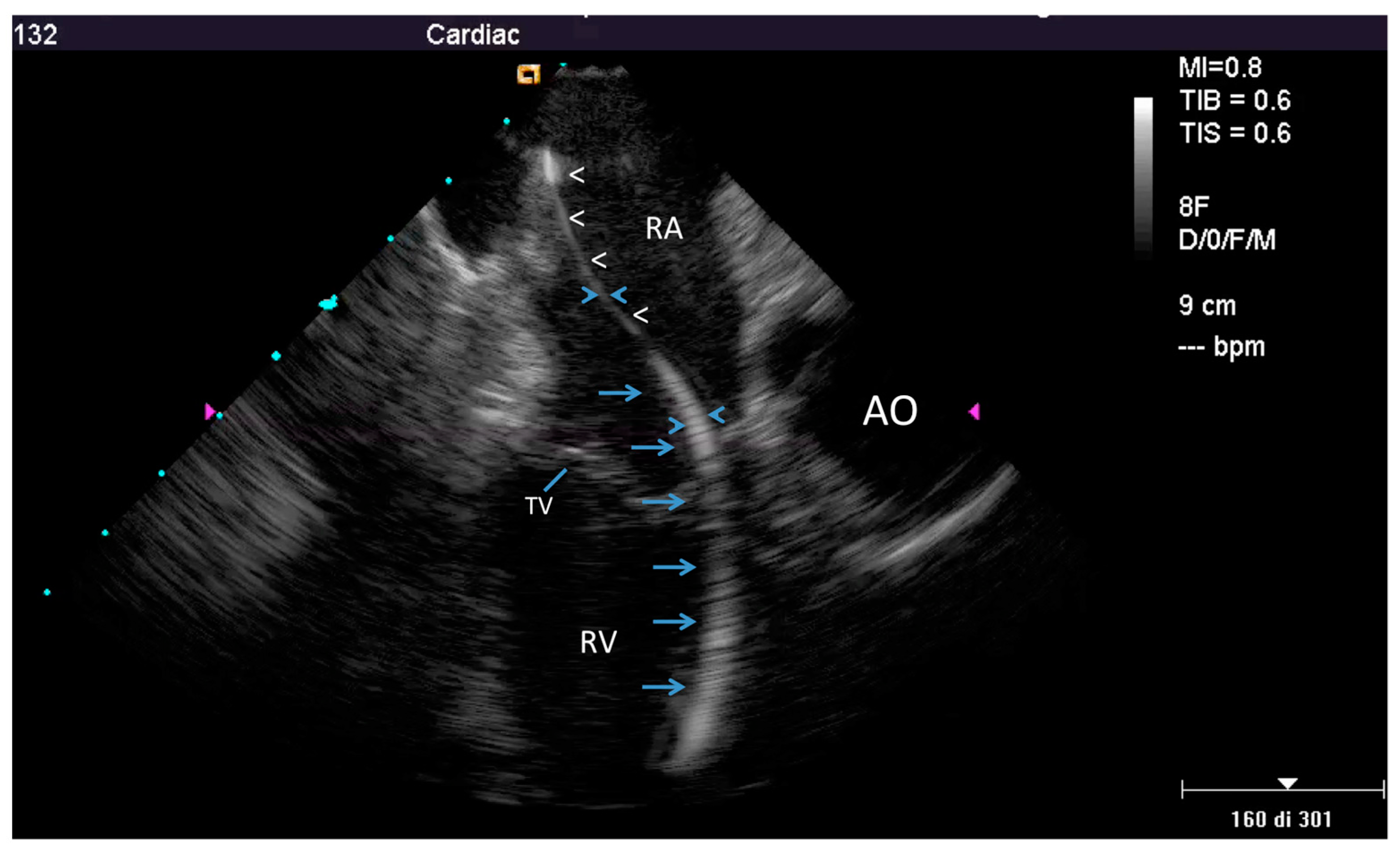
2.2.1. Before Extraction
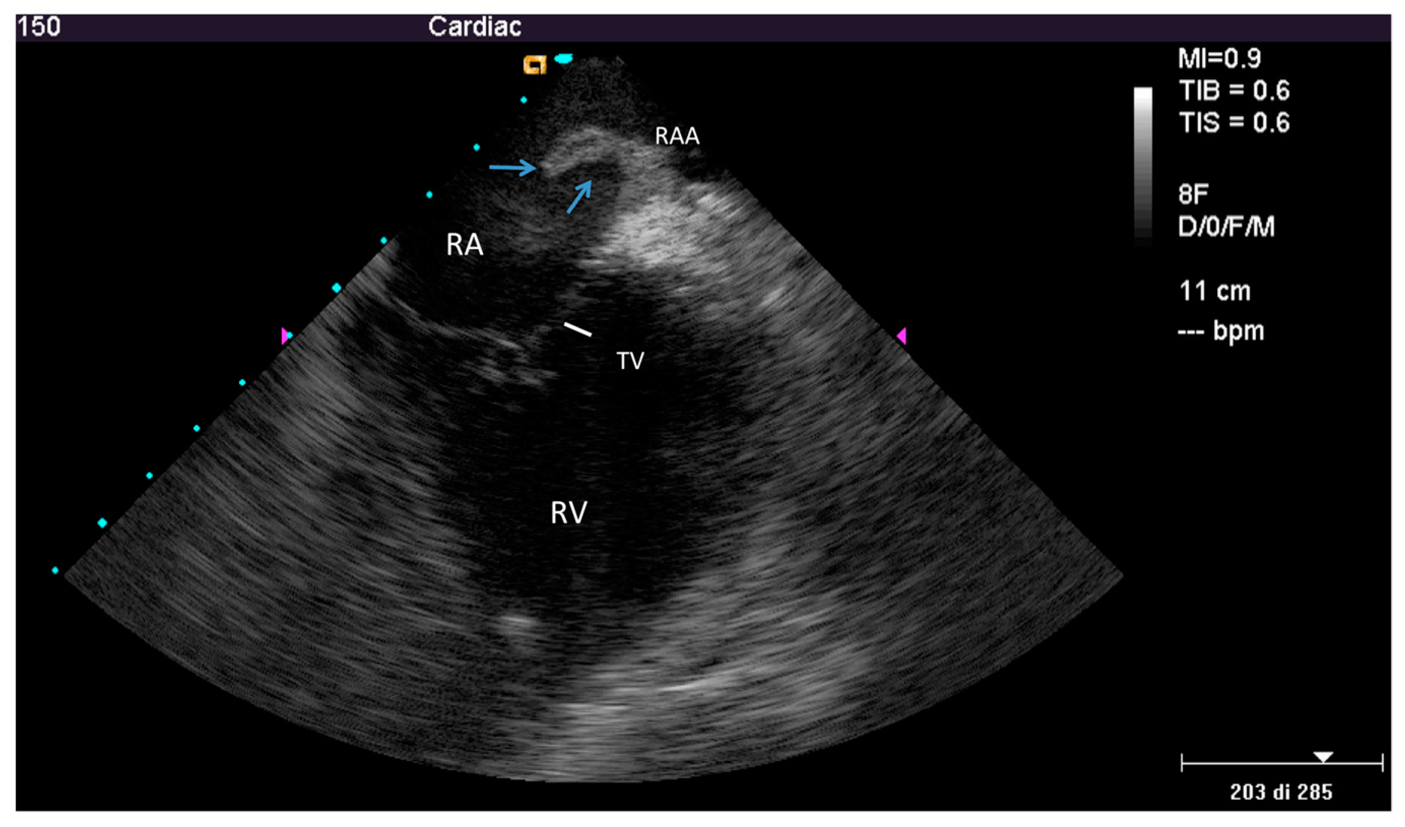
2.2.2. After the Extraction
2.3. Extraction Procedure
2.4. Transesophageal Echocardiography
2.5. End Points
2.6. Clinical Follow-up
2.7. Statistical Analysis
3. Results
3.1. Pre-extraction
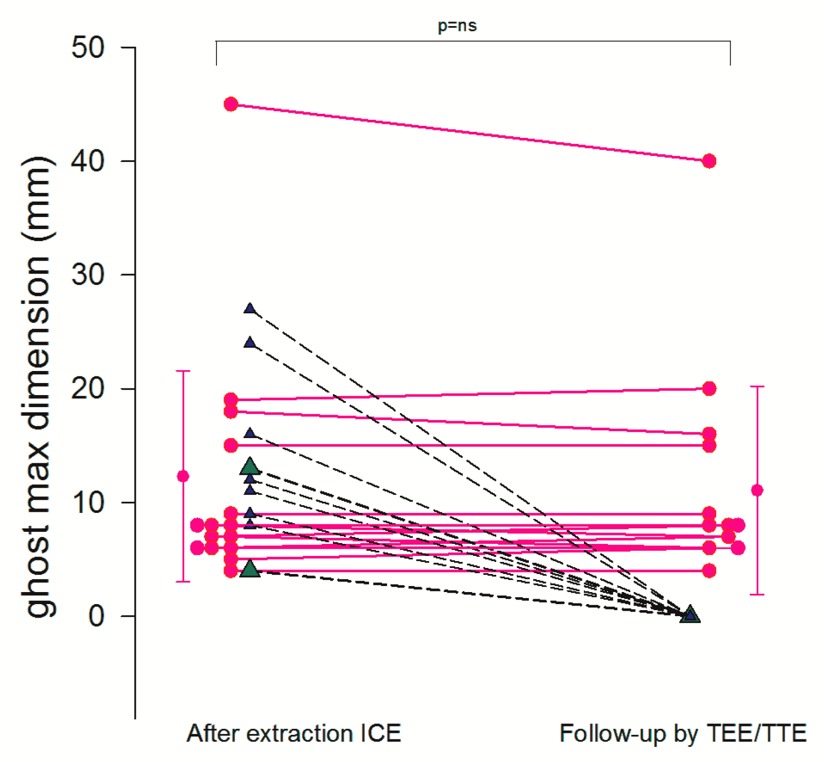
3.2. Post-extraction ICE
3.3. Prognostic Impact of Ghosts
4. Discussion
4.1. Fibrosis Involving Leads
4.2. Nature of the Ghost
4.3. Septic Risk Connected to Ghost
4.4. Previous Studies
4.5. Clinical Implication
4.6. Limitations
5. Conclusions
Supplementary Materials
Author Contributions
Funding
Acknowledgments
Conflicts of Interest
References
- Sohail, M.R.; Uslan, D.Z.; Khan, A.H.; Friedman, P.A.; Hayes, D.L.; Wilson, W.R.; Steckelberg, J.M.; Jenkins, S.M.; Baddour, L.M. Infective endocarditis complicating permanent pacemaker and implantable cardioverter-defibrillator infection. Mayo Clin. Proc. 2008, 83, 46–53. [Google Scholar] [CrossRef] [PubMed]
- Wazni, O.; Epstein, L.M.; Carrillo, R.G.; Love, C.; Adler, S.W.; Riggio, D.W.; Karim, S.S.; Bashir, J.; Geenspon, A.J.; DiMarco, J.P.; et al. Lead extraction in the contemporary setting: The LExICon study: An observational retrospective study of consecutive laser lead extractions. J. Am. Coll. Cardiol. 2010, 55, 579–586. [Google Scholar] [CrossRef] [PubMed]
- Le Dolley, Y.; Thuny, F.; Mancini, J.; Casalta, J.P.; Riberi, A.; Gouriet, F.; Bastard, E.; Ansaldi, S.; Franceschi, F.; Renard, S.; et al. Diagnosis of cardiac device-related infective endocarditis after device removal. JACC Cardiovasc. Imaging 2010, 3, 673–681. [Google Scholar] [CrossRef] [PubMed]
- Narducci, M.L.; Di Monaco, A.; Pelargonio, G.; Leoncini, E.; Boccia, S.; Mollo, R.; Perna, F.; Bencardino, G.; Pennestrì, F.; Scoppettuolo, G.; et al. Presence of ‘ghosts’ and mortality after transvenous lead extraction. Ep Eur. 2016, 19, 432–440. [Google Scholar] [CrossRef] [PubMed][Green Version]
- Poterała, M.; Kutarski, A.; Brzozowski, W.; Tomaszewski, M.; Gromadziński, L.; Tomaszewski, A. Echocardiographic assessment of residuals after transvenous intracardiac lead extraction. Int J. Cardiovasc. Imaging 2020, 36, 423–430. [Google Scholar] [CrossRef] [PubMed]
- Esposito, M.; Kennergren, C.; Holmstrom, N.; Nilsson, S.; Eckerdal, J.; Thomsen, P. Morphologic and immunohistochemical observations of tissues surrounding retrieved transvenous pacemaker leads. J. Biomed. Mater. Res. 2002, 63, 548–558. [Google Scholar] [CrossRef]
- Candinas, R.; Duru, F.; Schneider, J.; Luscher, T.F.; Stokes, K. Postmortem analysis of encapsulation around long-term ventricular endocardial pacing leads. Mayo Clin. Proc. 1999, 74, 120–125. [Google Scholar] [CrossRef]
- Stokes, K.; Anderson, J.; McVenes, R.; McClay, C. The encapsulation of polyurethane-insulated transvenous cardiac pacemaker leads. Cardiovasc. Pathol. 1995, 4, 163–171. [Google Scholar] [CrossRef]
- Kolodzinska, A.; Kutarski, A.; Koperski, L.; Grabowski, M.; Malecka, B.; Opolski, G. Differences in encapsulating lead tissue in patients who underwent transvenous lead removal. Ep Eur. 2012, 14, 994–1001. [Google Scholar] [CrossRef]
- Becker, A.E.; Becker, M.J.; Claudon, D.G.; Edwards, J.E. Surface thrombosis and fibrous encapsulation of intravenous pacemaker catheter electrode. Circulation 1972, 46, 409–412. [Google Scholar] [CrossRef]
- Becker, A.E.; Becker, M.J.; Martin, F.H.; Edwards, J.E. Bland Thrombosis and Infection in Relation to Intracardiac Catheter. Circulation 1972, 46, 200–203. [Google Scholar] [CrossRef] [PubMed][Green Version]
- Rizzello, V.; Dello Russo, A.; Casella, M.; Biddau, R. Residual fibrous tissue floating in the right atrium after percutaneous pacemaker lead extraction: An unusual complication early detected by intracardiac echocardiography. Int. J. Cardiol. 2008, 127, e67–e68. [Google Scholar] [CrossRef] [PubMed]
- Narducci, M.L.; Pelargonio, G.; Russo, E.; Marinaccio, L.; Di Monaco, A.; Perna, F.; Bencardino, G.; Casella, M.; Di Biase, L.; Santangeli, P.; et al. Usefulness of Intracardiac Echocardiography for the Diagnosis of Cardiovascular Implantable Electronic Device–Related Endocarditis. J. Am. Coll. Cardiol. 2013, 61, 1398–1405. [Google Scholar] [CrossRef] [PubMed]
- Wessler, S.; Freiman, D.G.; Ballon, J.D.; Katz, J.H.; Wolff, R.; Wolf, E. Experimental Pulmonary Embolism with Serum-Induced Thrombi. Am. J. Pathol. 1961, 38, 89–101. [Google Scholar]
- Durack, D.T.; Lukes, A.S.; Bright, D.K.; Service, D.E. New criteria for diagnosis of infective endocarditis: Utilization of specific echocardiographic findings. Am. J. Med. 1994, 96, 200–209. [Google Scholar] [CrossRef]
- Klug, D.; Lacroix, D.; Savoye, C.; Goullard, L.; Grandmougin, D.; Hennequin, J.L.; Kacet, S.; Lekieffre, J. Systemic infection related to endocarditis on pacemaker leads: Clinical presentation and management. Circulation 1997, 95, 95. [Google Scholar] [CrossRef]
- Bongiorni, M.G.; Soldati, E.; Zucchelli, G.; Di Cori, A.; Segreti, L.; De Lucia, R.; Solarino, G.; Balbarini, A.; Marzilli, M.; Mariani, M. Transvenous removal of pacing and implantable cardiac defibrillating leads using single sheath mechanical dilatation and multiple venous approaches: High success rate and safety in more than 2000 leads. Eur. Hear. J. 2008, 29, 2886–2893. [Google Scholar] [CrossRef]
- Victor, F.; De Place, C.; Camus, C.; Le Breton, H.; Leclercq, C.; Pavin, D.; Mabo, P.; Daubert, C. Pacemaker lead infection: Echocardiographic features, management, and outcome. Heart 1999, 81, 82–87. [Google Scholar] [CrossRef]
- Sanfilippo, A.J.; Picard, M.H.; Newell, J.B.; Rosas, E.; Davidoff, R.; Thomas, J.D.; Weyman, A.E. Echocardiographic assessment of patients with infectious endocarditis: Prediction of risk for complications. J. Am. Coll. Cardiol. 1991, 18, 1191–1199. [Google Scholar] [CrossRef]
- Altman, D.G. Practical Statistics for Medical Research; Chapman and Hall: London, UK, 1991. [Google Scholar]
- Bland, J.M.; Altman, D.G. Statistical methods for assessing agreement between two methods of clinical measurement. Lancet 1986, 1, 307–310. [Google Scholar] [CrossRef]
- Narducci, M.L.; Di Monaco, A.; Pelargonio, G.; Leoncini, E.; Boccia, S.; Mollo, R.; Perna, F.; Bencardino, G.; Pennestrì, F.; Scoppettuolo, G.; et al. Ghostbusters should come back to lead extraction arena in order to fight with ghosts: Author’s reply. Ep Eur. 2017, 19, 1585–1586. [Google Scholar] [CrossRef] [PubMed]
- Koneru, J.N.; Ellenbogen, K.A. Detection of transvenous pacemaker and ICD lead vegetations: The ICE cold facts. J. Am. Coll. Cardiol. 2013, 61, 1406–1408. [Google Scholar] [CrossRef] [PubMed]
- Caiati, C.; Pollice, P.; Lepera, M.E.; Favale, S. Pacemaker Lead Endocarditis Investigated with Intracardiac Echocardiography: Factors Modulating the Size of Vegetations and Larger Vegetation Embolic Risk during Lead Extraction. Antibiotics 2019, 8, 228. [Google Scholar] [CrossRef] [PubMed]
- Zhang, M.; Zhang, Y.; Pang, W.; Zhai, Z.; Wang, C. Circulating biomarkers in chronic thromboembolic pulmonary hypertension. Pulm. Circ. 2019, 9. [Google Scholar] [CrossRef]
- Bongiorni, M.G.; Di Cori, A.; Soldati, E.; Zucchelli, G.; Segreti, L.; Solarino, G.; De Lucia, R.; Sergi, D. [Iatrogenic risk of permanent pacemaker and defibrillator implantation]. G Ital Cardiol (Rome) 2009, 10, 395–406. [Google Scholar]
- Frost, J. Introduction to Statistics: An Intuitive Guide, 1st ed.; Statistics by Jim publishing: State College, PA, USA, 2019; pp. 196–204. [Google Scholar]
- Blumenthal, K.G.; Peter, J.G.; Trubiano, J.A.; Phillips, E.J. Antibiotic allergy. Lancet 2019, 393, 183–198. [Google Scholar] [CrossRef]
- Freedman, L.R. The pathogenesis of infective endocarditis. J. Antimicrob. Chemother. 1987, 20, 1–6. [Google Scholar] [CrossRef]
- Andreas, M.; Wiedemann, D.; Kocher, A.; Khazen, C. Materialization of ghosts: Severe intracardiac masses after pacemaker lead extraction requiring immediate surgical intervention. Heart Rhythm 2013, 10, 1826. [Google Scholar] [CrossRef]


| Demographic and clinical variables | Data |
|---|---|
| Enrolled patients, # | 40 |
| Age, years | 75 ± 11 |
| Gender | |
| Males, # | 33 |
| Females, # | 7 |
| BMI | 27 ± 5 |
| Chronic RF, # (%) | 15 (37%) |
| Diabetes, # (%) | 14 (35%) |
| Hypertension, # (%) | 35 (87%) |
| COPD, # (%) | 9 (22%) |
| LVEF,% | 46 |
| GFR, mL/min/1.73 m2 | 73 ± 23 |
| Number of leads removed during each procedure | 2 ± 0.7 |
| Atrial leads removed overall, # (%) | 33 (85%) |
| Ventricular leads removed overall, # (%) | 36 (95%) |
| CS leads removed overall, # (%) | 9 (23%) |
| Time from first implantation (m) | 113 ± 70 |
| Time from last operation (m) | 28 ± 28 |
| Variables | No TL/FAC Group (n = 15) | TL/FAC Group (n = 25) | Odds Ratio (95% CI) | p-Value | |
|---|---|---|---|---|---|
| Age | 78 ± 8 | 74 ± 12 | - | ns | |
| Sex, #pt (%) | male | 12 (80.0) | 21 (84.0) | ||
| female | 3 (20.0) | 4 (16.0) | ns | ||
| BMI | 25.94 ± 3.30 | 27.92 ± 5.91 | - | ns | |
| Diabetes, #pt (%) | - | 12 (80.0) | 14 (56.0) | 1 | |
| + | 3 (20.0) | 11 (44.0) | 3.14 (0.70–14) | =0.12 | |
| EF (%) | 47 ± 12 | 45 ± 11 | - | ns | |
| CRF, #pt (%) | - | 9 (60.0) | 16 (64.0) | 1 | |
| + | 6 (40.0) | 9 (36.0) | 0.8 (0.22–3.1) | ns | |
| Pocket culture positive, #pt (%) | - | 0 (0) | 10 (40) | 1 | |
| + | 15(100) | 15 (60) | 0.04(0.002–0.9) | =0.04 | |
| Local device infection, #pt (%) | - | 0 (0) | 7 (28) | 1 | |
| + | 15 (100) | 18 (72) | 0.07(0.004–1.5) | =0.09 | |
| Veg by ICE, #pt (%) | - | 7 (47) | 6 (24) | 1 | |
| + | 8 (53) | 19 (76) | 2.7(0.7–10) | ns | |
| CRP (mg/L) | 9.07 ± 10.37 | 17.83 ± 31.06 | - | ns | |
| White cells (count) | 5.77 ± 1.51 | 7.52 ± 2.40 | - | <0.05 | |
| ESR (mm/h) | 29.33 ± 22 | 29.88 ± 22.47 | - | ns | |
| Number of leads | 2.22 ± 0.6 | 2.13 ± 0.3 | - | ns | |
| Implantation time (months) | 77 ± 61 | 134 ± 67 | - | <0.05 | |
| Variables | No Ghost Group (n = 16) | Ghost Group (n = 24) | OR (95% CI) | p-Value | |
|---|---|---|---|---|---|
| Patient-related factors | |||||
| Age | 79 ± 9 | 73± 12 | - | ns | |
| Sex, # (%) | male | 13 (81.2%) | 20 (83.3%) | ||
| female | 3 (18.8%) | 4 (16.7%) | ns | ||
| BMI | 25.91 ± 3.19 | 28.03 ± 6.01 | - | ns | |
| Duke crit, # (%) | Possible endc | 5 (31.2%) | 5 (20.8%) | - | |
| Definite endc (2 criteria) | 9 (56.2%) | 13 (54.2%) | - | ||
| Definite endc (>2 criteria) | 2 (12.5%) | 6 (25%) | - | ns | |
| LVEF | 47 ± 12 | 45 ± 12 | - | ns | |
| CRF, # (%) | - | 10 (62.5%) | 15 (62.5%) | 1 | |
| + | 6 (37.5%) | 9 (37.5%) | 1.2(0.27–3.22) | ns | |
| Diabetes, # (%) | - | 13 (81%) | 13 (54%) | 1 | |
| + | 3 (18%) | 11 (46%) | 3.7(0.82–16.3) | =0.08 | |
| Infectious factors | |||||
| CRP (mg/L) | 9.26 ± 10.04 | 18.06 ± 31.7 | - | ns | |
| WBC (count*1000) | 5.98 ± 1.68 | 7.45 ± 2.43 | - | <0.05 | |
| ESR (mm/h) | 29.50 ± 21.27 | 29.79 ± 22.9 | - | ns | |
| Fever, # (%) | - | 12 (75%) | 18 (75%) | 1 | |
| + | 4 (25%) | 6 (25%) | 1.0(0.2–4.32) | ns | |
| Local device infection, # (%) | - | 0 (0%) | 7 (29%) | 1 | |
| + | 16 (100%) | 17 (71%) | 0.07(0.003–1.78) | ns | |
| Pocket culture positive, # (%) | - | 0 (0%) | 10 (42%) | 1 | |
| + | 16 (100%) | 14 (58.3%) | 0.04(0.002–.078) | <0.05 | |
| Lead tip culture* positive, #(%) | - | 16 (100%) | 19 (79%) | 1 | |
| + | 0 (0%) | 5 (21%) | 9.3(0.5–181) | ns | |
| BC pre-extraction positive, #(%) | - | 11 (69%) | 16 (67%) | 1 | |
| + | 5 (31%) | 8 (33%) | 2.1(0.08–54.9) | ns | |
| BC post-extraction positive, #(%) | - | 16 (100%) | 23 (96%) | 1 | |
| + | 0 (0%) | 1 (4.2%) | 2.1(0.08–54.9) | ns | |
| Endocarditis Vegetation by ICE, # (%) | - | 8 (50%) | 5 (21%) | 1 | |
| + | 8 (50%) | 19 (79%) | 2.7(0.90–8.1) | =0.073 | |
| Lead-related factors | |||||
| Number of leads | 2.25 ± 0.93 | 2.08 ± 0.58 | ns | ||
| lead implantation time (m) | 78 ± 59 | 135 ± 68 | <0.05 | ||
| Thickened lead, # (%) | - | 15 (94%) | 0 (0%) | 1 | |
| + | 1 (6.2%) | 24 (100%) | 506(19–13,324) | <0.0001 | |
| FAC by ICE, # (%) | - | 16 (100%) | 12 (50%) | 1 | |
| + | 0 (0%) | 12 (50%) | 33(1.8–612) | =0.002 | |
| Type of device, # (%) | PM | 8 (37%) | 14 (58%) | ||
| ICD | 10 (62%) | 10 (42%) | ns | ||
| Pt No | Age(years)/Gender | Type of Device | Heart Disease | LV EF | Complication of Extraction | Ghost Presence | Ghost Max Length (mm) | Days after Explantation, Cause of Death |
|---|---|---|---|---|---|---|---|---|
| 2 | 76/M | ICD | Ischemic DCM | 27% | - | no | - | 62, Acute HF |
| 4 | 78/M | ICD | Ischemic DCM | 25% | - | yes | 8 | 20, ARDS |
| 12 | 49/F | PM | Infiltrative CM | 45% | - | yes | 7 | 35, MOF |
| 24 | 81/M | ICD | Ischemic DCM | 30% | - | no | - | 81, Acute HF |
© 2020 by the authors. Licensee MDPI, Basel, Switzerland. This article is an open access article distributed under the terms and conditions of the Creative Commons Attribution (CC BY) license (http://creativecommons.org/licenses/by/4.0/).
Share and Cite
Caiati, C.; Luzzi, G.; Pollice, P.; Favale, S.; Lepera, M.E. A Novel Clinical Perspective on New Masses after Lead Extraction (Ghosts) by Means of Intracardiac Echocardiography. J. Clin. Med. 2020, 9, 2571. https://doi.org/10.3390/jcm9082571
Caiati C, Luzzi G, Pollice P, Favale S, Lepera ME. A Novel Clinical Perspective on New Masses after Lead Extraction (Ghosts) by Means of Intracardiac Echocardiography. Journal of Clinical Medicine. 2020; 9(8):2571. https://doi.org/10.3390/jcm9082571
Chicago/Turabian StyleCaiati, Carlo, Giovanni Luzzi, Paolo Pollice, Stefano Favale, and Mario Erminio Lepera. 2020. "A Novel Clinical Perspective on New Masses after Lead Extraction (Ghosts) by Means of Intracardiac Echocardiography" Journal of Clinical Medicine 9, no. 8: 2571. https://doi.org/10.3390/jcm9082571
APA StyleCaiati, C., Luzzi, G., Pollice, P., Favale, S., & Lepera, M. E. (2020). A Novel Clinical Perspective on New Masses after Lead Extraction (Ghosts) by Means of Intracardiac Echocardiography. Journal of Clinical Medicine, 9(8), 2571. https://doi.org/10.3390/jcm9082571