Article

Concomitant TP53 Mutation Confers Worse Prognosis in EGFR-Mutated Non-Small Cell Lung Cancer Patients Treated with TKIs

1
Biosciences Laboratory, Istituto Scientifico Romagnolo per lo Studio e la Cura dei Tumori (IRST) IRCCS, 47014 Meldola, Italy
2
Biostatistics and Clinical Trials Unit, Istituto Scientifico Romagnolo per lo Studio e la Cura dei Tumori (IRST) IRCCS, 47014 Meldola, Italy
3
Department of Medical Oncology, Istituto Scientifico Romagnolo per lo Studio e la Cura dei Tumori (IRST) IRCCS, 47014 Meldola, Italy
4
Department of Medical Oncology, Santa Maria della Misericordia Hospital, 06129 Perugia, Italy
5
Department of Pathology, Morgagni-Pierantoni Hospital, 47121 Forlì, Italy
6
Department of Medical Oncology, Per gli Infermi Hospital, Rimini 47923, Italy
7
UOS Oncology, Istituto per la Sicurezza Sociale, State Hospital, Cailungo 47893, San Marino, Italy
8
Department of Medical Oncology, Ospedali Riuniti Padova Sud “M. Teresa di Calcutta”, ULSS6 Euganea, 35131 Padova, Italy
9
Department of Medical Oncology, S. Maria delle Croci Hospital, 48121 Ravenna, Italy
*
Author to whom correspondence should be addressed.
J. Clin. Med. 2020, 9(4), 1047; https://doi.org/10.3390/jcm9041047
Submission received: 2 March 2020 / Revised: 1 April 2020 / Accepted: 3 April 2020 / Published: 7 April 2020

Abstract

:
Background: Non-small cell lung cancer (NSCLC) is the primary cause of cancer-related deaths worldwide. Epidermal Growth Factor Receptor (EGFR)-mutated patients usually benefit from TKIs treatment, but a significant portion show unresponsiveness due to primary resistance mechanisms. We investigated the role of TP53 mutations in predicting survival and response to EGFR-TKIs in EGFR-mutated NSCLC patients, to confirm, on an independent case series, our previous results. Methods: An independent retrospective cohort study was conducted, on a case series of 136 EGFR-mutated NSCLC patients receiving first or second generation TKIs as a first line therapy, and a smaller fraction of patients who acquired the T790M resistance mutation and were treated with third generation TKIs in the second or further line of treatment. TP53 mutations were evaluated in relation to disease control rate (DCR), objective response rate (ORR), progression-free survival (PFS) and overall survival (OS) of the patients. Results: Forty-two patients (30.9%) showed a TP53 mutation. Considered together, TP53 mutations had no significant impact on time-to-event endpoints. Considering the different TP53 mutations separately, exon 8 mutations confirmed their negative effect on PFS (HR 3.16, 95% 1.59–6.28, p = 0.001). In patients who developed the T790M resistance mutation, treated with third generation TKIs, the TP53 exon 8 mutations predicted worse PFS (even though not statistically significant), and OS (HR 4.86, 95% CI: 1.25–18.90, p = 0.023). Conclusions: TP53 exon 8 mutations confirmed their negative prognostic impact in patients treated with first and second generation TKIs and demonstrated a role in affecting clinical outcome in patients treated with third generation TKIs.

1. Introduction

Epidermal growth factor receptor (EGFR)-tyrosine kinase inhibitors (TKIs) have changed the natural history of non-small-cell lung cancer (NSCLC) patients harboring specific EGFR mutations at exons 18, 19 and 21. Randomized trials have demonstrated a median progression-free survival (PFS) of 9.7 and 9.5 months in patients harboring sensitizing EGFR mutations treated with first-generation EGFR-TKIs versus platinum-based chemotherapy [1,2], and 11.1 months for second generation TKIs [3]. Third generation TKI osimertinib, initially designed to overcome the arising of T790M resistance mutation in EGFR pre-treated patients [4], has recently become the gold standard for EGFR-mutated patients, reaching a median PFS of 18.9 months [5].
Despite the high sensitivity of EGFR-mutated patients to EGFR-TKIs, the objective response rate is of about 70–80% for 1st, 2nd and 3rd generation TKIs [2,3,5], meaning that a portion of patients do not respond to EGFR-TKI treatment, notwithstanding the presence of sensitizing EGFR mutation, suggesting the presence of primary resistance mechanisms.
Our previous study and several others showed that the concomitant presence of TP53 mutation confers a worse prognosis in EGFR-mutated patients treated with first and second generation TKIs [6,7,8]. Subsequent studies performed using next generation sequencing methodologies showed that the presence of concomitant mutation in different genes is associated with a lower response to EGFR-TKIs and, however, TP53 mutation confirms to be the most significant predictor of worse outcome. In particular, it seems that specific TP53 mutations are more implicated in predicting the worse prognosis [6,9,10], confirming that different TP53 mutations confer different p53 functions. Within the coding region of the TP53 gene, several studies have reported that a higher frequency of mutations occurs in the exons 5–8, and that mutations in these exons are associated to differential functions of p53 protein [9,10]. As the different published studies have analyzed principally patients treated with first and second generation TKIs, few data are available with regard to the role of TP53 mutation in relation to response to third generation TKIs.
The main purpose of this research was to confirm our previously published results on the role of TP53 mutations, in an independent cohort of advanced EGFR-mutated patients treated with first or second generation TKIs in the first line setting, and to investigate the role of TP53 mutations in predicting prognosis of patients with acquired T790M mutation treated with third generation TKIs.

2. Materials and Methods

To confirm our previous results on the role of TP53 mutations in relation to the effectiveness of TKIs, an independent retrospective cohort study was conducted. All consecutive patients with advanced EGFR-mutated NSCLC receiving a first line TKI treatment (i.e., gefitinib, erlotinib, or afatinib) from July 2010 to May 2018 at the Medical Oncology Units of the Romagna catchment area (Area Vasta Romagna, AVR) and at the S. Maria della Misericordia Hospital of Perugia, Italy, were included in this study. Demographic and clinical characteristics of the patients were obtained using a medical and radiographic records review including age, gender, smoking history, histology, and information on death and response to treatment. EGFR status had been routinely determined at the Biosciences Laboratory of IRST-IRCCS and the Laboratory of Molecular Biology of the S. Maria della Misericordia Hospital, Perugia, by MassARRAY, pyrosequencing, direct sequencing or Next-Generation Sequencing (NGS) methodologies.
To evaluate the independent role of TP53 mutations, that is, eventually adjusting for other covariates, and to obtain a more accurate estimate of their prognostic effect, an analysis combining the data of the present work with those from our previous one [6], was also performed, updating follow-ups of the previous case series to 30 June 2018.
Moreover, considering the two cohorts together, we identified a subgroup of 42 patients who developed the T790M resistance mutation and were treated with third generation TKI, osimertinib. All patients provided an informed consent, and the study was approved by the AVR Ethical Committee (study code IRST-B053).

2.1. EGFR and TP53 Mutation Analysis

EGFR mutation analyses were performed on both cytologic and histologic samples, accurately selected by a dedicated expert pathologist from each center at the time of diagnosis. The same DNA specimens were used for the determination of TP53 mutation status, blindly to the clinical outcomes. Quality controls were periodically performed during the course of the study to ensure concordance of molecular results.
DNA was extracted by macro-dissection of an area comprising at least 50% of tumor cells. Cells were lysed in a digestion buffer of 50 mmol/L KCl, 10 mmol/L Tris-HCl pH 8.0, 2.5 mmol/L MgCl2, and Tween-20 0.45%; proteinase K at 1.25 mg/mL were added to each specimen, with an overnight incubation at 56 °C. After proteinase K inactivation at 95 °C for 10 min, samples were centrifuged twice to eliminate debris and supernatant DNA quantity and quality was assessed by Nanodrop (Celbio) before molecular analyses.
Mutation status for exons 5–8 of TP53 gene was performed by PCR amplification and Direct Sequencing using 3130 Genetic Analyzer (Applied Biosystems, Monza, Italy), or Next-Generation Sequencing by Ion S5 platform (Thermofisher, Monza, Italy), or MySeq platform (Illumina, San Diego, CA, USA).

2.2. Response Evaluation

Best clinical response to treatment with TKI was classified on the basis of interval CT scans as complete response (CR), partial response (PR), stable disease (SD), or progressive disease (PD) using standard Response Evaluation Criteria in Solid Tumors criteria (RECIST) version 1.1. Patients with both baseline imaging and at least one repeated evaluation after continuous EGFR-TKI monotherapy were evaluable for radiographic response. The same criteria for response evaluation and periodicity were used by all centers taking part in the study.

2.3. Statistical Analyses

Data were summarized by mean ± standard deviation (SD) for continuous variables and through natural frequencies and percentages for categorical ones.
Treatment responses were reported as objective response rate (ORR) and disease control rate (DCR). The time-to-event endpoints examined were progression-free survival (PFS) and overall survival (OS). PFS was defined as the time from start of first line treatment (or from start of osimertinib for the subgroup analysis) to disease progression or death for any cause, whichever occurred first. Patients who were alive and progression-free at 30 June 2018, the last follow-up update, were censored at that date.
OS was defined as the time from start of first line treatment to death for any cause. Alive patients were censored at the date of the last follow-up update. PFS and OS functions were estimated using the Kaplan–Meier method, and the log-rank test was used to assess differences between groups. Median PFS and OS were reported as point estimates and 95% confidence intervals (CI) in round brackets. The Cox proportional hazards regression model was used to quantify the association between specific covariates and the time-to-event endpoints. Results are reported as HR and 95% CI in round brackets. To assess the association between mutations and the duration of response to TKIs, patients were divided into short-term responders (PFS less than 6 months), intermediate-term responders (PFS ≥6 months and ≤24 months) or long-term responders (PFS >24 months). The association between categorical variables was tested by the Pearson’s χ2 test or Fisher exact test, when appropriate, whereas those between a continuous variable and a categorical one was tested by means of the Student t-test or F test for more than two categories. To evaluate the independent role of TP53 mutations in a multivariate analysis and to obtain more accurate estimates of their prognostic effect, a combined analysis including data of the present work with those from our previous one, was performed. Follow-up of our previous cohort was updated on 30 June 2018. A multivariable model was obtained using backward stepwise variable selection, setting the significance level for variable removal from the model equal to 0.10. In a perspective of parsimonious modelling, when appropriate, categories of some study variables were grouped. The proportional hazards assumption was evaluated using a statistical test based on Schoenfeld residuals. In case of non-proportional hazards for a specific variable, a Cox model with time-dependent coefficient, β(t), was fitted. To simplify model interpretation, a step function for β(t) was used, dividing the follow-up period in three time periods since treatment started: the first 6 months, 6–12 months, and greater than 12 months.
Overall and when not otherwise specified, a two-sided p-value (p) <0.05 was considered statistically significant. All statistical analyses were performed using STATA 15.0 software (College Station, TX, USA) and R version 3.6.1.

3. Results

3.1. Clinico-Pathologic and Molecular Features of Patients

Patients characteristics, EGFR mutations, type of TKI received and TP53 mutations are reported in Table 1. All 136 patients carried an EGFR mutation (exon 19 deletions 53.7%, exon 21 L858R 35.3%, other EGFR mutations 11.0%) and received a first line EGFR-TKI (36.7% Erlotinib, 5.1% Erlotinib plus bevacizumab, 30.9% gefitinib, 27.2% afatinib). Of the patients with available information on smoking habit, half were never smokers (50.8%), and half were former or current smokers (28.8% and 20.3%, respectively). We found TP53 mutations in 42 (30.9%) of the 136 analyzed patients: 12 mutations were in exon 5 (28.6%), 6 in exon 6 (14.3%), 13 in exon 7 (31.0%) and 11 in exon 8 (26.2%). Following the classification of TP53 mutations into disruptive and non-disruptive ones [6], 11 patients had a disruptive mutation whereas 31 had a non-disruptive one (26.2% and 73.8%, respectively).
While for patients with exon 19 deletion the three different TKIs were used in an almost similar proportion (32.9% patients received erlotinib, 30.1% gefitinib, and 37.0% afatinib), patients with a mutation in exon 21 L858R were predominantly treated with erlotinib or gefitinib (52.1% and 39.6% of the patients, respectively), and those with uncommon mutations received predominantly erlotinib or afatinib (53.3% and 40.0%, respectively, Table S1).
No statistically significant associations were observed between type of TP53 mutation, type of EGFR mutation and patient characteristics (Table S2).

3.2. Patients Outcome in Relation to EGFR Mutations

Overall, ORR and DCR were 67.4%, and 89.3%, respectively. Considering the clinical responses by type of EGFR mutation, ORR was considerably higher in the subgroup of patients with exon 19 deletion (77.5%), with respect to patients with L585R mutation (55.3%), and the subgroup with other mutations (54.6%), p = 0.029. A higher percentage of long responders was observed in patients carrying exon 19 deletion (12.3%), with respect to patients with L858R point mutation (7.3%), or patients with the other EGFR mutations (6.6%), Table 2.
Median PFS and OS were 12.3 (95% CI: 9.9–13.8) and 27.3 (95% CI: 21.9–52.9) months, respectively. No statistically significant association between PFS, OS and type of EGFR mutations was found (p = 0.282 and p = 0.207, respectively).

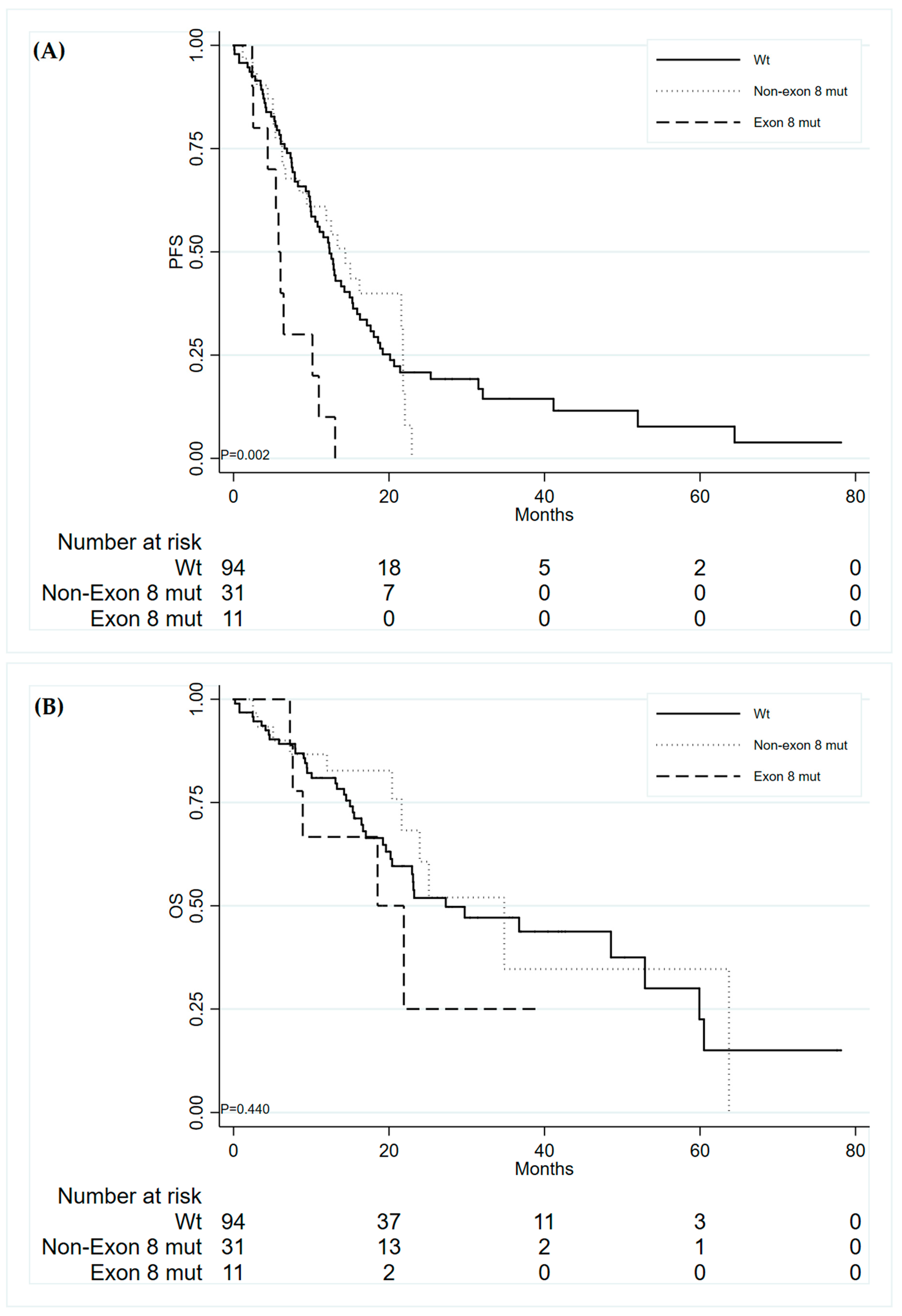
3.3. Patients Outcome in Relation to TP53 Mutations

No statistically significant associations were found between TP53 mutations and ORR and DCR (Table S3). When considering any type of TP53 mutation with regard to PFS, no association was found; however, significant results were observed considering only TP53 exon 8 mutations. As previously reported [6], patients with this gene mutation showed a shorter median PFS than non-exon 8 mutated and wild type TP53 patients: 5.8 months (95% CI: 2.4–10.2) vs. 14.4 (95% CI: 6.7–21.8) and 12.4 (95% CI: 10.0–15.0), respectively (Figure 1A). These patients also showed a poorer OS as compared with the other groups, even though this result was not statistically significant: median OS were 18.53 months (95% CI: 7.3–NR), 34.8 (95% CI: 21.6–NR), 27.3 (95% CI: 20.2–52.9), respectively (Figure 1B).
The presence of TP53 exon 8 mutation seemed to be associated with a worse prognosis in a similar way in the patients with the different EGFR mutations, both in terms of PFS and OS.
In particular, patients with wild type TP53 exon 8 had a better clinical outcome independently by EGFR status: median PFS and OS were 12.9 (95% CI: 10.0–16.3) and 29.7 months (95% CI: 23.0–60.5) for patients with EGFR exon 19 deletion vs. 12.4 months (95% CI: 7.9–15.0) and 23.2 months (95% CI: 19.2–63.7) for those with other EGFR mutations, respectively; in the subgroup of patients with TP53 exon 8 mutations, median PFS and OS were 5.8 months (95% CI: 2.5–NR) and 21.9 months (95% CI: 7.3–NR) for patients with EGFR exon 19 deletion vs. 6.4 (95% CI: 2.4–NR) and 18.5 months (95% CI: 7.6–NR) for those with other EGFR mutations. In Table S4, the univariate Cox analysis results are reported.

3.4. Multivariate Analysis of the Role of TP53 Mutation: Combined Cohorts of Patients

To obtain a more precise estimate of the effect of TP53 exon 8 mutation on PFS and OS, and to determine its potential independent role considering other information, a pooled analysis considering either data from our previously analyzed cohort and the one described in the present study, was performed.
The final multivariate model for PFS included both EGFR exon 19 deletion as well as TP53 mutation. As soon as the effect of exon 19 deletion on the hazard of disease progression or death is not constant over time, that is, the proportional hazards assumption underlying the Cox model was violated, to obtain a better model fit, this variable was entered into the model with a time-dependent coefficient. Table 3 shows that the effect of exon 19 mutation changes over time, showing a strong protective effect over the first six months that vanishes afterward.
Adjusting for presence of EGFR exon 19 deletion, TP53 mutations affecting exon 8 demonstrated to be the unique independent negative prognostic factor for PFS (HR 1.81, 95% CI: 1.13–2.92, Table 3). With regard to OS, only deletion in EGFR Exon 19 resulted associated to OS, probably due to data from our previous cohort (HR 0.52 (95% CI: 0.26–1.03) for the first 6 months of follow-up, HR 0.44 (95% CI: 0.22–0.90), for successive 6 months, and HR 1.08 (95% CI: 0.72–1.61) after 12 months).

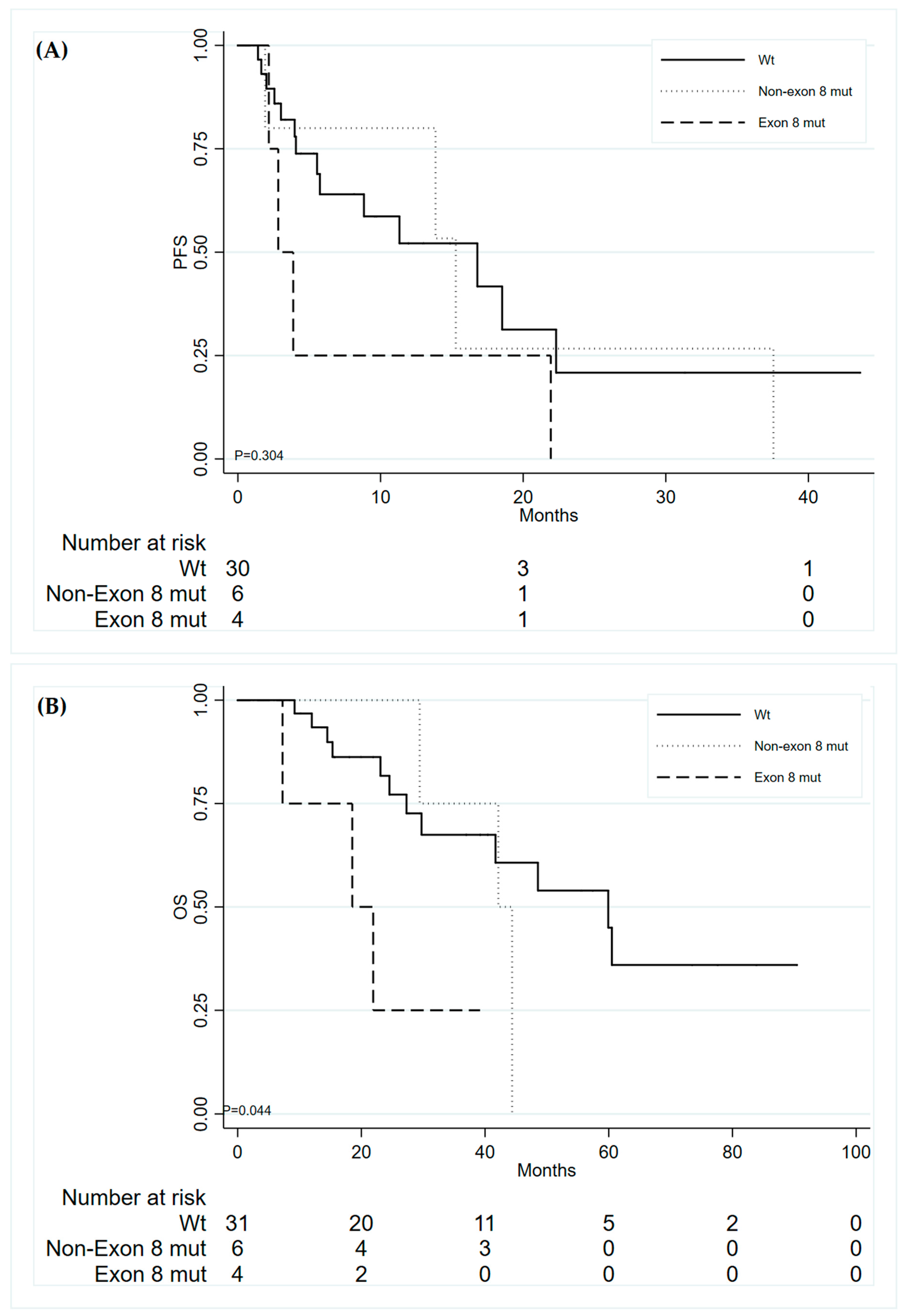
3.5. TP53 Mutations in Relation to Responsiveness to Third Generation TKIs: Combined Cohorts of Patients

Considering both patients’ cohorts (n = 272 patients), we considered 42 patients who developed a T790M resistance mutation and were treated with third generation TKI osimertinib, in the second or further lines of therapy. Of these, 41 were evaluable for TP53 mutation status; we found 10 TP53 mutated patients (24.4%): 3 mutations in exon 5 (30%), 1 in exon 6 (10%), 2 in exon 7 (20%) and 4 in exon 8 (40%). Within the 41 patients with available clinical information, median PFS and OS were 13.86 (95% CI: 5.5–18.53) and 44.38 months (95% CI: 10.64–24.28), respectively. Median PFS of exon 8 TP53 mutated patients was 2.83 (2.17–NR) months, with respect to a median PFS of 16.79 (5.55–22.31) and 15.28 (1.91–NR) months, for wt TP53 and patients with mutations in other exons of the gene, respectively (Figure 2A). Even though a good separation, the difference among curves was not statistically significant (p = 0.304), due to small numbers of the exon 8 mutated patients. On the other hand, exon 8 TP53 gene mutations significantly affected the survival of the patients, with a median OS for exon 8 TP53 mutated patients of 18.53 (7.26–NR) months, with respect to 42.15 (29.43–NR) and 59.92 (29.73–NR) months of patients with mutations in other exons of TP53 and wt TP53, respectively (p = 0.044) (Figure 2B). Table 4 shows the univariate hazard ratios for PFS and OS with respect to the presence of TP53 exon 8 mutation.

4. Discussion

In this study, we analyzed TP53 mutations in relation to clinical outcome in a large cohort of EGFR-mutated NSCLC patients receiving first or second generation TKIs as a first line therapy. Our results confirm that exon 8 TP53 mutations are associated with a shorter PFS, in all settings of treatment.
Moreover, such a negative effect was also observed in the subgroup of patients treated with third generation after the development of T790M mutation.
Numerous studies demonstrated the role of TP53 mutations in predicting poor prognosis of advanced NSCLC patients [9,11,12,13,14,15], and this was confirmed also in the subgroup of NSCLC patients carrying EGFR mutations [8,9,16]. In particular, different recent studies showed that the concurrent presence of TP53 mutations negatively affects response to TKIs in EGFR-mutated NSCLC patients, suggesting a role for these gene mutations in determining primary resistance to these drugs [6,7,17,18,19,20]. TP53 is the most frequently mutated gene in lung adenocarcinoma, with mutation rates reported up to 55% [13,21,22,23], with a predominantly clonal expression [24]. In our case series, we found 30% of patients carrying a TP53 mutation in the exons 5–8, the same percentage we previously reported in an independent case series. It is well known that different TP53 mutations lead to changes in the P53 protein that may have diverse biological significance [9,10,25], and mutations in the DNA-binding domain (exons 5–8), are frequently associated with gain-of-function properties, resulting in pro-oncogenic features of the P53 protein [26]. In our previous work, we found that exon 8 TP53 mutations were able to predict worse response to EGFR TKIs, especially in the subgroup of patients with EGFR exon 19 deletion. In the present study we confirmed the negative prognostic value of TP53 exon 8 mutation in an independent cohort of EGFR-mutated NSCLC treated with both first and second generation TKIs in the first line setting. In the present study, the prognostic value of exon 8 TP53 mutation was evident independently from the type of EGFR mutation. In a combined analysis, we showed that the effect was evident on overall survival in EGFR-mutated patients who developed T790M at progression after first line TKIs and osimertinib.
These results, in agreement with those reported by Kim et al. [7], suggest a negative predictive role of TP53 mutation in all lines of therapy and in relation to all TKIs. Furthermore, our results are consistent with a recent study that found that TP53 mutations in exon 8 are associated with shorter OS of patients receiving a TKI as a first line treatment [27].
In another study, missense mutations in TP53 gene resulted in shorter PFS in EGFR mutated patients treated with TKIs but showed no associations with PFS and OS in patients undergoing surgical resection [28]. According to Xu et al., who reported TP53 mutations in 88% of NSCLC EGFR-mutated patients that responded for <6 months to an EGFR TKIs, with respect to 13% of responders for >24 months [29], our results show a higher rate of TP53 mutations in non-responders group, with no TP53 mutated patients in the long responder group.
To investigate the role of TP53 mutations in predicting clinical outcome of patients treated with third generation TKIs, we considered 42 patients’ developed T790M mutation to first line treatment with first or second generation TKI and received a third generation drug in the second or further line of therapy. In this subgroup, we found a diminished PFS in patients carrying TP53 mutations in exon 8, even though without statistical significance, probably due to the small number of analyzed patients; exon 8 TP53 mutated patients had a significantly shorter OS, with respect to wt TP53 patients and patients with mutations in other exons of TP53. This observation is consistent with previous observations, that identified TP53 mutations (not only in exon 8) as a negative prognostic predictor [7,16]. This result was not confirmed by a study from Labbé et al., that found no differences in ORR of patients treated with third generation TKIs, based on TP53 mutation status; this could be for the small size of the analyzed case series [28]. In the light of the paradigm shift brought by FLAURA trial [5], there is a need to identify which biomarkers could predict primary resistance to osimertinib as a first line therapy; if confirmed in a larger case series treated with third generation TKI in the first line, these results could help to better stratify patients, suggesting an EGFR-independent mechanism of resistance, as others have already highlighted [30].

5. Conclusions

In conclusion, we confirmed that TP53 exon 8 mutations identify a subgroup of patients with primary resistance to EGFR TKIs, and that this is true also in relation to third generation TKIs such as osimertinib. These data suggest that patients with concomitant EGFR and exon 8 TP53 mutations should be candidates for more aggressive therapeutic schemes and should be monitored with a stricter follow-up.

Supplementary Materials

The following are available online at https://www.mdpi.com/2077-0383/9/4/1047/s1, Table S1: Demographic, clinicopathological and molecular characteristics of patients according to EGFR mutations (n = 136); Table S2: Demographic, clinicopathological and molecular characteristics by TP53 mutation; Table S3: Best clinical response according to TP53 mutations; Table S4: Univariate Cox analyses for PFS and OS.

Author Contributions

Conceptualization, A.D. (Angelo Delmonte) , L.C. and P.U.; methodology, M.C., E.P., E.C., V.L. and S.B.; validation, M.C., E.C., V.L., S.B. and P.U.; formal analysis, M.C., E.P., A.D. (Angelo Delmonte), A.D. (Alessandra Dubini) R.C., L.C. and P.U.; investigation, all authors; resources, A.D. (Angelo Delmonte), V.L., R.C., L.C. and P.U.; data curation, A.D. (Angelo Delmonte), A.D. (Alessandra Dubini) G.B., M.P., N.D.L., A.V., R.C., L.L., G.M. and M.A.B.; writing—original draft preparation, M.C., E.P., L.C. and P.U.; writing—review and editing, all authors; visualization, M.C., E.P. A.D. (Angelo Delmonte), L.C. and P.U.; supervision, A.D. (Angelo Delmonte), L.C. and P.U. All authors have read and agreed to the published version of the manuscript.

Funding

This research received no external funding.

Conflicts of Interest

The authors declare no conflict of interest.

References

  1. Rosell, R.; Carcereny, E.; Gervais, R.; Vergnenègre, A.; Massuti, B.; Felip, E.; Palmero, R.; Garcia-Gomez, R.; Pallares, C.; Sanchez, J.M.; et al. Erlotinib versus standard chemotherapy as first-line treatment for European patients with advanced EGFR mutation-positive non-small-cell lung cancer (EURTAC): a multicentre, open-label, randomised phase 3 trial. Lancet Oncol. 2012, 13, 239–246. [Google Scholar] [CrossRef]
  2. Mok, T.S.; Wu, Y.-L.; Thongprasert, S.; Yang, J.C.-H.; Chu, D.-T.; Saijo, N.; Sunpaweravong, P.; Han, B.; Margono, B.; Ichinose, Y.; et al. Gefitinib or Carboplatin–Paclitaxel in Pulmonary Adenocarcinoma. New Engl. J. Med. 2009, 361, 947–957. [Google Scholar] [CrossRef]
  3. Yang, J.C.-H.; Wu, Y.-L.; Schuler, M.; Sebastian, M.; Popat, S.; Yamamoto, N.; Zhou, C.; Hu, C.-P.; O’Byrne, K.; Feng, J.; et al. Afatinib versus cisplatin-based chemotherapy for EGFR mutation-positive lung adenocarcinoma (LUX-Lung 3 and LUX-Lung 6): analysis of overall survival data from two randomised, phase 3 trials. Lancet Oncol. 2015, 16, 141–151. [Google Scholar] [CrossRef] [Green Version]
  4. Mok, T.S.; Wu, Y.-L.; Ahn, M.-J.; Garassino, M.C.; Kim, H.R.; Ramalingam, S.R.; Shepherd, F.A.; He, Y.; Akamatsu, H.; Theelen, W.S.; et al. Osimertinib or Platinum-Pemetrexed in EGFR T790M-Positive Lung Cancer. New Engl. J. Med. 2016, 376, 629–640. [Google Scholar] [CrossRef] [Green Version]
  5. Soria, J.-C.; Ohe, Y.; Vansteenkiste, J.; Reungwetwattana, T.; Chewaskulyong, B.; Lee, K.H.; Dechaphunkul, A.; Imamura, F.; Nogami, N.; Kurata, T.; et al. Osimertinib in UntreatedEGFR-Mutated Advanced Non–Small-Cell Lung Cancer. New Engl. J. Med. 2018, 378, 113–125. [Google Scholar] [CrossRef]
  6. Canale, M.; Petracci, E.; Delmonte, A.; Chiadini, E.; Dazzi, C.; Papi, M.; Capelli, L.; Casanova, C.; De Luigi, N.; Mariotti, M.; et al. Impact of TP53 Mutations on Outcome in EGFR-Mutated Patients Treated with First-Line Tyrosine Kinase Inhibitors. Clin. Cancer Res. 2016, 23, 2195–2202. [Google Scholar] [CrossRef] [Green Version]
  7. Kim, Y.; Lee, B.; Shim, J.H.; Lee, S.-H.; Park, W.-Y.; Choi, Y.-L.; Sun, J.-M.; Ahn, J.S.; Ahn, M.-J.; Park, K. Concurrent Genetic Alterations Predict the Progression to Target Therapy in EGFR-Mutated Advanced NSCLC. J. Thorac. Oncol. 2019, 14, 193–202. [Google Scholar] [CrossRef] [Green Version]
  8. VanderLaan, P.A.; Rangachari, D.; Mockus, S.M.; Spotlow, V.; Reddi, H.V.; Malcolm, J.; Costa, D.B. Mutations in TP53, PIK3CA, PTEN and other genes in EGFR mutated lung cancers: Correlation with clinical outcomes. Lung Cancer 2018, 106, 17–21. [Google Scholar] [CrossRef] [Green Version]
  9. Molina-Vila, M.A.; Bertran-Alamillo, J.; Gascó, A.; Mayo-de-las-Casas, C.; Sánchez-Ronco, M.; Pujantell-Pastor, L.; Majem, M. Nondisruptive p53 Mutations Are Associated with Shorter Survival in Patients with Advanced Non–Small Cell Lung Cancer. Clin. Cancer Res. 2014, 2, 4647–4660. [Google Scholar] [CrossRef] [Green Version]
  10. Poeta, M.L.; Manola, J.; Goldwasser, M.A.; Forastiere, A.; Benoit, N.; Califano, J.A.; Ridge, J.A.; Goodwin, J.; Kenady, D.; Saunders, J.; et al. TP53 mutations and survival in squamous-cell carcinoma of the head and neck. N. Engl. J. Med. 2007, 357, 2552–2561. [Google Scholar] [CrossRef] [Green Version]
  11. La Fleur, L.; Falk-Sörqvist, E.; Smeds, P.; Berglund, A.; Sundström, M.; Mattsson, J.S.; Brandén, E.; Koyi, H.; Isaksson, J.; Brunnström, H.; et al. Mutation patterns in a population-based non-small cell lung cancer cohort and prognostic impact of concomitant mutations in KRAS and TP53 or STK11. Lung Cancer 2019, 130, 50–58. [Google Scholar] [CrossRef] [Green Version]
  12. Zhao, J.; Han, Y.; Li, J.; Chai, R.; Bai, C. Prognostic value of KRAS/TP53/PIK3CA in non-small cell lung cancer. Oncol Lett. 2019, 17, 3233–3240. [Google Scholar] [CrossRef] [Green Version]
  13. Volckmar, A.-L.; Leichsenring, J.; Kirchner, M.; Christopoulos, P.; Neumann, O.; Budczies, J.; De Oliveira, C.M.M.; Rempel, E.; Buchhalter, I.; Brandt, R.; et al. Combined targeted DNA and RNA sequencing of advanced NSCLC in routine molecular diagnostics: Analysis of the first 3,000 Heidelberg cases. Int. J. Cancer 2019, 145, 649–661. [Google Scholar] [CrossRef]
  14. Jiao, X.-D.; Qin, B.-D.; You, P.; Cai, J.; Zang, Y.-S. The prognostic value of TP53 and its correlation with EGFR mutation in advanced non-small cell lung cancer, an analysis based on cBioPortal data base. Lung Cancer 2018, 123, 70–75. [Google Scholar] [CrossRef]
  15. Gu, J.; Zhou, Y.; Huang, L.; Ou, W.; Wu, J.; Li, S.; Xu, J.; Feng, J.; Liu, B. TP53 mutation is associated with a poor clinical outcome for non-small cell lung cancer: Evidence from a meta-analysis. Mol. Clin. Oncol. 2016, 5, 705–713. [Google Scholar] [CrossRef] [Green Version]
  16. Aggarwal, C.; Davis, C.W.; Mick, R.; Thompson, J.C.; Ahmed, S.; Jeffries, S.; Bagley, S.; Gabriel, P.; Evans, T.L.; Bauml, J.M.; et al. Influence of TP53 Mutation on Survival in Patients With Advanced EGFR-Mutant Non–Small-Cell Lung Cancer. JCO Precis. Oncol. 2018, 1–29. [Google Scholar] [CrossRef]
  17. Yu, H.A.; Arcila, M.E.; Rekhtman, N.; Sima, C.S.; Zakowski, M.F.; Pao, W.; Kris, M.; Miller, V.A.; Ladanyi, M.; Riely, G.J. Analysis of tumor specimens at the time of acquired resistance to EGFR-TKI therapy in 155 patients with EGFR-mutant lung cancers. Clin. Cancer Res. 2013, 19, 2240–2247. [Google Scholar] [CrossRef] [Green Version]
  18. Hou, H.; Qin, K.; Liang, Y.; Zhang, C.; Liu, N.; Jiang, H.; Liu, K.; Zhu, J.; Lv, H.; Li, T.; et al. Concurrent TP53 mutations predict poor outcomes of EGFR-TKI treatments in Chinese patients with advanced NSCLC. Cancer Manag. Res. 2019, 11, 5665–5675. [Google Scholar] [CrossRef] [Green Version]
  19. Jin, Y.; Shi, X.; Zhao, J.; He, Q.; Chen, M.; Yan, J.; Ou, Q.; Wu, X.; Shao, Y.W.; Yu, X. Mechanisms of primary resistance to EGFR targeted therapy in advanced lung adenocarcinomas. Lung Cancer 2018, 124, 110–116. [Google Scholar] [CrossRef]
  20. Chen, M.; Xu, Y.; Zhao, J.; Zhong, W.; Zhang, L.; Bi, Y.; Wang, M. Concurrent Driver Gene Mutations as Negative Predictive Factors in Epidermal Growth Factor Receptor-Positive Non-Small Cell Lung Cancer. EBioMedicine 2019, 42, 304–310. [Google Scholar] [CrossRef] [Green Version]
  21. Comprehensive molecular characterization of gastric adenocarcinoma. Nature 2014, 513, 202–209. [CrossRef] [PubMed] [Green Version]
  22. Corrigendum: Comprehensive molecular profiling of lung adenocarcinoma. Nature 2014, 514, 262. [CrossRef] [Green Version]
  23. Shi, J.; Hua, X.; Zhu, B.; Ravichandran, S.; Wang, M.; Nguyen, C.; Brodie, S.A.; Palleschi, A.; Alloisio, M.; Pariscenti, G.; et al. Somatic Genomics and Clinical Features of Lung Adenocarcinoma: A Retrospective Study. PLoS Med. 2016, 13, 1–24. [Google Scholar] [CrossRef] [PubMed]
  24. Jamal-Hanjani, M.; Wilson, G.A.; McGranahan, N.; Birkbak, N.J.; Watkins, T.B.; Veeriah, S.; Shafi, S.; Johnson, D.H.; Mitter, R.; Rosenthal, R.; et al. Tracking the Evolution of Non-Small-Cell Lung Cancer. New Engl. J. Med. 2017, 376, 2109–2121. [Google Scholar] [CrossRef] [Green Version]
  25. Brosh, R.; Rotter, V. When mutants gain new powers: news from the mutant p53 field. Nat. Rev. Cancer 2009, 9, 701. [Google Scholar] [CrossRef]
  26. Muller, P.A.J.; Vousden, K.H. P53 mutations in cancer. Nat. Cell Biol. 2013, 15, 2–8. [Google Scholar] [CrossRef]
  27. Liu, Y.; Xu, F.; Wang, Y.; Wu, Q.; Wang, B.; Yao, Y.; Zhang, Y.; Han-Zhang, H.; Ye, J.; Zhang, L.; et al. Mutations in exon 8 of TP53 are associated with shorter survival in patients with advanced lung cancer. Oncol. Lett. 2019, 18, 3159–3169. [Google Scholar] [CrossRef]
  28. Labbé, C.; Cabanero, M.; Korpanty, G.J.; Tomasini, P.; Doherty, M.K.; Mascaux, C.; Leighl, N.B. Lung Cancer Prognostic and predictive e ff ects of TP53 co-mutation in patients with EGFR-mutated non-small cell lung cancer (NSCLC). Lung Cancer 2017, 111, 23–29. [Google Scholar] [CrossRef]
  29. Xu, Y.; Tong, X.; Yan, J.; Wu, X.; Shao, Y.W.; Fan, Y. Short-Term Responders of Non–Small Cell Lung Cancer Patients to EGFR Tyrosine Kinase Inhibitors Display High Prevalence of TP53 Mutations and Primary Resistance Mechanisms. Transl. Oncol. 2018, 11, 1364–1369. [Google Scholar] [CrossRef]
  30. Leonetti, A.; Sharma, S.; Minari, R.; Perego, P.; Giovannetti, E.; Tiseo, M. Resistance mechanisms to osimertinib in EGFR-mutated non-small cell lung cancer. Br. J. Cancer 2019, 121, 725–737. [Google Scholar] [CrossRef]
Figure 1. Progression-free survival (A) and Overall Survival (B) of patients according to TP53.
Jcm 09 01047 g001
Figure 2. Progression-free survival (A) and Overall Survival (B) in relation to TP53 mutations of patients with acquired T790M treated with third generation TKIs.
Jcm 09 01047 g002
Table 1. Demographic, clinicopathological and molecular characteristics of patients (n = 136).
Characteristicn(%)
Gender
Female86(62.2)
Male50(36.8)
Age at first line TKI
Mean ± SD67.6 ± 11.2
Smoking habit
Never smoker60(50.9)
Former smoker34(28.8)
Current smoker24(20.3)
Type of EGFR mutation
Exon 19 deletion73(53.7)
Exon 21 L858R48(35.3)
Other uncommon mutations15(11.0)
Type of EGFR exon 19 deletion
No exon 19 deletion63(46.3)
Deletion starts at codon 74662(45.6)
Deletion starts at codon 74711(8.1)
Type of TKI received in first line setting
Erlotinib *57(41.9)
Gefitinib42(30.9)
Afatinib37(27.2)
TP53 mutation
Wild type94(69.1)
Exon 512(8.8)
Exon 66(4.4)
Exon 713(9.6)
Exon 811(8.1)
Type of TP53 mutation
Wild type94(69.1)
Disruptive11(8.1)
Non-disruptive31(22.8)
The sum does not add up to the total due to missing values. * Of these patients, 7 received Erlotinib plus Bevacizumab as a first line therapy, as provided in the Beverly clinical trial.
Table 2. Best clinical response according to EGFR mutations.
All EGFR Mutations
(n = 136)
Exon 19 Deletion
(n = 73)
Exon 21 L858R
(n = 48)
Other EGFR Mutations
(n = 15)
p
n(%)n(%)n(%)n(%)
Best response 0.026
CR13(9.9)8(11.3)4(8.5)1(7.1)
PR76(57.6)47(66.2)22(46.8)7(50.0)
SD29(22.0)7(9.9)17(36.2)5(35.7)
PD14(10.6)9(12.7)4(8.5)1(7.1)
ORR89(67.4)55(77.5)26(55.3)6(54.6)0.029
DCR118(89.4)62(87.3)43(91.5)11(100.0)0.844
Duration of response 0.260
Short-term responders36(26.4)19(26.0)10(20.8)7(46.7)
Medium-term responders87(64.0)45(61.6)35(72.9)7(46.7)
Long-term responders13(9.6)9(12.3)2(6.3)1(6.6)
The sum does not add up to the total due to missing values.
Table 3. Multivariate Cox analysis of progression-free survival (PFS) (n = 272).
PFS
HR95% CIp
Exon 19 deletion
No1
Yes
0–6 months0.56(0.35–0.89)0.014
6–12 months0.67(0.40–1.12)0.123
>12 months1.27(0.80–2.03)0.314
TP53 exon 8 mutations
Wild-type TP531
Non-Exon 8 mutations1.020.73–1.420.905
Exon 8 mutations1.811.13–2.920.014
Table 4. TP53 mutations in relation to progression-free survival (PFS) and overall survival (OS) of patients receiving osimertinib in second or further lines of treatment.
PFSOS
HR(95% CI)pHR(95% CI)p
TP53 Exon 8
Wild type1 1
Non-Exon 8 mutations1.15(0.37–3.59)0.8111.55(0.42–5.76)0.514
Exon 8 mutations2.39(0.77–7.45)0.1344.86(1.25–18.90)0.023

