Endoscopic Submucosal Dissection of Gastric Neoplastic Lesions: An Italian, Multicenter Study


 , , ,
, , ,
Abstract
1. Introduction
2. Materials and Methods
2.1. Patients and Procedures
2.2. Statistics
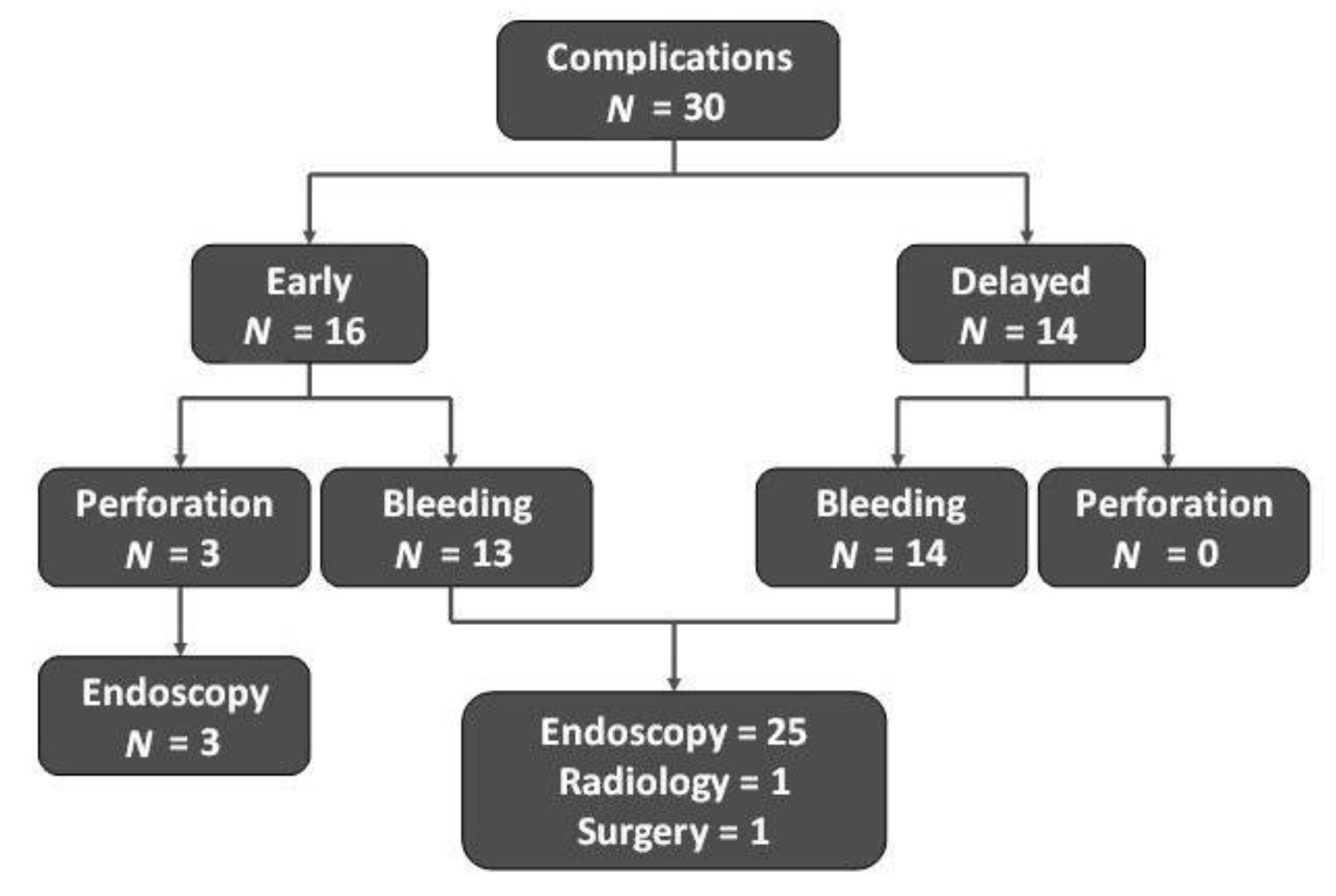
3. Results
4. Discussion
5. Conclusions
Author Contributions
Funding
Conflicts of Interest
References
- Liu, Q.; Ding, L.; Qiu, X.; Meng, F. Updated evaluation of endoscopic submucosal dissection versus surgery for early gastric cancer: A systematic review and meta-analysis. Int. J. Surg. 2019, 73, 28–41. [Google Scholar] [CrossRef] [PubMed]
- Ngamruengphong, S.; Abe, S.; Oda, I. Endoscopic management of early gastric adenocarcinoma and preinvasive gastric lesions. Surg. Clin. N. Am. 2017, 97, 371–385. [Google Scholar] [CrossRef] [PubMed]
- Bourke, M.J.; Neuhaus, H.; Bergman, J.J. Endoscopic submucosal dissection: Indications and application in Western endoscopy practice. Gastroenterology 2018, 154, 1887–1900. [Google Scholar] [CrossRef]
- Chan, W.L.; Lam, K.O.; Lee, V.H.F.; Davidson, M.; So, T.H.; Li, J.S.; Chau, I.; Kwong, D.L.W. Gastric cancer —From aetiology to management: Differences between the East and the West. Clin. Oncol. 2019, 31, 570–577. [Google Scholar] [CrossRef] [PubMed]
- Participants in the Paris Workshop. The Paris endoscopic classification of superficial neoplastic lesions: Esophagus, stomach, and colon: November 30 to December 1, 2002. Gastrointest. Endosc. 2003, 58, S3–S43. [Google Scholar] [CrossRef]
- Dixon, M.F. Gastrointestinal epithelial neoplasia: Vienna revisited. Gut 2002, 51, 130–131. [Google Scholar] [CrossRef] [PubMed]
- Japanese Gastric Cancer Association. Japanese gastric cancer treatment guidelines 2010 (ver. 3). Gastric Cancer 2011, 14, 113–123. [Google Scholar] [CrossRef] [PubMed]
- Pimentel-Nunes, P.; Dinis-Ribeiro, M.; Ponchon, T.; Repici, A.; Vieth, M.; De Ceglie, A.; Vieth, M.; De Ceglie, A.; Amato, A.; Berr, F.; et al. Endoscopic submucosal dissection: European Society of Gastrointestinal Endoscopy (ESGE) Guideline. Endoscopy 2015, 47, 829–854. [Google Scholar] [CrossRef] [PubMed]
- Bray, F.; Ferlay, J.; Soerjomataram, I.; Siegel, R.L.; Torre, L.A.; Jemal, A. Global cancer statistics 2018: GLOBOCAN estimates of incidence and mortality worldwide for 36 cancers in 185 countries. CA Cancer J. Clin. 2018, 68, 394–424. [Google Scholar] [CrossRef] [PubMed]
- Yue, H.; Shan, L.; Bin, L. The significance of OLGA and OLGIM staging systems in the risk assessment of gastric cancer: A systematic review and meta-analysis. Gastric Cancer 2018, 21, 579–587. [Google Scholar] [CrossRef] [PubMed]
- Pimentel-Nunes, P.; Libânio, D.; Marcos-Pinto, R.; Areia, M.; Leja, M.; Esposito, G.; Garrido, M.; Kikuste, I.; Megraud, F.; Matysiak-Budnik, T.; et al. Management of epithelial precancerous conditions and lesions in the stomach (MAPS II): European Society of Gastrointestinal Endoscopy (ESGE), European Helicobacter and Microbiota Study Group (EHMSG), European Society of Pathology (ESP), and Sociedade Portuguesa de Endoscopia Digestiva (SPED) guideline update 2019. Endoscopy 2019, 5, 365–388. [Google Scholar]
- Maselli, R.; Iacopini, F.; Azzolini, F.; Petruzziello, L.; Manno, M.; De Luca, L.; Cecinato, P.; Fiori, G.; Staiano, T.; Rizzotto, E.R.; et al. Endoscopic submucosal dissection: Italian national survey on current practices, training and outcomes. Dig. Liver Dis. 2020, 52, 64–71. [Google Scholar] [CrossRef] [PubMed]
- Kim, Y.; Kuan, J.Y.; Ratcliffe, E.; Baskind, S.; Prasad, N.; Assadsangabi, A.; Ang, J. Long-term follow-up of endoscopic submucosal dissection of gastric dysplasia and early neoplasia in a United Kingdom Caucasian population—A tertiary centre experience. Scand. J. Gastroenterol. 2020. [Google Scholar] [CrossRef] [PubMed]
- Probst, A.; Schneider, A.; Schaller, T.; Anthuber, M.; Ebigbo, A.; Messmann, H. Endoscopic submucosal dissection for early gastric cancer: Are expanded resection criteria safe for Western patients? Endoscopy 2017, 49, 855–865. [Google Scholar] [CrossRef] [PubMed]
- Karpińska-Kaczmarczyk, K.; Białek, A.; Lewandowska, M.; Dobak, E.; Ławniczak, M.; Urasińska, E. Histomorphologic features of early gastric carcinoma treated by endoscopic submucosal dissection: Relation to efficiency of endoscopic resection. Scand. J. Gastroenterol. 2016, 51, 1495–1501. [Google Scholar] [CrossRef] [PubMed]
- Mocker, L.; Hildenbrand, R.; Oyama, T.; Sido, B.; Yahagi, N.; Dumoulin, F.L. Implementation of endoscopic submucosal dissection for early upper gastrointestinal tract cancer after primary experience in colorectal endoscopic submucosal dissection. Endosc. Int. Open 2019, 7, E446–E451. [Google Scholar] [CrossRef]
- Catalano, F.; Trecca, A.; Rodella, L.; Lombardo, F.; Tomezzoli, A.; Battista, S.; Silano, M.; Gaj, F.; de Manzoni, G. The modern treatment of early gastric cancer: Our experience in an Italian cohort. Surg. Endosc. 2009, 23, 1581–1586. [Google Scholar] [CrossRef]
- Libanio, D.; Pimentel-Numes, P.; Afonso, L.P.; Henrique, R.; Dinis-Ribeiro, M. Long-term outcomes of gastric endoscopic submucosal-dissection: Focus on metachronous and non-curative resection management. GE Port. J. Gastroenterol. 2017, 24, 31–39. [Google Scholar] [CrossRef] [PubMed]


| Findings | N = 299 Lesions |
|---|---|
| Endoscopic features * | |
| 0–1 s | 20 |
| IIa | 110 |
| IIb | 42 |
| IIc | 9 |
| IIa + IIb | 18 |
| Is-IIa | 2 |
| Histological features | |
| EGC | 80 |
| High-grade dysplasia | 114 |
| Low-grade dysplasia | 103 |
| NET | 2 |
| Reference | Country | EGC (N) | Curative Rate (%) |
|---|---|---|---|
| Kim et al. [13] | UK | 13 | 7.7 |
| Probst et al. [14] | Germany | 122 | 63.9 |
| Petruzziello et al. [12] | Italy | 44 | 65.9 |
| Karpińska-Kaczmarczyk et al. [15] | Poland | 41 | 70.7 |
| Mocker et al. [16] | Germany | 19 | 73.1 |
| Maselli et al. [12] | Italy | 502 | 81.7 |
| Catalano et al. [17] | Italy | 12 | 91.7 |
© 2020 by the authors. Licensee MDPI, Basel, Switzerland. This article is an open access article distributed under the terms and conditions of the Creative Commons Attribution (CC BY) license (http://creativecommons.org/licenses/by/4.0/).
Share and Cite
Manta, R.; Galloro, G.; Pugliese, F.; Angeletti, S.; Caruso, A.; Zito, F.P.; Mangiafico, S.; Marmo, R.; Zullo, A.; Esposito, G.; et al. Endoscopic Submucosal Dissection of Gastric Neoplastic Lesions: An Italian, Multicenter Study. J. Clin. Med. 2020, 9, 737. https://doi.org/10.3390/jcm9030737
Manta R, Galloro G, Pugliese F, Angeletti S, Caruso A, Zito FP, Mangiafico S, Marmo R, Zullo A, Esposito G, et al. Endoscopic Submucosal Dissection of Gastric Neoplastic Lesions: An Italian, Multicenter Study. Journal of Clinical Medicine. 2020; 9(3):737. https://doi.org/10.3390/jcm9030737
Chicago/Turabian StyleManta, Raffaele, Giuseppe Galloro, Francesco Pugliese, Stefano Angeletti, Angelo Caruso, Francesco P. Zito, Santi Mangiafico, Riccardo Marmo, Angelo Zullo, Gianluca Esposito, and et al. 2020. "Endoscopic Submucosal Dissection of Gastric Neoplastic Lesions: An Italian, Multicenter Study" Journal of Clinical Medicine 9, no. 3: 737. https://doi.org/10.3390/jcm9030737
APA StyleManta, R., Galloro, G., Pugliese, F., Angeletti, S., Caruso, A., Zito, F. P., Mangiafico, S., Marmo, R., Zullo, A., Esposito, G., Annibale, B., Mutignani, M., & Conigliaro, R. (2020). Endoscopic Submucosal Dissection of Gastric Neoplastic Lesions: An Italian, Multicenter Study. Journal of Clinical Medicine, 9(3), 737. https://doi.org/10.3390/jcm9030737