Article

Low Levels of Low-Density Lipoprotein Cholesterol and Endothelial Function in Subjects without Lipid-Lowering Therapy

1
Department of Cardiovascular Medicine, Graduate School of Biomedical and Health Sciences, Faculty of Medicine, Hiroshima University, Hiroshima 734-8553, Japan
2
Division of Regeneration and Medicine, Medical Center for Translational and Clinical Research, Hiroshima University Hospital, Hiroshima 734-8551, Japan
3
Department of Biostatistics and Data Science, Graduate School of Medicine, Faculty of Medicine, Osaka University, Osaka 565-0871, Japan
4
Department of Gastroenterology and Metabolism, Graduate School of Biomedical and Health Sciences, Hiroshima University, Hiroshima 734-8551, Japan
5
Department of Physical Therapy, Hiroshima International University, Hiroshima 739-2695, Japan
6
Department of Cardiovascular Regeneration and Medicine, Research Institute for Radiation Biology and Medicine, Hiroshima University, Hiroshima 734-8553, Japan
7
Department of Stem Cell Biology and Medicine, Graduate School of Biomedical and Health Sciences, Faculty of Medicine, Hiroshima University, Hiroshima 734-8553, Japan
*
Author to whom correspondence should be addressed.
J. Clin. Med. 2020, 9(12), 3796; https://doi.org/10.3390/jcm9123796
Submission received: 21 October 2020 / Revised: 10 November 2020 / Accepted: 21 November 2020 / Published: 24 November 2020
(This article belongs to the Section Cardiology)

Abstract

:
An elevation of serum low-density lipoprotein cholesterol (LDL-C) levels has been associated with endothelial dysfunction in statin naïve subjects. However, there is no information on endothelial function in subjects with extremely low levels of LDL-C. The purpose of the present study was to determine the relationship of LDL-C levels, especially low levels of LDL-C, with endothelial function. Endothelial function assessed by flow-mediated vasodilation (FMD) measurement and LDL-C levels were evaluated in 7120 subjects without lipid-lowering therapy. We divided the subjects into five groups by LDL-C levels: <70 mg/dL, 70–99 mg/dL, 100–119 md/dL, 120–139 mg/dL, and ≥140 mg/dL. FMD values were significantly smaller in subjects with LDL-C levels of ≥140 mg/dL than in those with LDL-C levels of 70–99 mg/dL and 100–119 mg/dL (p < 0.001 and p = 0.004, respectively). The FMD values in the LDL-C of <70 mg/dL group were not significantly different from those in the other groups. To evaluate the relationship of extremely low LDL-C levels with endothelial function, we divided the subjects with LDL-C of <70 mg/dL into those with LDL-C levels of <50 mg/dL and 50–69 mg/dL. FMD values were similar in the LDL-C <50 mg/dL group and ≥50 mg/dL group in the propensity score-matched population (p = 0.570). A significant benefit was not found in subjects with low LDL-C levels from the aspect of endothelial function.

1. Introduction

Atherosclerotic cardiovascular disease (ASCVD) is the cause of morbidity and mortality worldwide [1]. An elevation of serum low-density lipoprotein cholesterol (LDL-C) levels is a risk factor of ASCVD [2]. It has been shown in large prospective epidemiologic cohort studies that there is a positive relationship between plasma LDL-C concentrations and ASCVD risk [3,4]. Randomized controlled trials have shown that lowering elevated LDL-C using statins significantly reduces the risk of major cardiovascular events [5,6,7,8]. These findings from epidemiologic and clinical studies indicate that elevation of LDL-C causes ASCVD [9]. It has also been shown that statins have pleiotropic effect. Statin therapy is recommended for cardiovascular disease in the 2018 American College Cardiology (ACC)/America Heart Association (AHA) cholesterol guidelines [10,11]. Higher dosages of statins for therapy are recommended for secondary prevention, and the effectiveness of drugs that lower LDL-C more strongly, including proprotein convertase subtilisin/kexin type 9 (PCSK9) inhibitor and ezetimibe, has been shown [12,13,14]. With the emergence of these new powerful LDL-lowering drugs, the excessive decrease in LDL-C levels is expected.
However, several studies have shown that high LDL-C levels are a good marker of longevity rather than a risk factor of mortality [15,16,17]. Moreover, a recent Korean epidemiological study has shown that low LDL-C levels (<70 mg/dL) without lipid-lowering agents were independently associated with the increase in risk of mortality and even cardiovascular disease mortality [18]. In a prospective observational study, subjects with low LDL-C levels (<70 mg/dL) had a significantly higher risk of intracerebral hemorrhage than did those with LDL-C levels of 70–99 mg/dL, and subjects with LDL-C levels of <50 mg/dL had the highest hazard ratio for risk of intracerebral hemorrhage [19]. Most randomized trials have been for subjects with LDL-C above a certain level, and subjects with very low LDL-C level have been excluded from analysis [12,13,14,20]. The degrees of atherosclerosis and cardiovascular mortality of subjects with extremely low LDL-C levels without lipid-lowering agents have been not elucidated.
The endothelium plays a very important role in maintenance of vascular homeostasis [21]. Endothelial cells synthesize various physiologically active substances including nitric oxide (NO) and these substances cause vascular smooth muscle relaxation and vasodilation [22]. Measurement of flow-mediated vasodilation (FMD) has been used as an assessment of endothelial function [23]. Endothelial dysfunction would be the initial step of atherosclerosis, leading to cardiovascular disease [24,25]. In addition, endothelial dysfunction assessed by impaired FMD is associated with future cardiovascular events [26,27].
We previously showed that vascular function was impaired in statin-naïve subjects with LDL-C levels of >100 mg/dL [28]. However, a significant benefit was not observed in subjects with LDL-C levels of <70 mg/dL. In addition, there is no information on endothelial function in subjects with extremely low levels of LDL-C (<50 mg/dL). Therefore, we evaluated the relationship of low levels of LDL-C with endothelial function in subjects without lipid-lowering therapy.

2. Materials and Methods

2.1. Subjects

A total of 10,247 subjects were enrolled from the FMD Japan registry (n = 7385) and from the Hiroshima University Vascular Function registry (n = 2862) between August 2007 and August 2016. The FMD-J study was a prospective multicenter study conducted at 22 university hospitals and affiliated clinics in Japan to examine the usefulness of FMD in risk stratification for cardiovascular disease in Japanese subjects. The rationale and design of the FMD-J study have been described previously [29]. Subjects with a history of cardiovascular disease (n = 1326), subjects without information on LDL-C levels (n = 728), subjects with triglycerides of more than 400 mg/dL (n = 40), subjects in whom FMD had not been measured accurately (n = 1), subjects who were taking lipid-lowering medicine (n = 1101), and subjects with an estimated glomerular filtration rate (eGFR) < 15 mL/min/1.73 m2 (n = 6) were excluded from this study. Finally, 7120 subjects were enrolled in this study (Figure S1). Cardiovascular disease was defined as cerebrovascular disease and coronary heart disease. Cardiovascular disease including included transient ischemic attack, ischemic stroke, and hemorrhagic stroke was defined as cerebrovascular disease and coronary heart disease. Coronary heart disease included angina pectoris, unstable angina, and myocardial infarction. Hypertension was defined as systolic blood pressure of 140 mm Hg or higher, diastolic pressure of 90 mm Hg or higher, or treatment with oral antihypertensive drugs. Diabetes mellitus was defined by the American Diabetes Association recommendation [30]. Dyslipidemia was defined by the third report of the National Cholesterol Education Program [31]. The Framingham risk score was calculated on the basis of the risk factors, including age, gender, systolic blood pressure, total cholesterol level, high-density lipoprotein cholesterol (HDL-C) level, diabetes mellitus, and smoking habit [32]. LDL-C concentration was calculated using the Friedewald equation. The protocol of the current study was approved by the ethical committees of the participating institutions and registered in the University Hospital Medical Information Network Clinical Trials Registry (UMIN000012950). Informed consent was obtained from all of the participants.

2.2. Measurement of FMD

Subjects fasted overnight for at least 12 h and abstained from caffeine, alcohol, smoking, and antioxidant vitamins on the day of the FMD examination [27,28]. The subjects were kept in the supine position in a quiet, dark, air-conditioned room (constant temperature of 22–25 °C) through the study [27,28]. The protocol of FMD measurement was previously described [27,28]. Briefly, vascular response to reactive hyperemia in the brachial artery was used for assessment of endothelium-dependent FMD. We used a high-resolution ultrasonography system (UNEXEF18G, UNEX Co, Nagoya, Japan) for evaluation of FMD. The brachial artery was scanned longitudinally 5 to 10 cm above the elbow. When the clearest B-mode image of the anterior and posterior intimal interfaces between the lumen and vessel wall was obtained, the transducer was held at the same point throughout the scan by a special probe holder (UNEX Co) to ensure consistency of the image. When the tracking gate was placed on the intima, the artery diameter was automatically tracked, and the waveform of diameter changes over the cardiac cycle was displayed in real-time using the FMD mode of the tracking system [27,28]. This allowed the ultrasound images to be optimized at the start of the scan and the transducer position to be adjusted immediately for optimal tracking performance throughout the scan. Baseline longitudinal imaging of the artery was performed for 30 s, and then the blood pressure cuff placed around the forearm was inflated to 50 mmHg above systolic pressure for 5 min. Longitudinal imaging of the artery was recorded continuously until 5 min after cuff deflation. At the same time as the recording of the longitudinal image of the artery, we also observed the short axis image using a high resolution ultrasonography system. Changes in brachial artery diameter were immediately expressed as the percentage change relative to the vessel diameter before cuff inflation [27,28]. FMD was automatically calculated as the percentage change in the peak vessel diameter from the baseline value. The percentage of FMD (peak diameter—baseline diameter)/baseline diameter) was used for analysis. The correlation coefficient between FMD analyzed at the core laboratory and at participant institutions was 0.84 (p < 0.001) [27,28]. We confirmed that the Bland–Altman analysis revealed no systemic bias in the variability of FMD measurement between the participating institutions and the core laboratory [33]. The observers were blind to the form of examination.

2.3. Statistical Analysis

Results are presented as means ± SDs or medians (interquartile range) for continuous variables and as percentages for categorical variables. All reported probability values were two-sided. A probability value of <0.05 was considered statistically significant. In accordance with the LDL-C quartiles and current guidelines, subjects were divided into five groups by LDL-C levels. To assess the association of extremely low LDL-C levels with endothelial function, the subjects with an LDL-C of <70 mg/dL were divided into <50 mg/dL and 50–69 mg/dL groups according to another prospective study [19]. Comparison of variables among two or more groups by differences in the LDL-C levels was performed using one-way analysis of variance (ANOVA). Categorical values, such as medications and medical histories, were compared by means of the χ2 test. Tukey’s post hoc test was used to compare the differences in FMD between these groups. As a sensitivity analysis, propensity score analysis was used to generate a set of matched cases and all remaining participants of this study. A logistic regression model was used to estimate the propensity of LDL-C levels < 70 mg/dL based on variables associated with LDL-C, including age, body mass index, gender, heart rate, glucose, triglycerides, HDL-C, hypertension (yes or no), diabetes mellitus (yes or no), smokers (yes or no), use of anti-hypertensive drugs (yes or no), and use of anti-hyperglycemic therapy (yes or no). With these propensity scores, using a caliper width of 0.2 SDs of the logit of the propensity score, two well-matched groups based on clinical characteristics were created for comparison of the ratio of FMD < 4.0%, which was the division point for the lowest FMD quartile in all subjects. All of the statistical analyses were conducted using JMP version 13.0 software (SAS Institute, Cary, NC, USA) and Stata version 17 software (Stata Corporation, College Station, TX, USA).

3. Results

3.1. Baseline Characteristics

The quartiles of LDL-C were 99 mg/dL, 118 mg/dL (median), and 139 mg/dL. According to these quartiles and current guidelines, we divided the subjects into five groups by LDL-C levels: < 70 mg/dL, 70–99 mg/dL, 100–119 mg/dL, 120–139 mg/dL, and ≥ 140 mg/dL. Baseline characteristics of all of the subjects according to LDL-C levels are summarized in Table 1. Of the 7120 subjects, 5465 (76.8%) were men, 2924 (41.1%) had hypertension, 2986 (41.9%) had dyslipidemia, 511 (7.2%) had diabetes mellitus, and 2165 (30.5%) were current smokers. There were significant differences among the five groups in age, body mass index, systolic blood pressure, diastolic blood pressure, heart rate, total cholesterol, triglycerides, HDL-C, glucose, use of anti-hypertensive drugs, prevalence of hypertension and dyslipidemia, and percentage of smokers.

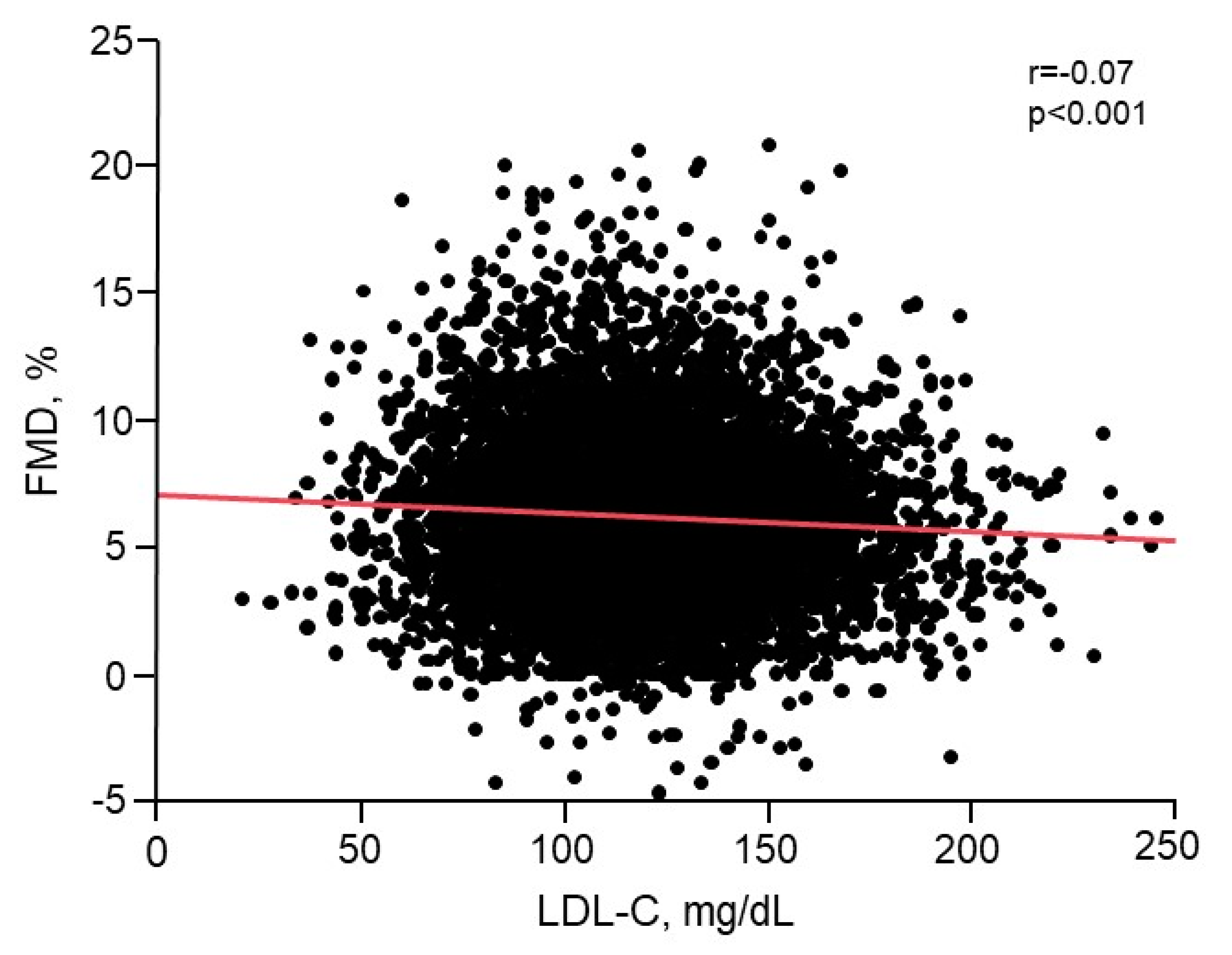
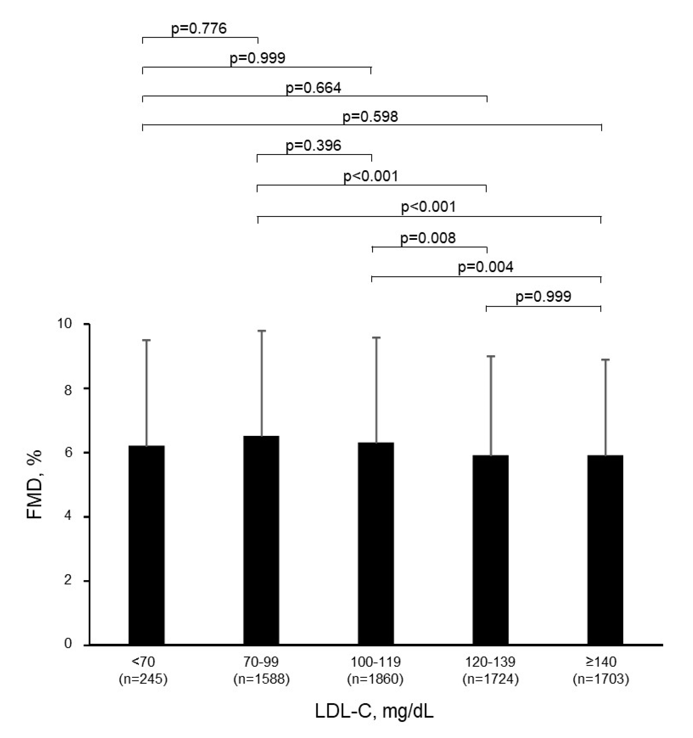
3.2. Relationship of LDL-C with Endothelial Function

There was a significant inverse relationship between FMD and LDL-C levels (r = −0.07, p < 0.001) (Figure 1 and Table S1). Multiple regression analyses revealed that LDL-C was an independent predictor of FMD (Table 2). FMD values were significantly smaller in the LDL-C ≥ 140 mg/dL group than in the 70–99 mg/dL group and the 100–119 mg/dL group (p < 0.001 and p = 0.004, respectively; Figure 2). FMD values were also significantly smaller in the 120–139 mg/dL group than in the 70–99 mg/dL group and the 100–119 mg/dL group (p < 0.001 and p = 0.008, respectively; Figure 2). Figure 1 shows the relationship between FMD and LDL-C.

3.3. Relationship of Extremely Low LDL-C with Endothelial Function

The FMD values in the LDL-C < 70 mg/dL group were not significantly different from those in the other groups (Table 1 and Figure 2). As a sensitivity analysis, propensity score analysis was used to generate a set of matched cases (subjects with LDL-C levels of <70 mg/dL) and all remaining participants of this study (≥70 mg/dL). A logistic regression model was used to estimate the propensity of LDL-C levels < 70 mg/dL based on variables associated with LDL-C, including age, body mass index, gender, heart rate, glucose, triglycerides, HDL-C, hypertension (yes or no), diabetes mellitus (yes or no), smokers (yes or no), use of anti-hypertensive drugs (yes or no), and use of anti-hyperglycemic therapy (yes or no). With these propensity scores, using a caliper width of 0.2 standard deviations of the logit of the propensity score, two well-matched groups based on clinical characteristics were created for comparison of the ratio of FMD < 4.0%, which was the division point for the lowest quartile of FMD in all subjects. FMD was not significantly different between the LDL-C < 70 mg/dL group and ≥70 mg/dL group in the propensity score-matched population (p = 0.386) (Table 3). To evaluate the relationship of extremely low LDL-C levels with endothelial function, we divided the subjects with an LDL-C of <70 mg/dL into a <50 mg/dL group (n = 35) and a 50–69 mg/dL group (n = 210). Baseline characteristics are summarized in Table 4. There were significant differences among the six groups in age, body mass index, systolic blood pressure, diastolic blood pressure, heart rate, total cholesterol, triglycerides, HDL-C, glucose, use of anti-hypertensive drugs, prevalence of hypertension and dyslipidemia, and percentage of smokers.
There were no significant differences among the LDL-C < 50 mg/dL group, LDL-C 50–69 mg/dL group, and LDL-C 70–99 mg/dL group in FMD, and there were no significant differences among the LDL-C < 50 mg/dL group, LDL-C 50–69 mg/dL group, and LDL-C ≥ 140 mg/dL group in FMD (Figure S2). The propensity score analysis was used to compare the <50 mg/dL group with all remaining participants. The variables used for the propensity score were the same as those described earlier. There was no significant difference in FMD values between the LDL-C < 50 mg/dL group and ≥ 50 mg/dL group in the propensity score-matched population (p = 0.570) (Table S2).

4. Discussion

In the present study, there was a significant inverse relationship between FMD and LDL-C levels in subjects without cardiovascular disease and not receiving lipid-lowering therapy. Although there was not much difference in FMD among the groups categorized by LDL-C levels, FMD was highest in subjects with LDL-C of 70–99 mg/dL. FMD in the low LDL-C groups, including <70 mg/dL and <50 mg/dL groups, were not significantly different from that in the other groups. FMD values were similar in the low LDL-C groups and all remaining participants in the propensity score-matched population.
In several epidemiological studies, there was an inverse relationship between total cholesterol and all-cause mortality, which could not be explained by reverse causality (i.e., serious diseases cause low cholesterol) [15,16,17]. A recent epidemiological study has shown the association of low levels of LDL-C (<70 mg/dL) with the increase in risk of cardiovascular disease in healthy Korean subjects without lipid-lowering therapy [18]. In high-risk primary prevention individuals in the REGARDS study, low high-sensitivity C reactive protein levels of <2 mg/dL appeared to be related to the decrease in risk of cardiovascular complications, whereas low LDL-C levels of <70 mg/dL were not related to prevention of cardiovascular complications [34]. Results of a 9-year follow-up of 96043 participants without cardiovascular complications and cancer at baseline showed that subjects with LDL-C levels <70 mg/dL had a higher risk of intracerebral hemorrhage than did those with LDL-C levels of 70–99 mg/dL, and adjusted hazard ratios were 1.65 for LDL-C levels of 50–69 mg/dL and 2.69 for LDL-C levels <50 mg/dL [19]. It is likely that low LDL-C levels do not always mean longevity and low cardiovascular morbidity.
In statin-naïve subjects, several investigators have reported an association of LDL-C levels with endothelial function assessed by FMD [28,35]. In a cross-sectional observational study of relatively young and healthy military men, FMD was correlated linearly with LDL-C, and FMD values were highest in subjects with an LDL-C of <100 mg/dL [35]. Our previous study showed that vascular function is impaired in subjects with an LDL-C of >100 mg/dL and that the optimal cutoff level for maintenance of vascular function is an LDL-C of 100 mg/dL [28]. In the present study, FMD values were highest in subjects with LDL levels of 70–99 mg/dL, consistent with the results of previous studies [28,35].
There are many reports showing that statin treatment improved endothelial function [36,37,38,39]. As the underlying mechanism, it has been shown that long-term statin treatment improves endothelial function via inhibition of vascular cell senescence, amelioration of oxidative stress, and normalization of endothelial and inducible NO synthase imbalance [40]. Statins have not only LDL-C-lowering effects but also pleiotropic effects for atherosclerosis [10]. A meta-analysis revealed that most of the anti-inflammatory effects of statins were related to the magnitude of LDL reduction [41]. On the other hand, there are some reports showing that endothelial function was not improved by treatment with a statin [42,43,44]. In patients with diabetes mellitus without manifest cardiovascular disease, 2-year statin therapy had no effect on FMD [42]. In patients with diabetes mellitus and dyslipidemia without cardiovascular disease, intensive lipid-lowering therapy by atorvastatin did not have beneficial effects on endothelial function [43]. In patients with insulin resistant familial combined hyperlipidemia, 12-week treatment with rosuvastatin did not improve endothelial function despite plasma cholesterol being significantly decreased [44]. In the present study, we selected subjects with reduced LDL-C who were not taking lipid-improving agents including statins. We were not able to directly compare our results with the results of studies using statins. In a previous study, there was no significant association of LDL-C levels with FMD in subjects receiving statins.
Meta-analyses of data from 27 randomized clinical trials in the Cholesterol Treatment Trialists’ Collaboration showed that statins decreased the risk of cardiovascular events by 21% for each 38.7 mg/dL reduction in LDL-C [45]. The efficacy of statin use for prevention of cardiovascular events has been established. We do not deny the beneficial effects of lowering LDL-C to <50 mg/dL by statin treatment on cardiovascular events. Indeed, the Improved Reduction of Outcomes: Vytorin Efficacy International Trial (IMPROVE-IT) showed that maintenance of a mean LDL-C level of 53.7 mg/dL by treatment with a statin and ezetimibe improved cardiovascular events compared to a mean LDL-C level of 69.5 mg/dL [12]. The further Cardiovascular Outcomes Research with PCSK9 Inhibition in Subjects with Elevated Risk (FOURIER) trial showed that a decrease in LDL-C level from 92 to 30 mg/dL by treatment with a PCSK-9 inhibitor in combination with statins decreased cardiovascular events by about 15% [13]. These findings suggest that for the prevention of cardiovascular events, the rule for the decrease in LDL-C levels is the earlier the better and the lower the better. We may have to consider separately the effects of LDL-C levels with lipid-lowering therapy and natural LDL-C levels on endothelial function and cardiovascular events.
There are a number of limitations in the present study. First, this study was a cross-sectional study. We could evaluate the association of LDL-C levels with endothelial function but not causality. Second, we did not know the accurate causes of extremely low LDL-C in the present study. In order to evaluate subjects without secondary causes of low LDL-C, we excluded subjects who were taking lipid-lowering medicine and eGFR < 15 mL/min/1.73 m2, BMI < 16 kg/m2, malignant tumor, and active infection. However, there may be residual unrecognized confounders between decrease in LDL-C and endothelial dysfunction. Confounding factors may remain even after the propensity score match. Our database had limited information on physical activity and educational status. These confounders can affect the relationship between FMD and LDL-C levels. In addition, lipoprotein disorders in which LDL-C concentrations are genetically reduced have been reported, and among them, patients with mutations in the ANGPTL3 and PCSK9 genes are asymptomatic [46]. Although sequence variants in the PCSK9 gene were associated with reduced LDL-C levels and reduced risk of cardiovascular events, LDL-C levels were approximately 100 mg/dL, which is not an extremely low level such as a level of <70 mg/dL or <50mg/dL [47]. One case of a homozygote for PCSK9 nonsense mutations that had an extremely low LDL-C concentration of approximately 16 mg/dL was reported [48]. However, there has been no report on the relationship between PCSK9 nonsense mutations and early atherosclerosis. Mutations of loss-of-function in the ANGPTL3 gene are associated with hypolipidemia, which is characterized by low levels of HDL-C, LDL-C, and triglycerides [49]. Although heterozygous ANGPTL3 loss-of-function variants were associated with a 41% lower odds of coronary artery disease, the median level of LDL-C in the heterozygous variants was approximately 112 mg/dL [50]. It has been reported that homozygotes for ANGPTL3 have been associated with the increase in carotid intima-media thickness and decreased FMD despite low plasma LDL-C levels (approximately 63 mg/dL) [51]. This increased risk for the development of early vascular atherosclerotic changes might be related to impaired HDL function. Assessment of genetic abnormalities related to LDL-C levels would draw more specific conclusions regarding the role of low LDL-C in vascular function. Third, in the present study, there was only a small number of subjects with extremely low LDL-C levels (two subjects with LDL-C levels of <30 mg/dL). Further studies involving a large number of subjects with extremely low LDL-C levels are needed to evaluate the relationship between low LDL-C levels and endothelial function.

5. Conclusions

In conclusion, FMD values were highest in subjects with LDL-C of 70–99 mg/dL. In the propensity score-matched population, FMD values were similar in the low LDL-C groups and all remaining participants. Although the precise mechanisms of the relationship between low LDL-C and endothelial function is not known, a significant benefit was not found in subjects with low LDL-C levels who were not receiving lipid-lowering treatment and had no history of cardiovascular disease from the aspect of endothelial function.

Supplementary Materials

The following are available online at https://www.mdpi.com/2077-0383/9/12/3796/s1, Table S1: Univariate analysis of the relationship between FMD and variables; Table S2: Clinical characteristics in propensity score mathced subjects; Figure S1: Flow chart of the study design; Figure S2: Comparison of FMD values of each group.

Author Contributions

Y.H. (Yukihito Higashi) and Y.T. drafting the article and conception of this study; M.K., T.M., T.Y., T.H., Y.H. (Yiming Han), S.K., C.G., Y.A., F.M.Y., and A.N., measuring the brachial artery diameter and FMD; Y.K., E.H. and K.C. revising the article critically for important intellectual content. All authors have read and agreed to the published version of the manuscript.

Funding

Grant-in-Aid for Scientific Research from the Ministry of Education, Science and Culture of Japan (18590815 and 21590898 to Y.H. (Yukihito Higashi)) and a Grant in Aid of Japanese Arteriosclerosis Prevention Fund (to Y.H. (Yukihito Higashi)).

Acknowledgments

The authors would like to thank all patients who participated in this study. In addition, we thank Miki Kumiji, Megumi Wakisaka, Ki-ichiro Kawano and Satoko Michiyama for their excellent secretarial assistance; FMD-J investigators of Takayuki Hidaka; Shuji Nakamura; Junko Soga; Yuichi Fujii; Naomi Idei; Noritaka Fujimura; Shinsuke Mikami; Yumiko Iwamoto; Akimichi Iwamoto; Takeshi Matsumoto; Nozomu Oda (Department of Cardiovascular Medicine, Hiroshima University Graduate School of Biomedical Sciences, Hiroshima, Japan); Kana Kanai; Hraruka Morimoto (Department of Cardiovascular Regeneration and Medicine, Research Institute for Radiation Biology and Medicine, Hiroshima University, Hiroshima, Japan); Tomohisa Sakashita; Yoshiki Kudo (Department of Obstetrics and Gynecology, Hiroshima University Graduate School of Biomedical Sciences, Hiroshima, Japan); Taijiro Sueda (Department of Surgery, Hiroshima University Graduate School of Biomedical Sciences, Hiroshima, Japan); Hirofumi Tomiyama; Akira Yamashina (Department of Cardiology, Tokyo Medical University, Tokyo, Japan); Bonpei Takase (Division of Biomedical Engineering, National Defense Medical College Research Institute, Tokorozawa, Japan); Takahide Kohro (Department of Cardiology, Tokyo Medical University, Tokyo, Japan); Toru Suzuki (Cardiovascular Medicine, University of Leicester, Leicester, UK); Tomoko Ishizu (Cardiovascular Division, Institute of Clinical Medicine, University of Tsukuba, Ibaraki, Japan); Shinichiro Ueda (Department of Clinical Pharmacology and Therapeutics, University of the Ryukyu School of Medicine, Okinawa, Japan); Tsutomu Yamazaki (Clinical Research Support Center, Faculty of Medicine, The University of Tokyo, Tokyo, Japan); Tomoo Furumoto (Department of Cardiovascular Medicine, Hokkaido University Graduate School of Medicine, Hokkaido, Japan); Kazuomi Kario (Division of Cardiovascular Medicine, Jichi Medical University School of Medicine, Tochigi, Japan); Teruo Inoue (Department of Cardiovascular Medicine, Dokkyo Medical University, Mibu, Tochigi, Japan); Shinji Koba (Department of Medicine, Division of Cardiology, Showa University School of Medicine, Tokyo, Japan); Kentaro Watanabe (Department of Neurology, Hematology, Metaboism, Endocrinology and Diabetology (DNHMED), Yamagata University School of Medicine, Yamagata, Japan); Yasuhiko Takemoto (Department of Internal Medicine and Cardiology, Osaka City University Graduate School of Medicine, Osaka, Japan); Takuzo Hano (Department of Medical Education and Population-based Medicine, Postgraduate School of Medicine, Wakayama Medical University, Wakayama, Japan); Masataka Sata (Department of Cardiovascular Medicine, Institute of Health Biosciences, The University of Tokushima Graduate School, Tokushima, Japan); Yutaka Ishibashi (Department of General Medicine, Shimane University Faculty of Medicine, Izumo, Japan); Koichi Node (Department of Cardiovascular and Renal Medicine, Saga University, Saga, Japan); Koji Maemura (Department of Cardiovascular Medicine, Nagasaki University Graduate School of Biomedical Sciences, Nagasaki, Japan); Yusuke Ohya (The Third Department of Internal Medicine, University of the Ryukyus, Okinawa, Japan); Taiji Furukawa (Department of Internal Medicine, Teikyo University School of Medicine, Tokyo, Japan); Hiroshi Ito (Department of Cardiovascular Medicine, Okayama University Graduate School of Medicine, Dentistry and Pharmaceutical Sciences, Japan); Hisao Ikeda (Faculty of Fukuoka Medical Technology, Teikyo University, Omuta, Japan).

Conflicts of Interest

The authors declare no conflict of interest.

References

  1. Mendis, S.; Davis, S.; Norrving, B. Organizational update: The world health organization global status report on noncommunicable diseases 2014; one more landmark step in the combat against stroke and vascular disease. Stroke 2015, 46, e121–e122. [Google Scholar] [CrossRef]
  2. Lloyd-Jones, D.M.; Huffman, M.D.; Karmali, K.N.; Sanghavi, D.M.; Wright, J.S.; Pelser, C.; Gulati, M.; Masoudi, F.A.; Goff, D.C., Jr. Estimating Longitudinal Risks and Benefits from Cardiovascular Preventive Therapies Among Medicare Patients: The Million Hearts Longitudinal ASCVD Risk Assessment Tool: A Special Report from the American Heart Association and American College of Cardiology. Circulation 2017, 135, e793–e813. [Google Scholar] [CrossRef] [Green Version]
  3. Di Angelantonio, E.; Gao, P.; Pennells, L.; Kaptoge, S.; Caslake, M.; Thompson, A.; Butterworth, A.S.; Sarwar, N.; Wormser, D.; Saleheen, D.; et al. Lipid-related markers and cardiovascular disease prediction. JAMA 2012, 307, 2499–2506. [Google Scholar] [CrossRef] [PubMed] [Green Version]
  4. Abdullah, S.M.; Defina, L.F.; Leonard, D.; Barlow, C.E.; Radford, N.B.; Willis, B.L.; Rohatgi, A.; McGuire, D.K.; de Lemos, J.A.; Grundy, S.M.; et al. Long-Term Association of Low-Density Lipoprotein Cholesterol with Cardiovascular Mortality in Individuals at Low 10-Year Risk of Atherosclerotic Cardiovascular Disease. Circulation 2018, 138, 2315–2325. [Google Scholar] [CrossRef]
  5. Mihaylova, B.; Emberson, J.; Blackwell, L.; Keech, A.; Simes, J.; Barnes, E.H.; Voysey, M.; Gray, A.; Collins, R.; Baigent, C. The effects of lowering LDL cholesterol with statin therapy in people at low risk of vascular disease: Meta-analysis of individual data from 27 randomised trials. Lancet 2012, 380, 581–590. [Google Scholar] [CrossRef] [PubMed]
  6. Collins, R.; Reith, C.; Emberson, J.; Armitage, J.; Baigent, C.; Blackwell, L.; Blumenthal, R.; Danesh, J.; Smith, G.D.; DeMets, D.; et al. Interpretation of the evidence for the efficacy and safety of statin therapy. Lancet 2016, 388, 2532–2561. [Google Scholar] [CrossRef] [Green Version]
  7. Cholesterol Treatment Trialists’ Collaboration. Efficacy and safety of statin therapy in older people: A meta-analysis of individual participant data from 28 randomised controlled trials. Lancet 2019, 393, 407–415. [Google Scholar] [CrossRef] [Green Version]
  8. Amarenco, P.; Kim, J.S.; Labreuche, J.; Charles, H.; Abtan, J.; Bejot, Y.; Cabrejo, L.; Cha, J.K.; Ducrocq, G.; Giroud, M.; et al. A Comparison of Two LDL Cholesterol Targets after Ischemic Stroke. N. Engl. J. Med. 2019. [Google Scholar] [CrossRef]
  9. Ference, B.A.; Ginsberg, H.N.; Graham, I.; Ray, K.K.; Packard, C.J.; Bruckert, E.; Hegele, R.A.; Krauss, R.M.; Raal, F.J.; Schunkert, H.; et al. Low-density lipoproteins cause atherosclerotic cardiovascular disease. 1. Evidence from genetic, epidemiologic, and clinical studies. A consensus statement from the European Atherosclerosis Society Consensus Panel. Eur. Heart J. 2017, 38, 2459–2472. [Google Scholar] [CrossRef] [Green Version]
  10. Oesterle, A.; Laufs, U.; Liao, J.K. Pleiotropic Effects of Statins on the Cardiovascular System. Circ. Res. 2017, 120, 229–243. [Google Scholar] [CrossRef] [Green Version]
  11. Grundy, S.M.; Stone, N.J.; Bailey, A.L.; Beam, C.; Birtcher, K.K.; Blumenthal, R.S.; Braun, L.T.; de Ferranti, S.; Faiella-Tommasino, J.; Forman, D.E.; et al. 2018 AHA/ACC/AACVPR/AAPA/ABC/ACPM/ADA/AGS/APhA/ASPC/NLA/PCNA Guideline on the Management of Blood Cholesterol: A Report of the American College of Cardiology/American Heart Association Task Force on Clinical Practice Guidelines. Circulation 2019, 139, e1082–e1143. [Google Scholar] [CrossRef] [PubMed]
  12. Cannon, C.P.; Blazing, M.A.; Giugliano, R.P.; McCagg, A.; White, J.A.; Theroux, P.; Darius, H.; Lewis, B.S.; Ophuis, T.O.; Jukema, J.W.; et al. Ezetimibe Added to Statin Therapy after Acute Coronary Syndromes. N. Engl. J. Med. 2015, 372, 2387–2397. [Google Scholar] [CrossRef] [PubMed] [Green Version]
  13. Sabatine, M.S.; Giugliano, R.P.; Keech, A.C.; Honarpour, N.; Wiviott, S.D.; Murphy, S.A.; Kuder, J.F.; Wang, H.; Liu, T.; Wasserman, S.M.; et al. Evolocumab and Clinical Outcomes in Patients with Cardiovascular Disease. N. Engl. J. Med. 2017, 376, 1713–1722. [Google Scholar] [CrossRef] [PubMed]
  14. Schwartz, G.G.; Steg, P.G.; Szarek, M.; Bhatt, D.L.; Bittner, V.A.; Diaz, R.; Edelberg, J.M.; Goodman, S.G.; Hanotin, C.; Harrington, R.A.; et al. Alirocumab and Cardiovascular Outcomes after Acute Coronary Syndrome. N. Engl. J. Med. 2018, 379, 2097–2107. [Google Scholar] [CrossRef]
  15. Petursson, H.; Sigurdsson, J.A.; Bengtsson, C.; Nilsen, T.I.; Getz, L. Is the use of cholesterol in mortality risk algorithms in clinical guidelines valid? Ten years prospective data from the Norwegian HUNT 2 study. J. Eval. Clin. Pract. 2012, 18, 159–168. [Google Scholar] [CrossRef]
  16. Nago, N.; Ishikawa, S.; Goto, T.; Kayaba, K. Low cholesterol is associated with mortality from stroke, heart disease, and cancer: The Jichi Medical School Cohort Study. J. Epidemiol. 2011, 21, 67–74. [Google Scholar] [CrossRef] [Green Version]
  17. Liang, Y.; Vetrano, D.L.; Qiu, C. Serum total cholesterol and risk of cardiovascular and non-cardiovascular mortality in old age: A population-based study. BMC Geriatr. 2017, 17, 294. [Google Scholar] [CrossRef] [Green Version]
  18. Sung, K.C.; Huh, J.H.; Ryu, S.; Lee, J.Y.; Scorletti, E.; Byrne, C.D.; Kim, J.Y.; Hyun, D.S.; Ko, S.B. Low Levels of Low-Density Lipoprotein Cholesterol and Mortality Outcomes in Non-Statin Users. J. Clin. Med. 2019, 8, 1571. [Google Scholar] [CrossRef] [Green Version]
  19. Ma, C.; Gurol, M.E.; Huang, Z.; Lichtenstein, A.H.; Wang, X.; Wang, Y.; Neumann, S.; Wu, S.; Gao, X. Low-density lipoprotein cholesterol and risk of intracerebral hemorrhage: A prospective study. Neurology 2019, 93, e445–e457. [Google Scholar] [CrossRef]
  20. Vallejo-Vaz, A.J.; Robertson, M.; Catapano, A.L.; Watts, G.F.; Kastelein, J.J.; Packard, C.J.; Ford, I.; Ray, K.K. Low-Density Lipoprotein Cholesterol Lowering for the Primary Prevention of Cardiovascular Disease Among Men with Primary Elevations of Low-Density Lipoprotein Cholesterol Levels of 190 mg/dL or Above: Analyses from the WOSCOPS (West of Scotland Coronary Prevention Study) 5-Year Randomized Trial and 20-Year Observational Follow-Up. Circulation 2017, 136, 1878–1891. [Google Scholar] [CrossRef]
  21. Pi, X.; Xie, L.; Patterson, C. Emerging Roles of Vascular Endothelium in Metabolic Homeostasis. Circ. Res. 2018, 123, 477–494. [Google Scholar] [CrossRef] [PubMed]
  22. Vanhoutte, P.M.; Zhao, Y.; Xu, A.; Leung, S.W. Thirty Years of Saying NO: Sources, Fate, Actions, and Misfortunes of the Endothelium-Derived Vasodilator Mediator. Circ. Res. 2016, 119, 375–396. [Google Scholar] [CrossRef] [PubMed] [Green Version]
  23. Corretti, M.C.; Anderson, T.J.; Benjamin, E.J.; Celermajer, D.; Charbonneau, F.; Creager, M.A.; Deanfield, J.; Drexler, H.; Gerhard-Herman, M.; Herrington, D.; et al. Guidelines for the ultrasound assessment of endothelial-dependent flow-mediated vasodilation of the brachial artery: A report of the International Brachial Artery Reactivity Task Force. J. Am. Coll. Cardiol. 2002, 39, 257–265. [Google Scholar] [CrossRef] [Green Version]
  24. Ross, R. Atherosclerosis—An inflammatory disease. N. Engl. J. Med. 1999, 340, 115–126. [Google Scholar] [CrossRef] [PubMed]
  25. Higashi, Y.; Noma, K.; Yoshizumi, M.; Kihara, Y. Endothelial function and oxidative stress in cardiovascular diseases. Circ. J. 2009, 73, 411–418. [Google Scholar] [CrossRef] [PubMed] [Green Version]
  26. Matsuzawa, Y.; Kwon, T.G.; Lennon, R.J.; Lerman, L.O.; Lerman, A. Prognostic Value of Flow-Mediated Vasodilation in Brachial Artery and Fingertip Artery for Cardiovascular Events: A Systematic Review and Meta-Analysis. J. Am. Heart Assoc. 2015, 4. [Google Scholar] [CrossRef] [Green Version]
  27. Maruhashi, T.; Soga, J.; Fujimura, N.; Idei, N.; Mikami, S.; Iwamoto, Y.; Iwamoto, A.; Kajikawa, M.; Matsumoto, T.; Oda, N.; et al. Endothelial Dysfunction, Increased Arterial Stiffness, and Cardiovascular Risk Prediction in Patients with Coronary Artery Disease: FMD-J (Flow-Mediated Dilation Japan) Study A. J. Am. Heart Assoc. 2018, 7. [Google Scholar] [CrossRef] [Green Version]
  28. Matsui, S.; Kajikawa, M.; Hida, E.; Maruhashi, T.; Iwamoto, Y.; Iwamoto, A.; Oda, N.; Kishimoto, S.; Hidaka, T.; Kihara, Y.; et al. Optimal Target Level of Low-density Lipoprotein Cholesterol for Vascular Function in Statin Naive Individuals. Sci. Rep. 2017, 7, 8422. [Google Scholar] [CrossRef] [Green Version]
  29. Tomiyama, H.; Kohro, T.; Higashi, Y.; Takase, B.; Suzuki, T.; Ishizu, T.; Ueda, S.; Yamazaki, T.; Furumoto, T.; Kario, K.; et al. A multicenter study design to assess the clinical usefulness of semi-automatic measurement of flow-mediated vasodilatation of the brachial artery. Int. Heart J. 2012, 53, 170–175. [Google Scholar] [CrossRef] [Green Version]
  30. Expert Committee on the Diagnosis and Clasification of Diabetes Mellitus. American Diabetes Association: Clinical practice recommendations 1999. Diabetes Care 1999, 22 (Suppl. S1), S1–S114. [Google Scholar]
  31. Expert Panel on Detection, Evaluation, and Treatment of High Blood Cholesterol in Adults. Executive Summary of the Third Report of The National Cholesterol Education Program (NCEP) Expert Panel on Detection, Evaluation, And Treatment of High Blood Cholesterol in Adults (Adult Treatment Panel III). JAMA 2001, 285, 2486–2497. [Google Scholar] [CrossRef] [PubMed]
  32. Wilson, P.W.; D’Agostino, R.B.; Levy, D.; Belanger, A.M.; Silbershatz, H.; Kannel, W.B. Prediction of coronary heart disease using risk factor categories. Circulation 1998, 97, 1837–1847. [Google Scholar] [CrossRef] [PubMed] [Green Version]
  33. Tomiyama, H.; Kohro, T.; Higashi, Y.; Takase, B.; Suzuki, T.; Ishizu, T.; Ueda, S.; Yamazaki, T.; Furumoto, T.; Kario, K.; et al. Reliability of measurement of endothelial function across multiple institutions and establishment of reference values in Japanese. Atherosclerosis 2015, 242, 433–442. [Google Scholar] [CrossRef] [PubMed] [Green Version]
  34. Penson, P.E.; Long, D.L.; Howard, G.; Toth, P.P.; Muntner, P.; Howard, V.J.; Safford, M.M.; Jones, S.R.; Martin, S.S.; Mazidi, M.; et al. Associations between very low concentrations of low density lipoprotein cholesterol, high sensitivity C-reactive protein, and health outcomes in the Reasons for Geographical and Racial Differences in Stroke (REGARDS) study. Eur. Heart J. 2018, 39, 3641–3653. [Google Scholar] [CrossRef]
  35. Laclaustra, M.; Frangi, A.F.; Frangi, A.G.; Casasnovas, J.A.; Cia, P. Association of endothelial function and vascular data with LDL-c and HDL-c in a homogeneous population of middle-aged, healthy military men: Evidence for a critical role of optimal lipid levels. Int. J. Cardiol. 2008, 125, 376–382. [Google Scholar] [CrossRef]
  36. Brunetti, N.D.; Maulucci, G.; Casavecchia, G.P.; Distaso, C.; De Gennaro, L.; Luigi Pellegrino, P.; Di Biase, M. Improvement in Endothelium Dysfunction in Diabetics Treated with Statins: A Randomized Comparison of Atorvastatin 20 mg versus Rosuvastatin 10 mg. J. Interv. Cardiol. 2007, 20, 481–487. [Google Scholar] [CrossRef]
  37. Frick, M.; Alber, H.F.; Hügel, H.; Schwarzacher, S.P.; Pachinger, O.; Weidinger, F. Short- and long-term changes of flow-mediated vasodilation in patients under statin therapy. Clin. Cardiol. 2002, 25, 291–294. [Google Scholar] [CrossRef]
  38. Karatzis, E.; Lekakis, J.; Papamichael, C.; Andreadou, I.; Cimponeriu, A.; Aznaouridis, K.; Papaioannou, T.G.; Protogerou, A.; Mavrikakis, M. Rapid effect of pravastatin on endothelial function and lipid peroxidation in unstable angina. Int. J. Cardiol. 2005, 101, 65–70. [Google Scholar] [CrossRef]
  39. Kabaklić, A.; Fras, Z. Moderate-dose atorvastatin improves arterial endothelial function in patients with angina pectoris and normal coronary angiogram: A pilot study. Arch. Med Sci. AMS 2017, 13, 827–836. [Google Scholar] [CrossRef] [Green Version]
  40. Gong, X.; Ma, Y.; Ruan, Y.; Fu, G.; Wu, S. Long-term atorvastatin improves age-related endothelial dysfunction by ameliorating oxidative stress and normalizing eNOS/iNOS imbalance in rat aorta. Exp. Gerontol. 2014, 52, 9–17. [Google Scholar] [CrossRef]
  41. Kinlay, S. Low-density lipoprotein-dependent and -independent effects of cholesterol-lowering therapies on C-reactive protein: A meta-analysis. J. Am. Coll. Cardiol. 2007, 49, 2003–2009. [Google Scholar] [CrossRef] [PubMed]
  42. Beishuizen, E.D.; Tamsma, J.T.; Jukema, J.W.; van de Ree, M.A.; van der Vijver, J.C.; Meinders, A.E.; Huisman, M.V. The effect of statin therapy on endothelial function in type 2 diabetes without manifest cardiovascular disease. Diabetes Care 2005, 28, 1668–1674. [Google Scholar] [CrossRef] [PubMed] [Green Version]
  43. van Venrooij, F.V.; van de Ree, M.A.; Bots, M.L.; Stolk, R.P.; Huisman, M.V.; Banga, J.D.; Group, D.S. Aggressive lipid lowering does not improve endothelial function in type 2 diabetes: The Diabetes Atorvastatin Lipid Intervention (DALI) Study: A randomized, double-blind, placebo-controlled trial. Diabetes Care 2002, 25, 1211–1216. [Google Scholar] [CrossRef] [Green Version]
  44. ter Avest, E.; Abbink, E.J.; Holewijn, S.; de Graaf, J.; Tack, C.J.; Stalenhoef, A.F.H. Effects of rosuvastatin on endothelial function in patients with familial combined hyperlipidaemia (FCH). Curr. Med. Res. Opin. 2005, 21, 1469–1476. [Google Scholar] [CrossRef] [PubMed]
  45. Fulcher, J.; O’Connell, R.; Voysey, M.; Emberson, J.; Blackwell, L.; Mihaylova, B.; Simes, J.; Collins, R.; Kirby, A.; Colhoun, H.; et al. Efficacy and safety of LDL-lowering therapy among men and women: Meta-analysis of individual data from 174,000 participants in 27 randomised trials. Lancet 2015, 385, 1397–1405. [Google Scholar] [CrossRef] [PubMed]
  46. Hooper, A.J.; Burnett, J.R. Update on primary hypobetalipoproteinemia. Curr. Atheroscler. Rep. 2014, 16, 423. [Google Scholar] [CrossRef] [PubMed] [Green Version]
  47. Cohen, J.C.; Boerwinkle, E.; Mosley, T.H., Jr.; Hobbs, H.H. Sequence variations in PCSK9, low LDL, and protection against coronary heart disease. N. Engl. J. Med. 2006, 354, 1264–1272. [Google Scholar] [CrossRef] [PubMed]
  48. Hooper, A.J.; Marais, A.D.; Tanyanyiwa, D.M.; Burnett, J.R. The C679X mutation in PCSK9 is present and lowers blood cholesterol in a Southern African population. Atherosclerosis 2007, 193, 445–448. [Google Scholar] [CrossRef] [PubMed]
  49. Musunuru, K.; Pirruccello, J.P.; Do, R.; Peloso, G.M.; Guiducci, C.; Sougnez, C.; Garimella, K.V.; Fisher, S.; Abreu, J.; Barry, A.J.; et al. Exome sequencing, ANGPTL3 mutations, and familial combined hypolipidemia. N. Engl. J. Med. 2010, 363, 2220–2227. [Google Scholar] [CrossRef] [Green Version]
  50. Dewey, F.E.; Gusarova, V.; Dunbar, R.L.; O’Dushlaine, C.; Schurmann, C.; Gottesman, O.; McCarthy, S.; Van Hout, C.V.; Bruse, S.; Dansky, H.M.; et al. Genetic and Pharmacologic Inactivation of ANGPTL3 and Cardiovascular Disease. N. Engl. J. Med. 2017, 377, 211–221. [Google Scholar] [CrossRef]
  51. Minicocci, I.; Cantisani, V.; Poggiogalle, E.; Favari, E.; Zimetti, F.; Montali, A.; Labbadia, G.; Pigna, G.; Pannozzo, F.; Zannella, A.; et al. Functional and morphological vascular changes in subjects with familial combined hypolipidemia: An exploratory analysis. Int. J. Cardiol. 2013, 168, 4375–4378. [Google Scholar] [CrossRef] [PubMed]
Figure 1. Scatter plot showing the relationship between FMD and LDL-C.
Jcm 09 03796 g001
Figure 2. Comparison of FMD values of each group. Bar graphs show flow-mediated vasodilation (FMD) in low-density lipoprotein cholesterol (LDL-C) < 70 mg/dL, 70−99 mg/dL, 100−119 mg/dL, 120−139 mgdL, and ≥ 140 mg/dL groups. The error bars indicate the standard deviation.
Jcm 09 03796 g002
Table 1. Clinical characteristics of the subjects on the basis of LDL-C.
VariablesTotal
(n = 7120)
<70 mg/dL
(n = 245)
70–99 mg/dL
(n = 1588)
100–119 mg/dL
(n = 1860)
120–139 mg/dL
(n = 1724)
≥140 mg/dL
(n = 1703)
p-Value
Age, year50.2 ± 1247.5 ± 1447.1 ± 1350.4 ± 1251.2 ± 1152.3 ± 10<0.001
Body mass index, kg/m223.3 ± 3.422.2 ± 3.422.3 ± 3.323.1 ± 3.323.8 ± 3.324.1 ± 3.4<0.001
Gender, men/women5465/1655182/631222/3661428/4321361/3631272/4310.050
Systolic blood pressure, mmHg 127 ± 17125 ± 19124 ± 17127 ± 17129 ± 16130 ± 17<0.001
Diastolic blood pressure, mmHg 79 ± 1278 ± 1377 ± 1279 ± 1281 ± 1281 ± 12<0.001
Heart rate, bpm 65 ± 1166 ± 1364 ± 1165 ± 1164 ± 1066 ± 11<0.001
Total cholesterol, mg/dL203 ± 33146 ± 23172 ± 19193 ± 16211 ± 16242 ± 23<0.001
Triglycerides, mg/dL104 (72, 149)87 (57, 145)85 (61, 130)95 (67, 136)111 (79, 155)123 (90, 162)<0.001
HDL-C, mg/dL60 ± 1664 ± 2063 ± 1761 ± 1558 ± 1557 ± 14<0.001
LDL-C, mg/dL120 ± 3060 ± 988 ± 8110 ± 6129 ± 6159 ± 18-
Glucose, mg/dL100 ± 20100 ± 2197 ± 18100 ± 21100 ± 19102 ± 22<0.001
Medications, n (%)
Anti-hypertensive therapy1669 (23.5)59 (24.0)312 (19.7)443 (23.8)460 (26.7)395 (23.2)<0.001
Anti-hyperglycemic therapy311 (4.4)16 (6.5)70 (4.4)80 (4.3)70 (4.1)75 (4.4)0.544
Framingham risk score, %8.1 ± 7.22.7 ± 2.23.3 ± 2.97.9 ± 6.49.4 ± 7.012.3 ± 8.5<0.001
Medical history, n (%)
Hypertension 2924 (41.1)90 (36.6)543 (34.2)767 (41.2)769 (44.6)755 (44.3)<0.001
Dyslipidemia2986 (41.9)63 (25.7)320 (20.2)392 (21.1) 508 (29.5)1703 (100.0)<0.001
Diabetes mellitus511 (7.2)22 (9.0)96 (6.1)132 (7.1) 117 (6.8)144 (8.5)0.066
Smokers2165 (30.5)82 (33.5)530 (33.4)542 (29.2)524 (30.5)487 (28.7)0.021
FMD, %6.1 ± 3.26.2 ± 3.36.5 ± 3.36.3 ± 3.35.9 ± 3.15.9 ± 3.0<0.001
HDL-C indicates high-density lipoprotein cholesterol; LDL-C, low-density lipoprotein cholesterol; FMD, flow-mediated vasodilation.
Table 2. Multiple linear regression analysis of the relationships between FMD and variables.
VariablesβVIFParameter EstimateStandard Errorp-Value
Age, year−0.2841.45−0.0770.004<0.001
Body mass index, kg/m2 −0.1141.30−0.1090.012<0.001
Systolic blood pressure, mmHg −0.1122.85−0.0210.004<0.001
Diastolic blood pressure, mmHg 0.0742.660.0200.005<0.001
Heart rate, bpm 0.0481.110.0140.003<0.001
Total cholesterol, mg/dL0.86070.450.0830.009<0.001
Triglycerides, mg/dL−0.38610.69−0.0190.002<0.001
HDL-C, mg/dL−0.42016.78−0.0850.009<0.001
LDL-C, mg/dL−0.74458.13−0.0800.009<0.001
Glucose, mg/dL −0.0351.21−0.0060.0020.004
Framingham risk score, %−0.0362.40−0.0160.0080.037
The adjusted r2 was 0.16. FMD indicates flow-mediated vasodilation; HDL-C, high-density lipoprotein cholesterol; LDL-C, low-density lipoprotein cholesterol; VIF, variance inflation factor.
Table 3. Clinical characteristics in propensity score-matched subjects.
Variables<70 mg/dL
(n = 244)
≥70 mg/dL
(n = 244)
p-Value
Age, year47.5 ± 1447.5 ± 130.997
Body mass index, kg/m2 22.2 ± 3.422.2 ± 3.50.856
Gender, men/women182/61196/470.127
Systolic blood pressure, mmHg 125 ± 19126 ± 170.354
Diastolic blood pressure, mmHg 78 ± 1378 ± 120.795
Heart rate, bpm 66 ± 1366 ± 110.619
Total cholesterol, mg/dL147 ± 22203 ± 32<0.001
Triglycerides, mg/dL87 (56, 145)95 (67, 144)0.826
HDL-C, mg/dL65 ± 2065 ± 190.998
LDL-C, mg/dL60 ± 9117 ± 28<0.001
Glucose, mg/dL100 ± 2299 ± 230.759
Medications, n (%)
Anti-hypertensive therapy59 (24.3)59 (24.3)1.000
Anti-hyperglycemic therapy16 (6.6)17 (7.0)0.857
Framingham risk score, %2.7 ± 2.17.2 ± 6.9<0.001
Medical history, n (%)
Hypertension 90 (37.0)82 (33.7)0.448
Dyslipidemia62 (25.5)84 (34.6)0.030
Diabetes mellitus22 (9.1)24 (9.9)0.757
Smokers82 (33.7)79 (32.5)0.773
FMD, %6.2 ± 3.26.4 ± 3.30.386
Data are presented as mean ± SD or median (interquartile range). HDL-C indicates high-density lipoprotein cholesterol; LDL-C, low-density lipoprotein cholesterol; FMD, flow-mediated vasodilation. Variables used for propensity score-matched analysis: age, body mass index, gender, heart rate, glucose, triglycerides, HDL-C, hypertension (yes or no), diabetes mellitus (yes or no), smokers (yes or no), use of anti-hypertensive drugs (yes or no), and use of anti-hyperglycemic therapy (yes or no).
Table 4. Clinical characteristics of the subjects on the basis of LDL-C.
VariablesTotal
(n = 7120)
<50 mg/dL
(n = 35)
50–69 mg/dL
(n = 210)
70–99 mg/dL
(n = 1588)
100–119 mg/dL
(n = 1860)
120–139 mg/dL
(n = 1724)
≥140 mg/dL
(n = 1703)
p-Value
Age, year50.2 ± 1249.1 ± 1347.2 ± 1447.1 ± 1350.4 ± 1251.2 ± 1152.3 ± 10<0.001
Body mass index, kg/m223.3 ± 3.422.5 ± 4.022.1 ± 3.322.3 ± 3.323.1 ± 3.323.8 ± 3.324.1 ± 3.4<0.001
Gender, men/women5465/165528/7154/561222/3661428/4321361/3631272/4310.067
Systolic blood pressure, mmHg 127 ± 17126 ± 21124 ± 18124 ± 17127 ± 17129 ± 16130 ± 17<0.001
Diastolic blood pressure, mmHg 79 ± 1278 ± 1378 ± 1377 ± 1279 ± 1281 ± 1281 ± 12<0.001
Heart rate, bpm 65 ± 1166 ± 1166 ± 1364 ± 1165 ± 1164 ± 1066 ± 11<0.001
Total cholesterol, mg/dL203 ± 33132 ± 22149 ± 22172 ± 19193 ± 16211 ± 16242 ± 23<0.001
Triglycerides, mg/dL104 (72, 149)87 (65, 168)87 (56, 141)85 (61, 130)95 (67, 136)111 (79, 155)123 (90, 162)<0.001
HDL-C, mg/dL60 ± 1665 ± 2164 ± 2063 ± 1761 ± 1558 ± 1557 ± 14<0.001
LDL-C, mg/dL120 ± 3043 ± 662 ± 588 ± 8110 ± 6129 ± 6159 ± 18-
Glucose, mg/dL100 ± 20100 ± 12100 ± 2397 ± 18100 ± 21100 ± 19102 ± 22<0.001
Medications, n (%)
Anti-hypertensive therapy1669 (23.5)10 (28.6)49 (23.3)312 (19.7)443 (23.8)460 (26.7)395 (23.2)<0.001
Anti-hyperglycemic therapy 311 (4.4)3 (8.6)13 (6.2)70 (4.4)80 (4.3)70 (4.1)75 (4.4)0.617
Framingham risk score, %8.1 ± 7.22.6 ± 2.02.7 ± 2.23.3 ± 2.97.9 ± 6.49.4 ± 7.012.3 ± 8.5<0.001
Medical history, n (%)
Hypertension 2924 (41.1)13 (37.1)77 (36.7)543 (34.2)767 (41.2)769 (44.6)755 (44.3)<0.001
Dyslipidemia2986 (41.9)12 (34.3)51 (24.3)320 (20.2)392 (21.1)508 (29.5)1703 (100.0)<0.001
Diabetes mellitus511 (7.2)3 (8.6)19 (9.1)96 (6.1)132 (7.1)117 (6.8)144 (8.5)0.115
Smokers2165 (30.5)12 (34.3)70 (33.3)530 (33.4)542 (29.2)524 (30.5)487 (28.7)0.040
FMD, %6.1 ± 3.26.2 ± 3.56.2 ± 3.26.5 ± 3.36.3 ± 3.35.9 ± 3.15.9 ± 3.0<0.001
Data are presented as mean ± SD or median (interquartile range); LDL-C indicates low-density lipoprotein cholesterol; HDL-C, high-density lipoprotein cholesterol; FMD, flow-mediated vasodilation.
Publisher’s Note: MDPI stays neutral with regard to jurisdictional claims in published maps and institutional affiliations.

