Functional Assessment of Outer and Middle Macular Layers in Multiple Sclerosis

 ,
,  ,
,  ,
, 
Abstract
1. Introduction
2. Materials and Methods
2.1. Study Design and Participants
- Age between 28 and 45 years;
- Diagnosis of RR MS according to validated 2010 McDonald criteria [24];
- MS disease duration (MS-DD), estimated as the number of years from onset to the most recent assessment of disability, ranging from 5 and 15 years;
- Treatment with disease-modifying therapies (DMT) currently approved for preventing MS relapses. DMT considered in our study were Interferon-β-1a, Interferon-β-1b, Peginterferon beta-1a, Glatiramer acetate, Natalizumab, Dimethyl fumarate and Teriflunomide [27];
- Absence of ON, or a single episode of ON without recurrence, that elapsed from the onset of the disease at least 12 months (ranging from 13 to 20 months) before the inclusion in the study. For MS patients with ON, this criterion was chosen, since it is known that the retrograde degeneration following ON occurs over a period of 6 months [28]. When an MS patient was affected by ON in both eyes, we studied the eye affected longer that met the inclusion criteria;
- Based on the ophthalmological examination, other inclusion criteria were: mean refractive error (when present) between −3.00 and +3.00 spherical equivalent; intraocular pressure less than 18 mmHg, absence of glaucoma or other diseases involving cornea, lens (lens opacity classification system, LOCS III, stage < 1), uvea, retina; BCVA between 0.0 and 1.0 LogMAR of the Early Treatment of Diabetic Retinopathy (ETDRS) charts; absence of central visual field defects and ability to maintain a stable fixation that allowed performing multifocal ERG (see below); absence of other systemic diseases (i.e., diabetes, systemic hypertension, rheumatologic disorders) that may influence the retinal function.
2.2. Multifocal Electroretinogram Recordings
- Ring analysis: the averaged response obtained from five concentric annular retinal areas (rings) centered on the fovea: from 0 to 5 degrees (ring 1, R1), from 5 to 10 degrees (ring 2, R2), from 10 to 15 degrees (ring 3, R3), from 15 to 20 degrees (ring 4, R4) and from 20 to 25 degrees (ring 5, R5) (Figure 1).
- Sector analysis 1: the averaged bioelectrical response obtained from the central macular region up to 15 degrees (0–15 degrees) sectioning it in four sectors: superior (S1-S), nasal (S1-N), inferior (S1-I) and temporal (S1-T) with respect to the fovea. In each sector, we included also the responses obtained from the more central macular area (0–5 degrees) (Figure 2).
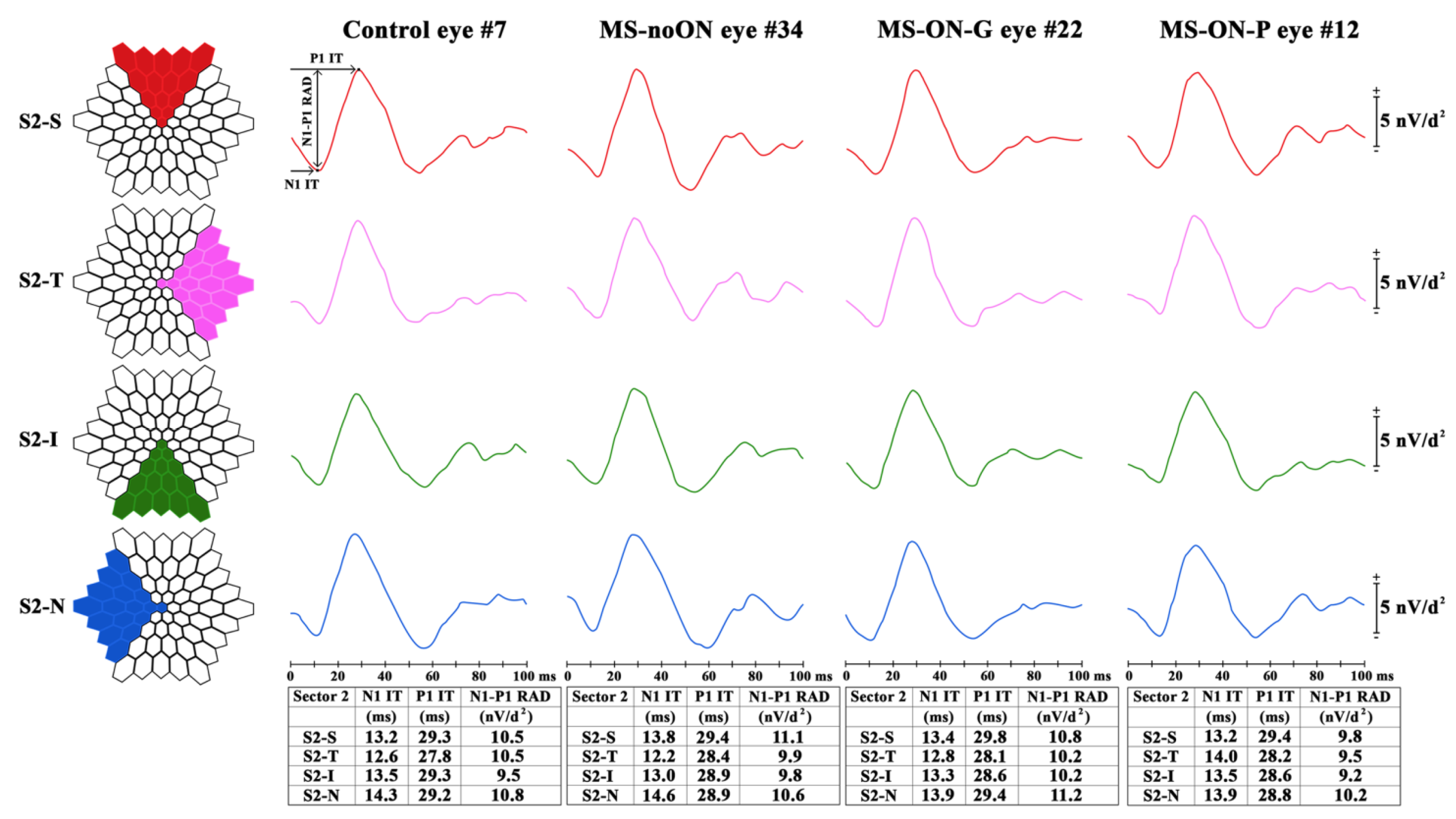
- Sector analysis 2: the averaged bioelectrical response obtained from the retinal area from the fovea up to 25 degrees (0–25 degrees) sectioning it in four sectors: S2-S, S2-N, S2-I and S2-T with respect to the fovea. In each sector, we included also the responses obtained from the more central macular area (0–5 degrees) (Figure 3).
2.3. Sd-OCT Assessment
- (1)
- the 1 mm central area (named as Area 1, directly provided by the Sd-OCT machine)
- (2)
- the middle 1–3 mm ring (named as Area 2, obtained by subtracting from the displayed volume within 3 mm the ones within the 1 mm),
- (3)
- the external 3–6 mm ring (named as Area 3, obtained by subtracting from the displayed volume within 6 mm the one within 3 mm directly provided by the Sd-OCT machine),
- (4)
- the whole 6 mm area (named as Area 1 + Area 2 + Area 3, directly provided by the Sd-OCT machine).
2.4. Statistical Analysis
3. Results
3.1. Demographic and Clinical Features
3.2. Multifocal Electroretinogram Ring Analysis
3.3. Multifocal Electroretinogram Sector Analysis 1 (0–15 Degrees)
3.4. Multifocal Electroretinogram Sector Analysis 2 (0–25 Degrees)
3.5. Morphological Data in MS-ON-P Group and Correlations with mfERG Findings
4. Discussion
5. Conclusions
Author Contributions
Funding
Acknowledgments
Conflicts of Interest
Abbreviations
| MS | multiple sclerosis |
| ON | optic neuritis |
| MS-noON | multiple sclerosis patients without optic neuritis |
| MS-ON-G | multiple sclerosis patients with optic neuritis followed by good recovery of best corrected visual acuity |
| MS-ON-P | multiple sclerosis patients with optic neuritis followed by poor recovery of best corrected visual acuity |
| BCVA | best corrected visual acuity |
| MfERG | multifocal electroretinogram |
| IT | implicit time |
| RAD | response amplitude density |
| P-ERG | pattern electroretinogram |
| Ff-ERG | Full-field electroretinogram |
| F-ERG | focal electroretinogram |
| IML | innermost retinal layers |
| O-MR | outer and in middle retinal |
| SD | one standard deviation of the mean |
| N | number of eyes of each group |
| A | one-way analysis of variance |
| MV | macular volume |
| MT | macular thickness |
| WR | whole retina |
| IR | inner retina |
| OR | outer retina |
| S-S | sector-superior |
| S-T | sector-temporal |
| S-I | sector-inferior |
| S-N | sector -nasal |
References
- Miller, D.; Barkhof, F.; Montalban, X.; Thompson, A.; Filippi, M. Clinically isolated syndromes suggestive of multiple sclerosis, part 2: Non-conventional MRI, recovery processes, and management. Lancet Neurol. 2005, 4, 341–348. [Google Scholar] [CrossRef]
- Dinkin, M. Trans-synaptic Retrograde Degeneration in the Human Visual System: Slow, Silent, and Real. Curr. Neurol. Neurosci. Rep. 2017, 17, 16. [Google Scholar] [CrossRef] [PubMed]
- Britze, J.; Pihl-Jensen, G.; Frederiksen, J.L. Retinal ganglion cell analysis in multiple sclerosis and optic neuritis: A systematic review and meta-analysis. J. Neurol. 2017, 264, 1837–1853. [Google Scholar] [CrossRef] [PubMed]
- Janáky, M.; Jánossy, Á.; Horváth, G.; Benedek, G.; Braunitzer, G. VEP and PERG in patients with multiple sclerosis, with and without a history of optic neuritis. Doc. Ophthalmol. 2017, 134, 185–193. [Google Scholar] [CrossRef]
- Parisi, V.; Manni, G.; Spadaro, M.; Colacino, G.; Restuccia, R.; Marchi, S.; Bucci, M.G.; Pierelli, F. Correlation between morphological and functional retinal impairment in multiple sclerosis patients. Investig. Ophthalmol. Vis. Sci. 1999, 40, 2520–2527. [Google Scholar]
- Trip, S.A.; Schlottmann, P.G.; Jones, S.J.; Altmann, D.R.; Garway-Heath, D.F.; Thompson, A.J.; Plant, G.T.; Miller, D.H. Retinal nerve fiber layer axonal loss and visual dysfunction in optic neuritis. Ann. Neurol. 2005, 58, 383–391. [Google Scholar] [CrossRef]
- Monsalve, P. Decoding PERG: A neuro-ophthalmic retinal ganglion cell function review. Curr. Ophthalmol. Rep. 2019, 7, 51–58. [Google Scholar] [CrossRef]
- Robson, A.G.; Nilsson, J.; Li, S.; Jalali, S.; Fulton, A.B.; Tormene, A.P.; Holder, G.E.; Brodie, S.E. ISCEV guide to visual electrodiagnostic procedures. Doc. Ophthalmol. 2018, 136, 1–26. [Google Scholar] [CrossRef]
- McCulloch, D.L.; Marmor, M.F.; Brigell, M.G.; Hamilton, R.; Holder, G.E.; Tzekov, R.; Bach, M. ISCEV Standard for full-field clinical electroretinography (2015 update). Doc. Ophthalmol. 2015, 130, 1–12. [Google Scholar] [CrossRef]
- Parisi, V.; Falsini, B. Electrophysiological evaluation of the macular cone system: Focal electroretinography and visual evoked potentials after photostress. Semin. Ophthalmol. 1998, 13, 178–188. [Google Scholar] [CrossRef]
- Hood, D.C.; Bach, M.; Brigell, M.; Keating, D.; Kondo, M.; Lyons, J.S.; Marmor, M.F.; McCulloch, D.F.; Palmowski-Wolfe, A.M. International Society For Clinical Electrophysiology of Vision. ISCEV standard for clinical multifocal electroretinography (mfERG) (2011 edition). Doc. Ophthalmol. 2012, 124, 1–13. [Google Scholar] [CrossRef] [PubMed]
- Bearse, M.A., Jr.; Sutter, E.E. Imaging localized retinal dysfunction with the multifocal electroretinogram. J. Opt. Soc. Am. A Opt. Image Sci. Vis. 1996, 13, 634–640. [Google Scholar] [CrossRef] [PubMed]
- Hood, D.C. Assessing retinal function with the multifocal technique. Prog. Retin. Eye Res. 2000, 19, 607–646. [Google Scholar] [CrossRef]
- Parisi, V.; Ziccardi, L.; Stifano, G.; Montrone, L.; Gallinaro, G.; Falsini, B. Impact of regional retinal responses on cortical visually evoked responses: Multifocal ERGs and VEPs in the retinitis pigmentosa model. Clin. Neurophysiol. 2010, 121, 380–385. [Google Scholar] [CrossRef] [PubMed]
- Papakostopoulos, D.; Fotiou, F.; Hart, J.C.; Banerji, N.K. The electroretinogram in multiple sclerosis and demyelinating optic neuritis. Electroencephalogr. Clin. Neurophysiol. 1989, 74, 1–10. [Google Scholar] [CrossRef]
- Hamurcu, M.; Orhan, G.; Sarıcaoğlu, M.S.; Mungan, S.; Duru, Z. Analysis of multiple sclerosis patients with electrophysiological and structural tests. Int. Ophthalmol. 2017, 37, 649–653. [Google Scholar] [CrossRef]
- Forooghian, F.; Sproule, M.; Westall, C.; Gordon, L.; Jirawuthiworavong, G.; Shimazaki, K.; O’Connor, P. Electroretinographic abnormalities in multiple sclerosis: Possible role for retinal autoantibodies. Doc. Ophthalmol. 2006, 113, 123–132. [Google Scholar] [CrossRef]
- Hanson, J.V.M.; Hediger, M.; Manogaran, P.; Landau, K.; Hagenbuch, N.; Schippling, S.; Gerth-Kahlert, C. Outer Retinal Dysfunction in the Absence of Structural Abnormalities in Multiple Sclerosis. Investig. Ophthalmol. Vis. Sci. 2018, 59, 549–560. [Google Scholar] [CrossRef]
- Falsini, B.; Bardocci, A.; Porciatti, V.; Bolzani, R.; Piccardi, M. Macular dysfunction in multiple sclerosis revealed by steady-state flicker and pattern ERGs. Electroencephalogr. Clin. Neurophysiol. 1992, 82, 53–59. [Google Scholar] [CrossRef]
- Saidha, S.; Syc, S.B.; Ibrahim, M.A.; Eckstein, C.; Warner, C.V.; Farrell, S.K.; Oakley, J.D.; Durbin, M.K.; Meyer, S.A.; Balcer, L.J.; et al. Primary retinal pathology in multiple sclerosis as detected by optical coherence tomography. Brain 2011, 134, 518–533. [Google Scholar] [CrossRef]
- Filgueiras, T.G.; Oyamada, M.K.; Preti, R.C.; Apóstolos-Pereira, S.L.; Callegaro, D.; Monteiro, M.L.R. Outer Retinal Dysfunction on Multifocal Electroretinography May Help Differentiating Multiple Sclerosis From Neuromyelitis Optica Spectrum Disorder. Front. Neurol. 2019, 10, 928. [Google Scholar] [CrossRef] [PubMed]
- Gundogan, F.C.; Demirkaya, S.; Sobaci, G. Is optical coherence tomography really a new biomarker candidate in multiple sclerosis?-A structural and functional evaluation. Investig. Ophthalmol. Vis. Sci. 2007, 48, 5773–5781. [Google Scholar] [CrossRef] [PubMed]
- ZiccardI, L.; Barbano, L.; Boffa, L.; Albanese, M.; Grzybowski, A.; Centonze, D.; Parisi, V. Morphological Outer Retina Findings in Multiple Sclerosis Patients With or Without Optic Neuritis. Front. Neurol. 2020, 11, 858. [Google Scholar] [CrossRef] [PubMed]
- Polman, C.H.; Reingold, S.C.; Banwell, B.; Clanet, M.; Cohen, J.A.; Filippi, M.; Fujihara, K.; Havrdova, E.; Hutchinson, M.; Kappos, L.; et al. Diagnostic criteria for multiple sclerosis: 2010 revisions to the McDonald criteria. Ann. Neurol. 2011, 69, 292–302. [Google Scholar] [CrossRef] [PubMed]
- Kurtzke, J.F. Rating neurologic impairment in multiple sclerosis: An Expanded Disability Status Scale (EDSS). Neurology 1983, 33, 1444–1452. [Google Scholar] [CrossRef] [PubMed]
- Neurostatus.net. Available online: http://www.neurostatus.net/index.php?file=start (accessed on 6 July 2020).
- Williams, U.E.; Oparah, S.K.; Philip-Ephraim, E.E. Disease Modifying Therapy in Multiple Sclerosis. Int. Sch. Res. Not. 2014, 2014, 307064. [Google Scholar] [CrossRef]
- Huang-Link, Y.M.; Al-Hawasi, A.; Lindehammar, H. Acute optic neuritis: Retinal ganglion cell loss precedes retinal nerve fiber thinning. Neurol. Sci. 2015, 36, 617–620. [Google Scholar] [CrossRef]
- Parisi, V.; Ziccardi, L.; Sadun, F.; De Negri, A.M.; La Morgia, C.; Barbano, L.; Carelli, V.; Barboni, P. Functional Changes of Retinal Ganglion Cells and Visual Pathways in Patients with Chronic Leber’s Hereditary Optic Neuropathy during One Year of Follow-up. Ophthalmology 2019, 126, 1033–1044. [Google Scholar] [CrossRef]
- Cascavilla, M.L.; Parisi, V.; Triolo, G.; Ziccardi, L.; Borrelli, E.; Di Renzo, A.; Balducci, N.; Lamperti, C.; Bianchi Marzoli, S.; Darvizeh, F.; et al. Retinal dysfunction characterizes subtypes of dominant optic atrophy. Acta Ophthalmol. 2018, 96, e156–e163. [Google Scholar] [CrossRef]
- Parisi, V.; Ziccardi, L.; Centofanti, M.; Tanga, L.; Gallinaro, G.; Falsini, B.; Bucci, M.G. Macular function in eyes with open-angle glaucoma evaluated by multifocal electroretinogram. Investig. Ophthalmol. Vis. Sci. 2012, 53, 6973–6980. [Google Scholar] [CrossRef][Green Version]
- Curcio, C.A.; Sloan, K.R.; Kalina, R.E.; Hendrickson, A.E. Human photoreceptor topography. J. Comp. Neurol. 1990, 292, 497–523. [Google Scholar] [CrossRef] [PubMed]
- Hanson, J.V.M.; Schippling, S.; Gerth-Kahlert, C. Commentary: Outer retinal dysfunction on multifocal electroretinography may help differentiating multiple sclerosis from neuromyelitis optica spectrum disorder. Front. Neurol. 2020, 11, 282. [Google Scholar] [CrossRef] [PubMed]
- Ziccardi, L.; Parisi, V.; Picconi, F.; Di Renzo, A.; Lombardo, M.; Frontoni, S.; Parravano, M. Early and localized retinal dysfunction in patients with type 1 diabetes mellitus studied by multifocal electroretinogram. Acta Diabetol. 2018, 55, 1191–1200. [Google Scholar] [CrossRef] [PubMed]
- Boquete, L.; López-Guillén, E.; Vilades, E.; Miguel-Jiménez, J.M.; Pablo, L.E.; De Santiago, L.; Ortiz Del Castillo, M.; Alonso-Rodríguez, M.C.; Sánchez Morla, E.M.; López-Dorado, A.; et al. Diagnostic ability of multifocal electroretinogram in early multiple sclerosis using a new signal analysis method. PLoS ONE 2019, 14, e0224500. [Google Scholar] [CrossRef] [PubMed]
- Verdon, W.A.; Haegerstrom-Portnoy, G. Topography of the multifocal electroretinogram. Doc. Ophthalmol. 1998, 95, 73–90. [Google Scholar] [CrossRef] [PubMed]
- Fairless, R.; Williams, S.K.; Hoffmann, D.B.; Stojic, A.; Hochmeister, S.; Schmitz, F.; Storch, M.K.; Diem, R. Preclinical retinal neurodegeneration in a model of multiple sclerosis. J. Neurosci. 2012, 32, 5585–5597. [Google Scholar] [CrossRef]
- McGregor, J.E.; Yin, L.; Yang, Q.; Godat, T.; Huynh, K.T.; Zhang, G.; Williams, D.G.; Merigan, W.H. Functional architecture of the foveola revealed in the living primate. PLoS ONE 2018, 13, e0207102. [Google Scholar] [CrossRef]
- Hood, D.C.; Frishman, L.J.; Saszik, S.; Viswanathan, S. Retinal origins of the primate multifocal ERG: Implications for the human response. Investig. Ophthalmol. Vis. Sci. 2002, 43, 1673–1685. [Google Scholar]
- Garcia-Martin, E.; Pueyo, V.; Almarcegui, C.; Martin, J.; Ara, J.R.; Sancho, E.; Pablo, L.E.; Dolz, I.; Fernandez, J. Risk factors for progressive axonal degeneration of the retinal nerve fibre layer in multiple sclerosis patients. Br. J. Ophthalmol. 2011, 95, 1577–1582. [Google Scholar] [CrossRef]
- Mousa, M.F.; Cubbidge, R.P.; Al-Mansouri, F.; Bener, A. Evaluation of hemifield sector analysis protocol in multifocal visual evoked potential objective perimetry for the diagnosis and early detection of glaucomatous field defects. Korean J. Ophthalmol. 2014, 28, 49–65. [Google Scholar] [CrossRef][Green Version]
- Satue, M.; Obis, J.; Alarcia, R.; Orduna, E.; Rodrigo, M.J.; Vilades, E.; Gracia, H.; Otin, S.; Fuertes, M.I.; Polo, V.; et al. Retinal and Choroidal Changes in Patients with Parkinson’s Disease Detected by Swept-Source Optical Coherence Tomography. Curr. Eye Res. 2018, 43, 109–115. [Google Scholar] [CrossRef] [PubMed]
- Polo, V.; Garcia-Martin, E.; Bambo, M.P.; Pinilla, J.; Larrosa, J.M.; Satue, M.; Otin, S.; Pablo, L.E. Reliability and validity of Cirrus and Spectralis optical coherence tomography for detecting retinal atrophy in Alzheimer’s disease. Eye (Lond.) 2014, 28, 680–690. [Google Scholar] [CrossRef] [PubMed]
- Hollander, H.; Bisti, S.; Maffei, L.; Hebel, R. Electroretinographic responses and retrograde changes of retinal morphology after intracranial optic nerve section. Exp. Brian Res. 1984, 55, 483–494. [Google Scholar]
- Petzold, A.; Balcer, L.J.; Calabresi, P.A.; Costello, F.; Frohman, T.C.; Frohman, E.M.; Martinez-Lapiscina, E.H.; Green, A.J.; Kardon, R.; Outteryck, O.; et al. Retinal layer segmentation in multiple sclerosis: A systematic review and meta-analysis. Lancet Neurol. 2017, 16, 797–812. [Google Scholar] [CrossRef]
- Saidha, S.; Sotirchos, E.S.; Ibrahim, M.A.; Crainiceanu, C.M.; Gelfand, J.M.; Sepah, Y.J.; Ratchford, J.N.; Oh, J.; Seigo, M.A.; Newsome, S.D.; et al. Microcystic macular oedema, thickness of the inner nuclear layer of the retina, and disease characteristics in multiple sclerosis: A retrospective study. Lancet Neurol. 2012, 11, 963–972. [Google Scholar] [CrossRef]
- Gelfand, J.M.; Nolan, R.; Schwartz, D.M.; Graves, J.; Green, A.J. Microcystic macular oedema in multiple sclerosis is associated with disease severity. Brain 2012, 135, 1786–1793. [Google Scholar] [CrossRef]
- Gorczyca, W.A.; Ejma, M.; Witkowska, D.; Misiuk-Hojło, M.; Kuropatwa, M.; Mulak, M.; Szymaniec, S. Retinal antigens are recognized by antibodies present in sera of patients with multiple sclerosis. Ophthalmic Res. 2004, 36, 120–123. [Google Scholar] [CrossRef]
- Dembla, M.; Kesharwani, A.; Natarajan, S.; Fecher-Trost, C.; Fairless, R.; Williams, S.K.; Flockerzi, V.; Diem, R.; Schwarz, K.; Schmitz, F. Early auto-immune targeting of photoreceptor ribbon synapses in mouse models of multiple sclerosis. EMBO Mol. Med. 2018, 10, e8926. [Google Scholar] [CrossRef]
- Stathopoulos, P.; Alexopoulos, H.; Dalakas, M.C. Autoimmune antigenic targets at the node of Ranvier in demyelinating disorders. Nat. Rev. Neurol. 2015, 11, 143–156. [Google Scholar] [CrossRef]
- Hood, D.C.; Seiple, W.; Holopigian, K.; Greenstein, V. A comparison of the components of the multifocal and full-field ERGs. Vis. Neurosci. 1997, 14, 533–544. [Google Scholar] [CrossRef]


| Control (N a = 41) (Mean ± 1SD b) | MS-noON (N a = 41) (Mean ± 1SD b) | MS-ON-G (N a = 27) (Mean ± 1SD b) | MS-ON-P (N a = 20) (Mean ± 1SD b) | |
|---|---|---|---|---|
| Age (years) | 40.64 ± 4.83 | 41.32 ± 3.72 | 39.92 ± 4.86 § | 41.08 ± 4.66 §,# |
| Male/Female (Ratio) | 15/26 (0.57) | 14/27 (0.51) | 10/17 (0.58) | 8/12 (0.66) |
| MS-DD c (years) | - | 8.53 ± 4.19 | 9.06 ± 5.58 § | 9.96 ± 6.03 §,# |
| EDSS d score | - | 1.43 ± 1.06 | 1.53 ± 1.22 § | 1.49 ± 1.18 §,# |
| Number of ON e episodes | - | - | 1.00 ± 0.00 | 1.00 ± 0.00 # |
| Time elapsed from ON to the mfERG f and BCVA g assessments (months) | - | - | 14.12 ± 2.72 | 15.87 ± 3.46 # |
| Ring 1: 0–5 Degrees | Ring 2: 5–10 Degrees | Ring 3: 10–15 Degrees | Ring 4: 15–20 Degrees | Ring 5: 20–25 Degrees | ||||||||||||
|---|---|---|---|---|---|---|---|---|---|---|---|---|---|---|---|---|
| N1 IT a | P1 IT a | RAD b | N1 IT a | P1 IT a | RAD b | N1 IT a | P1 IT a | RAD b | N1 IT a | P1 IT a | RAD b | N1 IT a | P1 IT a | RAD b | ||
| Controls N d = 41 | Mean | 14.693 | 29.785 | 56.137 | 13.863 | 28.793 | 22.037 | 12.890 | 28.110 | 12.012 | 12.815 | 28.168 | 8.724 | 13.459 | 28.944 | 7.129 |
| SD c | 2.666 | 2.678 | 10.771 | 1.712 | 1.349 | 4.816 | 1.312 | 1.394 | 3.090 | 2.396 | 1.331 | 2.090 | 1.247 | 1.611 | 1.778 | |
| MS-noON N d = 41 | Mean | 15.078 | 30.035 | 54.273 | 13.743 | 28.393 | 21.505 | 13.193 | 27.413 | 12.525 | 13.115 | 28.455 | 9.050 | 13.198 | 28.650 | 7.535 |
| SD c | 2.383 | 1.958 | 11.665 | 2.177 | 1.661 | 4.556 | 2.505 | 3.076 | 3.409 | 1.020 | 1.285 | 2.573 | 1.156 | 1.115 | 2.321 | |
| A e vs. C | f(1.81) | 0.491 | 0.234 | 0.568 | 0.078 | 1.432 | 0.262 | 0.464 | 0.913 | 0.063 | 0.582 | 1.002 | 0.409 | 0.949 | 0.902 | 0.812 |
| P | 0.487 | 0.638 | 0.453 | 0.782 | 0.235 | 0.610 | 0.498 | 0.343 | 0.810 | 0.449 | 0.319 | 0.525 | 0.332 | 0.345 | 0.372 | |
| MS-ON-G N d = 27 | Mean | 15.748 | 30.156 | 53.467 | 13.741 | 29.104 | 21.081 | 12.800 | 28.041 | 12.344 | 13.089 | 28.585 | 8.926 | 12.963 | 28.596 | 7.511 |
| SD c | 2.934 | 2.739 | 11.053 | 1.903 | 1.761 | 5.121 | 1.522 | 1.831 | 2.755 | 1.042 | 1.484 | 1.906 | 0.692 | 1.196 | 1.622 | |
| A e vs. C | f(1.67) | 2.371 | 0.029 | 1.000 | 0.071 | 0.672 | 0.609 | 0.068 | 0.032 | 0.262 | 0.332 | 1.423 | 0.183 | 3.591 | 0.879 | 0.799 |
| P | 0.128 | 0.563 | 0.321 | 0.787 | 0.415 | 0.463 | 0.796 | 0.859 | 0.611 | 0.570 | 0.238 | 0.676 | 0.063 | 0.352 | 0.376 | |
| A e vs. MS-noON | f(1.67) | 1.072 | 0.042 | 0.079 | 0.000 | 2.841 | 0.164 | 0.532 | 0.913 | 0.063 | 0.013 | 0.153 | 0.042 | 0.932 | 0.029 | 0.009 |
| P | 0.304 | 0.834 | 0.776 | 1.000 | 0.097 | 0.693 | 0.470 | 0.343 | 0.810 | 0.907 | 0.703 | 0.836 | 0.337 | 0.861 | 0.954 | |
| MS-ON-P N d = 20 | Mean | 15.657 | 30.012 | 43.136 | 13.976 | 29.464 | 21.362 | 12.984 | 28.524 | 13.486 | 13.002 | 28.648 | 8.322 | 13.892 | 28.027 | 6.994 |
| SD c | 2.572 | 2.923 | 10.964 | 2.023 | 1.941 | 6.013 | 1.937 | 1.641 | 3.904 | 2.474 | 1.823 | 3.566 | 1.721 | 2.526 | 2.843 | |
| A e vs. C | f(1.60) | 1.802 | 0.091 | 19.36 | 0.053 | 2.473 | 0.221 | 0.053 | 1.053 | 2.570 | 0.084 | 1.363 | 0.311 | 1.262 | 2.962 | 0.051 |
| P | 0.185 | 0.764 | 0.000 f | 0.821 | 0.121 | 0.638 | 0.824 | 0.309 | 0.115 | 0.778 | 0.248 | 0.581 | 0.267 | 0.090 | 0.821 | |
| A e vs. MS-noON | f(1.60) | 0.753 | 0.002 | 12.73 | 0.162 | 5.003 | 0.012 | 0.111 | 2.283 | 0.972 | 0.064 | 0.233 | 0.831 | 3.482 | 1.812 | 0.630 |
| P | 0.389 | 0.971 | 0.000 f | 0.690 | 0.029 | 0.918 | 0.744 | 0.137 | 0.328 | 0.801 | 0.634 | 0.366 | 0.067 | 0.184 | 0.431 | |
| A e vs. MS-ON-G | f(1.46) | 0.012 | 0.033 | 11.41 | 0.172 | 0.443 | 0.033 | 0.133 | 0.871 | 1.383 | 0.033 | 0.021 | 0.562 | 6.552 | 1.062 | 0.626 |
| P | 0.912 | 0.862 | 0.002 f | 0.686 | 0.510 | 0.864 | 0.717 | 0.355 | 0.245 | 0.870 | 0.897 | 0.458 | 0.014 | 0.309 | 0.434 | |
| 0–15 Central Degrees Superior Sector | 0–15 Central Degrees Temporal Sector | 0–15 Central Degrees Inferior Sector | 0–15 Central Degrees Nasal Sector | ||||||||||
|---|---|---|---|---|---|---|---|---|---|---|---|---|---|
| N1 IT a | P1 IT a | RAD b | N1 IT a | P1 IT a | RAD b | N1 IT a | P1 IT a | RAD b | N1 IT a | P1 IT a | RAD b | ||
| Controls N d = 41 | Mean | 13.266 | 28.800 | 17.910 | 13.251 | 28.917 | 17.944 | 13.373 | 28.327 | 17.573 | 13.195 | 27.698 | 19.039 |
| SD c | 1.712 | 1.588 | 4.254 | 1.659 | 1.776 | 4.746 | 1.747 | 1.432 | 4.424 | 1.742 | 1.300 | 4.406 | |
| MS-noON N d = 41 | Mean | 13.758 | 28.319 | 17.442 | 13.972 | 28.508 | 18.119 | 13.881 | 27.997 | 16.489 | 13.831 | 27.506 | 19.369 |
| SD c | 2.058 | 1.438 | 3.982 | 1.996 | 1.677 | 4.094 | 2.167 | 1.525 | 4.131 | 1.679 | 1.587 | 4.508 | |
| A e vs. C | f(1.81) | 1.379 | 2.069 | 0.262 | 3.158 | 1.148 | 0.029 | 1.368 | 1.018 | 1.308 | 2.028 | 0.362 | 0.108 |
| P | 0.243 | 0.154 | 0.608 | 0.079 | 0.287 | 0.859 | 0.246 | 0.316 | 0.255 | 0.096 | 0.551 | 0.738 | |
| MS-ON-G N d = 27 | Mean | 13.256 | 28.459 | 17.241 | 13.774 | 28.356 | 18.278 | 13.419 | 28.648 | 17.007 | 13.570 | 28.037 | 17.933 |
| SD c | 1.430 | 1.554 | 3.932 | 2.042 | 2.237 | 4.843 | 2.027 | 1.289 | 4.129 | 1.610 | 1.806 | 4.246 | |
| A e vs. C | f(1.67) | 0.003 | 0.758 | 0.432 | 1.352 | 1.320 | 0.079 | 0.009 | 0.878 | 0.282 | 0.801 | 0.809 | 1.002 |
| P | 0.988 | 0.385 | 0.516 | 0.250 | 0.255 | 0.779 | 0.921 | 0.351 | 0.598 | 0.374 | 0.371 | 0.321 | |
| A e vs. MS-noON | f(1.67) | 1.222 | 0.138 | 0.039 | 0.162 | 0.102 | 0.019 | 0.779 | 3.340 | 0.258 | 0.408 | 1.632 | 1.642 |
| P | 0.274 | 0.705 | 0.838 | 0.693 | 0.750 | 0.885 | 0.381 | 0.072 | 0.615 | 0.526 | 0.206 | 0.205 | |
| MS-ON-P N d = 20 | Mean | 13.519 | 29.004 | 17.828 | 13.987 | 28.763 | 17.874 | 13.287 | 28.736 | 17.232 | 14.122 | 28.006 | 14.892 |
| SD c | 2.391 | 1.738 | 5.008 | 2.674 | 2.222 | 4.586 | 2.562 | 2.876 | 5.023 | 1.936 | 1.964 | 3.225 | |
| A e vs. C | f(1.60) | 0.222 | 0.212 | 0.002 | 1.75 | 0.092 | 0.002 | 0.022 | 0.552 | 0.072 | 3.542 | 0.532 | 14.00 |
| P | 0.637 | 0.645 | 0.947 | 0.191 | 0.771 | 0.957 | 0.878 | 0.459 | 0.788 | 0.065 | 0.468 | 0.000 f | |
| A e vs. MS-noON | f(1.60) | 0.161 | 2.663 | 0.111 | 0.002 | 0.253 | 0.042 | 0.901 | 1.731 | 0.383 | 0.372 | 1.642 | 15.73 |
| P | 0.688 | 0.108 | 0.745 | 0.980 | 0.619 | 0.834 | 0.348 | 0.193 | 0.542 | 0.548 | 0.205 | 0.000 f | |
| A e vs. MS-ON-G | f(1.46) | 0.222 | 1.284 | 0.202 | 0.101 | 0.386 | 0.081 | 0.042 | 0.022 | 0.032 | 1.142 | 0.002 | 7.192 |
| P | 0.641 | 0.264 | 0.655 | 0.758 | 0.539 | 0.774 | 0.345 | 0.888 | 0.867 | 0.292 | 0.956 | 0.009 f | |
| 0–25 Degrees Superior Sector | 0–25 Degrees Temporal Sector | 0–25 Degrees Inferior Sector | 0–25 Degrees Nasal Sector | ||||||||||
|---|---|---|---|---|---|---|---|---|---|---|---|---|---|
| N1 IT a | P1 IT a | RAD b | N1 IT a | P1 IT a | RAD b | N1 IT a | P1 IT a | RAD b | N1 IT a | P1 IT a | RAD b | ||
| Controls (N d = 41) | Mean | 13.090 | 28.783 | 9.759 | 13.402 | 28.027 | 9.176 | 13.283 | 28.680 | 8.132 | 13.268 | 27.985 | 9.388 |
| SD c | 1.504 | 1.357 | 2.463 | 1.240 | 1.405 | 2.793 | 1.378 | 1.613 | 2.217 | 1.479 | 1.227 | 2.197 | |
| MS-noON (N d = 41) | Mean | 13.133 | 28.258 | 10.192 | 13.336 | 28.467 | 9.181 | 13.394 | 28.281 | 7.994 | 12.964 | 27.661 | 9.994 |
| SD c | 1.154 | 1.240 | 2.789 | 1.365 | 1.132 | 2.606 | 1.793 | 1.640 | 2.558 | 0.858 | 1.217 | 3.022 | |
| A e vs. C | f(1.81) | 0.021 | 3.339 | 0.561 | 0.049 | 2.439 | 0.001 | 0.100 | 1.229 | 0.069 | 1.129 | 1.442 | 1.082 |
| P | 0.896 | 0.071 | 0.450 | 0.819 | 0.122 | 0.993 | 0.754 | 0.270 | 0.795 | 0.258 | 0.234 | 0.302 | |
| MS-ON-G (N d = 27) | Mean | 12.904 | 28.763 | 10.022 | 13.333 | 28.307 | 9.437 | 13.296 | 28.900 | 8.322 | 12.793 | 27.856 | 9.552 |
| SD c | 1.121 | 1.055 | 1.857 | 1.775 | 1.407 | 2.224 | 1.308 | 1.450 | 2.246 | 0.998 | 1.260 | 1.963 | |
| A e vs. C | f(1.67) | 0.301 | 0.009 | 0.219 | 0.039 | 0.649 | 0.168 | 0.002 | 0.329 | 0.118 | 2.761 | 0.182 | 0.104 |
| P | 0.585 | 0.949 | 0.638 | 0.851 | 0.424 | 0.685 | 0.969 | 0.569 | 0.732 | 0.101 | 0.676 | 0.755 | |
| A e vs. MS-noON | f(1.67) | 0.659 | 3.028 | 0.079 | 0.022 | 0.272 | 0.178 | 0.059 | 2.538 | 0.092 | 0.567 | 0.409 | 0.448 |
| P | 0.421 | 0.086 | 0.782 | 0.994 | 0.607 | 0.676 | 0.808 | 0.116 | 0.589 | 0.454 | 0.526 | 0.504 | |
| MS-ON-P N d = 20 | Mean | 13.834 | 28.916 | 9.786 | 14.003 | 28.237 | 9.924 | 13.977 | 28.471 | 8.976 | 13.219 | 28.104 | 9.812 |
| SD c | 1.345 | 1.723 | 2.923 | 2.656 | 1.579 | 3.512 | 1.422 | 1.765 | 2.784 | 1.806 | 3.245 | 2.782 | |
| A e vs. C | f(1.60) | 3.522 | 0.11 | 0.00 | 1.437 | 0.28 | 0.812 | 3.342 | 0.211 | 1.644 | 0.012 | 0.040 | 0.422 |
| P | 0.066 | 0.744 | 0.970 | 0.231 | 0.601 | 0.371 | 0.073 | 0.647 | 0.205 | 0.911 | 0.838 | 0.520 | |
| A e vs. MS-noON | f(1.60) | 4.454 | 2.910 | 0.281 | 1.690 | 0.430 | 0.871 | 1.615 | 0.173 | 1.874 | 0.564 | 0.60 | 0.051 |
| P | 0.039 | 0.093 | 0.601 | 0.198 | 0.517 | 0.356 | 0.209 | 0.680 | 0.177 | 0.456 | 0.442 | 0.822 | |
| A e vs. MS-ON-G | f(1.46) | 6.673 | 0.143 | 0.111 | 1.073 | 0.031 | 0.345 | 2.892 | 0.845 | 0.082 | 1.076 | 0.132 | 0.141 |
| P | 0.013 | 0.708 | 0.737 | 0.305 | 0.874 | 0.564 | 0.096 | 0.365 | 0.377 | 0.307 | 0.718 | 0.709 | |
| A | WR-MV (mm3) | IR-MV (mm3) | OR-MV (mm3) | ||||||||||
| AREA 1 | AREA 2 | AREA 3 | AREA 1 + 2 + 3 | AREA 1 | AREA 2 | AREA 3 | AREA 1 + 2 + 3 | AREA 1 | AREA 2 | AREA 3 | AREA 1 + 2 + 3 | ||
| Controls N b = 41 | Mean | 0.212 | 2.025 | 3.656 | 5.893 | 0.065 | 0.824 | 1.451 | 2.342 | 0.146 | 1.201 | 2.205 | 3.552 |
| SD a | 0.019 | 0.135 | 0.245 | 0.372 | 0.018 | 0.075 | 0.127 | 0.199 | 0.016 | 0.088 | 0.233 | 0.328 | |
| MS-ON-P N b = 20 | Mean | 0.189 | 1.599 | 2.087 | 3.875 | 0.056 | 0.632 | 1.217 | 1.905 | 0.133 | 0.967 | 2.008 | 3.108 |
| SD a | 0.015 | 0.086 | 1.412 | 0.289 | 0.012 | 0.038 | 0.122 | 0.272 | 0.008 | 0.174 | 0.077 | 0.131 | |
| A c vs. C | f(1.60) | 22.422 | 165.532 | 40.471 | 453.482 | 0.112 | 115.824 | 46.800 | 50.661 | 11.701 | 49.074 | 13.482 | 33.771 |
| P | 0.000 d | 0.000 d | 0.000 d | 0.000 d | 0.742 | 0.000 d | 0.000 d | 0.000 d | 0.001 d | 0.000 d | <0.01 d | 0.000 d | |
| B | WR-MT (μ) | IR-MT (μ) | OR-MT (μ) | ||||||||||
| AREA 1 | AREA 2 | AREA 3 | AREA 1 | AREA 2 | AREA 3 | AREA 1 | AREA 2 | AREA 3 | |||||
| Controls N b = 41 | Mean | 263.866 | 327.674 | 299.975 | 81.134 | 138.427 | 114.422 | 182.732 | 189.247 | 185.553 | |||
| SD a | 12.512 | 12.913 | 9.572 | 10.561 | 8.946 | 6.566 | 9.884 | 9.983 | 7.002 | ||||
| MS-ON-P N b = 20 | Mean | 243.834 | 275.179 | 263.253 | 69.417 | 106.667 | 96.833 | 174.417 | 168.512 | 166.417 | |||
| SD a | 12.999 | 13.665 | 10.146 | 10.227 | 7.183 | 9.737 | 7.225 | 7.816 | 5.979 | ||||
| A c vs. C | f(1.60) | 38.672 | 213.901 | 190.280 | 16.893 | 191.321 | 69.202 | 11.199 | 86.811 | 110.000 | |||
| P | 0.000 d | 0.000 d | 0.000 d | 0.000 d | 0.000 d | 0.000 d | 0.001 d | 0.000 d | 0.000 d | ||||
| SUPERIOR SECTOR | TEMPORAL SECTOR | INFERIOR SECTOR | NASAL SECTOR | ||||||||||
| A | WR-MV | IR-MV | OR-MV | WR-MV | IR-MV | OR-MV | WR-MV | OR-MV | IR-MV | WR-MV | IR-MV | OR-MV | |
| (mm3) | (mm3) | (mm3) | (mm3) | (mm3) | (mm3) | (mm3) | (mm3) | (mm3) | (mm3) | (mm3) | (mm3) | ||
| Controls N b = 41 | Mean | 0.551 | 0.214 | 0.337 | 0.537 | 0.203 | 0.334 | 0.551 | 0.216 | 0.335 | 0.579 | 0.231 | 0.348 |
| SD a | 0.029 | 0.017 | 0.019 | 0.031 | 0.015 | 0.02 | 0.028 | 0.012 | 0.021 | 0.048 | 0.018 | 0.024 | |
| MS-ON-P N b = 20 | Mean | 0.475 | 0.168 | 0.307 | 0.479 | 0.172 | 0.307 | 0.487 | 0.177 | 0.310 | 0.478 | 0.175 | 0.303 |
| SD a | 0.029 | 0.021 | 0.011 | 0.028 | 0.012 | 0.014 | 0.021 | 0.016 | 0.016 | 0.028 | 0.015 | 0.015 | |
| A c vs. C | f(1.60) | 92.321 | 84.173 | 42.642 | 50.022 | 64.942 | 29.312 | 81.756 | 113.55 | 22.032 | 75.577 | 144.312 | 58.801 |
| P | 0.000 d | 0.000 d | 0.000 d | 0.000 d | 0.000 d | 0.000 d | 0.000 d | 0.000 d | 0.000 d | 0.000 d | 0.000 d | 0.000 d | |
| B | WR-MT | IR-MT | OR-MT | WR-MT | IR-MV | OR-MT | WR-MT | OR-MT | IR-MT | WR-MT | IR-MT | OR-MT | |
| (μ) | (μ) | (μ) | (μ) | (μ) | (μ) | (μ) | (μ) | (μ) | (μ) | (μ) | (μ) | ||
| Controls N b = 41 | Mean | 297.124 | 111.358 | 185.766 | 292.804 | 108.243 | 184.561 | 297.078 | 112.518 | 184.56 | 306.789 | 117.604 | 189.185 |
| SD a | 8.573 | 7.217 | 6.755 | 8.239 | 7.862 | 6.561 | 6.072 | 6.756 | 6.366 | 7.541 | 8.822 | 6.341 | |
| MS-ON-P N b = 20 | Mean | 271.297 | 94.442 | 176.857 | 271.135 | 92.052 | 178.995 | 268.411 | 94.191 | 174.221 | 274.021 | 97.834 | 176.190 |
| SD a | 6.580 | 7.065 | 5.417 | 7.742 | 6.325 | 5.444 | 8.341 | 9.344 | 7.085 | 8.033 | 7.898 | 5.253 | |
| A c vs. C | f(1.60) | 140.612 | 68.222 | 26.427 | 96.632 | 64.326 | 10.755 | 233.066 | 76.457 | 32.933 | 243.281 | 72.150 | 69.390 |
| P | 0.000 d | 0.000 d | 0.000 d | 0.000 d | 0.000 d | 0.002 d | 0.000 d | 0.000 d | 0.000 d | 0.000 d | 0.000 d | 0.000 d | |
| A | AREA 1 vs. mfERG Ring1 | AREA 2 vs. mfERG Ring 2 | AREA 3 vs. mfERG Ring 3 | AREA 1 + 2 + 3 vs. Rings 1 + 2 + 3 | ||||||||
| N1 IT a | P1 IT a | RAD b | N1 IT a | P1 IT a | RAD b | N1 IT a | P1 IT a | RAD b | N1 IT a | P1 IT a | RAD b | |
| r; p c | r; p c | r; p c | r; p c | r; p c | r; p c | r; p c | r; p c | r; p c | r; p c | r; p c | r; p c | |
| WR-MV | 0.198; 0.535 | −0.055; 0.862 | 0.512; 0.088 | 0.192; 0.549 | −0.103; 0.748 | 0.245; 0.441 | −0.324; 0.303 | −0.509; 0.090 | −0.163; 0.611 | −0.169; 0.599 | −0.474; 0.118 | 0.037; 0.907 |
| IR-MV | 0.338; 0.282 | 0.086; 0.794 | 0.511; 0.090 | 0.132; 0.683 | −0.160; 0.621 | 0.283; 0.372 | −0.288; 0.365 | −0.437; 0.156 | −0.061; 0.851 | 0.185; 0.565 | −0.265; 0.405 | 0.261; 0.413 |
| OR-MV | 0.037; 0.910 | −0.193; 0.548 | 0.443; 0.168 | 0.228; 0.475 | −0.348; 0.267 | −0.498; 0.099 | −0.345; 0.271 | −0.567; 0.055 | −0.309; 0.328 | −0.173; 0.591 | −0.308; 0.331 | −0.074; 0.819 |
| WR-MT | 0.201; 0.532 | −0.053; 0.870 | 0.409; 0.131 | 0.184; 0.567 | −0.100; 0.756 | 0.254; 0.426 | −0.324; 0.304 | −0.517; 0.086 | −0.167; 0.604 | - | - | - |
| IR-MT | 0.331; 0.293 | 0.073; 0.822 | 0.507; 0.093 | 0.134; 0.677 | −0.176; 0.585 | 0.281; 0.376 | −0.298; 0.347 | −0.435; 0.158 | −0.061; 0.851 | - | - | - |
| OR-MT | 0.042; 0.896 | −0.181; 0.574 | 0.369; 0.204 | 0.223; 0.486 | −0.021; 0.947 | 0.169; 0.600 | −0.336; 0.285 | −0.571; 0.052 | −0.309; 0.329 | - | - | - |
| B | SUPERIOR SECTOR | TEMPORAL SECTOR | INFERIOR SECTOR | NASAL SECTOR | ||||||||
| N1 IT a | P1 IT a | RAD b | N1 IT a | P1 IT a | RAD b | N1 IT a | P1 IT a | RAD b | N1 IT a | P1 IT a | RAD b | |
| r; p c | r; p c | r; p c | r; p c | r; p c | r; p c | r; p c | r; p c | r; p c | r; p c | r; p c | r; p c | |
| WR-MV | 0.167; 0.602 | −0.113; 0.726 | 0.281; 0.376 | −0.331; 0.292 | −0.119; 0.711 | −0.331; 0.292 | −0.045; 0.888 | −0.569; 0.053 | 0.193; 0.547 | −0.119; 0.710 | −0.036; 0.910 | −0.056; 0.861 |
| IR-MV | 0.156; 0.627 | −0.166; 0.606 | 0.349; 0.266 | −0.363; 0.245 | −0.078; 0.809 | −0.363; 0.245 | 0.100; 0.756 | −0.341; 0.277 | 0.171; 0.595 | −0.169; 0.598 | −0.109; 0.734 | 0.082; 0.798 |
| OR-MV | 0.163; 0.611 | 0.020; 0.949 | 0.085; 0.791 | −0.125; 0.698 | −0.063; 0.845 | −0.125; 0.698 | −0.168; 0.601 | −0.228; 0.407 | 0.073; 0.819 | −0.034; 0.916 | −0.060; 0.852 | −0.224; 0.483 |
| WR-MT | −0.162; 0.614 | 0.001; 0.999 | 0.742; 0.006 | −0.159; 0.620 | −0.418; 0.175 | −0.159; 0.620 | 0.110; 0.731 | −0.549; 0.064 | 0.131; 0.684 | −0.099; 0.759 | −0.137; 0.669 | −0.074; 0.817 |
| IR-MT | 0.145; 0.650 | −0.208; 0.516 | 0.313; 0.321 | −0.374; 0.230 | −0.082; 0.799 | −0.374; 0.230 | 0.265; 0.404 | −0.318; 0.312 | 0.201; 0.529 | −0.117; 0.716 | −0.093; 0.771 | 0.052; 0.870 |
| OR-MT | 0.060; 0.852 | 0.002; 0.994 | 0.088; 0.785 | −0.325; 0.302 | −0.164; 0.608 | −0.325; 0.302 | −0.028; 0.929 | −0.391; 0.312 | 0.055; 0.865 | −0.065; 0.840 | −0.159; 0.621 | −0.186; 0.561 |
Publisher’s Note: MDPI stays neutral with regard to jurisdictional claims in published maps and institutional affiliations. |
© 2020 by the authors. Licensee MDPI, Basel, Switzerland. This article is an open access article distributed under the terms and conditions of the Creative Commons Attribution (CC BY) license (http://creativecommons.org/licenses/by/4.0/).
Share and Cite
Ziccardi, L.; Barbano, L.; Boffa, L.; Albanese, M.; Nicoletti, C.G.; Landi, D.; Grzybowski, A.; Falsini, B.; Marfia, G.A.; Centonze, D.; et al. Functional Assessment of Outer and Middle Macular Layers in Multiple Sclerosis. J. Clin. Med. 2020, 9, 3766. https://doi.org/10.3390/jcm9113766
Ziccardi L, Barbano L, Boffa L, Albanese M, Nicoletti CG, Landi D, Grzybowski A, Falsini B, Marfia GA, Centonze D, et al. Functional Assessment of Outer and Middle Macular Layers in Multiple Sclerosis. Journal of Clinical Medicine. 2020; 9(11):3766. https://doi.org/10.3390/jcm9113766
Chicago/Turabian StyleZiccardi, Lucia, Lucilla Barbano, Laura Boffa, Maria Albanese, Carolina Gabri Nicoletti, Doriana Landi, Andrzej Grzybowski, Benedetto Falsini, Girolama Alessandra Marfia, Diego Centonze, and et al. 2020. "Functional Assessment of Outer and Middle Macular Layers in Multiple Sclerosis" Journal of Clinical Medicine 9, no. 11: 3766. https://doi.org/10.3390/jcm9113766
APA StyleZiccardi, L., Barbano, L., Boffa, L., Albanese, M., Nicoletti, C. G., Landi, D., Grzybowski, A., Falsini, B., Marfia, G. A., Centonze, D., & Parisi, V. (2020). Functional Assessment of Outer and Middle Macular Layers in Multiple Sclerosis. Journal of Clinical Medicine, 9(11), 3766. https://doi.org/10.3390/jcm9113766