Article

Betaglycan Gene (TGFBR3) Polymorphism Is Associated with Increased Risk of Endometrial Cancer

by
Piotr K. Zakrzewski
1,*,
Ewa Forma
1,
Adam I. Cygankiewicz
1,
Magdalena Bryś
1,
Katarzyna Wójcik-Krowiranda
2,
Andrzej Bieńkiewicz
2,
Andrzej Semczuk
3 and
Wanda M. Krajewska
1
1
Department of Cytobiochemistry, Faculty of Biology and Environmental Protection, University of Lodz, Pomorska 141/143, 90-236 Lodz, Poland
2
Department of Gynecological Oncology, Medical University of Lodz, Pabianicka 62, 93-513 Lodz, Poland
3
IInd Department of Gynecology, Medical University of Lublin, Jaczewskiego 8, 20-090 Lublin, Poland
*
Author to whom correspondence should be addressed.
J. Clin. Med. 2020, 9(10), 3082; https://doi.org/10.3390/jcm9103082
Submission received: 29 July 2020 / Revised: 12 September 2020 / Accepted: 22 September 2020 / Published: 24 September 2020
(This article belongs to the Special Issue Screening and Tests for Gynecologic Cancer)

Abstract

:
We investigated single nucleotide polymorphism (SNP) of the betaglycan gene (TGFBR3) encoding the TGFβ co-receptor in endometrial cancer (EC) and its association with betaglycan expression. The study group included 153 women diagnosed with EC and 248 cancer-free controls. SNP genotyping and gene expression were analyzed using TaqMan probes. Three out of the eight SNPs tested, i.e., rs12566180 (CT; OR = 2.22; 95% CI = 1.15–4.30; p = 0.0177), rs6680463 (GC; OR = 2.34; 95% CI = 1.20–4.53; p = 0.0120) and rs2296621 (TT; OR = 6.40; 95% CI = 1.18–34.84; p = 0.0317) were found to be significantly associated with increased risk of EC (adjusted to age, body mass index, menarche and parity). Among the analyzed SNPs, only rs2296621 demonstrated the impact on the increased cancer aggressiveness evaluated by the WHO grading system (G3 vs. G1/2, GT—OR = 4.04; 95% CI = 1.56–10.51; p = 0.0026; T—OR = 2.38; 95% CI = 1.16–4.85; p = 0.0151). Linkage disequilibrium (LD) analysis revealed high LD (r2 ≥ 0.8) in two haploblocks, constructed by rs2770186/rs12141128 and rs12566180/rs6680463, respectively. In the case of C/C haplotype (OR = 4.82; 95% CI = 1.54–15.07; p = 0.0116—Bonferroni corrected) and T/G haplotype (OR = 3.25; 95% CI = 1.29–8.15; p = 0.0328—Bonferroni corrected) in haploblock rs12566180/rs6680463, significantly higher frequency was observed in patients with EC as compared to the control group. The genotype-phenotype studies showed that SNPs of the TGFBR3 gene associated with an increased risk of EC, i.e., rs12566180 and rs2296621 may affect betaglycan expression at the transcriptomic level (rs12566180—CC vs. TT, p < 0.01; rs2296621—GG vs. TT, p < 0.001, GT vs. TT, p < 0.05). Functional consequences of evaluated TGFBR3 gene SNPs were supported by RegulomeDB search. In conclusion, polymorphism of the TGFBR3 gene may be associated with an increased EC occurrence, as well as may be the molecular mechanism responsible for observed betaglycan down-regulation in EC patients.

1. Introduction

Endometrial cancer (EC) is one of the leading female cancer-related causes of death with around 382,069 new cases and 89,929 deaths worldwide each year. Significantly higher incidence rate is observed in developed countries in contrast to less-developed ones with the world morbidity around 8.4/100,000 of female population [1]. In Poland, its incidence rate takes the third position among cancers and occurs predominantly in women in their menopausal and post-menopausal age (30.2/100,000). However, EC occurrence in Poland has risen rapidly in the last three decades, although the mortality trend is stable, as it is observed in global population [2]. In the future, the incidence of endometrial cancer is expected to increase due to the gradual aging of the population.
According to the clinico-pathological features and different pathogenesis, endometrial cancer is commonly classified into type I—endometrioid and type II—non-endometrioid. Type I is the most diagnosed type of endometrial cancer (75–90%) and develops from glandular cells in the endometrium lining. Endometrioid tumors are represented predominantly by endometrial adenocarcinomas, which are estrogen-dependent and tend to be low grade with favorable prognosis. Non-endometrioid cancers typically include papillary serous or clear cell carcinomas, in general, histological subtypes characterized by more aggressive phenotypes with poor outcome [3,4]. Endometrial cancer is mostly diagnosed in the early stages (FIGO I and II) as it is observed in 75% of patients. In this stage, 5-year overall survival is 74–91%, whereas for more advanced stages, 5-year overall survival rates are 57–66% and 20–26%, for FIGO III and FIGO IV, respectively [5]. Molecular classification distinguished based on a large scale, comprehensive genetic analysis of EC according to The Cancer Genome Atlas includes four subgroups, i.e., DNA polymerase epsilon ultramutated (POLE), microsatellite instability hypermutated (MSI), copy-number low and copy-number high subgroup. Each of categories is characterized by distinct clinical, pathological and molecular alterations. The POLE subgroup displays polymerase epsilon mutations in exonuclease domain, which results in a remarkable high mutation rate (232 × 10−6 mutations per Mb). The MSI subgroup is related to deficiencies in a DNA mismatch repair system leading to common mutations of ARID5B, PTEN, PIK3CA and PIK3R1 genes. The copy-number low subgroup is described also as microsatellite stable and corresponds to more than half of low-grade endometrioid tumors, whereas copy-number high subgroup reflects to serous histopathology [6,7,8]. Moreover, up to 5% of ECs are described as familial ones, due to the loss-of-function or expression alterations of DNA mismatch repair genes, i.e., (MLH1, MSH2, MSH6 or PMS2). The most frequent form of inherited EC is associated with Lynch syndrome, which increases the risk of developing EC to 25–60% [9,10].
According to the molecular findings, impaired TGFβ signaling has been reported in ECs [11,12,13]. The canonical signal, mediated via TGFβ factors, occurs through TGFβ membrane receptors type I (TGFβRI) and type II (TGFβRII), which possess serine/threonine kinase activity. Among huge number of TGFβ factors, three classical TGFβ isoforms, i.e., TGFβ1, TGFβ2 and TGFβ3, were identified. Dimeric TGFβ factors bind to the TGFβRII receptor, which in turn activates TGFβRI receptor. The activated TGFβRII/TGFβRI complex trans-phosphorylates cytoplasmic effectors, i.e., Smad2/3 proteins, forming a heterocomplex with the Smad4 protein, are translocated to the nucleus, where together with other transcription factors, regulate gene expression [14]. Appropriate signaling in TGFβ pathway requires the presence of co-receptors termed TGFβ receptors type III (TGFβRIII). TGFβ co-receptors are deprived of any known enzymatic activity; however, they are anchored in the cell membrane, and they are responsible for TGFβ ligand presentation to their canonical TGFβ receptors. The plethora of TGFβ ligands and receptors results in the regulation of many cellular processes, such as growth and proliferation, survival, apoptosis, cells adhesion, remodeling of extracellular matrix, angiogenesis and embryonic development [15].
The first identified TGFβ co-receptor was betaglycan [16]. Betaglycan gene (TGFBR3), located on chromosome 1, encodes a transmembrane proteoglycan. Literature data and our previous studies indicate the contribution of betaglycan loss to the development and progression of cancers originated from different tissue types, i.e., breast, endometrium, ovary, prostate, lung, bladder, liver, pancreas, kidney, and neuroblastoma [17,18,19,20,21,22,23,24,25,26,27,28]. Down-regulation of betaglycan expression seems to be engaged in the impaired TGFβ signaling initiated by the TGFβ2 isoform to which it displays the highest affinity.
Until now, there is no efficient and rapid molecular method suitable for neither early diagnosis nor prediction of EC risk. This became a basis for betaglycan (TGFBR3) gene single nucleotide polymorphism (SNP) investigations. In the current study, we examined eight SNPs within the TGFBR3 gene and their association with primary EC and their clinico-pathological variables, as well as potential impact on betaglycan expression.

2. Materials and Methods

2.1. Study Population

In the study, we enrolled only Caucasian women born and living in Poland. The case-control study involved 153 women who underwent surgery of EC and 248 healthy individuals which served as cancer-free controls. Biological material (cancer group—endometrial tissue samples and peripheral blood; cancer-free control—peripheral blood) was collected in the IInd Department of Gynecology, Lublin Medical University, Lublin, Poland and in the Department of Gynecological Oncology, Medical University of Lodz, Lodz, Poland, between 2012–2017. Inclusion criteria for case group, included women with diagnosed primary endometrial adenocarcinomas, who had not received neither hormonal therapy, radiation therapy nor chemotherapy prior surgery; whereas control group was recruited from non-related women during periodic health check-ups, who have never been diagnosed with endometrial cancer or other tumors.
Cancer tissue specimens, after the surgery, were divided into two portions; one was fixed in buffered formalin (pH 7.4) for routine histological assessment while the other was immediately placed at −70 °C. Clinical stage was assigned based on surgico-pathological findings according to the revised FIGO staging, while WHO classification was applied to determine the histological type and grade. Table 1 presents socio-demographic and clinical characteristics of the patients and examined samples. All studied cancer samples were classified as endometrioid cancers (type I)—endometrial adenocarcinomas.

2.2. Ethical Approval

The study was conducted in accordance with the ethical principles of the 1975 Helsinki Declaration and its later amendments. The local Independent Committees of Bioethics of Lublin Medical University, Medical University of Lodz and University of Lodz approved the tissues collections and study protocols. All methods in the study were performed in accordance with above-mentioned bioethical permissions. All participating subject gave written, informed consent prior to enrolment.

2.3. Lifestyle Risk Factors

Study participants were interviewed during the examination about socio-demographic, health related information and reproductive history (parity and menarche). Body mass index (BMI) was calculated as current weight in kilograms divided by square of height expressed in meters. Any missing survey data were subsequently completed using patient’s query.

2.4. Genomic DNA Isolation

Genomic DNA was extracted from peripheral blood collected in the presence of anti-coagulant (EDTA) using PureLink Genomic DNA Mini Kit (Thermo Fisher Scientific, Waltham, MA, USA) and stored at −70 °C. The quality and quantity of DNA was estimated spectrophotometrically with BioPhotometer plus (Eppendorf, Hamburg, Germany). DNA samples were characterized with A260 nm/A280 nm ratio, which was in the range of 1.8–2.0.

2.5. SNP Selection and Genotyping

Eight SNPs in the TGFBR3 gene were selected according to NCBI SNPs database: rs883873, rs2770186, rs12141128, rs12566180, rs6680463, rs1805110, rs1805113, rs2296621. All SNPs were supposed to have minor allele frequency (MAF) ≥5% and localization assigned as 5′ regulatory region, intron or exon of the TGFBR3 gene, which is located on chromosome 1. Characteristics of studied SNPs are presented in Table 2.
Real-Time PCR method with TaqMan Genotyping Assays (Thermo Fisher Scientific, Waltham, MA, USA) was applied for SNPs genotyping. The characteristics and sequences of used TaqMan probes are shown in Supplementary Table S1. PCR amplifications were conducted in a total volume of 10 µL and consisted of 5 µL (2×) of TaqMan Genotyping Master Mix buffer (Thermo Fisher Scientific, Waltham, MA, USA), 0.25 µL (40×) TaqMan Genotyping Assay (Thermo Fisher Scientific, Waltham, MA, USA) and 10 ng of template DNA. Thermal conditions were as follows: initial denaturation at 95 °C for 10 min, followed by 40 cycles of sequential incubations at 95 °C for 15 s and at 60 °C for 1 min and final endpoint measurement of fluorescence. Real-Time PCR amplifications and allelic discrimination were performed using Mastercycler®ep realplex (Eppendorf, Hamburg, Germany).

2.6. Expression of the TGFBR3 Gene

Total RNA was extracted from frozen endometrial tissues using PureLink RNA Mini kit (Thermo Fisher Scientific, Waltham, MA, USA) according to dedicated protocol. The amount and quantity of isolated RNA was assessed spectrophotometrically with BioPhotometer plus (Eppendorf, Hamburg, Germany) based on A260 nm/A280 nm ratio, which was in the range 1.8–2.0. Total RNA (1 μg) was transcribed using RevertAid™ H Minus First Strand cDNA Synthesis kit (Thermo Fisher Scientific, Waltham, MA, USA) according to manufacturer’s recommendation. cDNA synthesis was performed in Thermocycler 2720 (Applied Biosystems, Foster City, CA, USA) with the following incubations: 10 min at 25 °C, 120 min at 37 °C and 5 min at 85 °C. The obtained cDNA was stored at −70 °C. Real time PCR was performed using TaqMan probes (Thermo Fisher Scientific, Waltham, MA, USA) in line with the manufacturer’s protocol on Mastercycler® Epgradient S Realplex (Eppendorf, Hamburg, Germany) in the presence of TaqMan Gene Expression Master Mix Thermo Fisher Scientific, Waltham, MA, USA). GAPDH served as a reference gene. The catalogue numbers of probes were Hs00234259_m1 for TGFBR3 and Hs99999905_m1 for GAPDH. The relative expression level was normalized to GAPDH and was calculated using the following equation: 2−ΔCt × 1000.

2.7. Statistical Analysis

The genotype frequency was tested for agreement with Hardy–Weinberg equilibrium (HWE) and assessed by chi-square goodness-of-fit test. Case-control differences in genotype and allelic distribution were analyzed using Pearson’s χ2 (chi-square) or Fisher’s exact tests against the homozygote of the common allele as the reference group (OR = 1.00). Dominant and recessive genetic models were also implemented in the analysis. Variants of homozygotes and heterozygotes were combined to evaluate the dominant effect. SNPs distribution and their association with the clinico-pathological parameters were evaluated by multiple logistic regression. Genotype and allelic associations with endometrial cancer risk were expressed as odds ratio (ORs) and 95% confidence interval (95% CI) in crude and multivariate model including age, BMI, parity, and age at menarche.
Linkage disequilibrium (LD) and haplotypes distribution analysis were performed using the powerful online platform SHEsis (http://analysis.bio-x.cn/myAnalysis.php) [29]. Haplotypes with frequency less than 0.03 were excluded from the analysis. Bonferroni correction was applied for multiple comparisons of SNPs haplotypes.
To assess inter-group differences of socio-demographic parameters (age, BMI, parity and menarche), as well as the TGFBR3 gene expression levels between respective genotypes of the analyzed SNPs, the first Shapiro–Wilk test was applied to determine the normality of obtained data. Following, the statistical significance of difference was evaluated using either Student’s t-test, for normally distributed data, or Mann–Whitney test, for non-normally distributed data. p < 0.05 in a two-tailed test was considered statistically significant. A statistical analysis of obtained data was conducted using GraphPad Prism version 5.00 for Windows (GraphPad Software, La Jolla, CA, USA) and PQStat version 1.6.8 (PQStat Software, Poland).

2.8. Bioinformatic Analysis

The functional consequences of significant SNPs were examined in RegulomeDB, which is a public database dedicated for noncoding SNP and annotates SNPs with known and putative regulatory elements in non-coding regions of human genome, such as regulatory DNA elements including regions of DNAase hypersensitivity, binding sites of transcription factors, and promoter regions that have been biochemically characterized to regulate transcription. RegulomeDB annotations are based on an integration of data from ENCODE project and other published literature, combined together by self-developed score system ranging from 1–6. A higher rank corresponds to a less functional significance [30].

3. Results

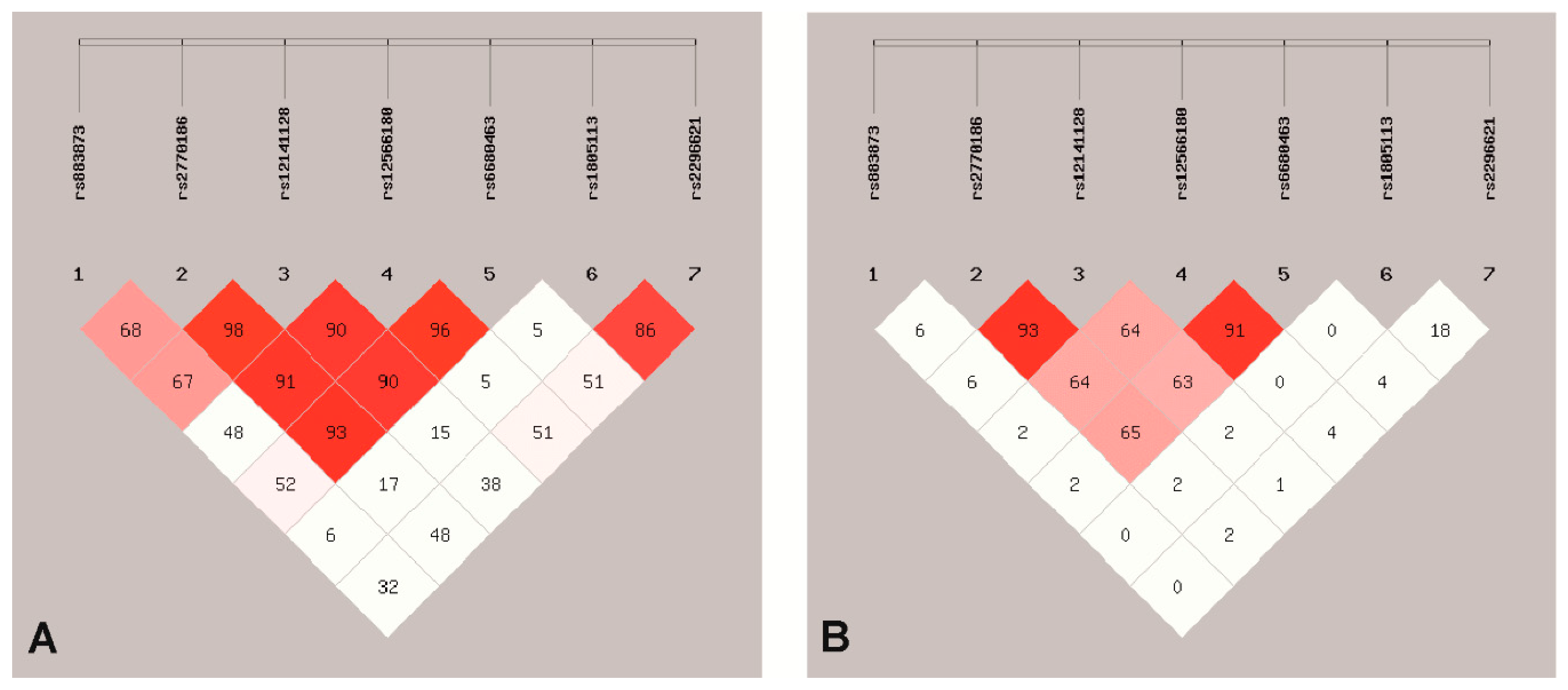
3.1. SNPs Association with EC

Eight single nucleotide polymorphisms in the TGFBR3 gene and their association with EC risk and invasiveness were evaluated. Three of analyzed polymorphisms, i.e., rs883873 (g.92380302A > G), rs2770186 (g.92378843T > C) and rs12141128 (g.92373747A > G) are located in 5′ regulatory region, whereas the other five polymorphisms, i.e., rs12566180 (c.−114 + 2392C > T), rs6680463 (c.−114 + 7008C > G), rs1805110 (p.Ser15Phe), rs1805113 (p.Phe675=) and rs2296621 (c.2285 − 99G > T) are located downstream start codon of the TGFBR3 gene.
Based on a comparison of 153 women diagnosed with endometrial cancer and 248 healthy controls, we found significant differences in the distribution of the three studied SNPs (Table 3) adjusted to the following covariates, i.e., age, BMI, menarche and parity. Significant differences between endometrial cancer patients and control women were noted in body mass index (BMI; p < 0.001), menarche (p < 0.001) and parity (p < 0.001). In addition, the subjects’ age showed significant differences between case and control group when analyzed in 10 years subgroups (p = 0.009) (Table 1). Accordingly, age, BMI, menarche and parity were selected as main covariates in further analysis. All studied SNPs were in Hardy–Weinberg equilibrium (HWE) except polymorphic site rs1805110 (p.Ser15Phe), which was excluded from further analysis. The obtained results indicate that the polymorphisms of the highest importance for the increased endometrial cancer risk are rs12566180 (c.−114 + 2392C > T), rs6680463 (c.−114 + 7008C > G) and rs2296621 (c.2285 − 99G > T). SNPs, rs12566180 (c.-114 + 2392C > T) and rs6680463 (c.−114 + 7008C > G) were found to be more frequent as heterozygous variants in the study group as compared to the controls with respective frequencies 55.6% vs. 44.0% (p = 0.0177) and 58.2% vs. 45.2% (p = 0.0120), increasing the risk of endometrial cancer about 2.3 times. In turn, the polymorphic site rs2296621 (c.2285 − 99G > T) was found to be 6.4-fold more frequent in the case of study group compared to the controls (4.6% vs. 0.8%, p = 0.0317) as homozygous variant TT (Table 3).
Analysis of significantly altered SNPs in the cancer group in the context of clinico-pathological parameters revealed that in the case of rs2296621 (c.2285−99G > T) the genotype GT and allele T are associated with an increased histological grade according to the WHO grading system (G3 vs. G1/2). The GT genotype was observed in 54.5% of high-graded tumors (G3) compared to 23.7% of less-graded (G1/2) (OR = 4.04; 95% CI = 1.56–10.51; p = 0.0026). Respectively, allele T carriers demonstrated G3 tumors more frequent than G1/2, i.e., 31.8% vs. 16.4% (OR = 2.38; 95% CI = 1.16–4.85; p = 0.0151) (Supplementary Table S2).
Linkage disequilibrium (LD) analysis revealed that among eight studied SNPs in the TGFBR3 gene, four of them were in high LD (r2 ≥ 0.8) (Figure 1) and were arranged in two haploblocks constructed by rs2770186/rs12141128 and rs12566180/rs6680463. The frequency of haplotypes rs12566180/rs6680463 C/C and T/G were significantly higher in endometrial cancer patients as compared to healthy controls: for C/C haplotype 0.038 vs. 0.008 (OR = 4.82; 95% CI = 1.54–15.07; p = 0.0116—Bonferroni corrected) and for T/G haplotype 0.045 vs. 0.014 (OR = 3.25; 95% CI = 1.29–8.15; p = 0.0328—Bonferroni corrected), respectively (Table 4).

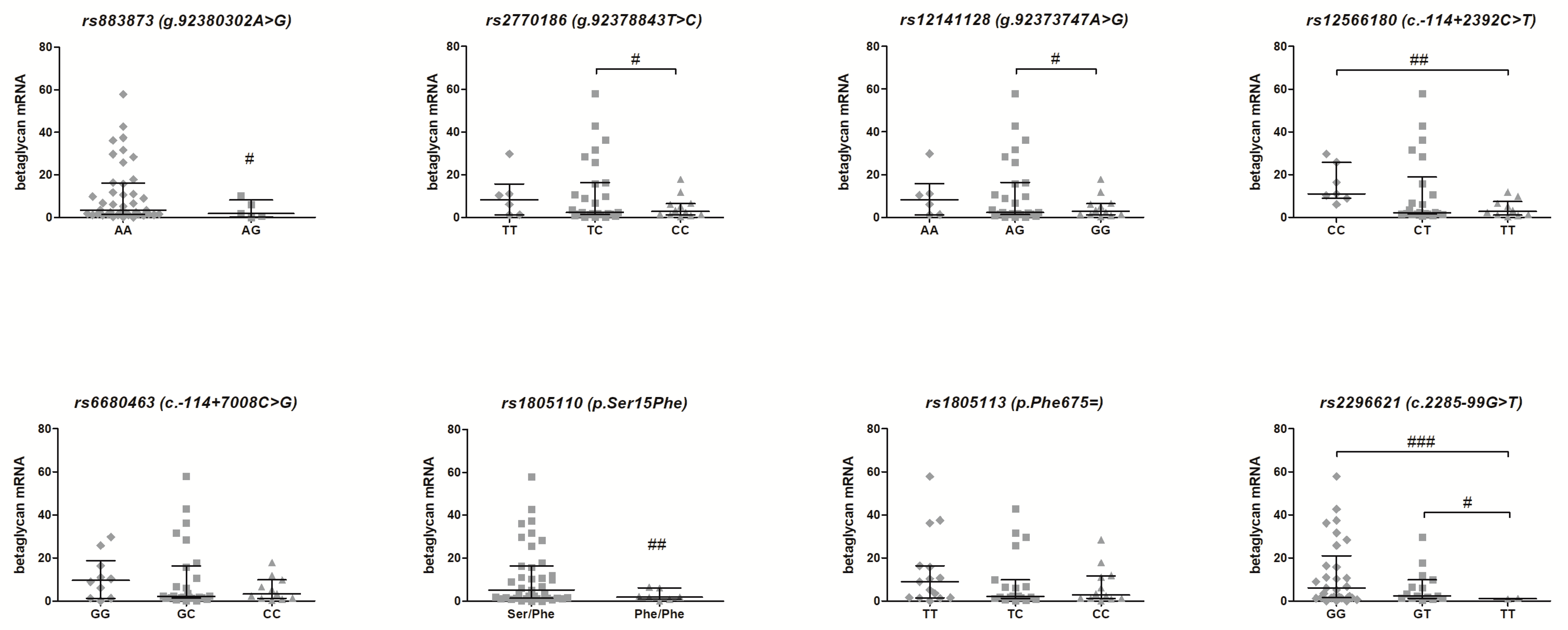
3.2. Association of the TGFBR3 Gene SNPs and Betaglycan mRNA Expression—Genotype-Phenotype Analysis

Figure 2 shows the mRNA expression level of the TGFBR3 gene in 50 EC patients in relation to the genotypes of eight studied SNPs of the TGFBR3 gene. Six SNPs of the TGFBR3 gene were found to modulate its expression. The significant down-regulation of TGFBR3 mRNA was observed in the case of homozygous variant of rs2770186 for genotype CC (p < 0.05), rs12141128 for genotype GG (p < 0.05), rs1805110 for phenotype variant Phe/Phe (p < 0.01) and rs2296621 for genotype TT (p < 0.05) as compared to heterozygous variants. Furthermore, rs883873 polymorphism for genotype AG (p < 0.05), rs12566180 for genotype TT (p < 0.01) and rs2296621 for genotype TT (p < 0.001) demonstrated a statistically lower TGFBR3 mRNA level with regard to the wild-type carriers.

4. Discussion

Transforming growth factors β isoforms, i.e., TGFβ1, TGFβ2 and TGFβ3, belong to a large superfamily of cytokines, which were identified due to their important role in normal development and homeostasis. TGFβ pathway controls many opposed processes, which is known as the pleiotropic effect on cell and tissue physiology. TGFβ cascade is responsible for both suppression or induction of cell proliferation and apoptosis, as well as regulates autophagy, cell dormancy and senescence. Deregulation of TGFβ signaling, both at induction step and downstream signaling contributes to developmental anomalies and diseases, in particular fibrosis and cancer, which is associated with overexpression of TGFβ isoforms [31,32]. Moreover, in cancer cells, disturbed signal mediation in TGFβ pathway triggers its role from a tumor suppressor, early in neoplastic transformation, to a cancer-promoting and metastatic agent in advanced clinical stages of the disease [33].
In cancer cells, alteration of TGFβ signaling, which plays the pleiotropic role during carcinogenesis, may be influenced by gene polymorphism. Knowledge of the potential role of polymorphism of the TGFBR3 gene encoding betaglycan and its relation to development of EC is elusive. Risk of endometrial cancer development is highly associated with different lifestyle and socio-demographic factors including obesity, onset of menarche, reproductive history, ethnicity and patient’s age [34,35,36,37,38,39,40]. Overweight, young age at menarche or nulliparity cause prolonged exposure to estrogens, which possess high proliferative potential, in particular to uterus lining. In the case of obesity, unopposed estrogen stimulation is the result of reduction of progesterone synthesis and higher levels of circulating estrogens. During pregnancy, the estrogen exposure is balanced by the shift toward progesterone signaling. For this reason, nulliparous women have a higher risk of developing endometrial cancer due to extended estrogen stimuli. Moreover, the increased number of births shows a protective effect on endometrial cancer occurrence [40,41,42]. Our study has demonstrated that rs12566180 (c.−114 + 2392C > T), rs6680463 (c.−114 + 7008C > G) and rs2296621 (c.2285 – 99G > T) polymorphisms of the TGFBR3 gene are associated with increased risk of EC, both as crude or adjusted for socio-demographic risk factors, such as age, body mass index (BMI), menarche and parity. None of the patients had received EC related hormonal therapy prior to surgery; however, the patients’ records concerning hormonal replacement therapy as well as Lynch syndrome history were not available. In spite of the fact, that Lynch syndrome significantly increases the risk of EC to 25–60%, its impact on development of EC can be excluded as its occurrence ranges between 0.5% and 4.6% of all EC cases [9,10].
The rs12566180 (c.-114 + 2392C > T) and rs2296621 (c.2285 − 99G > T) polymorphisms significantly altered in endometrial cancer are located within intronic regions of the TGFBR3 gene, which may indicate their potential impact on transcription and stability of the primary transcript. Obtained results suggest that studied SNPs are involved in the observed betaglycan down-regulation in endometrial cancer; however, the only one of them, i.e., rs2296621 (c.2285 − 99G > T), seems to be related to pronounced tumor aggressiveness. Bioinformatic analysis using RegulomeDB showed that rs12566180 (c.-114 + 2392C > T) and rs6680463 (c.−114 + 7008C > G) have a score of 4, whereas rs2296621 (c.2285 − 99G > T) has a score of 2 [30]. Taking into account SNPs case-control study, haplotype analysis and genotype-phenotype findings, the observed results indicate that rs12566180 (c.-114 + 2392C > T), rs6680463 (c.−114 + 7008C > G) and rs2296621 (c.2285 − 99G > T) could be regarded as potential markers for EC. However, further studies are required.
According to the literature data, polymorphism of the TGFBR3 gene is considered as a mechanism responsible for betaglycan down-regulation of HBV-infection related hepatocellular carcinoma and ovarian cancer [17,43,44,45]. Bae et al. [17] evaluated six SNPs, i.e., rs1805110 (p.Ser15Phe), rs2810904 (p.Ala72=), rs2306888 (p.Ser173=), rs1805113 (p.Phe675=), rs284878 (p.Thr749=) and newly identified SNP p.Thr711=, in the TGFBR3 gene in a group consisting of 67 patients with hepatocellular carcinoma. In their study, rs1805110 (p.Ser15Phe) polymorphism was found to be present at a high frequency, i.e., in about 98.5% of examined cancer samples; however, the lack of case-control comparison made it impossible to confirm the association between rs1805110 (p.Ser15Phe) SNP occurrence and betaglycan down-regulation [17]. Similar results were presented by Xin et al. [45]. Among 16 different SNPs in genes encoding components of TGFβ pathway, i.e., TGFβ1, TGFβR1/2 and betaglycan, significantly changed frequency of rs1805110 polymorphic site was found to be associated with incidence of HBV-related hepatocellular carcinoma (T allele, OR = 1.33; 95% CI = 1.09–1.63; p = 0.005) for male Chinese patients of Han ethnicity. According to the study by Kim et al. [43] rs1805113 (Phe676Phe) in exon 13 and rs1805117 in 3′-UTR (p = 0.009 and p = 0.008, respectively) polymorphisms were significantly associated with HBV clearance. In addition, Cox relative hazards analysis revealed that the GGTCAA haplotype of rs2306888, rs1805112, rs1805113, rs284878, rs1805117 and rs1804506 polymorphisms showed a significant association with the age of HCC occurrence among chronic HBV patients (relative hazard = 1.38; p = 0.007). In turn, the study presented by Charbonneau et al. [44] showed that in the case of mucinous invasive epithelial ovarian cancer (EOC), significantly altered distribution of polymorphisms rs12129174 (c.384 + 1320G > A) and rs4658265 (c.247 – 16378G > A), located in the introns of the TGFBR3 gene, was moderately correlated with patients’ survival (rs12129174—HR = 1.61; 95% CI = 1.18–2.19; p = 0.0038; rs4658265—HR = 1.56; 95% CI = 1.20–2.05; p = 0.0012).
Furthermore, besides above-mentioned relationship between SNPs of the TGFBR3 gene and neoplastic transformation, the importance of polymorphism in betaglycan encoding gene was reported for other non-cancerous diseases, i.e., premature ovarian failure (POF), testicular dysgenesis, sickle cell anemia, pulmonary emphysema and primary open angle glaucoma [46,47,48,49,50,51]. It is suggested that the TGFBR3 gene polymorphism may play a potential role in determining bone mineral density, as well as optic disc area parameters [52,53,54].
Interestingly, in our study, six out of eight analyzed SNPs of the TGFBR3 gene were found to have an impact on betaglycan expression in EC. Altered betaglycan expression may be responsible for impaired TGFβ signaling initiated by TGFβ isoforms and simultaneous redirection of this signal to Smad-independent pathways. TGFBR3 gene downregulation has been stated in the case of different cancers, and the observed decline in the expression of the TGFBR3 gene appears to be correlated with cancer progression, when tumor cells demonstrate an increase invasiveness and metastatic potential [17,18,19,20,21,22,23,25,26,27,28].
As previously described by different research groups, TGFβ signaling induced by TGFβ isoforms may be engaged in the induction of epithelial-mesenchymal transition (EMT). EMT is a biological process characterized by the reorganization of the epithelial tissue structure and is manifested by the acquisition of the mesenchymal phenotype resulting in the loss of polarity and adhesion by cells together with ability to migration and invasion. This process plays a vital role during physiological events, i.e., embryogenesis, organogenesis and morphogenesis of different tissues, wound healing, as well as inflammation. In cancer progression, EMT is responsible for the development of drug resistance and metastasis due to the increased cancer cell motility [55,56,57].
In summary, our study has demonstrated for the first time the role of the TGFBR3 gene polymorphism and its association with the increased risk of EC development. Moreover, we have shown that the TGFBR3 gene SNPs may modulate betaglycan expression at the transcriptomic level. Our findings contribute to a better understanding of the importance of gene polymorphism in the TGFβ signaling, especially at the level of signal initiation through TGFβ2 isoform mediated exclusively by betaglycan. Along with our previous findings concerning the significance of allelic loss in the TGFBR3 gene, where LOH was reported in 52% of examined cancer samples, SNPs may be an additional mechanism responsible for betaglycan deregulation in EC [28,58]. What is more, obtained results strongly support the view of individual variability among EC patients and suggest the necessity of developing personalized diagnostic and/or therapeutic approach in the treatment of endometrial cancer.

Supplementary Materials

Table S1: Characteristics and sequences of TaqMan probes used for genotyping of TGFBR3 gene; Table S2: The association between significantly altered SNPs, i.e., rs12566180, rs6680463 and rs2296621 polymorphisms and clinico-pathological parameters of studied cancer samples.

Author Contributions

Conception and design: P.K.Z., W.M.K.; collecting human samples and clinical findings, K.W.-K., A.B., A.S., M.B.; acquisition of data, P.K.Z., E.F., A.I.C.; analysis and interpretation of data, P.K.Z., W.M.K.; drafting the article or revising it critically for important intellectual content, P.K.Z., W.M.K., E.F., M.B., A.S., A.B.; final approval of the version to be published, P.K.Z., E.F., A.I.C., M.B., K.W.-K., A.B., A.S., W.M.K. All authors have read and agreed to the published version of the manuscript.

Funding

The work was supported by grant UMO-2011/01/N/NZ4/01723 (to PKZ) from The National Science Centre (NCN), Poland.

Conflicts of Interest

The authors declare no conflict of interest.

References

  1. Ferlay, J.; Colombet, M.; Soerjomataram, I.; Mathers, C.; Parkin, D.M.; Piñeros, M.; Znaor, A.; Bray, F. Estimating the global cancer incidence and mortality in 2018: GLOBOCAN sources and methods. Int. J. Cancer 2019, 144, 1941–1953. [Google Scholar] [CrossRef] [Green Version]
  2. Didkowska, J.; Wojciechowska, U.; Czaderny, K.; Olasek, P.; Ciuba, A. Cancer in Poland in 2017; Polish National Cancer Registry: Warsaw, Poland, 2019. [Google Scholar]
  3. Bokhman, J.V. Two pathogenetic types of endometrial carcinoma. Gynecol. Oncol. 1983, 15, 10–17. [Google Scholar] [CrossRef]
  4. Webb, P.M. Environmental (nongenetic) factors in gynecological cancers: Update and future perspectives. Future Oncol. 2015, 11, 295–307. [Google Scholar] [CrossRef] [PubMed]
  5. Murali, R.; Soslow, R.A.; Weigelt, B. Classification of endometrial carcinoma: More than two types. Lancet Oncol. 2014, 15, e268–e278. [Google Scholar] [CrossRef]
  6. Yen, T.T.; Wang, T.L.; Fader, A.N.; Shih, I.M.; Gaillard, S. Molecular Classification and Emerging Targeted Therapy in Endometrial Cancer. Int. J. Gynecol. Pathol. 2020, 39, 26–35. [Google Scholar] [CrossRef]
  7. Lee, Y.C.; Lheureux, S.; Oza, A.M. Treatment strategies for endometrial cancer: Current practice and perspective. Curr. Opin. Obstet. Gynecol. 2017, 29, 47–58. [Google Scholar] [CrossRef]
  8. Getz, G.; Gabriel, S.B.; Cibulskis, K.; Lander, E.; Sivachenko, A.; Sougnez, C.; Lawrence, M.; Kandoth, C.; Dooling, D.; Fulton, R.; et al. Integrated genomic characterization of endometrial carcinoma. Nature 2013, 497, 67–73. [Google Scholar] [CrossRef] [Green Version]
  9. Bell, D.W.; Ellenson, L.H. Molecular Genetics of Endometrial Carcinoma. Annu. Rev. Pathol. Mech. Dis. 2019, 14, 339–367. [Google Scholar] [CrossRef]
  10. Rossi, L.; Le Frere-Belda, M.A.; Laurent-Puig, P.; Buecher, B.; De Pauw, A.; Stoppa-Lyonnet, D.; Canlorbe, G.; Caron, O.; Borghese, B.; Colas, C.; et al. Clinicopathologic characteristics of endometrial cancer in lynch syndrome A French multicenter study. Int. J. Gynecol. Cancer 2017, 27, 953–960. [Google Scholar] [CrossRef]
  11. Parekh, T.V.; Gama, P.; Wen, X.; Demopoulos, R.; Munger, J.S.; Carcangiu, M.L.; Reiss, M.; Gold, L.I. Transforming growth factor β signaling is disabled early in human endometrial carcinogenesis concomitant with loss of growth inhibition. Cancer Res. 2002, 62, 2778–2790. [Google Scholar]
  12. Piestrzeniewicz-Ulanska, D.; Brys, M.; Semczuk, A.; Jakowicki, J.A.; Krajewska, W.M. Expression of TGF-β type I and II receptors in normal and cancerous human endometrium. Cancer Lett. 2002, 186, 231–239. [Google Scholar] [CrossRef]
  13. Piestrzeniewicz-Ulanska, D.; Brys, M.; Semczuk, A.; Rechberger, T.; Jakowicki, J.A.; Krajewska, W.M. TGF-β signaling is disrupted in endometrioid-type endometrial carcinomas. Gynecol. Oncol. 2004, 95, 173–180. [Google Scholar] [CrossRef] [PubMed]
  14. Drabsch, Y.; Ten Dijke, P. TGF-β signalling and its role in cancer progression and metastasis. Cancer Metastasis Rev. 2012, 31, 553–568. [Google Scholar] [CrossRef] [PubMed]
  15. Gordon, K.J.; Blobe, G.C. Role of transforming growth factor-β superfamily signaling pathways in human disease. Biochim. Biophys. Acta Mol. Basis Dis. 2008, 1782, 197–228. [Google Scholar] [CrossRef] [Green Version]
  16. Cheifetz, S.; Andres, J.L.; Massagué, J. The transforming growth factor-beta receptor type III is a membrane proteoglycan. Domain structure of the receptor. J. Biol. Chem. 1988, 263, 16984–16991. [Google Scholar]
  17. Bae, H.J.; Eun, J.W.; Noh, J.H.; Kim, J.K.; Jung, K.H.; Xie, H.J.; Park, W.S.; Lee, J.Y.; Nam, S.W. Down-regulation of transforming growth factor β receptor type III in hepatocellular carcinoma is not directly associated with genetic alterations or loss of heterozygosity. Oncol. Rep. 2009, 22, 475–480. [Google Scholar] [CrossRef]
  18. Bilandzic, M.; Chu, S.; Farnworth, P.G.; Harrison, C.; Nicholls, P.; Wang, Y.; Escalona, R.M.; Fuller, P.J.; Findlay, J.K.; Stenvers, K.L. Loss of betaglycan contributes to the malignant properties of human granulosa tumor cells. Mol. Endocrinol. 2009, 23, 539–548. [Google Scholar] [CrossRef]
  19. Copland, J.A.; Luxon, B.A.; Ajani, L.; Maity, T.; Campagnaro, E.; Guo, H.; LeGrand, S.N.; Tamboli, P.; Wood, C.G. Genomic profiling identifies alterations in TGFβ signaling through loss of TGFβ receptor expression in human renal cell carcinogenesis and progression. Oncogene 2003, 22, 8053–8062. [Google Scholar] [CrossRef] [Green Version]
  20. Dong, M.; How, T.; Kirkbride, K.C.; Gordon, K.J.; Lee, J.D.; Hempel, N.; Kelly, P.; Moeller, B.J.; Marks, J.R.; Blobe, G.C. The type III TGF-β receptor suppresses breast cancer progression. J. Clin. Investig. 2007, 117, 206–217. [Google Scholar] [CrossRef] [Green Version]
  21. Finger, E.C.; Turley, R.S.; Dong, M.; How, T.; Fields, T.A.; Blobe, G.C. TβRIII suppresses non-small cell lung cancer invasiveness and tumorigenicity. Carcinogenesis 2008, 29, 528–535. [Google Scholar] [CrossRef]
  22. Gordon, K.J.; Dong, M.; Chislock, E.M.; Fields, T.A.; Blobe, G.C. Loss of type III transforming growth factor beta receptor expression increases motility and invasiveness associated with epithelial to mesenchymal transition during pancreatic cancer progression. Carcinogenesis 2008, 29, 252–262. [Google Scholar] [CrossRef] [PubMed]
  23. Hempel, N.; How, T.; Dong, M.; Murphy, S.K.; Fields, T.A.; Blobe, G.C. Loss of betaglycan expression in ovarian cancer: Role in motility and invasion. Cancer Res. 2007, 67, 5231–5238. [Google Scholar] [CrossRef] [PubMed] [Green Version]
  24. Iolascon, A.; Giordani, L.; Borriello, A.; Carbone, R.; Izzo, A.; Tonini, G.P.; Gambini, C.; Della Ragione, F. Reduced expression of transforming growth factor-beta receptor type III in high stage neuroblastomas. Br. J. Cancer 2000, 82, 1171–1176. [Google Scholar] [CrossRef] [PubMed]
  25. Liu, X.L.; Xiao, K.; Xue, B.; Yang, D.; Lei, Z.; Shan, Y.; Zhang, H.T. Dual role of TGFBR3 in bladder cancer. Oncol. Rep. 2013, 30, 1301–1308. [Google Scholar] [CrossRef] [Green Version]
  26. Sharifi, N.; Hurt, E.M.; Kawasaki, B.T.; Farrar, W.L. TGFBR3 loss and consequences in prostate cancer. Prostate 2007, 67, 301–311. [Google Scholar] [CrossRef]
  27. Turley, R.S.; Finger, E.C.; Hempel, N.; How, T.; Fields, T.A.; Blobe, G.C. The type III transforming growth factor-beta receptor as a novel tumor suppressor gene in prostate cancer. Cancer Res. 2007, 67, 1090–1098. [Google Scholar] [CrossRef] [Green Version]
  28. Zakrzewski, P.K.; Mokrosinski, J.; Cygankiewicz, A.I.; Semczuk, A.; Rechberger, T.; Skomra, D.; Krajewska, W.M. Dysregulation of betaglycan expression in primary human endometrial carcinomas. Cancer Investig. 2011, 29, 137–142. [Google Scholar] [CrossRef]
  29. Shi, Y.Y.; He, L. SHEsis, a powerful software platform for analyses of linkage disequilibrium, haplotype construction, and genetic association at polymorphism loci. Cell Res. 2005, 15, 97–98. [Google Scholar] [CrossRef]
  30. Boyle, A.P.; Hong, E.L.; Hariharan, M.; Cheng, Y.; Schaub, M.A.; Kasowski, M.; Karczewski, K.J.; Park, J.; Hitz, B.C.; Weng, S.; et al. Annotation of functional variation in personal genomes using RegulomeDB. Genome Res. 2012, 22, 1790–1797. [Google Scholar] [CrossRef] [Green Version]
  31. Akhurst, R.J. Targeting TGF-β Signaling for Therapeutic Gain. Cold Spring Harb. Perspect. Biol. 2017, 9, a022301. [Google Scholar] [CrossRef] [Green Version]
  32. Zhang, Y.; Alexander, P.B.; Wang, X.F. TGF-β family signaling in the control of cell proliferation and survival. Cold Spring Harb. Perspect. Biol. 2017, 9, a022145. [Google Scholar] [CrossRef] [PubMed] [Green Version]
  33. Xie, F.; Ling, L.; Van Dam, H.; Zhou, F.; Zhang, L. TGF-β signaling in cancer metastasis. Acta Biochim. Biophys. Sin. 2018, 50, 121–132. [Google Scholar] [CrossRef] [PubMed] [Green Version]
  34. Jenabi, E.; Poorolajal, J. The effect of body mass index on endometrial cancer: A meta-analysis. Public Health 2015, 129, 872–880. [Google Scholar] [CrossRef] [PubMed]
  35. Gao, Y.; Dai, X.; Chen, L.; Lee, A.C.; Tong, M.; Wise, M.; Chen, Q. Body Mass Index Is Positively Associated with Endometrial Cancer in Chinese Women, Especially Prior to Menopause. J. Cancer 2016, 7, 1169–1173. [Google Scholar] [CrossRef] [Green Version]
  36. Althubiti, M. Mutation frequencies in endometrial cancer patients of different ethnicities and tumor grades: An analytical study. Saudi J. Med. Med. Sci. 2019, 7, 16. [Google Scholar] [CrossRef]
  37. Park, S.L.; Goodman, M.T.; Zhang, Z.F.; Kolonel, L.N.; Henderson, B.E.; Setiawan, V.W. Body size, adult BMI gain and endometrial cancer risk: The multiethnic cohort. Int. J. Cancer 2010, 126, 490–499. [Google Scholar] [CrossRef] [Green Version]
  38. Garg, K.; Soslow, R.A. Endometrial carcinoma in women aged 40 years and younger. Arch. Pathol. Lab. Med. 2014, 138, 335–342. [Google Scholar] [CrossRef]
  39. O’Mara, T.A.; Glubb, D.M.; Kho, P.F.; Thompson, D.J.; Spurdle, A.B. Genome-Wide Association Studies of Endometrial Cancer: Latest Developments and Future Directions. Cancer Epidemiol. Biomark. Prev. 2019, 28, 1095–1102. [Google Scholar] [CrossRef]
  40. Chen, Q.; Tong, M.; Guo, F.; Lau, S.; Zhao, M. Parity correlates with the timing of developing endometrial cancer, but not subtype of endometrial cancer. J. Cancer 2015, 6, 1087–1092. [Google Scholar] [CrossRef]
  41. Karageorgi, S.; Hankinson, S.E.; Kraft, P.; De Vivo, I. Reproductive factors and postmenopausal hormone use in relation to endometrial cancer risk in the Nurses’ Health Study cohort 1976–2004. Int. J. Cancer 2010, 126, 208–216. [Google Scholar] [CrossRef]
  42. Dossus, L.; Allen, N.; Kaaks, R.; Bakken, K.; Lund, E.; Tjonneland, A.; Olsen, A.; Overvad, K.; Clavel-Chapelon, F.; Fournier, A.; et al. Reproductive risk factors and endometrial cancer: The European prospective investigation into cancer and nutrition. Int. J. Cancer 2010, 127, 442–451. [Google Scholar] [CrossRef] [PubMed]
  43. Kim, J.-H.; Jong, S.; Park, B.-L.; Sub, H.; Yu, S.J.; Cheong, H.S.; Pasaje, C.F.A.; Bae, J.S.; Lee, H.-S.; Shin, H.D.; et al. TGFBR3 Polymorphisms and Its Haplotypes Associated with Chronic Hepatitis B Virus Infection and Age of Hepatocellular Carcinoma. Dig. Dis. 2011, 744, 278–283. [Google Scholar] [CrossRef]
  44. Charbonneau, B.; Moysich, K.B.; Kalli, K.R.; Oberg, A.L.; Vierkant, R.A.; Fogarty, Z.C.; Block, M.S.; Maurer, M.J.; Goergen, K.M.; Fridley, B.L.; et al. Large-scale evaluation of common variation in regulatory T cell-related genes and ovarian cancer outcome. Cancer Immunol. Res. 2014, 2, 332–340. [Google Scholar] [CrossRef] [PubMed] [Green Version]
  45. Xin, Z.; Zhang, W.; Xu, A.; Zhang, L.; Yan, T.; Li, Z.; Wu, X.; Zhu, X.; Ma, J.; Li, K.; et al. Polymorphisms in the potential functional regions of the TGF-β 1 and TGF-β receptor genes and disease susceptibility in HBV-related hepatocellular carcinoma patients. Mol. Carcinog. 2012, 51, E123–E131. [Google Scholar] [CrossRef] [PubMed]
  46. Chand, A.L.; Robertson, D.M.; Shelling, A.N.; Harrison, C.A. Mutational analysis of betaglycan/TGF-βRIII in premature ovarian failure. Fertil. Steril. 2007, 87, 210–212. [Google Scholar] [CrossRef]
  47. Dalgaard, M.D.; Weinhold, N.; Edsgärd, D.; Silver, J.D.; Pers, T.H.; Nielsen, J.E.; Jørgensen, N.; Juul, A.; Gerds, T.A.; Giwercman, A.; et al. A genome-wide association study of men with symptoms of testicular dysgenesis syndrome and its network biology interpretation. J. Med. Genet. 2012, 49, 58–65. [Google Scholar] [CrossRef] [Green Version]
  48. Elliott, L.; Ashley-Koch, A.E.; De Castro, L.; Jonassaint, J.; Price, J.; Ataga, K.I.; Levesque, M.C.; Brice Weinberg, J.; Eckman, J.R.; Orringer, E.P.; et al. Genetic polymorphisms associated with priapism in sickle cell disease. Br. J. Haematol. 2007, 137, 262–267. [Google Scholar] [CrossRef]
  49. Flanagan, J.M.; Frohlich, D.M.; Howard, T.A.; Schultz, W.H.; Driscoll, C.; Nagasubramanian, R.; Mortier, N.A.; Kimble, A.C.; Aygun, B.; Adams, R.J.; et al. Genetic predictors for stroke in children with sickle cell anemia. Blood 2011, 117, 6681–6684. [Google Scholar] [CrossRef] [Green Version]
  50. Hersh, C.P.; Hansel, N.N.; Barnes, K.C.; Lomas, D.A.; Pillai, S.G.; Coxson, H.O.; Mathias, R.A.; Rafaels, N.M.; Wise, R.A.; Connett, J.E.; et al. Transforming growth factor-β receptor-3 is associated with pulmonary emphysema. Am. J. Respir. Cell Mol. Biol. 2009, 41, 324–331. [Google Scholar] [CrossRef] [Green Version]
  51. Li, Z.; Allingham, R.R.; Nakano, M.; Jia, L.; Chen, Y.; Ikeda, Y.; Mani, B.; Chen, L.J.; Kee, C.; Garway-Heath, D.F.; et al. A common variant near TGFBR3 is associated with primary open angle glaucoma. Hum. Mol. Genet. 2015, 24, 3880–3892. [Google Scholar] [CrossRef]
  52. Duncan, E.L.; Danoy, P.; Kemp, J.P.; Leo, P.J.; McCloskey, E.; Nicholson, G.C.; Eastell, R.; Prince, R.L.; Eisman, J.A.; Jones, G.; et al. Genome-wide association study using extreme truncate selection identifies novel genes affecting bone mineral density and fracture risk. PLoS Genet. 2011, 7, e1001372. [Google Scholar] [CrossRef] [PubMed] [Green Version]
  53. Khor, C.C.; Ramdas, W.D.; Vithana, E.N.; Cornes, B.K.; Sim, X.; Tay, W.T.; Saw, S.M.; Lavanya, Y.Z.; Wu, R.; Wang, J.J.; et al. Genome-wide association studies in Asians confirm the involvement of ATOH7 and TGFBR3, and further identify CARD10 as a novel locus influencing optic discarea. Hum. Mol. Genet. 2011, 20, 1864–1872. [Google Scholar] [CrossRef] [PubMed]
  54. Xiong, D.H.; Liu, X.G.; Guo, Y.F.; Tan, L.J.; Wang, L.; Sha, B.Y.; Tang, Z.H.; Pan, F.; Yang, T.L.; Chen, X.D.; et al. Genome-wide Association and Follow-Up Replication Studies Identified ADAMTS18 and TGFBR3 as Bone Mass Candidate Genes in Different Ethnic Groups. Am. J. Hum. Genet. 2009, 84, 388–398. [Google Scholar] [CrossRef] [PubMed] [Green Version]
  55. Hanahan, D.; Weinberg, R.A. Hallmarks of cancer: The next generation. Cell 2011, 144, 646–674. [Google Scholar] [CrossRef] [PubMed] [Green Version]
  56. Makker, A.; Goel, M.M. Tumor progression, metastasis, and modulators of epithelial-mesenchymal transition in endometrioid endometrial carcinoma: An update. Endocr. Relat. Cancer 2016, 23, R85–R111. [Google Scholar] [CrossRef] [PubMed] [Green Version]
  57. Zhang, J.; Tian, X.-J.; Xing, J. Signal Transduction Pathways of EMT Induced by TGF-β, SHH, and WNT and Their Crosstalks. J. Clin. Med. 2016, 5, 41. [Google Scholar] [CrossRef]
  58. Zakrzewski, P.K.; Nowacka-Zawisza, M.; Semczuk, A.; Rechberger, T.; Gałczyński, K.; Krajewska, W.M. Significance of TGFBR3 allelic loss in the deregulation of TGF signaling in primary human endometrial carcinomas. Oncol. Rep. 2016, 35, 932–938. [Google Scholar] [CrossRef] [Green Version]
Figure 1. Analysis of linkage disequilibrium of rs883873, rs2770186, rs12141128, rs12566180, rs6680463, rs1805113, rs2296621 polymorphisms in the TGFBR3 gene. (A). Pairwise D’ values × 100. (B). Pairwise r2 values × 100. r2 ≥ 0.8—high LD.
Jcm 09 03082 g001
Figure 2. Impact of single-nucleotide polymorphisms related to the TGFBR3 gene on betaglycan mRNA expression in women with EC. Data are shown as scatter dot plots, horizontal lines represent median, whereas whiskers correspond to interquartile range. # p < 0.05, ## p < 0.01, ### p < 0.001.
Jcm 09 03082 g002
Table 1. Socio-demographic and clinical characteristics of investigated subjects.
Cases (n = 153)Controls (n = 248)p
Socio-Demographic Characteristics
  AGE (years) a62.1 ± 8.663.4 ± 9.00.1896
    min3535
    max8585
      35–44 b2 (1.3)10 (4.0)
      45–54 b25 (16.3)36 (14.5)
      55–64 b75 (49.0)84 (33.9)
      65–74 b38 (24.8)78 (31.5)
      74–85 b13 (8.5)40 (16.1)0.009
  BMI (kg/m2) a28.5 ± 6.624.9 ± 4.0<0.001
    obesity (BMI > 30 kg/m2) b56 (36.6)11 (4.4)<0.001
  Menarche (years) a13.8 ± 2.011.9 ± 1.6<0.001
  Parity (childbirths) a1.9 ± 1.41.1 ± 0.9<0.001
Clinico-Pathological Characteristics
  Histological Diagnosisendometrial adenocarcinoma
  Tumor Stage c
    I b89 (58.2)
    II b38 (24.8)
    III b18 (11.8)
    IV b8 (5.2)
  Histological Grade d
    G1 b37 (24.2)
    G2 b94 (61.4)
    G3 b22 (14.4)
Depth OF Myometrial Invasion
    <1/2 b82 (53.6)
    >1/2 b71 (46.4)
  Vascular Space Invasion
    not present b85 (55.6)
    present b23 (15.0)
    data not available b45 (29.4)
BMI—body mass index, a Mean ± SD. b Number of subjects (percent total). c International Federation of Gynecology and Obstetrics staging system (FIGO). d World Health Organization grading system.
Table 2. Characteristics of studied polymorphisms.
rs NumberPolymorphismLocalizationMaf
rs883873g.92380302A > G5′ regulatory region0.1394
rs2770186g.92378843T > C5′ regulatory region0.4730
rs12141128g.92373747A > G5′ regulatory region0.4736
rs12566180c.−114 + 2392C > Tintron0.4209
rs6680463c. −114 + 7008C > Gintron0.4687
rs1805110c.44C > T (p.Ser15Phe)exon0.1859
rs1805113c.2025T > C (p.Phe675=)exon0.2798
rs2296621c.2285 − 99G > Tintron0.1050
Table 3. Genotype distribution and allelic frequencies of investigated SNPs in the TGFBR3 gene for women with diagnosed endometrial cancer and healthy controls.
SNP Genotype/AlleleCancer (n = 123)Control (n = 248)OR a95% CIp valueOR b95% CIp valueHWE c
NumberFrequency (%)NumberFrequency (%)
rs883873
(g.92380302A > G)
  AA10769.920080.61.00(ref.) 1.00(ref.) 0.3110
  AG4529.44719.01.791.12–2.870.01491.260.68–2.330.4607
  GG10.710.41.870.12–30.181--1
χ2 = 6.049; p = 0.0486
  AG or GG vs. AA d46 48 1.791.12–2.860.01391.280.70–2.370.4242
  AG or AA vs. GG e152 247 0.620.04–9.911---
  A25984.644790.10.600.39–0.930.02030.770.42–1.400.3846
  G4715.4499.91.661.08–2.540.02031.310.72–2.390.3846
rs2770186
(g.92378843T > C)
  TT2214.44618.51.00(ref.) 1.00(ref.) 0.8572
  TC8958.212048.41.550.87–2.760.13451.540.74–3.220.2469
  CC4227.58233.11.070.57–2.010.82311.090.49–2.410.8286
χ2 = 3.672; p = 0.1595
  TC or CC vs. TT d131 202 1.360.78–2.360.27941.270.73–2.230.3961
  TC or TT vs. CC e111 166 1.310.84–2.030.23671.350.67–2.730.3930
  T13343.521242.71.030.77–1.370.84151.020.70–1.500.9100
  C17356.528457.30.970.73–1.290.84150.980.67–1.440.9100
rs12141128
(g.92373747A > G)
  AA2315.04919.81.00(ref.) 1.00(ref.) 0.6113
  AG8857.511847.61.590.90–2.800.10821.650.80–3.380.1742
  GG4227.58132.71.100.59–2.050.75181.140.52–2.470.7418
χ2= 3.833; p = 0.1471
  AG or GG vs. AA d130 199 1.390.81–2.390.23181.430.72–2.830.3018
  AG or AA vs. GG e111 167 1.280.82–2.000.27131.270.72–2.230.4023
  A13443.821643.51.010.76–1.351.00001.000.68–1.460.9920
  G17256.228056.50.990.74–1.321.00001.000.69–1.460.9920
rs12566180
(c.−114 + 2392C > T)
  CC3019.66827.41.00(ref.) 1.00(ref.) 0.0570
  CT8555.610944.01.771.06–2.960.03932.221.15–4.300.0177
  TT3824.87128.61.210.68–2.170.51691.180.58–2.410.6484
χ2 = 5.497; p = 0.0640
  CT or TT vs. CC d123 180 1.550.95–2.520.07691.730.94–3.170.0775
  CT or CC vs. TT e115 177 1.210.77–1.920.40621.420.80–2.510.2256
  C14547.424549.40.920.69–1.230.57770.950.67–1.350.7761
  T16152.625150.61.080.81–1.440.57771.050.74–1.500.7761
rs6680463
(c.−114 + 7008C > G)
  GG2919.06827.41.00(ref.) 1.00(ref.) 0.1275
  GC8958.211245.21.861.11–3.120.01742.341.20–4.530.0120
  CC3522.96827.41.210.67–2.190.53761.090.53–2.240.8243
χ2 = 6.758; p = 0.0341
  GC or CC vs. GG d124 180 1.610.99–2.640.05441.740.95–3.200.0749
  GC or GG vs. CC e118 180 1.270.80–2.040.31251.600.90–2.860.1114
  G14748.024850.00.920.70–1.230.59020.990.69–1.410.9470
  C15952.024850.01.080.81–1.440.59021.010.71–1.450.9470
rs1805110 c.44C > T (p.Ser15Phe)
  CC00.000.0------<0.001
  CT11273.221285.50.460.28–0.770.00240.790.42–1.500.4771
  TT4126.83614.52.161.30–3.560.00241.260.66–2.390.4771
χ2 = 9.199; p = 0.0100
  CT or TT vs. CC d153 248 ------
  CT or CC vs. TT e112 212 0.460.28–0.770.00240.790.42–1.500.4771
  C11236.621242.70.770.58–1.040.08530.790.42–1.500.4771
  T19463.428457.31.290.96–1.730.08531.260.66–2.390.4771
rs1805113 c.2025T > C (p.Phe675=)
  TT5636.68735.11.00(ref.) 1.00(ref.) 0.6117
  TC6844.412349.60.860.55–1.340.50550.620.34–1.120.1115
  CC2919.03815.31.190.66–2.140.57070.930.44–1.960.8513
χ2 = 1.336; p = 0.5127
  TC or CC vs. TT d97 161 0.940.62–1.420.75180.700.40–1.210.2001
  TC or TT vs. CC e124 210 0.770.45–1.320.34380.810.42–1.570.5405
  T18058.829759.90.960.72–1.280.76421.100.77–1.580.6083
  C12641.219940.11.040.78–1.400.76420.910.63–1.310.6083
rs2296621
c.2285 − 99G > T
  GG10367.317871.81.00(ref.) 1.00(ref.) 0.0988
  GT4328.16827.41.090.70–1.720.69850.870.49–1.530.6226
  TT74.620.86.051.23–29.660.01786.401.18–34.840.0317
χ2 = 6.272; p = 0.0435
  GT or TT vs. GG d50 70 1.230.80–1.910.34281.040.60–1.800.8951
  GT or GG vs. TT e146 246 0.170.03–0.830.01780.150.03–0.800.0267
  G24981.442485.50.740.51–1.090.12370.800.50–1.300.3703
  T5718.67214.51.350.92–1.970.12371.240.77–2.000.3703
a Crude. b Adjusted for age, BMI, parity and menarche. c Hardy–Weinberg equilibrium test for controls. d Testing dominant genetic model. e Testing recessive genetic model; p < 0.05 along with corresponding ORs are in bold.
Table 4. Distribution of combined haplotype of rs12566180, rs6680463 and rs2296621 polymorphisms and risk of the endometrial cancer. (Bold indicates statistically significant results.).
Combined HaplotypeCancerControlOR95% CIp Value (Bonferroni Corrected)
NumberFrequency (%)NumberFrequency (%)
rs2770186/rs12141128
 C/A10.0036.020.012---
 C/G1720.562277.980.5600.980.73–1.301.0000
 T/A1330.435209.980.4231.020.77–1.371.0000
 T/G00.0002.020.004---
rs12566180/rs6680463
C/C11.690.0384.050.0084.821.54–15.070.0116
 C/G133.310.436240.950.4860.820.61–1.090.6676
 T/C147.310.481243.950.4920.960.72–1.281.0000
T/G13.690.0457.050.0143.251.29–8.150.0328

