Review

Clinical Relevance of Immunobiology in Umbilical Cord Blood Transplantation

1
Division of Hematology, Oncology and Cellular Therapy, Department of Medicine, Rush University, Chicago, IL 60091, USA
2
Division of Hematology, Oncology and Transplantation, Department of Medicine, University of Minnesota, Minneapolis, MN 60612, USA
*
Author to whom correspondence should be addressed.
J. Clin. Med. 2019, 8(11), 1968; https://doi.org/10.3390/jcm8111968
Submission received: 9 September 2019 / Revised: 28 October 2019 / Accepted: 7 November 2019 / Published: 14 November 2019
(This article belongs to the Special Issue Hematopoietic Stem Cell Transplant for Blood Cancer)

Abstract

:
Umbilical cord blood transplantation (UCBT) has been an important donor source for allogeneic hematopoietic stem cell transplantation, especially for patients who lack suitable matched donors. UCBT provides unique practical advantages, such as lower risks of graft-versus-host-disease (GVHD), permissive HLA mismatch, and ease of procurement. However, there are clinical challenges in UCBT, including high infection rates and treatment-related mortality in selected patient groups. These clinical advantages and challenges are tightly linked with cell-type specific immune reconstitution (IR). Here, we will review IR, focusing on T and NK cells, and the impact of IR on clinical outcomes. Better understanding of the immune biology in UCBT will allow us to further advance this field with improved clinical practice.

1. Introduction

Since umbilical cord blood transplantation (UCBT) was first implemented for children and adults, it has been a valuable alternative donor source for allogeneic transplantation given its logistic advantages and comparable clinical outcomes to other types of hematopoietic stem cell transplantations (HCTs). Umbilical cord blood (UCB) grafts contain a unique cell composition in lymphocytes, and immune reconstitution (IR) of T and NK cells following UCBT appears to be somewhat different from other donor types. Here, we review immune cell composition in UCB, IR with focus on T and NK cells, and clinical relevance of IR in UCBT outcomes.

2. Lymphocyte Subsets in the UCB Graft

The immune cell composition and properties of UCB units is different from peripheral blood or bone marrow. Functional and physiological relevance of the lymphocytes in UCB graft can be assessed by comparing to adult peripheral blood (PB) [1]. The absolute numbers of T, B, and NK cells per volume and the fraction of NK and B cells are higher in UCB than PB (Figure 1) [1]. The median T cells, NK cells, and B cells are 61%, 23%, and 16% in UCB, respectively, whereas 75%, 13%, and 12% in PB. In UCB, there are two lymphocyte populations (CD45dim and CD45bright), whereas in PB, lymphocytes are all CD45bright population [2]. The CD45dim lymphocyte population contains higher fractions of B and NK cells than CD45bright lymphocytes in UCB. The phenotypic and functional characteristics of T and NK cells in UCB grafts are further discussed below and summarized in Figure 2.

2.1. CD3+ T cells

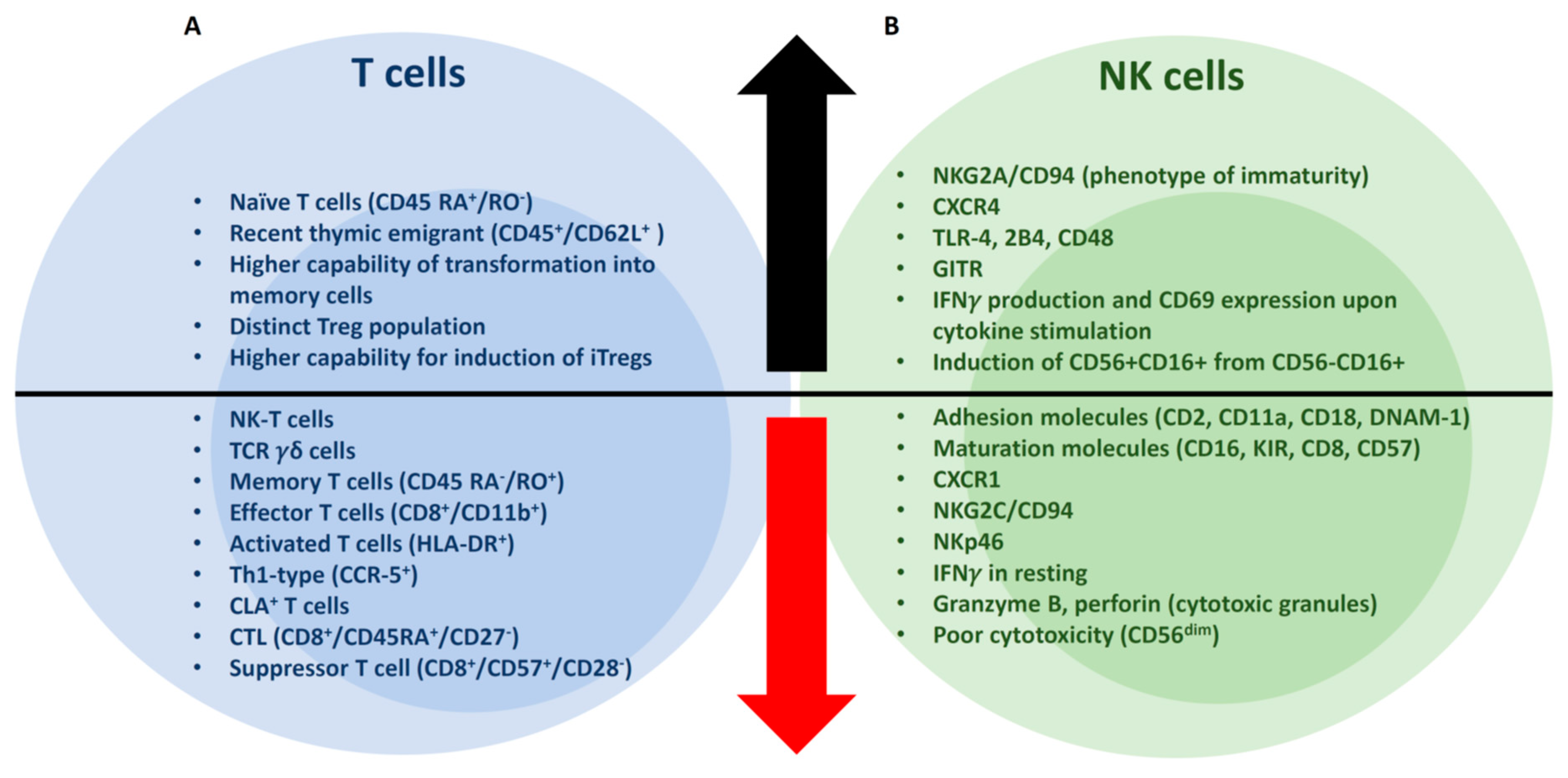
As compared to PB, the UCB CD3+ compartment has a lower number of NK-T and TCRγδ+ T cells [1]. CD4+ and CD8 T cells are 72% and 28%, respectively, in UCB, whereas 65% and 35% in PB. The fractions of naïve (CD45 RA+/RO) and memory T cells (CD45 RA/RO+) are also different from PB (Figure 1); the median naïve T cells is 85% and memory T cells is 6% in UCB, whereas naïve T cells 39% and memory T cells 50% in PB. UCB contains a significantly greater percentage of CD45+/CD62L+ “recent thymic emigrants”, naïve CD4 and naïve CD8 T cell subsets with lower numbers of memory CD8+ T cells [1,3]. Cytotoxic T lymphocyte (CTL, CD8+/CD45RA+/CD27) and suppressor T cell (CD8+/CD57+/CD28) populations are absent, with a lower percentage of effector (CD8+CD11b+), activated (HLA-DR+), Th1-type (CCR-5+) T cells [1]. Cutaneous lymphoid antigen (CLA) is expressed on T cells preferentially homing to cutaneous inflammatory areas. Interestingly, CLA is not expressed on UCB T cells, potentially relevant to a lower incidence of GVHD in UCBT [1,4]. Upon phytohemagglutinin (PHA) and interleukin-2 (IL-2) stimulation, newborn CD4+CD45RA+ naïve T cells convert more rapidly into CD4+CD45RO+ memory T cells than the adult counterpart, suggesting a greater capability of UCB naïve T cells to transform into memory T cells [5]. However, IFNγ and TNF⍺, as well as IL-2 production of UCB lymphocytes, are markedly lower than adult PB lymphocytes [3], possibly due to the reduced expression of NFAT-dependent genes [6]. Notably, UCB T cells have powerful allogeneic antitumor activities with a higher tumor-infiltrating CD8/Treg ratio and rapid differentiation into memory/effector cells than adult PB T cells [7].

2.2. Tregs

Tregs are conventionally defined as a subset of CD4+CD25+ T cells that maintain self-tolerance and immune suppression. Forkhead box protein P3 (FoxP3) is the master transcription factor for development of Tregs [8,9]. Tregs are either developed in the thymus (nTregs) or transformed from CD4+CD25 naïve T cells in the presence of transforming growth factor beta (TGFβ) in peripheral lymphoid tissues (iTregs) [8,10]. UCB possesses a distinct CD25+ population within the CD4+ T cell compartment [11]. The majority of UCB regulatory T cells (Tregs) are inexperienced with antigen stimulation, hence, cannot suppress “antigen-specific” alloreactive T cells [12,13]. However, after UCB Tregs are expanded and activated by TCR engagement and cytokine stimulation (e.g., stimulation with anti-CD3/CD28 mAb-coated beads and IL-2), their suppressive potency for allogeneic T cells become significantly greater than Tregs isolated from adult PB [11]. Furthermore, UCB Tregs possess a higher capacity for expansion [14], and Tregs (CD4+CD25+CTLA4+) are more readily inducible from CD4+CD25 T cells in UCB than adult PB [13]. Notably, early phase clinical trials using ex vivo UCB-derived nTreg expansion have shown promising clinical efficacy to prevent acute GVHD [15,16].

2.3. NK Cells

NK cells are known to be the main effector for graft-versus-leukemia (GVL) reactions early after HCT [17,18], and are enriched comprising up to 30% in the UCB graft (Figure 1) [1,19,20,21]. The CD56bright/CD56dim ratio of UCB NK cells is similar or slightly higher than that of PB NK cells (Figure 1) [20,22,23]. Notably, the cytotoxicity of CD56dim NK subset is poorer than that of CD56bright NK cells in the UCB graft, whereas CD56dim NK cells exert stronger cytotoxic effects in PB [22]. Cytotoxicity of CD56bright subset of UCB NK cells is comparable to that of PB NK cells, whereas cytotoxicity of CD56dim NK cells of UCB is greatly diminished compared to the counterpart of PB. A conjugate forming assay revealed that binding of CD56dim NK cells of UCB to leukemic targets was significantly impaired secondary to a lower expression of adhesion molecules, including CD2, CD11a, CD18, and DNAM-1 [22]. In another report, L-selectin, ICAM-1 expression was significantly lower in NK cells of UCB than PB [20,23]. The expression of chemokine receptors in UCB NK cells are different from PB NK cells. CXCR1 expression in UCB CD56dim NK cells is significantly reduced, whereas CXCR4 expression in both UCB CD56dim and CD56bright NK cells is increased [23]. Hence, UCB NK cells may be less responsive to inflammatory stimulation (involved in CXCR1 expression), but better capable of homing to the bone marrow (CXCR4), perhaps accounting for the effective GVL of UCBT [24]. Compared to PB NK cells, UCB NK cells less express maturation markers such as KIR, CD16, and CD57, whereas the expression of NKG2A/CD94, a phenotypic marker of NK cell immaturity, is higher in UCB NK cells [20,21,23]. In terms of NK cell activation receptors, the expression of NKG2C/CD94 and NKp46 (in CD56bright) is lower, but TLR-4, GITR, 2B4, and CD48 (in CD56bright) expression is higher in UCB NK cells than PB NK cells [2,23].
Immature CD56/CD16+ NK cells are a distinct population identified in the UCB graft, but rarely found in healthy adults [18,25,26]. CD56/CD16+ NK cells can differentiate into CD56+/CD16+ NK cells under IL-2 or IL-15 stimulation, acquiring enhanced cytotoxicity [26]. The expansion of UCB NK cells is less responsive to low-dose IL-2 stimulation (200 IU/mL) than PB NK cells. This is likely secondary to lower expression of CD25 (IL-2Rα) in CD56dim NK cells in UCB [23]. While resting, UCB CD56dim NK cells express lower levels of CD107a, IFNγ, granzyme B, perforin, and FAS-L, thereby exert a lower cytotoxicity than PB NK cells. UCB NK cells can acquire a potent cytotoxicity with an enhanced production of IFNγ and cytotoxic granules by high-dose IL-2 stimulation (1000 IU/mL) [23]. Furthermore, the response of UCB NK cells to IL-12 and IL-18 stimulation measured by IFNγ production and CD69 expression is higher than that of PB NK cells [27,28].

3. Immune Reconstitution in UCBT

Immune reconstitution after HCT may impact clinical outcomes such as incidence of transplant-related mortality and GVHD, the risks of infections and relapse, and, ultimately, survival. Numerous factors influence IR after HCT, including conditioning regimen (myeloablative vs. nonmyeloablative, use of antithymocyte globulin, and total body irradiation), immune suppression regimen, the graft source, cell compositions of the graft, and viral infections in HCT [29]. In this section, we will focus on T and NK cell reconstitution after UCBT, and compare with other donor types when possible.

3.1. T cells

T cell reconstitution is delayed after UCBT as compared to bone marrow transplantation (BMT) [30] and peripheral blood stem cell transplantation (PBSCT) [31]. T cell reconstitution after HCT occurs in two distinct pathways: (1) Peripheral expansion of mature T cells (thymus-independent pathway), and (2) thymopoiesis from donor hematopoietic progenitors (thymus-dependent pathway) [32,33]. Early after HCT, T cell reconstitution takes place through peripheral expansion by T cells transferred from the graft or recipient T cells which have survived conditioning therapy (thymus-independent pathway). The mature T cells compete for homeostatic cytokines, such as IL-7 or IL-15, and self-MHC molecules presented by antigen-presenting cells (APCs) [34]. In the lymphopenic condition, IL-7 and IL-15 are constantly produced by immune and non-immune cells, but little is consumed. Hence, there are high plasma levels of these cytokines early after HCT [35]. The mature T cells transferred from the graft have greater access to IL-7 or IL-15 and self-MHC on APCs, which, in the context of lymphopenia, promotes expansion with limited competition [34,36]. Naturally, the T cells undergo rapid expansion with multiple cell divisions, leading to accelerated telomere shortening in the first year post-HCT [37]. In contrast to memory T cells, naïve T cells require TCR engagement with MHC molecules presented by APC, in addition to cytokine stimulation, for survival and expansion [34,38]. As a result, peripheral expansion of memory T cells is greater than naïve T cells after HCT. Another unique aspect of early immune reconstitution is the inverted CD4/CD8 ratio, secondary to better peripheral expansion of memory CD8 T cells [29]. In certain donor/recipient pairs, seropositivity to viral pathogens like CMV can polarize T cell expansion towards the viral antigens, narrowing the T cell repertoire that predominantly proliferates towards antigen-specific memory T cells, limiting polyclonal expansion [39]. In UCBT, early T cell reconstitution is primarily dependent on the peripheral expansion and may have limited T cell repertoire due to delayed thymopoiesis (especially in adults), as compared to other donor types [40]. Furthermore, CD4+ and CD8+ T cells at day +100 after UCBT have reduced capability to produce IFNγ upon superantigen and CMV stimulation, possibly indicating impaired T cell functions early after UCBT [41]. In a pediatric study using myeloablative conditioning (MAC) with antithymocyte globulin (ATG), median time to T cell recovery (CD3+ T cells >0.5 × 10⁹/L) was 6.3 months in UCBT group vs. 3.2 months for unrelated BM group (p = 0.008) [30]. This was apparently driven by CD8+ T cell reconstitution (>0.25 × 10⁹/L) that took a median of 7.7 months after UCB vs. 2.8 months after unrelated BM. However, recovery of the CD4+ T cell (CD4+ T cells >0.5 × 10⁹/L) numbers was faster after UCBT, with median time for CD4+ T cell recovery 9.3 months vs. 12 months in unrelated BMT (p = 0.003). Hence, the inverted CD4/CD8 ratio is not observed early after UCBT because of the delayed CD8+ T cell recovery after UCBT [42].
The reconstitution of T cell repertoire diversity from donor-derived naïve T cells occurs in the thymus following peripheral expansion of mature T cells post HCT (thymus-dependent pathway). This process, termed thymopoiesis, requires a longer period of time in which donor-derived lymphoid progenitors enter the thymus and undergo maturation processes (positive and negative selection) [43]. Thymopoiesis occurs weeks after HCT and can last up to 6 years [44]. After the sequential positive and negative selections, only small fractions of T cells can survive and exit the thymus, so-called recent thymus emigrant (RTE) [45]. This de novo process can be measured by T cell receptor (TCR) excision circle (TREC), naïve T cell counts, and T cell repertoire diversity [44,46,47], and is critical for broad and self-tolerant T cell immunity [32,46]. After UCBT, TREC levels correlate with CD3+CD4+45RO naïve T cell counts (r = 0.83, p = 0.0001), and TCR repertoire diversity (r = 0.83, p = 0.0001) [48]. Long-term T cell reconstitution (CD3 >1.5 × 10⁹/L) is similar between UCBT and unrelated BMT (9.3 vs. 10 months) in the pediatric population [30]. In an age- and GVHD-matched comparison of children and young adults between UCB [median age 12.6 years (3–34.6)] and matched sibling recipients, TREC and CD4+CD45RO naïve T cells were significantly higher, whereas CD8+ activated and memory T cells were lower at 2 years in UCB as compared to matched sibling donor group, indicating efficient thymopoiesis in UCBT [48].

3.2. NK Cells

Natural killer cells are the first lymphocytes reconstituting after HCT. NK cell immunity plays a critical role in GVL, especially early after UCBT, because of the low absolute counts and functional immaturity of T cells transferred with the UCB graft. The time to NK cell reconstitution (>0.1 × 10⁹/L) was similar between UCBT (1 month) and unrelated BMT (1.4 months), when both groups received ATG as part of the conditioning regimen [30]. Notably, after UCB with no ATG in the conditioning regimen, NK cell count reconstitution at 1 month after UCBT was similar to healthy controls [49,50]. Moreover, a better NK cell reconstitution with higher NK cell counts was observed over a 24-month period in UCBT than PBSCT [31,51].
NK cell reconstitution 1–3 months after UCBT is polarized to CD56bright NK cells (approximately 40% of the total NK cells), as compared to healthy donor controls [49,50]. Three months after UCBT, NK cells express high levels of NKG2A and CD62L and low levels of CD16, CD8, and CD57 [49]. Even in CD56dim NK cells, the expression of CD94/NKG2A, an inhibitory receptor recognizing HLA-E antigen, is higher early after UCBT, but gradually returns to levels similar to that of healthy controls’ by 1 year after UCBT [50]. The expression of KIR2DL2/3 and KIR3DL1 of NK cells is significantly lower in the UCB graft, but becomes comparable within 3 months after UCBT to healthy donors, indicating acquisition of NK cell education [49,50]. However, KIR2DL1 levels of CD56dim NK cells are persistently lower than that of healthy controls during the first 6 months after UCBT, consistent with the sequential acquisition of KIR commonly observed in other types of HCT [49,50,52,53]. Interestingly, NKp30, NKp46 (natural cytotoxicity receptors involving NK cell activation), and CD69 (an activation marker) of CD56dim NK cells are transiently higher for the first couple of months after UCBT than healthy controls [50], potentially providing advantages in GVL reactions. The HLA-DR expression of NK cells is significantly higher during the first year of UCBT than that of healthy controls and UCB grafts [49]. DNAM-1 (an activating NK cell receptor) expression of CD56dim NK cells is significantly lower in the UCB graft, but gradually increases, and becomes similar to the level of healthy control NK cells within a year after UCBT [50].
NK cells acquire unique functional characteristics after UCBT, as evidenced by high IFNγ production in the first 1–3 months [49,50]. Direct cytotoxicity of NK cells during the first 6 months post-UCBT against K562 targets and HLA mismatched primary acute myeloid leukemia (AML) samples is robust, and similar to that of healthy controls [50]. However, antibody-dependent cellular cytotoxicity (ADCC) of NK cells within 3 months after UCBT is significantly impaired [50], consistent with low expression of CD16 early after UCBT [49].

4. Clinical Factors Associated with Immune Reconstitution in UCBT

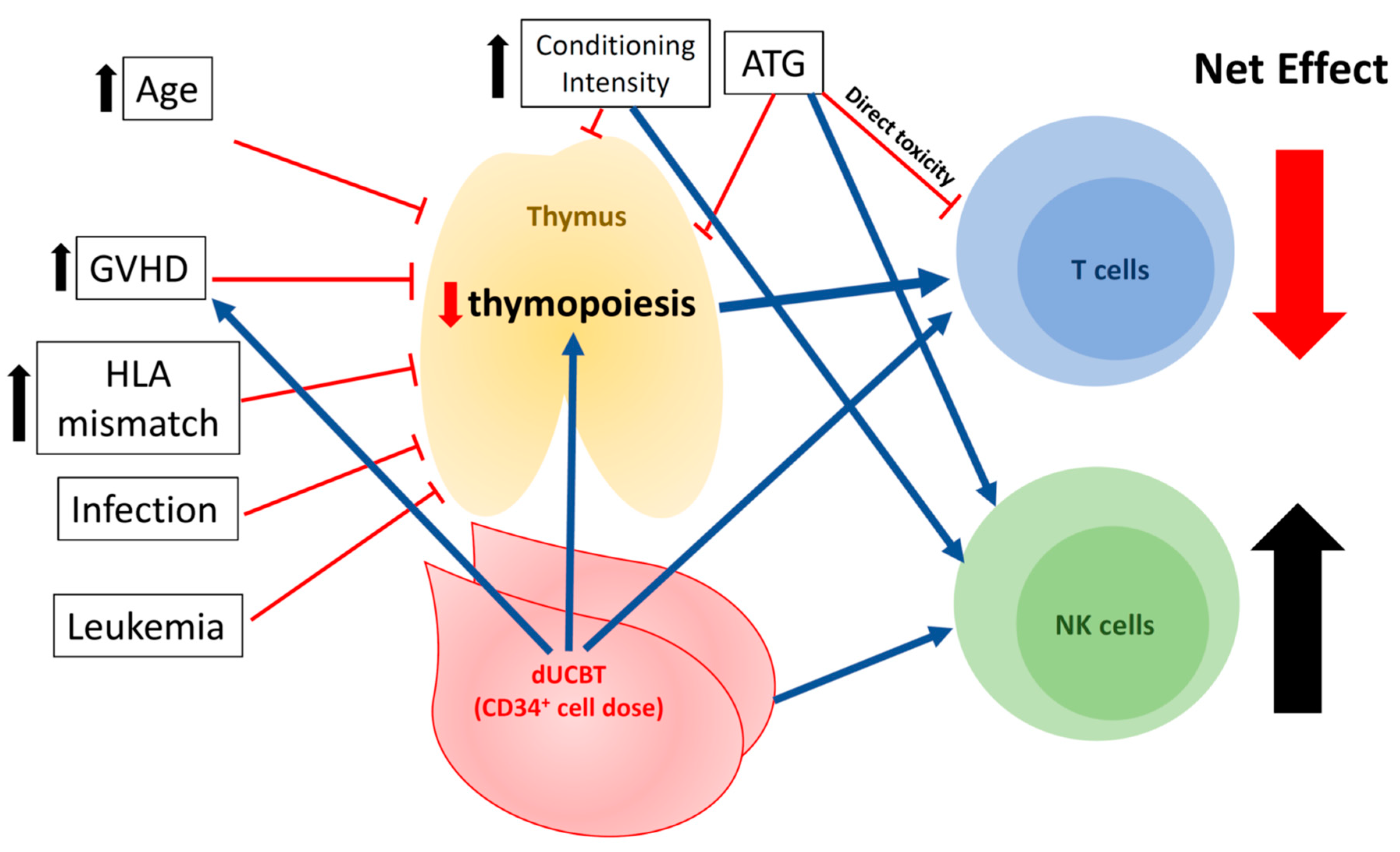
As summarized in Figure 3, multiple clinical factors potentially influence the reconstitution of T and NK cells after UCBT. Selected factors are reviewed below.

4.1. Viral Infections

High incidence of HHV-6 (up to 70–80%) has been associated with delayed engraftment after UCBT in multiple reports [54,55]. HHV-6 infection can also interfere with T cell reconstitution, both in a thymus-dependent and independent pathways, resulting in dysfunctional T cell population after HCT [56]. A recent retrospective study with time-dependent analysis revealed that high HHV6 viral load (>105 copies/mL) was associated with impairment of both CD4+ and CD8+ T cell reconstitution after HCT, including UCBT [57]. Interestingly, HHV6 infection negatively affected reconstitution of naïve, but not effector memory CD4+ T cells. In addition, reactivation of HHV-6 early after UCBT was associated with T cells expressing CD57, NKG2A, and KIR2DL2/3, surface markers of T cell senescence, and hypofunction, and it was associated in inferior clinical outcomes [58]. Other viral infections may result in limited and skewed TCR diversity towards a specific viral antigen, as observed in individuals with Epstein-Barr virus (EBV) and cytomegalovirus (CMV) infections than those without these infections [59].

4.2. GVHD

Glucocorticoid (GC) is the cornerstone of treatment for acute and chronic GVHD. GC is known to induce in vivo Treg expansion [60,61], and inhibit the JAK-STAT signaling pathway induced by IL-2, IL-4, IL-7, and IL-15 in T cells [62]. A murine HCT model suggests CD8+ T cells as the main target of GC [63]. Taken together, CG used for treatment of GVHD profoundly affects in the T cell function. GC also prevents upregulation of MHC class II and costimulatory molecules on dendritic cells [64,65], hence, compromises T cell responses against foreign or allo-antigens. High-dose GC can also induce T cell apoptosis [65]. Furthermore, GVHD can directly damage the thymus by inducing apoptosis of thymocytes (thymic GVHD) [66,67]. Among allo-HCT recipients, absolute counts of naïve T cells were significantly lower at 12 months, with narrower and more skewed TCR repertoires in patients with aGVHD than without aGVHD [47,48]. Furthermore, both sjTREC and βTREC were significantly lower in the aGVHD group, but sj/βTREC ratio was comparable between groups with and without aGVHD, indicating that aGVHD primarily impairs early-stage thymopoiesis [47]. In addition, patients with cGVHD developed markedly lower TREC [44,48]. Interestingly, steady increases in TREC levels in both CD4+ and CD8+ T cells were observed in UCBT patients receiving immunosuppression without active GVHD, suggesting that GVHD prophylaxis with immunosuppression alone does not necessarily cease thymopoiesis [44].

4.3. Conditioning Regimen

ATG has been frequently used in the conditioning regimen in UCBT. ATG delays T cell reconstitution by depleting naïve and memory T cells transferred with the graft required for early peripheral expansion of mature T cells [68]. In contrast, ATG exposure is associated with strong recovery of B and NK cells 30 days after transplant, enabling B and NK cells to compensate T cell defects in UCBT [41,43]. In addition, the timing, dose of ATG administration, and serum level at the time of the allograft infusion may influence incidence and grade of GVHD and T cell subset reconstitution [68,69]. In the absence of exposure to ATG, better T cell reconstitution after UCBT is observed, which may contribute, at least in part, to better leukemia control and lower all-cause mortality [68,70,71].
Reduced intensity regimen (RIC, Fludarabine 30 mg/m2 for 5 days, Cyclophosphamide 50 mg/kg, and TBI 200cGy) without ATG in adults employed at the University of Minnesota was associated with comparable lymphoid reconstitution at day 180 post-HCT and significantly lower chronic GVHD at 1 year post-HCT in UCBT compared to matched sibling donor HCT. In a pediatric UCBT study where all patients received ATG in the conditioning [72], NK cell counts after transplant were higher in the MAC group. Multivariate analysis revealed that the MAC group had a higher risk of developing acute GVHD (HR 6.1, p = 0.002), increased treatment-related mortality (TRM) (OR 26.8, p = 0.008), and overall mortality (HR = 4.1, p = 0.0001). In another adult study [73], CD3+ T cell recovery was observed at 6–12 months after UCBT with higher number of CD45RA+ T cells, more diverse T cell repertoire in patients treated with nonmyeloablative regimen (NMA) (fludarabine 30 mg/m2, cyclophosphamide 500 mg/m2, and ATG 30 mg/kg) when compared to a historical MA group [40]. Furthermore, TREC was detected at 12 months in NMA group, whereas at 18–24 months in the MA group [40,73]. Taken together, RIC may provide better T cell reconstitution, whereas MAC may favor NK cell reconstitution.

4.4. Age

Age is one of the most significant host factors to influence T cell reconstitution and thymopoiesis following UCBT. In adult patients with TBI- and ATG-based conditioning regimen, the number of CD8+ T cells reached normal ranges a year after UCBT, but total T cell counts remained below normal for 2 years from UCBT, and memory T cells remained 70% of the total T cell population until 12 months post-UCBT [40,41]. These data suggest that T cell reconstitution and thymopoiesis are delayed in adult UCBT. In contrast, the long-term immune recovery, including after UCBT, was similar to those who underwent adult unrelated donor [30,74] and haploientical donor HCT [75] in children, suggesting faster thymus-dependent T cell reconstitution. In a pediatric population, TREC numbers recovered to the pre-UCBT levels by 6 months [75], and reached within normal limits by 1 year [40,45], whereas in adults, sjTREC recovery took a median of 3 years after UCBT [40,41]. Furthermore, diverse T cell repertoires were observed at 1–2 years post-UCBT in children, whereas it took 3–4 years in adult patients [40]. Hence, in part, driven by reduced thymopoiesis, the delay of T cell numbers and repertoire reconstitution is a challenge in adults undergoing UCBT.

4.5. Cell Dose: CD34+ Progenitor Counts in Grafts and Single vs. Double Unit(s) of UCBT

CD34+ count (>107/kg vs. <107/kg) in the allograft correlates with the TREC levels after HCT in children, suggesting that CD34+ cell dose plays a role in thymopoiesis [76]. However, despite a higher combined CD34 cell dose in children receiving double (dUCBT) UCB grafts, reconstitution of lymphocyte subsets was similar to that of single UCBT (sUCBT) in children (and young adults) at 1–2 years after UCBT [77,78]. While data directly comparing single with double UCBT in adults are not available, in adult dUCBT without ATG, the TCR diversity measured by TCR deep sequencing at 6 months after HCT approached that observed in a healthy control group [59]. Others reported that, despite administration of ATG, adult patients undergoing dUCBT had a steep rise in TREC numbers between 6 and 12 months after transplant [79], while the recovery of T numbers between 6 and 12 months approached that of recipients of sibling and unrelated donors grafts in the absence of ATG [80,81]. In summary, dUCBT seems to provide better T cell reconstitution in adults. At least in part, this may be explained by the “threshold effect” of CD34+ cell doses in thymopoiesis [76] (i.e., a single UCB graft may contain sufficient CD34+ progenitors to reach the “threshold” of thymopoiesis for pediatric but not for adult recipients). This effect, however, has to be considered in the context of conditioning regimen intensity and administration of ATG.

5. Clinical Impacts of Immune Reconstitution on Outcomes of UCBT

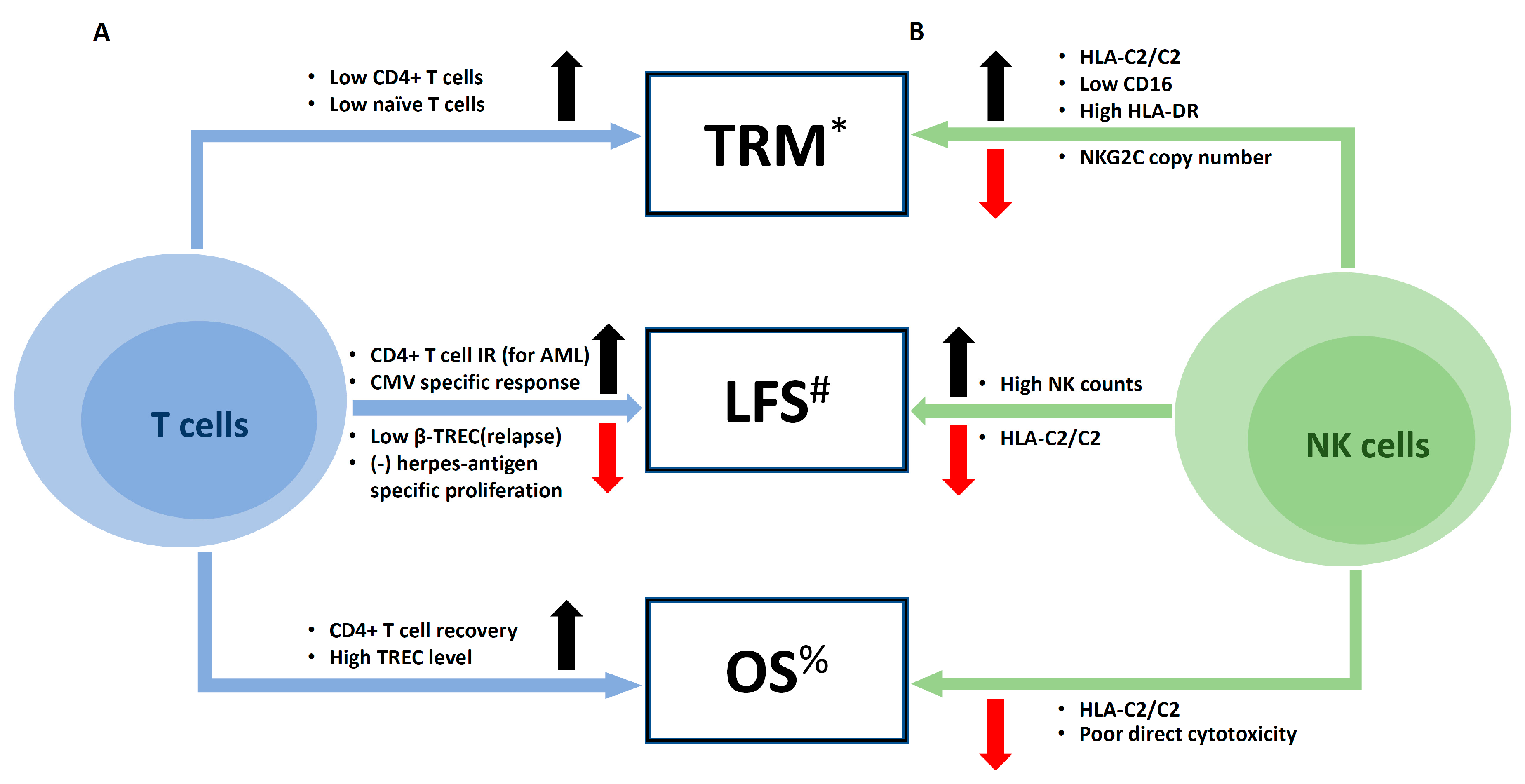
Reconstitution of lymphocyte subsets after UCBT is influenced by multiple clinical factors, as discussed above. Conversely, immunological and genetic characteristics of T and NK cells are critical determinants of clinical outcomes in HCT. Figure 4 summarizes the potential impact of lymphocyte reconstitution on UCBT outcomes.

5.1. Infections

Infection is a major cause of death in UCBT. Szabolcs et al. reported that 58% of deaths within 6 months in UCBT were due to infection in children [82]. A better T cell reconstitution at day 50 post-UCBT, including higher absolute CD4+ T cell counts, was observed in the group without subsequent opportunistic infections (OI) than with OI at day 100 post-UCBT. This finding indicates that T cells play a critical role to prevent opportunistic infection. Additionally, both CD34+ and CD3+ cell doses were associated with lower death from OI at 6 months. In a retrospective study where UCBT recipients constituted more than a half of the entire cohort, delayed CD4+ T cell reconstitution was predictive of adenovirus, EBV, and HHV6 infections [83]. Moreover, CD4+ T cell reconstitution (≥50 × 106/L) within 100 days was associated with a shorter duration of adenovirus infection. CMV infection is a major life-threatening complication in HCT. In a large cohort study (n = 332) at the University of Minnesota, 51% of recipients with hematological malignancies undergoing UCBT developed CMV reactivation [84]. CMV-specific CD8+ T cells transferred from the UCB graft alone could not eradicate CMV viremia, but clearance of CMV viremia occurred later and seemed to depend on CD4+CD45RA+ T cells by thymopoiesis [79]. However, the Seattle group identified intact CMV-specific T cell priming early after UCBT (at day +42), suggesting that failure to control CMV reactivation is likely due to insufficient numbers of these T cells in vivo [85]. Nevertheless, successful T cell reconstitution by thymopoiesis is required for an optimal control of CMV reactivation. Additionally, NK cells may play an important role in CMV control. A recent study demonstrated that low NKG2C copy number of NK cells in the UCB graft was independently associated with increased risk of developing CMV reactivation (HR = 2.72, p < 0.0001) [86].

5.2. Major Clinical Outcomes: Relapse, Mortality, and Survival

Poor T cell reconstitution is associated with increased risks of relapse in HCT. Clave et al. reported that relapsed pediatric patients with hematological malignancy had lower β-TREC levels at 6 months before and after HCT (including UCBT), suggesting an association between impairment of early intra-thymic T cell development and increased relapse risks [75]. Regardless of graft types, both low CD4+ and naïve T cell reconstitution are significantly associated with increased risks of treatment-related mortality at day 100 post-HCT [87]. Successful CD4+ T cell reconstitution at day 100 after UCBT by less exposure to ATG is associated with lower non-relapse mortality (NRM), lower relapse-related mortality (particularly for AML), better event-free survival, and better overall survival (OS) [68]. Furthermore, early CD4+ T cell reconstitution (defined by CD4+ T cell >50 × 106/L within 100 days after UCBT) resulted in better leukemia-free survival (LFS) (HR = 0.24, p = 0.003), improved OS (HR = 0.16, p = 0.0014) with lower NRM (HR = 0.20, p = 0.0072). Again, lower RI (HR = 0.31, p = 0.041) in association with improved T cell reconstitution was observed in AML [88]. Successful CD8+ T cell reconstitution and high TREC levels, as well as CD4+ T cell reconstitution, are associated with improved OS [79]. Moreover, CMV-specific T cell response and high NK cell counts are independently associated with better progression-free survival (PFS). As T cells in the UCB graft are unlikely to have encountered antigens of herpes viruses, herpes antigen-specific T cell response represents T cell reconstitution occurring in vivo. Hence, Parkman et al. measured herpes antigen-specific proliferative T cell responses as measurement of successful T cell IR [89]. In pediatric patients with acute leukemias undergoing ATG-based MA sUCBT, the earliest herpes-antigen specific T cell response was observed within the first month. Notably, negative antigen-specific T cell response was independently associated with higher leukemia relapse (HR = 3.7, p = 0.003) and lower relapse-free survival (HR = 3.6, p = 0.0002), indicating that successful T cell reconstitution plays a critical role in relapse prevention.
Transplant outcomes in UCBT are also tightly associated with NK cell IR. In RIC UCBT for AML, the low CD16 and high HLA-DR expression on NK cells are significantly associated with increased risks of TRM [49]. In addition, KIR-HLA typing is associated with overall survival (OS) [49]. HLA C2 homozygous recipients have much poorer event-free survival (EFS) (HR = 6.19, p = 0.002), OS (HR = 6.12, p = 0.001), and higher TRM (HR = 9.44, p = 0.026) than HLA C1/x recipients. Furthermore, poor direct cytotoxicity of NK cells against K562 measured by CD107a expression was significantly associated with poor overall survival as well.

6. Closing Remarks

UCBT has been a valuable alternative donor for transplantation for the past several decades. It has clinical advantages, including readily available grafts, relatively lower incidence of GVHD, and lower disease relapse [4,24,77,90]. While there have been some challenges in UCBT, advances have been made by modifying conditioning regimens [87,91], double UCB grafts [77]. More sophisticated utilization of ATG has substantially improved clinical outcomes [68,69,70]. Emergence of more robust ex vivo expansion techniques have enabled to meet the adequate cell doses for larger patients [92,93]. In addition, a recent report described the powerful GVL effect of UCB in those with acute leukemia and minimal residual disease [24]. Better understanding of the immune biology in UCBT will lead to improved graft engineering in the future.

Author Contributions

H.D.Y. and C.B. conceived the idea. H.D.Y. analyzed and synthesized literature data. C.B. supervised the entire project. H.D.Y. and C.B. wrote and edited the manuscript. A.V., M.J.H., S.N. did literature search and edited the manuscript. All authors approved the publication.

Funding

This work was supported by no external funding.

Acknowledgments

We appreciate commitment and support in cancer research from both institutions, Rush University Medical Center and University of Minnesota.

Conflicts of Interest

The authors declare no conflicts of interest.

References

  1. Szabolcs, P.; Park, K.D.; Reese, M.; Marti, L.; Broadwater, G.; Kurtzberg, J. Coexistent naive phenotype and higher cycling rate of cord blood T cells as compared to adult peripheral blood. Exp. Hematol. 2003, 31, 708–714. [Google Scholar] [CrossRef]
  2. Lopez, M.C.; Palmer, B.E.; Lawrence, D.A. Phenotypic differences between cord blood and adult peripheral blood. Cytom. Part B Clin. Cytom. 2009, 76, 37–46. [Google Scholar] [CrossRef] [PubMed]
  3. Chalmers, I.M.; Janossy, G.; Contreras, M.; Navarrete, C. Intracellular cytokine profile of cord and adult blood lymphocytes. Blood 1998, 92, 11–18. [Google Scholar] [CrossRef] [PubMed]
  4. Barker, J.N.; Wagner, J.E. Umbilical-cord blood transplantation for the treatment of cancer. Nat. Rev. Cancer 2003, 3, 526–532. [Google Scholar] [CrossRef]
  5. Early, E.; Reen, D.J. Rapid conversion of naive to effector T cell function counteracts diminished primary human newborn T cell responses. Clin. Exp. Immunol. 1999, 116, 527–533. [Google Scholar] [CrossRef]
  6. Kaminski, B.A.; Kadereit, S.; Miller, R.E.; Leahy, P.; Stein, K.R.; Topa, D.A.; Radivoyevitch, T.; Veigl, M.L.; Laughlin, M.J. Reduced expression of NFAT-associated genes in UCB versus adult CD4+ T lymphocytes during primary stimulation. Blood 2003, 102, 4608–4617. [Google Scholar] [CrossRef]
  7. Hiwarkar, P.; Qasim, W.; Ricciardelli, I.; Gilmour, K.; Quezada, S.; Saudemont, A.; Amrolia, P.; Veys, P. Cord blood T cells mediate enhanced antitumor effects compared with adult peripheral blood T cells. Blood 2015, 126, 2882–2891. [Google Scholar] [CrossRef]
  8. Hori, S.; Nomura, T.; Sakaguchi, S. Control of regulatory T cell development by the transcription factor Foxp3. Science 2003, 299, 1057–1061. [Google Scholar] [CrossRef]
  9. Fontenot, J.D.; Gavin, M.A.; Rudensky, A.Y. Foxp3 programs the development and function of CD4+CD25+ regulatory T cells. Nat. Immunol. 2003, 4, 330–336. [Google Scholar] [CrossRef]
  10. Chen, W.; Jin, W.; Hardegen, N.; Lei, K.J.; Li, L.; Marinos, N.; McGrady, G.; Wahl, S.M. Conversion of peripheral CD4+CD25- naive T cells to CD4+CD25+ regulatory T cells by TGF-beta induction of transcription factor Foxp3. J. Exp. Med. 2003, 198, 1875–1886. [Google Scholar] [CrossRef]
  11. Godfrey, W.R.; Spoden, D.J.; Ge, Y.G.; Baker, S.R.; Liu, B.; Levine, B.L.; June, C.H.; Blazar, B.R.; Porter, S.B. Cord blood CD4(+)CD25(+)-derived T regulatory cell lines express FoxP3 protein and manifest potent suppressor function. Blood 2005, 105, 750–758. [Google Scholar] [CrossRef] [PubMed]
  12. Wing, K.; Lindgren, S.; Kollberg, G.; Lundgren, A.; Harris, R.A.; Rudin, A.; Lundin, S.; Suri-Payer, E. CD4 T cell activation by myelin oligodendrocyte glycoprotein is suppressed by adult but not cord blood CD25+ T cells. Eur. J. Immunol. 2003, 33, 579–587. [Google Scholar] [CrossRef] [PubMed]
  13. Chang, C.C.; Satwani, P.; Oberfield, N.; Vlad, G.; Simpson, L.L.; Cairo, M.S. Increased induction of allogeneic-specific cord blood CD4+CD25+ regulatory T (Treg) cells: A comparative study of naive and antigenic-specific cord blood Treg cells. Exp. Hematol. 2005, 33, 1508–1520. [Google Scholar] [CrossRef] [PubMed]
  14. Torelli, G.F.; Maggio, R.; Peragine, N.; Chiaretti, S.; De Propris, M.S.; Lucarelli, B.; Screnci, M.; Mascolo, M.G.; Milano, F.; Iori, A.P.; et al. Functional analysis and gene expression profile of umbilical cord blood regulatory T cells. Ann. Hematol. 2012, 91, 155–161. [Google Scholar] [CrossRef]
  15. Brunstein, C.G.; Miller, J.S.; McKenna, D.H.; Hippen, K.L.; DeFor, T.E.; Sumstad, D.; Curtsinger, J.; Verneris, M.R.; MacMillan, M.L.; Levine, B.L.; et al. Umbilical cord blood-derived T regulatory cells to prevent GVHD: Kinetics, toxicity profile, and clinical effect. Blood 2016, 127, 1044–1051. [Google Scholar] [CrossRef]
  16. Brunstein, C.G.; Miller, J.S.; Cao, Q.; McKenna, D.H.; Hippen, K.L.; Curtsinger, J.; Defor, T.; Levine, B.L.; June, C.H.; Rubinstein, P.; et al. Infusion of ex vivo expanded T regulatory cells in adults transplanted with umbilical cord blood: Safety profile and detection kinetics. Blood 2011, 117, 1061–1070. [Google Scholar] [CrossRef]
  17. Sarvaria, A.; Jawdat, D.; Madrigal, J.A.; Saudemont, A. Umbilical Cord Blood Natural Killer Cells, Their Characteristics, and Potential Clinical Applications. Front. Immunol. 2017, 8, 329. [Google Scholar] [CrossRef]
  18. Verneris, M.R.; Miller, J.S. The phenotypic and functional characteristics of umbilical cord blood and peripheral blood natural killer cells. Br. J. Haematol. 2009, 147, 185–191. [Google Scholar] [CrossRef]
  19. Kotylo, P.K.; Baenzinger, J.C.; Yoder, M.C.; Engle, W.A.; Bolinger, C.D. Rapid analysis of lymphocyte subsets in cord blood. Am. J. Clin. Pathol. 1990, 93, 263–266. [Google Scholar] [CrossRef]
  20. Dalle, J.H.; Menezes, J.; Wagner, E.; Blagdon, M.; Champagne, J.; Champagne, M.A.; Duval, M. Characterization of cord blood natural killer cells: Implications for transplantation and neonatal infections. Pediatric Res. 2005, 57, 649–655. [Google Scholar] [CrossRef]
  21. Wang, Y.; Xu, H.; Zheng, X.; Wei, H.; Sun, R.; Tian, Z. High expression of NKG2A/CD94 and low expression of granzyme B are associated with reduced cord blood NK cell activity. Cell. Mol. Immunol. 2007, 4, 377–382. [Google Scholar] [PubMed]
  22. Tanaka, H.; Kai, S.; Yamaguchi, M.; Misawa, M.; Fujimori, Y.; Yamamoto, M.; Hara, H. Analysis of natural killer (NK) cell activity and adhesion molecules on NK cells from umbilical cord blood. Eur. J. Haematol. 2003, 71, 29–38. [Google Scholar] [CrossRef] [PubMed]
  23. Luevano, M.; Daryouzeh, M.; Alnabhan, R.; Querol, S.; Khakoo, S.; Madrigal, A.; Saudemont, A. The unique profile of cord blood natural killer cells balances incomplete maturation and effective killing function upon activation. Hum. Immunol. 2012, 73, 248–257. [Google Scholar] [CrossRef] [PubMed]
  24. Milano, F.; Gooley, T.; Wood, B.; Woolfrey, A.; Flowers, M.E.; Doney, K.; Witherspoon, R.; Mielcarek, M.; Deeg, J.H.; Sorror, M.; et al. Cord-Blood Transplantation in Patients with Minimal Residual Disease. N. Engl. J. Med. 2016, 375, 944–953. [Google Scholar] [CrossRef] [PubMed]
  25. Della Chiesa, M.; Falco, M.; Podesta, M.; Locatelli, F.; Moretta, L.; Frassoni, F.; Moretta, A. Phenotypic and functional heterogeneity of human NK cells developing after umbilical cord blood transplantation: A role for human cytomegalovirus? Blood 2012, 119, 399–410. [Google Scholar] [CrossRef] [PubMed]
  26. Gaddy, J.; Broxmeyer, H.E. Cord blood CD16+56- cells with low lytic activity are possible precursors of mature natural killer cells. Cell. Immunol. 1997, 180, 132–142. [Google Scholar] [CrossRef] [PubMed]
  27. Nomura, A.; Takada, H.; Jin, C.H.; Tanaka, T.; Ohga, S.; Hara, T. Functional analyses of cord blood natural killer cells and T cells: A distinctive interleukin-18 response. Exp. Hematol. 2001, 29, 1169–1176. [Google Scholar] [CrossRef]
  28. Mehta, R.S.; Shpall, E.J.; Rezvani, K. Cord Blood as a Source of Natural Killer Cells. Front. Med. 2015, 2, 93. [Google Scholar] [CrossRef]
  29. Ogonek, J.; Kralj Juric, M.; Ghimire, S.; Varanasi, P.R.; Holler, E.; Greinix, H.; Weissinger, E. Immune Reconstitution after Allogeneic Hematopoietic Stem Cell Transplantation. Front. Immunol. 2016, 7, 507. [Google Scholar] [CrossRef]
  30. Renard, C.; Barlogis, V.; Mialou, V.; Galambrun, C.; Bernoux, D.; Goutagny, M.P.; Glasman, L.; Loundou, A.D.; Poitevin-Later, F.; Dignat-George, F.; et al. Lymphocyte subset reconstitution after unrelated cord blood or bone marrow transplantation in children. Br. J. Haematol. 2011, 152, 322–330. [Google Scholar] [CrossRef]
  31. Jacobson, C.A.; Turki, A.T.; McDonough, S.M.; Stevenson, K.E.; Kim, H.T.; Kao, G.; Herrera, M.I.; Reynolds, C.G.; Alyea, E.P.; Ho, V.T.; et al. Immune reconstitution after double umbilical cord blood stem cell transplantation: Comparison with unrelated peripheral blood stem cell transplantation. Biol. Blood Marrow Transplant. 2012, 18, 565–574. [Google Scholar] [CrossRef] [PubMed] [Green Version]
  32. Chaudhry, M.S.; Velardi, E.; Malard, F.; van den Brink, M.R. Immune Reconstitution after Allogeneic Hematopoietic Stem Cell Transplantation: Time To T Up the Thymus. J. Immunol. 2017, 198, 40–46. [Google Scholar] [CrossRef] [PubMed]
  33. Servais, S.; Hannon, M.; Peffault de Latour, R.; Socie, G.; Beguin, Y. Reconstitution of adaptive immunity after umbilical cord blood transplantation: Impact on infectious complications. Stem Cell Investig. 2017, 4, 40. [Google Scholar] [CrossRef] [PubMed] [Green Version]
  34. Jameson, S.C. Maintaining the norm: T-cell homeostasis. Nat. Rev. Immunol. 2002, 2, 547–556. [Google Scholar] [CrossRef]
  35. Thiant, S.; Yakoub-Agha, I.; Magro, L.; Trauet, J.; Coiteux, V.; Jouet, J.P.; Dessaint, J.P.; Labalette, M. Plasma levels of IL-7 and IL-15 in the first month after myeloablative BMT are predictive biomarkers of both acute GVHD and relapse. Bone Marrow Transpl. 2010, 45, 1546–1552. [Google Scholar] [CrossRef] [Green Version]
  36. Williams, K.M.; Hakim, F.T.; Gress, R.E. T cell immune reconstitution following lymphodepletion. Semin. Immunol. 2007, 19, 318–330. [Google Scholar] [CrossRef] [Green Version]
  37. Rufer, N.; Brummendorf, T.H.; Chapuis, B.; Helg, C.; Lansdorp, P.M.; Roosnek, E. Accelerated telomere shortening in hematological lineages is limited to the first year following stem cell transplantation. Blood 2001, 97, 575–577. [Google Scholar] [CrossRef] [Green Version]
  38. Tanchot, C.; Lemonnier, F.A.; Perarnau, B.; Freitas, A.A.; Rocha, B. Differential requirements for survival and proliferation of CD8 naive or memory T cells. Science 1997, 276, 2057–2062. [Google Scholar] [CrossRef]
  39. Suessmuth, Y.; Mukherjee, R.; Watkins, B.; Koura, D.T.; Finstermeier, K.; Desmarais, C.; Stempora, L.; Horan, J.T.; Langston, A.; Qayed, M.; et al. CMV reactivation drives posttransplant T-cell reconstitution and results in defects in the underlying TCRbeta repertoire. Blood 2015, 125, 3835–3850. [Google Scholar] [CrossRef]
  40. Klein, A.K.; Patel, D.D.; Gooding, M.E.; Sempowski, G.D.; Chen, B.J.; Liu, C.; Kurtzberg, J.; Haynes, B.F.; Chao, N.J. T-Cell recovery in adults and children following umbilical cord blood transplantation. Biol. Blood Marrow Transplant. 2001, 7, 454–466. [Google Scholar] [CrossRef] [Green Version]
  41. Komanduri, K.V.; St John, L.S.; de Lima, M.; McMannis, J.; Rosinski, S.; McNiece, I.; Bryan, S.G.; Kaur, I.; Martin, S.; Wieder, E.D.; et al. Delayed immune reconstitution after cord blood transplantation is characterized by impaired thymopoiesis and late memory T-cell skewing. Blood 2007, 110, 4543–4551. [Google Scholar] [CrossRef] [PubMed]
  42. Ruggeri, A.; Peffault de Latour, R.; Carmagnat, M.; Clave, E.; Douay, C.; Larghero, J.; Cayuela, J.M.; Traineau, R.; Robin, M.; Madureira, A.; et al. Outcomes, infections, and immune reconstitution after double cord blood transplantation in patients with high-risk hematological diseases. Transpl. Infect. Dis. 2011, 13, 456–465. [Google Scholar] [CrossRef] [PubMed]
  43. Klein, L.; Kyewski, B.; Allen, P.M.; Hogquist, K.A. Positive and negative selection of the T cell repertoire: What thymocytes see (and don’t see). Nat. Rev. Immunol. 2014, 14, 377–391. [Google Scholar] [CrossRef] [PubMed] [Green Version]
  44. Weinberg, K.; Blazar, B.R.; Wagner, J.E.; Agura, E.; Hill, B.J.; Smogorzewska, M.; Koup, R.A.; Betts, M.R.; Collins, R.H.; Douek, D.C. Factors affecting thymic function after allogeneic hematopoietic stem cell transplantation. Blood 2001, 97, 1458–1466. [Google Scholar] [CrossRef] [PubMed]
  45. Politikos, I.; Boussiotis, V.A. The role of the thymus in T-cell immune reconstitution after umbilical cord blood transplantation. Blood 2014, 124, 3201–3211. [Google Scholar] [CrossRef] [Green Version]
  46. Douek, D.C.; Vescio, R.A.; Betts, M.R.; Brenchley, J.M.; Hill, B.J.; Zhang, L.; Berenson, J.R.; Collins, R.H.; Koup, R.A. Assessment of thymic output in adults after haematopoietic stem-cell transplantation and prediction of T-cell reconstitution. Lancet 2000, 355, 1875–1881. [Google Scholar] [CrossRef]
  47. Clave, E.; Busson, M.; Douay, C.; Peffault de Latour, R.; Berrou, J.; Rabian, C.; Carmagnat, M.; Rocha, V.; Charron, D.; Socie, G.; et al. Acute graft-versus-host disease transiently impairs thymic output in young patients after allogeneic hematopoietic stem cell transplantation. Blood 2009, 113, 6477–6484. [Google Scholar] [CrossRef] [Green Version]
  48. Talvensaari, K.; Clave, E.; Douay, C.; Rabian, C.; Garderet, L.; Busson, M.; Garnier, F.; Douek, D.; Gluckman, E.; Charron, D.; et al. A broad T-cell repertoire diversity and an efficient thymic function indicate a favorable long-term immune reconstitution after cord blood stem cell transplantation. Blood 2002, 99, 1458–1464. [Google Scholar] [CrossRef] [Green Version]
  49. Nguyen, S.; Achour, A.; Souchet, L.; Vigouroux, S.; Chevallier, P.; Furst, S.; Sirvent, A.; Bay, J.O.; Socie, G.; Ceballos, P.; et al. Clinical impact of NK-cell reconstitution after reduced intensity conditioned unrelated cord blood transplantation in patients with acute myeloid leukemia: Analysis of a prospective phase II multicenter trial on behalf of the Societe Francaise de Greffe de Moelle Osseuse et Therapie Cellulaire and Eurocord. Bone Marrow Transpl. 2017, 52, 1428–1435. [Google Scholar] [CrossRef]
  50. Beziat, V.; Nguyen, S.; Lapusan, S.; Hervier, B.; Dhedin, N.; Bories, D.; Uzunov, M.; Boudifa, A.; Trebeden-Negre, H.; Norol, F.; et al. Fully functional NK cells after unrelated cord blood transplantation. Leukemia 2009, 23, 721–728. [Google Scholar] [CrossRef]
  51. Oshrine, B.R.; Li, Y.; Teachey, D.T.; Heimall, J.; Barrett, D.M.; Bunin, N. Immunologic recovery in children after alternative donor allogeneic transplantation for hematologic malignancies: Comparison of recipients of partially T cell-depleted peripheral blood stem cells and umbilical cord blood. Biol. Blood Marrow Transplant. 2013, 19, 1581–1589. [Google Scholar] [CrossRef] [PubMed] [Green Version]
  52. Foley, B.; Felices, M.; Cichocki, F.; Cooley, S.; Verneris, M.R.; Miller, J.S. The biology of NK cells and their receptors affects clinical outcomes after hematopoietic cell transplantation (HCT). Immunol. Rev. 2014, 258, 45–63. [Google Scholar] [CrossRef] [PubMed]
  53. Fischer, J.C.; Ottinger, H.; Ferencik, S.; Sribar, M.; Punzel, M.; Beelen, D.W.; Schwan, M.A.; Grosse-Wilde, H.; Wernet, P.; Uhrberg, M. Relevance of C1 and C2 epitopes for hemopoietic stem cell transplantation: Role for sequential acquisition of HLA-C-specific inhibitory killer Ig-like receptor. J. Immunol. 2007, 178, 3918–3923. [Google Scholar] [CrossRef] [PubMed] [Green Version]
  54. Chevallier, P.; Hebia-Fellah, I.; Planche, L.; Guillaume, T.; Bressolette-Bodin, C.; Coste-Burel, M.; Rialland, F.; Mohty, M.; Imbert-Marcille, B.M. Human herpes virus 6 infection is a hallmark of cord blood transplant in adults and may participate to delayed engraftment: A comparison with matched unrelated donors as stem cell source. Bone Marrow Transpl. 2010, 45, 1204–1211. [Google Scholar] [CrossRef] [PubMed]
  55. Sashihara, J.; Tanaka-Taya, K.; Tanaka, S.; Amo, K.; Miyagawa, H.; Hosoi, G.; Taniguchi, T.; Fukui, T.; Kasuga, N.; Aono, T.; et al. High incidence of human herpesvirus 6 infection with a high viral load in cord blood stem cell transplant recipients. Blood 2002, 100, 2005–2011. [Google Scholar] [CrossRef] [PubMed]
  56. Phan, T.L.; Pritchett, J.C.; Leifer, C.; Zerr, D.M.; Koelle, D.M.; Di Luca, D.; Lusso, P. HHV-6B infection, T-cell reconstitution, and graft-vs-host disease after hematopoietic stem cell transplantation. Bone Marrow Transpl. 2018, 53, 1508–1517. [Google Scholar] [CrossRef]
  57. De Koning, C.; Admiraal, R.; Nierkens, S.; Boelens, J.J. Human herpesvirus 6 viremia affects T-cell reconstitution after allogeneic hematopoietic stem cell transplantation. Blood Adv. 2018, 2, 428–432. [Google Scholar] [CrossRef] [Green Version]
  58. Rashidi, A.; Ebadi, M.; Said, B.; Cao, Q.; Miller, J.S.; Weisdorf, D.J. Weisdorf Early HHV-6 Reactivation in CMV-Seronegative Cord Blood Transplant Recipients is Associated with Inferior Relapse-Free and Overall Survival. Biol. Blood Marrow Transplant. 2018, 24, S368–S369. [Google Scholar] [CrossRef]
  59. Van Heijst, J.W.; Ceberio, I.; Lipuma, L.B.; Samilo, D.W.; Wasilewski, G.D.; Gonzales, A.M.; Nieves, J.L.; van den Brink, M.R.; Perales, M.A.; Pamer, E.G. Quantitative assessment of T cell repertoire recovery after hematopoietic stem cell transplantation. Nat. Med. 2013, 19, 372–377. [Google Scholar] [CrossRef] [Green Version]
  60. Xie, Y.; Wu, M.; Song, R.; Ma, J.; Shi, Y.; Qin, W.; Jin, Y. A glucocorticoid amplifies IL-2-induced selective expansion of CD4(+)CD25(+)FOXP3(+) regulatory T cells in vivo and suppresses graft-versus-host disease after allogeneic lymphocyte transplantation. Acta Biochim. Biophys. Sin. 2009, 41, 781–791. [Google Scholar] [CrossRef] [Green Version]
  61. Chen, X.; Oppenheim, J.J.; Winkler-Pickett, R.T.; Ortaldo, J.R.; Howard, O.M. Glucocorticoid amplifies IL-2-dependent expansion of functional FoxP3(+)CD4(+)CD25(+) T regulatory cells in vivo and enhances their capacity to suppress EAE. Eur. J. Immunol. 2006, 36, 2139–2149. [Google Scholar] [CrossRef] [PubMed]
  62. Bianchi, M.; Meng, C.; Ivashkiv, L.B. Inhibition of IL-2-induced Jak-STAT signaling by glucocorticoids. Proc. Natl. Acad. Sci. USA 2000, 97, 9573–9578. [Google Scholar] [CrossRef] [PubMed] [Green Version]
  63. Theiss-Suennemann, J.; Jorss, K.; Messmann, J.J.; Reichardt, S.D.; Montes-Cobos, E.; Luhder, F.; Tuckermann, J.P.; AWolff, H.; Dressel, R.; Grone, H.J.; et al. Glucocorticoids attenuate acute graft-versus-host disease by suppressing the cytotoxic capacity of CD8(+) T cells. J. Pathol. 2015, 235, 646–655. [Google Scholar] [CrossRef] [PubMed]
  64. Vanderheyde, N.; Verhasselt, V.; Goldman, M.; Willems, F. Inhibition of human dendritic cell functions by methylprednisolone. Transplantation 1999, 67, 1342–1347. [Google Scholar] [CrossRef] [PubMed]
  65. Franchimont, D. Overview of the actions of glucocorticoids on the immune response: A good model to characterize new pathways of immunosuppression for new treatment strategies. Ann. N. Y. Acad. Sci. 2004, 1024, 124–137. [Google Scholar] [CrossRef] [PubMed]
  66. Krenger, W.; Rossi, S.; Hollander, G.A. Apoptosis of thymocytes during acute graft-versus-host disease is independent of glucocorticoids. Transplantation 2000, 69, 2190–2193. [Google Scholar] [CrossRef] [PubMed]
  67. Krenger, W.; Hollander, G.A. The immunopathology of thymic GVHD. Semin. Immunopathol. 2008, 30, 439–456. [Google Scholar] [CrossRef]
  68. Admiraal, R.; Lindemans, C.A.; van Kesteren, C.; Bierings, M.B.; Versluijs, A.B.; Nierkens, S.; Boelens, J.J. Excellent T-cell reconstitution and survival depend on low ATG exposure after pediatric cord blood transplantation. Blood 2016, 128, 2734–2741. [Google Scholar] [CrossRef] [Green Version]
  69. Lindemans, C.A.; Chiesa, R.; Amrolia, P.J.; Rao, K.; Nikolajeva, O.; de Wildt, A.; Gerhardt, C.E.; Gilmour, K.; Bierings, M.; Veys, P.; et al. Impact of thymoglobulin prior to pediatric unrelated umbilical cord blood transplantation on immune reconstitution and clinical outcome. Blood 2014, 123, 126–132. [Google Scholar] [CrossRef] [Green Version]
  70. Zheng, C.; Luan, Z.; Fang, J.; Sun, X.; Chen, J.; Li, C.K.; Hu, S.; Zhu, Y.; Sun, Z. Comparison of conditioning regimens with or without antithymocyte globulin for unrelated cord blood transplantation in children with high-risk or advanced hematological malignancies. Biol. Blood Marrow Transplant. 2015, 21, 707–712. [Google Scholar] [CrossRef] [Green Version]
  71. Tong, J.; Xuan, L.; Sun, Y.; Huang, D.; Liu, H.; Zheng, C.; Zhu, X.; Tang, B.; Song, K.; Zhang, X.; et al. Umbilical Cord Blood Transplantation without Antithymocyte Globulin Results in Similar Survival but Better Quality of Life Compared with Unrelated Peripheral Blood Stem Cell Transplantation for the Treatment of Acute Leukemia-A Retrospective Study in China. Biol. Blood Marrow Transplant. 2017, 23, 1541–1548. [Google Scholar] [CrossRef] [PubMed] [Green Version]
  72. Geyer, M.B.; Jacobson, J.S.; Freedman, J.; George, D.; Moore, V.; van de Ven, C.; Satwani, P.; Bhatia, M.; Garvin, J.H.; Bradley, M.B.; et al. A comparison of immune reconstitution and graft-versus-host disease following myeloablative conditioning versus reduced toxicity conditioning and umbilical cord blood transplantation in paediatric recipients. Br. J. Haematol. 2011, 155, 218–234. [Google Scholar] [CrossRef] [PubMed]
  73. Chao, N.J.; Liu, C.X.; Rooney, B.; Chen, B.J.; Long, G.D.; Vredenburgh, J.J.; Morris, A.; Gasparetto, C.; Rizzieri, D.A. Nonmyeloablative regimen preserves “niches” allowing for peripheral expansion of donor T-cells. Biol. Blood Marrow Transplant. 2002, 8, 249–256. [Google Scholar] [CrossRef] [PubMed] [Green Version]
  74. Moretta, A.; Maccario, R.; Fagioli, F.; Giraldi, E.; Busca, A.; Montagna, D.; Miniero, R.; Comoli, P.; Giorgiani, G.; Zecca, M.; et al. Analysis of immune reconstitution in children undergoing cord blood transplantation. Exp. Hematol. 2001, 29, 371–379. [Google Scholar] [CrossRef] [Green Version]
  75. Clave, E.; Lisini, D.; Douay, C.; Giorgiani, G.; Busson, M.; Zecca, M.; Moretta, F.; Acquafredda, G.; Brescia, L.P.; Locatelli, F.; et al. Thymic function recovery after unrelated donor cord blood or T-cell depleted HLA-haploidentical stem cell transplantation correlates with leukemia relapse. Front. Immunol. 2013, 4, 54. [Google Scholar] [CrossRef] [PubMed] [Green Version]
  76. Eyrich, M.; Wollny, G.; Tzaribaschev, N.; Dietz, K.; Brugger, D.; Bader, P.; Lang, P.; Schilbach, K.; Winkler, B.; Niethammer, D.; et al. Onset of thymic recovery and plateau of thymic output are differentially regulated after stem cell transplantation in children. Biol. Blood Marrow Transplant. 2005, 11, 194–205. [Google Scholar] [CrossRef] [PubMed] [Green Version]
  77. Wagner, J.E., Jr.; Eapen, M.; Carter, S.; Wang, Y.; Schultz, K.R.; Wall, D.A.; Bunin, N.; Delaney, C.; Haut, P.; Margolis, D.; et al. One-unit versus two-unit cord-blood transplantation for hematologic cancers. N. Engl. J. Med. 2014, 371, 1685–1694. [Google Scholar] [CrossRef] [PubMed]
  78. Michel, G.; Galambrun, C.; Sirvent, A.; Pochon, C.; Bruno, B.; Jubert, C.; Loundou, A.; Yakoub-Agha, I.; Milpied, N.; Lutz, P.; et al. Single- vs double-unit cord blood transplantation for children and young adults with acute leukemia or myelodysplastic syndrome. Blood 2016, 127, 3450–3457. [Google Scholar] [CrossRef] [Green Version]
  79. Brown, J.A.; Stevenson, K.; Kim, H.T.; Cutler, C.; Ballen, K.; McDonough, S.; Reynolds, C.; Herrera, M.; Liney, D.; Ho, V.; et al. Clearance of CMV viremia and survival after double umbilical cord blood transplantation in adults depends on reconstitution of thymopoiesis. Blood 2010, 115, 4111–4119. [Google Scholar] [CrossRef] [Green Version]
  80. Verneris, M.R.; Brunstein, C.G.; Barker, J.; MacMillan, M.L.; DeFor, T.; McKenna, D.H.; Burke, M.J.; Blazar, B.R.; Miller, J.S.; McGlave, P.B.; et al. Relapse risk after umbilical cord blood transplantation: Enhanced graft-versus-leukemia effect in recipients of 2 units. Blood 2009, 114, 4293–4299. [Google Scholar] [CrossRef]
  81. Sauter, C.; Abboud, M.; Jia, X.; Heller, G.; Gonzales, A.M.; Lubin, M.; Hawke, R.; Perales, M.A.; van den Brink, M.R.; Giralt, S.; et al. Serious infection risk and immune recovery after double-unit cord blood transplantation without antithymocyte globulin. Biol. Blood Marrow Transplant. 2011, 17, 1460–1471. [Google Scholar] [CrossRef] [PubMed] [Green Version]
  82. Szabolcs, P.; Niedzwiecki, D. Immune reconstitution after unrelated cord blood transplantation. Cytotherapy 2007, 9, 111–122. [Google Scholar] [CrossRef] [PubMed]
  83. Admiraal, R.; de Koning, C.C.H.; Lindemans, C.A.; Bierings, M.B.; Wensing, A.M.J.; Versluys, A.B.; Wolfs, T.F.W.; Nierkens, S.; Boelens, J.J. Viral reactivations and associated outcomes in the context of immune reconstitution after pediatric hematopoietic cell transplantation. J. Allergy Clin. Immunol. 2017, 140, 1643–1650.e1496. [Google Scholar] [CrossRef] [PubMed] [Green Version]
  84. Beck, J.C.; Wagner, J.E.; DeFor, T.E.; Brunstein, C.G.; Schleiss, M.R.; Young, J.A.; Weisdorf, D.H.; Cooley, S.; Miller, J.S.; Verneris, M.R. Impact of cytomegalovirus (CMV) reactivation after umbilical cord blood transplantation. Biol. Blood Marrow Transplant. 2010, 16, 215–222. [Google Scholar] [CrossRef] [PubMed] [Green Version]
  85. McGoldrick, S.M.; Bleakley, M.E.; Guerrero, A.; Turtle, C.J.; Yamamoto, T.N.; Pereira, S.E.; Delaney, C.S.; Riddell, S.R. Cytomegalovirus-specific T cells are primed early after cord blood transplant but fail to control virus in vivo. Blood 2013, 121, 2796–2803. [Google Scholar] [CrossRef] [Green Version]
  86. Cao, K.; Marin, D.; Sekine, T.; Rondon, G.; Zhao, W.; Smith, N.T.; Daher, M.; Wang, Q.; Li, L.; Saliba, R.M.; et al. Donor NKG2C Copy Number: An Independent Predictor for CMV Reactivation After Double Cord Blood Transplantation. Front. Immunol. 2018, 9, 2444. [Google Scholar] [CrossRef] [Green Version]
  87. Bejanyan, N.; Brunstein, C.G.; Cao, Q.; Lazaryan, A.; Luo, X.; Curtsinger, J.; Mehta, R.S.; Warlick, E.; Cooley, S.A.; Blazar, B.R.; et al. Delayed immune reconstitution after allogeneic transplantation increases the risks of mortality and chronic GVHD. Blood Adv. 2018, 2, 909–922. [Google Scholar] [CrossRef]
  88. Admiraal, R.; Chiesa, R.; Lindemans, C.A.; Nierkens, S.; Bierings, M.B.; Versluijs, A.B.; Hiwarkar, P.; Furtado Silva, J.M.; Veys, P.; Boelens, J.J. Leukemia-free survival in myeloid leukemia, but not in lymphoid leukemia, is predicted by early CD4+ reconstitution following unrelated cord blood transplantation in children: A multicenter retrospective cohort analysis. Bone Marrow Transpl. 2016, 51, 1376–1378. [Google Scholar] [CrossRef]
  89. Parkman, R.; Cohen, G.; Carter, S.L.; Weinberg, K.I.; Masinsin, B.; Guinan, E.; Kurtzberg, J.; Wagner, J.E.; Kernan, N.A. Successful immune reconstitution decreases leukemic relapse and improves survival in recipients of unrelated cord blood transplantation. Biol. Blood Marrow Transplant. 2006, 12, 919–927. [Google Scholar] [CrossRef] [Green Version]
  90. Brunstein, C.G.; Gutman, J.A.; Weisdorf, D.J.; Woolfrey, A.E.; Defor, T.E.; Gooley, T.A.; Verneris, M.R.; Appelbaum, F.R.; Wagner, J.E.; Delaney, C. Allogeneic hematopoietic cell transplantation for hematologic malignancy: Relative risks and benefits of double umbilical cord blood. Blood 2010, 116, 4693–4699. [Google Scholar] [CrossRef] [Green Version]
  91. Barker, J.N.; Weisdorf, D.J.; DeFor, T.E.; Blazar, B.R.; Miller, J.S.; Wagner, J.E. Rapid and complete donor chimerism in adult recipients of unrelated donor umbilical cord blood transplantation after reduced-intensity conditioning. Blood 2003, 102, 1915–1919. [Google Scholar] [CrossRef] [PubMed] [Green Version]
  92. Wagner, J.E., Jr.; Brunstein, C.G.; Boitano, A.E.; DeFor, T.E.; McKenna, D.; Sumstad, D.; Blazar, B.R.; Tolar, J.; Le, C.; Jones, J.; et al. Phase I/II Trial of StemRegenin-1 Expanded Umbilical Cord Blood Hematopoietic Stem Cells Supports Testing as a Stand-Alone Graft. Cell Stem Cell 2016, 18, 144–155. [Google Scholar] [CrossRef] [PubMed] [Green Version]
  93. Horwitz, M.E.; Chao, N.J.; Rizzieri, D.A.; Long, G.D.; Sullivan, K.M.; Gasparetto, C.; Chute, J.P.; Morris, A.; McDonald, C.; Waters-Pick, B.; et al. Umbilical cord blood expansion with nicotinamide provides long-term multilineage engraftment. J. Clin. Investig. 2014, 124, 3121–3128. [Google Scholar] [CrossRef] [PubMed] [Green Version]
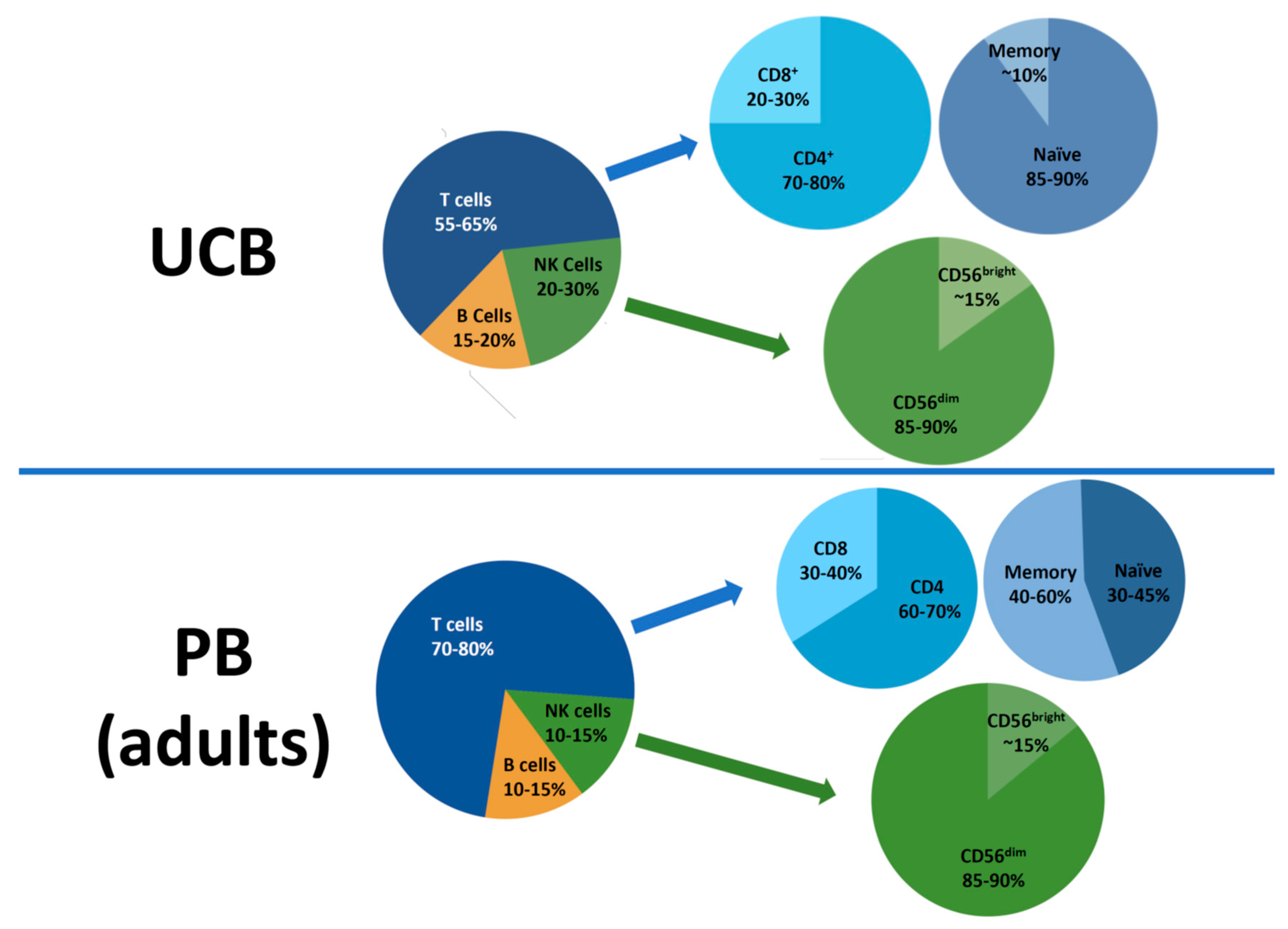
Figure 1. Cell composition in the umbilical cord blood (UCB) graft. The majority of T cells are CD4+ and naïve T cells in the UCB graft. The ratio of CD56dim/CD56bight NK cells is similar to that of PB. However, the cytotoxicity of CD56dim NK cells in the UCB graft is poor compared to that of PB NK cells. UCB, umbilical cord blood; PB, peripheral blood; CD, cluster of differentiation; NK, natural killer.
Jcm 08 01968 g001
Figure 2. The immunophenotypic and functional characteristics of T and NK cells in the UCB graft. (A) Naïve T cells, the major subset of UCB T cells have a better plasticity to transform into memory T cells and iTreg cells than PB T cells. The UCB graft contains a distinct Treg population. The fractions of memory, effector, activated T cells are low in UCB graft. (B) NK cells of the UCB graft are characterized by the phenotype of immaturity. The cytotoxicity of resting UCB NK cells is markedly low. However, the UCB NK cells acquire potent cytotoxicity with phenotypic maturation upon cytokine stimulation.
Jcm 08 01968 g002
Figure 3. Impacts of clinical factors on immune reconstitution of T and NK cells in UCBT. Advanced age, acute and chronic GVHD, HLA mismatch, infection, myeloablative conditioning, and ATG result in impaired thymopoiesis. Furthermore, relatively low T cells in the UCB graft cause delayed T cell recovery. Double UCBT enhances early T cell IR and thymopoiesis (in adults). Contrarily, NK cell IR in UCBT is comparable or better, compared to other types of HCT, given relatively high fractions of NK cells in the UCB graft. ATG and myeloablative conditioning are known to further prompt NK cell IR. GVHD, graft-versus-host disease; UCB, umbilical cord blood; UCBT, umbilical cord blood transplantation; NK, natural killer; IR, immune reconstitution; HCT, hematopoietic cell transplantation.
Jcm 08 01968 g003
Figure 4. Impacts of T and NK cell IR on major clinical outcomes in UCBT. (A) Impacts of T cell IR on clinical outcomes. CD4+, naïve T cell IR, thymopoiesis measured by TREC, viral antigen-specific T cell immunity significantly impact clinical outcomes. (B) NK cell effects on clinical outcomes in UCBT. Immunogenetic factors such as recipients’ HLA typing play a critical role in clinical outcomes in UCBT by KIR–HLA interactions. Furthermore, the NK cell function (e.g., cytotoxicity against K562) and the phenotype (CD16, HLA-DR expression) can affect clinical outcomes. There are conflicting data in the literature on effects of KIR-L mismatch in clinical outcomes. * treatment-related mortality; # leukemia-free survival; % overall survival.
Jcm 08 01968 g004