Article

Impact of Body Composition on the Risk of Hepatocellular Carcinoma Recurrence After Liver Transplantation

1
Second Department of Clinical Radiology, Medical University of Warsaw, 02-097 Warsaw, Poland
2
Department of General, Transplant and Liver Surgery, Medical University of Warsaw, 02-097 Warsaw, Poland
*
Author to whom correspondence should be addressed.
J. Clin. Med. 2019, 8(10), 1672; https://doi.org/10.3390/jcm8101672
Submission received: 30 August 2019 / Revised: 20 September 2019 / Accepted: 11 October 2019 / Published: 13 October 2019
(This article belongs to the Special Issue Liver Transplantation)

Abstract

:
Background: Body composition parameters are reported to influence the risk of hepatocellular carcinoma (HCC) recurrence after liver resection, yet data on patients undergoing liver transplantation are scarce. The aim of this study was to evaluate the impact of the amount of abdominal adipose tissue and skeletal muscles on the risk of HCC recurrence after liver transplantation. Methods: This was a retrospective observational study performed on 77 HCC patients after liver transplantation. Subcutaneous fat area (SFA), visceral fat area, psoas muscle area and total skeletal muscle area were assessed on computed tomography on the level of L3 vertebra and divided by square meters of patient height. The primary outcome measure was five-year recurrence-free survival. Results: Recurrence-free survival in the entire cohort was 95.7%, 90.8%, and 86.5% after one, three, and five years post-transplantation, respectively. SFA was significantly associated with the risk of HCC recurrence (p = 0.013), whereas no significant effects were found for visceral fat and skeletal muscle indices. The optimal cut-off for SFA for prediction of recurrence was 71.5 cm2/m2. Patients with SFA < 71.5 cm2/m2 and ≥71.5 cm2/m2 exhibited five-year recurrence-free survival of 96.0% and 55.4%, respectively (p = 0.001). Conclusions: Excessive amount of subcutaneous adipose tissue is a risk factor for HCC recurrence after liver transplantation and may be considered in patient selection process.

1. Introduction

Liver transplantation remains the mainstay of treatment for patients with hepatocellular carcinoma (HCC) at low risk of post-transplant tumor recurrence [1]. For more than two decades, appropriate selection of HCC patients for transplantation was accomplished through strict utilization of the Milan criteria (one tumor < 5 cm or two to three tumors < 3 cm, no macrovascular invasion, no extrahepatic spread) [2]. Currently, however, there is a shift towards more precise selection of patients using a combination of morphological criteria with surrogates of tumor biological aggressiveness, such as serum alpha-fetoprotein concentration [3,4,5,6,7,8]. Despite these improvements, a remarkable proportion of patients beyond the Milan criteria at increased risk of HCC recurrence remains eligible for transplantation according to recent proposals [9,10]. Therefore, further refinement of patient selection strategy is warranted.
Obesity is a known factor predisposing to development of cancer [11]. Increased amount of adipose tissue is widely reported to be a negative prognostic factor in patients with various malignancies [12,13,14]. Aside from obesity, generalized loss of skeletal muscle mass (sarcopenia) is also predictive of negative outcomes in cancer patients [15,16,17,18,19]. The negative effects of both sarcopenia and accumulation of adipose tissue were confirmed in patients undergoing liver resections for HCC with respect to recurrence and survival, yet the results of available studies are inconsistent [20,21,22]. The potential mechanisms responsible for the influence of the quantity of adipose tissue and skeletal muscles comprise systemic and tumor-directed actions of various molecules, namely adipokines and myokines [23,24]. Increased levels of leptin and decreased levels of adiponectin are common in obesity and related to increased inflammation, angiogenesis, cell proliferation, and metastatic potential of cancer cells [25]. Decreased levels of interleukin-15 observed in sarcopenia contribute to immune senescence. Notably, altered function of natural killer cells was also found in case of obesity [24,26]. In contrast to several previous studies focused on the impact of body composition on the outcomes of HCC patients undergoing hepatectomy, data with respect to patients undergoing liver transplantation is scarce. Liver transplantation is associated with removal of the underlying liver disease along with potential intrahepatic micrometastases, occurrence of ischemia-reperfusion injury directly modifying the risk of recurrence [27], and immune suppression. Therefore, the findings from liver resection studies cannot be extrapolated. The aim of this study was to evaluate the impact of body adipose and muscle contents on the risk of HCC recurrence after liver transplantation.

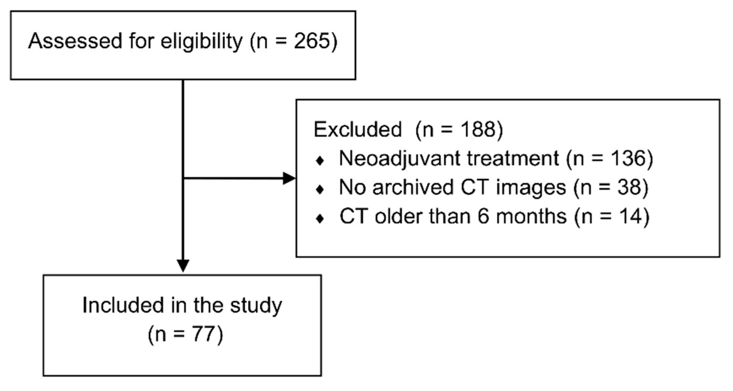
2. Materials and Methods

This retrospective observational study was performed on the data of 77 HCC patients after liver transplantations performed in the period between January 2006 and June 2017 in the Department of General, Transplant and Liver Surgery of the Medical University of Warsaw. The sole inclusion criterion was histologically confirmed HCC in the explanted liver. Exclusion criteria comprised a lack of available images from multiphasic computed tomography (CT) performed within 6 months preceding transplantation and utilization of neoadjuvant treatment (Figure 1). The study protocol was approved by the local ethics committee of the Medical University of Warsaw (AKBE/231/2017). Due to the retrospective nature, the need for informed consent was waived.
The primary outcome measure was 5-year recurrence-free survival, defined as time between liver transplantation and tumor recurrence. Observations were censored at last available follow-up, death for non-recurrence related causes, or 5 years post-transplantation, whichever occurred earlier. Details on surgical technique, perioperative care, immunosuppression protocol and post-transplant follow up were described elsewhere [28,29]. The primary factors of interest were subcutaneous (SFA), visceral (VFA), and total fat area (TFA), psoas muscle area (PMA), and total skeletal muscle area (TSMA) expressed in cm2 per 1 m2 of patient height. The areas of tissues of interest were calculated in Osirix software (Pixmeo, Switzerland) in a selected region of interest based on the range of Hounsfield units (HU). All calculations were performed using computed tomography scans on the level of the transverse processes of the third lumbar vertebra. The HU ranges were: −150 to −30 HU for VFA; −150 to −50 HU for SFA, and −30 to +150 HU for PMA and TSMA. TFA was defined as the sum of VFA and SFA. Examples of calculation of SFA, VFA, PMA, and TSMA are presented in Figure 2a–d, respectively.
Quantitative variables were expressed as medians with interquartile ranges (IQRs) and qualitative variables were expressed as numbers with percentages. Chi2 test and Kruskall–Wallis test were used for intergroup comparisons of qualitative and quantitative variables, respectively. Spearman’s correlation coefficient was applied for analyses of correlations. Kaplan–Meier method was used for calculation of recurrence-free survival. Log-rank test was used for comparison of survival curves. Cox proportional hazards regression was used to evaluate risk factors for inferior recurrence-free survival. Receiver operating characteristics (ROC) analyses were performed to determine optimal cut-offs for selected body composition parameters in prediction of post-transplant HCC recurrence. Hazard ratios (HRs) and areas under the curve (AUCs) were presented with 95% confidence intervals (95% CI). Two-tailed p < 0.05 was considered to indicate statistical significance. All statistical analyses were performed using STATISTICA version 13 software (Dell Inc., Tulsa, USA).

3. Results

Baseline characteristics of the 77 patients included in the study cohort are summarized in Table 1. Median VFA, SFA, and TFA were 27.8 cm2/m2 (IQR: 20.3–46.6), 51.7 cm2/m2 (IQR: 37.3–72.3), and 83.4 cm2/m2 (IQR: 58.7–110.9), respectively. Median Body Mass Index (BMI) was 25.2 kg/m2 with IQR of 23.3–28.3.
Body mass index was significantly correlated to VFA (p < 0.001), SFA (p < 0.001), TFA (p < 0.001), PMA (p < 0.001), and TSMA (p < 0.001) in all patients and separately in males and females (Table 2). None of the analyzed body composition parameters was significantly correlated with patient age or model for end-stage liver disease score.
Median follow-up period was 42 months. There were a total of seven patients who developed post-transplant HCC recurrence after a median of 12 months post-transplantation. Recurrence-free survival rates at one, three, and five years post-transplantation were 95.7%, 90.8%, and 86.5%, respectively.
Both SFA (p = 0.013) and TFA (p = 0.017) were significantly associated with the risk of post-transplant HCC recurrence. The risk of recurrence was not associated with VFA (p = 0.264), PMA (p = 0.141), and TSMA (p = 0.618). Out of the other factors, significant predictors of recurrence comprised diameter of the largest nodule (p = 0.005), alpha-fetoprotein concentration (p = 0.006), fulfillment of Milan criteria (p < 0.001), and presence of microvascular invasion (p = 0.022). The results of analyses of risk factors for tumor recurrence are summarized in Table 3.
The optimal cut-offs for prediction of post-transplant HCC recurrence were 71.5 cm2/m2 for SFA and 90.5 cm2/m2 for TFA with AUCs of 0.772 and 0.767, respectively. Five-year recurrence-free survival rates were 96.0% in patients with SFA < 71.5 cm2/m2 as compared to 55.4% in patients with SFA ≥71.5 cm2/m2 (p = 0.001; Figure 3a). Five-year recurrence-free survival rates were 100% in patients with TFA <90.5 cm2/m2 as compared to 72.5% in patients with TFA ≥90.5 cm2/m2 (p = 0.004; Figure 3b).
In 33 patients with BMI within the range of 18.5–24.9 kg/m2, there were 3 (9.1%) with SFA ≥ 71.5 cm2/m2 and 7 (21.2%) with TFA ≥ 90.5 cm2/m2 (Table 4). On the contrary, out of 35 overweight patients (BMI category 25–29.9), there were 25 (71.4%) and 14 (40.0%) patients with SFA < 71.5 cm2/m2 and TFA < 90.5 cm2/m2, respectively. In nine obese patients (BMI ≥ 30 kg/m2), there was only one (11.1%) with SFA < 71.5 cm2/m2 and none with TFA < 90.5 cm2/m2.

4. Discussion

Both obesity and sarcopenia are highly prevalent in a population of HCC patients with liver cirrhosis. Given the imperfect accuracy of current systems for evaluating patient eligibility, evaluation of the impact of body composition on post-transplant risk of recurrence seems clinically important. In contrast to studies performed in patients undergoing liver resection, no significant impact of sarcopenia on HCC recurrence risk was observed. Interestingly, the most important body composition factor influencing this risk was the quantity of subcutaneous adipose tissue.
Patients with SFA exceeding 71.5 cm2/m2 were characterized by approximately 11-fold higher risk for post-transplant tumor recurrence than those with smaller amount of subcutaneous tissue. While a total amount of adipose tissue was also significantly associated with the risk of tumor recurrence, these seems to be attributable to the effects of subcutaneous fat given no significant effects of visceral fat area. This is clearly in contrast to previous studies, which results pointed towards the negative impact of visceral fat and irrelevance of subcutaneous tissue. In a recent study on 78 HCC liver transplant recipients increased visceral fat amount was significantly associated with post-transplant tumor recurrence, whereas no significant effects were found for the amount of subcutaneous adipose tissue [30]. Similarly, visceral to subcutaneous fat amount ratio was recently reported to increase the risk of HCC recurrence in patients undergoing liver resection [31]. In contrast, several studies identified high visceral fat amount to be a protective factor with respect to HCC recurrence after liver resection [21,32]. Nevertheless, given the high metabolic activity of visceral adipose tissue, it was expected to yield more effects on the risk of recurrence than the subcutaneous tissue [33]. The major negative impact of increased amount of subcutaneous adipose tissue is, therefore, a rather unexpected finding. Although this is the first study reporting the negative consequences of excessive subcutaneous tissue with respect to the risk of HCC recurrence, it was previously described as a risk factor for patients with prostate cancer [34,35].
While visceral adipose tissue is commonly considered to be metabolically more active than subcutaneous adipose tissue, recent findings point towards the important role in secretion of adipokines and potential influence on immune response of the latter. A recent study performed in middle-aged men indicated that expression of adipokines in subcutaneous adipose tissue is directly related to circulating adipokine profile, including leptin and adiponectin [36]. In a study on lung transplant recipients, subcutaneous but not visceral adipose tissue was associated with primary graft dysfunction [37]. In that study, pre-transplant leptin levels were highly correlated with subcutaneous adipose tissue index. Importantly, leptin was previously found to increase the metastatic potential of HCC cells through activation of several signaling pathways and to promote angiogenesis [38,39,40]. Further, leptin may promote creation of protumorigenic microenvironment by influencing cytokine profile and activity of matrix metalloproteinases [41]. In contrast, the actions of adiponectin, which levels decrease in obesity, include promotion of HCC cells apoptosis and anti-inflammatory effects [38,42]. Excessive adipose tissue is known to promote systemic pro-inflammatory response through activation of macrophages and secretion of multiple pro-inflammatory cytokines, including tumor necrosis factor α and interleukin 6, and therefore, promote post-transplant HCC recurrence [43]. Nevertheless, the mechanisms underlying the association between excessive subcutaneous adipose tissue and elevated risk of post-transplant HCC recurrence should be elucidated.
Sarcopenia was identified as a predictor of post-transplant HCC recurrence in a single study performed in recipients of living-donor liver transplantations [44]. The results of another study on recipients of living-donor liver transplantations pointed towards the negative impact of low skeletal muscle mass to visceral fat ratio, which is indicative of sarcopenic obesity [45]. Both studies were performed in Asian population, and similarly for the majority of those indicating the association between sarcopenia and HCC recurrence after liver resection [20,46,47,48,49,50]. The results of the present study indicate that sarcopenia does not increase the risk of HCC recurrence after deceased donor liver transplantation, at least in a European population.
Notably, all of the body composition parameters analyzed on computed tomography images were significantly associated with patient BMI. Accordingly, increased BMI was associated with increased probability of excessive amount of both subcutaneous and visceral adipose tissue. Nevertheless, the results indicate that BMI itself is a highly imprecise parameter for evaluation of these two body composition components, as 9% and 21% of patients with normal weight had excessive amount of subcutaneous and visceral fat, respectively. Further, majority of overweight patients had low amount of subcutaneous adipose tissue and 40% of these patients did not have excessive amount of visceral fat. This is in line with the finding of previous studies [32]. Further, BMI was not found to be associated with outcomes of HCC patients after liver transplantation neither in present nor previous studies [51]. The necessity of evaluating subcutaneous and visceral adipose tissue on computed tomography in obese patients seems more controversial, as there was only one patient with SFA below the established limit in this population. However, these findings are limited by a relatively low number of patients with obesity in the present study and this, should be interpreted with caution, especially considering patient with excessive ascites.
The method utilized in the present study for quantification of adipose tissue is similar to those described in other studies, except for a higher threshold for HU units (−150 HU versus −190 HU) [21,30,32]. However, as all the images were assessed individually, this was unlikely to bring remarkable differences. While psoas muscle thickness was previously assessed instead of psoas muscle area, the method used to evaluate muscular tissue in the present study was commonly utilized previously [44,46,47,48,49,50].
Neoadjuvant treatment was commonly utilized in the study performed in liver transplant recipients that pointed towards the significant prognostic role of visceral adipose tissue [30]. Conversely, patients in the present study did not undergo neoadjuvant treatment and due to selection bias, had generally lower tumor burden with less biologically aggressive tumors, as indicated by lower rate of microvascular invasion and poor differentiation. Therefore, the results need to be validated in a larger cohort of patients in order to evaluate potential differences in prognostic roles of visceral and subcutaneous adipose tissue depending on tumor burden and biological aggressiveness. Similarly, patients included in the study pointing towards the association between sarcopenia and tumor recurrence were all beyond the limits of Milan criteria and had more excessive tumor burden than those included in the present study [44]. Further, they were recipients of living-donor liver transplantation. Accordingly, the results of the present study are limited to more homogeneous group of patients at generally lower risk of post-transplant HCC recurrence.
The present study is subject to similar limitations. First, it was retrospective in nature and thus prone to all limitations of retrospective studies. Second, there was a low absolute number of the observed tumor recurrences, precluding performance of multivariable analyses. This also results in a higher risk of type II error for evaluating the effects of visceral fat amount and sarcopenia on the risk of HCC recurrence. On the other hand, this was related to selection of more homogeneous population of HCC patients for the present study, as all patients undergoing pre-transplant neoadjuvant treatment were excluded. Moreover, the number of patients included in the present study is similar to those included in other studies focused on liver transplantation [30,44].

5. Conclusions

In conclusion, the results of the present study indicate that excessive amount of subcutaneous adipose tissue is associated with increased risk of tumor recurrence and may be considered as another factor potentially useful in selection of HCC patients for liver transplantation. Neither visceral adipose tissue nor sarcopenia seem to be related with the risk of HCC recurrence following liver transplantation.

Author Contributions

Conceptualization, K.G.; Data curation, K.G., R.P., M.G., M.K., K.Z. and O.R.; Formal analysis, K.G. and M.G.; Investigation, K.G.; Methodology, K.G.; Resources, R.P., M.K, K.Z. and O.R.; Supervision, R.P., M.K., K.Z. and O.R.; Writing—original draft, K.G.; Writing—review & editing, R.P., M.G., M.K., K.Z. and O.R.

Funding

This research received no external funding.

Conflicts of Interest

The authors declare no conflicts of interest.

References

  1. Puigvehí, M.; Hashim, D.; Haber, P.K.; Dinani, A.; Schiano, T.D.; Asgharpour, A.; Kushner, T.; Kakked, G.; Tabrizian, P.; Schwartz, M.; et al. Liver Transplant for Hepatocellular Carcinoma in the United States: Evolving Trends over the Last Three Decades. Am. J. Transplant. 2019. [Google Scholar] [CrossRef] [PubMed]
  2. Mazzaferro, V.; Regalia, E.; Doci, R.; Andreola, S.; Pulvirenti, A.; Bozzetti, F.; Montalto, F.; Ammatuna, M.; Morabito, A.; Gennari, L. Liver transplantation for the treatment of small hepatocellular carcinomas in patients with cirrhosis. N. Engl. J. Med. 1996, 334, 693–699. [Google Scholar] [CrossRef] [PubMed]
  3. Grąt, M.; Wronka, K.M.; Stypułkowski, J.; Bik, E.; Krasnodębski, M.; Masior, Ł.; Lewandowski, Z.; Grąt, K.; Patkowski, W.; Krawczyk, M. The Warsaw Proposal for the Use of Extended Selection Criteria in Liver Transplantation for Hepatocellular Cancer. Ann. Surg. Oncol. 2017, 24, 526–534. [Google Scholar] [CrossRef] [PubMed]
  4. Mazzaferro, V.; Sposito, C.; Zhou, J.; Pinna, A.D.; De Carlis, L.; Fan, J.; Cescon, M.; Di Sandro, S.; Yi-Feng, H.; Lauterio, A.; et al. Metroticket 2.0 Model for Analysis of Competing Risks of Death After Liver Transplantation for Hepatocellular Carcinoma. Gastroenterology 2018, 154, 128–139. [Google Scholar] [CrossRef] [PubMed] [Green Version]
  5. Duvoux, C.; Roudot-Thoraval, F.; Decaens, T.; Pessione, F.; Badran, H.; Piardi, T.; Francoz, C.; Compagnon, P.; Vanlemmens, C.; Dumortier, J.; et al. Liver transplantation for hepatocellular carcinoma: A model including α-Fetoprotein improves the performance of Milan criteria. Gastroenterology 2012, 143, 986–994. [Google Scholar] [CrossRef] [PubMed]
  6. Mehta, N.; Dodge, J.L.; Roberts, J.P.; Hirose, R.; Yao, F.Y. Alpha-Fetoprotein Decrease from >1000 to <500 ng/mL in Patients with Hepatocellular Carcinoma Leads to Improved Posttransplant Outcomes. Hepatology 2019, 69, 1193–1205. [Google Scholar] [PubMed]
  7. Halazun, K.J.; Tabrizian, P.; Najjar, M.; Florman, S.; Schwartz, M.; Michelassi, F.; Samstein, B.; Brown, R.S., Jr.; Emond, J.C.; Busuttil, R.W.; et al. Is it Time to Abandon the Milan Criteria?: Results of a Bicoastal US Collaboration to Redefine Hepatocellular Carcinoma Liver Transplantation Selection Policies. Ann. Surg. 2018, 268, 690–699. [Google Scholar] [CrossRef] [PubMed]
  8. Halazun, K.J.; Najjar, M.; Abdelmessih, R.M.; Samstein, B.; Griesemer, A.D.; Guarrera, J.V.; Kato, T.; Verna, E.C.; Emond, J.C.; Brown, R.S., Jr. Recurrence After Liver Transplantation for Hepatocellular Carcinoma: A New MORAL to the Story. Ann. Surg. 2017, 265, 557–564. [Google Scholar] [CrossRef]
  9. Grąt, M.; Stypułkowski, J.; Morawski, M.; Wronka, K.M.; Wasilewicz, M.; Lewandowski, Z.; Grąt, K.; Wójcik, Z.; Patkowski, W.; Zieniewicz, K. Shadows Behind Using Simple Risk Models in Selection of Hepatocellular Carcinoma Patients for Liver Transplantation. Ann. Surg. 2018. [Google Scholar] [CrossRef]
  10. Grąt, M.; Krasnodębski, M.; Krawczyk, M.; Stypułkowski, J.; Morawski, M.; Wasilewicz, M.; Lewandowski, Z.; Grąt, K.; Patkowski, W.; Zieniewicz, K. Extremes of Liver Transplantation for Hepatocellular Carcinoma. J. Clin. Med. 2019, 8, 787. [Google Scholar] [CrossRef]
  11. Karczewski, J.; Begier-Krasińska, B.; Staszewski, R.; Popławska, E.; Gulczynska-Elhadi, K.; Dobrowolska, A. Obesity and the Risk of Gastrointestinal Cancers. Dig. Dis. Sci. 2019, 64, 2740–2749. [Google Scholar] [CrossRef] [PubMed] [Green Version]
  12. Okumura, S.; Kaido, T.; Hamaguchi, Y.; Kobayashi, A.; Shirai, H.; Yao, S.; Yagi, S.; Kamo, N.; Hatano, E.; Okajima, H.; et al. Visceral Adiposity and Sarcopenic Visceral Obesity are Associated with Poor Prognosis After Resection of Pancreatic Cancer. Ann. Surg. Oncol. 2017, 24, 3732–3740. [Google Scholar] [CrossRef] [PubMed]
  13. Kambara, Y.; Yuasa, N.; Takeuchi, E.; Miyake, H.; Nagai, H.; Yoshioka, Y.; Okuno, M.; Miyata, K. Overweight or Obesity is an Unfavorable Long-Term Prognostic Factor for Patients who Underwent Gastrectomy for Stage II/III Gastric Cancer. World J. Surg. 2019, 43, 1766–1776. [Google Scholar] [CrossRef] [PubMed]
  14. Shiota, M.; Takeuchi, A.; Sugimoto, M.; Kashiwagi, E.; Dejima, T.; Kiyoshima, K.; Inokuchi, J.; Tatsugami, K.; Yokomizo, A.; Eto, M. The Differential Impact of Body Mass Index and the Feature of Metabolic Syndrome on Oncological Outcomes Following Different Surgical Procedures in Japanese Men with Prostate Cancer. Ann. Surg. Oncol. 2017, 24, 1443–1450. [Google Scholar] [CrossRef] [PubMed]
  15. Zheng, Z.F.; Lu, J.; Zheng, C.H.; Li, P.; Xie, J.W.; Wang, J.B.; Lin, J.X.; Chen, Q.Y.; Lin, M.; Huang, C.M. A Novel Prognostic Scoring System Based on Preoperative Sarcopenia Predicts the Long-Term Outcome for Patients After R0 Resection for Gastric Cancer: Experiences of a High-Volume Center. Ann. Surg. Oncol. 2017, 24, 1795–1803. [Google Scholar] [CrossRef] [PubMed]
  16. Kudou, K.; Saeki, H.; Nakashima, Y.; Edahiro, K.; Korehisa, S.; Taniguchi, D.; Tsutsumi, R.; Nishimura, S.; Nakaji, Y.; Akiyama, S.; et al. Prognostic Significance of Sarcopenia in Patients with Esophagogastric Junction Cancer or Upper Gastric Cancer. Ann. Surg. Oncol. 2017, 24, 1804–1810. [Google Scholar] [CrossRef] [PubMed]
  17. Lu, J.; Zheng, Z.F.; Li, P.; Xie, J.W.; Wang, J.B.; Lin, J.X.; Chen, Q.Y.; Cao, L.L.; Lin, M.; Tu, R.H.; et al. A Novel Preoperative Skeletal Muscle Measure as a Predictor of Postoperative Complications, Long-Term Survival and Tumor Recurrence for Patients with Gastric Cancer After Radical Gastrectomy. Ann. Surg. Oncol. 2018, 25, 439–448. [Google Scholar] [CrossRef] [PubMed]
  18. Chakedis, J.; Spolverato, G.; Beal, E.W.; Woelfel, I.; Bagante, F.; Merath, K.; Sun, S.H.; Chafitz, A.; Galo, J.; Dillhoff, M.; et al. Pre-Operative Sarcopenia Identifies Patients at Risk for Poor Survival After Resection of Biliary Tract Cancers. J. Gastrointest. Surg. 2018, 22, 1697–1708. [Google Scholar] [CrossRef] [PubMed]
  19. Jung, A.R.; Roh, J.L.; Kim, J.S.; Kim, S.B.; Choi, S.H.; Nam, S.Y.; Kim, S.Y. Prognostic value of body composition on recurrence and survival of advanced-stage head and neck cancer. Eur. J. Cancer 2019, 116, 98–106. [Google Scholar] [CrossRef]
  20. Kobayashi, A.; Kaido, T.; Hamaguchi, Y.; Okumura, S.; Shirai, H.; Yao, S.; Kamo, N.; Yagi, S.; Taura, K.; Okajima, H.; et al. Impact of Sarcopenic Obesity on Outcomes in Patients Undergoing Hepatectomy for Hepatocellular Carcinoma. Ann. Surg. 2019, 269, 924–931. [Google Scholar] [CrossRef]
  21. Itoh, S.; Shirabe, K.; Matsumoto, Y.; Yoshiya, S.; Muto, J.; Harimoto, N.; Yamashita, Y.; Ikegami, T.; Yoshizumi, T.; Nishie, A.; et al. Effect of body composition on outcomes after hepatic resection for hepatocellular carcinoma. Ann. Surg. Oncol. 2014, 21, 3063–3068. [Google Scholar] [CrossRef] [PubMed]
  22. Guo, Z.; Zhang, J.; Jiang, J.H.; Li, L.Q.; Xiang, B.D. Obesity Does Not Influence Outcomes in Hepatocellular Carcinoma Patients following Curative Hepatectomy. PLoS ONE 2015, 10, e0125649. [Google Scholar] [CrossRef] [PubMed]
  23. Wang, Y.X.; Zhu, N.; Zhang, C.J.; Wang, Y.K.; Wu, H.T.; Li, Q.; Du, K.; Liao, D.F.; Qin, L. Friend or foe: Multiple roles of adipose tissue in cancer formation and progression. J. Cell. Physiol. 2019, 234, 21436–21449. [Google Scholar] [CrossRef] [PubMed]
  24. Lutz, C.T.; Quinn, L.S. Sarcopenia, obesity, and natural killer cell immune senescence in aging: Altered cytokine levels as a common mechanism. Aging 2012, 4, 535–546. [Google Scholar] [CrossRef] [PubMed] [Green Version]
  25. Parida, S.; Siddharth, S.; Sharma, D. Adiponectin, Obesity, and Cancer: Clash of the Bigwigs in Health and Disease. Int. J. Mol. Sci. 2019, 20, 2519. [Google Scholar] [CrossRef] [PubMed]
  26. O’Shea, D.; Hogan, A.E. Dysregulation of Natural Killer Cells in Obesity. Cancers 2019, 11, 573. [Google Scholar] [CrossRef] [PubMed]
  27. Grąt, M.; Krawczyk, M.; Wronka, K.M.; Stypułkowski, J.; Lewandowski, Z.; Wasilewicz, M.; Krawczyk, P.; Grąt, K.; Patkowski, W.; Zieniewicz, K. Ischemia-Reperfusion injury and the risk of hepatocellular carcinoma recurrence after deceased donor liver transplantation. Sci. Rep. 2018, 8, 8935. [Google Scholar] [CrossRef]
  28. Grąt, M.; Kornasiewicz, O.; Lewandowski, Z.; Skalski, M.; Zieniewicz, K.; Pączek, L.; Krawczyk, M. The impact of surgical technique on the results of liver transplantation in patients with hepatocellular carcinoma. Ann. Transplant. 2013, 18, 448–459. [Google Scholar]
  29. Krawczyk, M.; Grąt, M.; Barski, K.; Ligocka, J.; Antczak, A.; Kornasiewicz, O.; Skalski, M.; Patkowski, W.; Nyckowski, P.; Zieniewicz, K.; et al. 1000 liver transplantations at the Department of General, Transplant and Liver Surgery, Medical University of Warsaw—Analysis of indications and results. Pol. Przegl. Chir. 2012, 84, 304–312. [Google Scholar] [CrossRef]
  30. Montano-Loza, A.J.; Mazurak, V.C.; Ebadi, M.; Meza-Junco, J.; Sawyer, M.B.; Baracos, V.E.; Kneteman, N. Visceral adiposity increases risk for hepatocellular carcinoma in male patients with cirrhosis and recurrence after liver transplant. Hepatology 2018, 67, 914–923. [Google Scholar] [CrossRef]
  31. Hamaguchi, Y.; Kaido, T.; Okumura, S.; Kobayashi, A.; Shirai, H.; Yao, S.; Yagi, S.; Kamo, N.; Seo, S.; Taura, K.; et al. Preoperative Visceral Adiposity and Muscularity Predict Poor Outcomes after Hepatectomy for Hepatocellular Carcinoma. Liver Cancer 2019, 8, 92–109. [Google Scholar] [CrossRef] [PubMed]
  32. Higashi, T.; Hayashi, H.; Kaida, T.; Arima, K.; Takeyama, H.; Taki, K.; Izumi, D.; Tokunaga, R.; Kosumi, K.; Nakagawa, S.; et al. Prognostic Impact of Visceral Fat Amount and Branched-Chain Amino Acids (BCAA) in Hepatocellular Carcinoma. Ann. Surg. Oncol. 2015, 22, S1041–S1047. [Google Scholar] [CrossRef] [PubMed]
  33. Fujihara, S.; Mori, H.; Kobara, H.; Nishiyama, N.; Kobayashi, M.; Oryu, M.; Masaki, T. Metabolic syndrome, obesity, and gastrointestinal cancer. Gastroenterol. Res. Pract. 2012, 2012, 483623. [Google Scholar] [CrossRef] [PubMed]
  34. McDonald, A.M.; Fiveash, J.B.; Kirkland, R.S.; Cardan, R.A.; Jacob, R.; Kim, R.Y.; Dobelbower, M.C.; Yang, E.S. Subcutaneous adipose tissue characteristics and the risk of biochemical recurrence in men with High-Risk prostate cancer. Urol. Oncol. 2017, 35, e15–e663. [Google Scholar] [CrossRef] [PubMed]
  35. Delouya, G.; Tiberi, D.; Bhatnagar, S.R.; Campeau, S.; Saad, F.; Taussky, D. Impact of adipose tissue on prostate cancer aggressiveness—Analysis of a high-risk population. Horm. Mol. Biol. Clin. Investig. 2018, 36. [Google Scholar] [CrossRef] [PubMed]
  36. Lee, S.; Norheim, F.; Langleite, T.M.; Gulseth, H.L.; Birkeland, K.I.; Drevon, C.A. Effects of Long-Term exercise on plasma adipokine levels and inflammation-related gene expression in subcutaneous adipose tissue in sedentary dysglycaemic, overweight men and sedentary normoglycaemic men of healthy weight. Diabetologia 2019, 62, 1048–1064. [Google Scholar] [CrossRef] [PubMed]
  37. Anderson, M.R.; Udupa, J.K.; Edwin, E.; Diamond, J.M.; Singer, J.P.; Kukreja, J.; Hays, S.R.; Greenland, J.R.; Ferrante, A.; Lippel, M.; et al. Adipose tissue quantification and primary graft dysfunction after lung transplantation: The Lung Transplant Body Composition study. J. Heart Lung Transplant. 2019, 10, S1053–S2498. [Google Scholar] [CrossRef] [PubMed]
  38. Duan, X.F.; Tang, P.; Li, Q.; Yu, Z.T. Obesity, adipokines and hepatocellular carcinoma. Int. J. Cancer 2013, 133, 1776–1783. [Google Scholar] [CrossRef] [Green Version]
  39. Saxena, N.K.; Sharma, D.; Ding, X.; Lin, S.; Marra, F.; Merlin, D.; Anania, F.A. Concomitant activation of the JAK/STAT, PI3K/AKT, and ERK signaling is involved in Leptin-Mediated promotion of invasion and migration of hepatocellular carcinoma cells. Cancer Res. 2007, 67, 2497–2507. [Google Scholar] [CrossRef]
  40. Ribatti, D.; Belloni, A.S.; Nico, B.; Di Comite, M.; Crivellato, E.; Vacca, A. Leptin-Leptin receptor are involved in angiogenesis in human hepatocellular carcinoma. Peptides 2008, 29, 1596–1602. [Google Scholar] [CrossRef]
  41. Stefanou, N.; Papanikolaou, V.; Furukawa, Y.; Nakamura, Y.; Tsezou, A. Leptin as a critical regulator of hepatocellular carcinoma development through modulation of human telomerase reverse transcriptase. BMC Cancer 2010, 10, 442. [Google Scholar] [CrossRef] [PubMed]
  42. Sharma, D.; Wang, J.; Fu, P.P.; Sharma, S.; Nagalingam, A.; Mells, J.; Handy, J.; Page, A.J.; Cohen, C.; Anania, F.A.; et al. Adiponectin antagonizes the oncogenic actions of leptin in hepatocellular carcinogenesis. Hepatology 2010, 52, 1713–1722. [Google Scholar] [CrossRef] [PubMed] [Green Version]
  43. Corrêa, L.H.; Heyn, G.S.; Magalhaes, K.G. The Impact of the Adipose Organ Plasticity on Inflammation and Cancer Progression. Cells 2019, 8, E662. [Google Scholar] [CrossRef] [PubMed]
  44. Kim, Y.R.; Park, S.; Han, S.; Ahn, J.H.; Kim, S.; Sinn, D.H.; Jeong, W.K.; Ko, J.S.; Gwak, M.S.; Kim, G.S. Sarcopenia as a predictor of post-transplant tumor recurrence after living donor liver transplantation for hepatocellular carcinoma beyond the Milan criteria. Sci. Rep. 2018, 8, 7157. [Google Scholar] [CrossRef] [PubMed]
  45. Itoh, S.; Yoshizumi, T.; Kimura, K.; Okabe, H.; Harimoto, N.; Ikegami, T.; Uchiyama, H.; Shirabe, K.; Nishie, A.; Maehara, Y. Effect of Sarcopenic Obesity on Outcomes of Living-Donor Liver Transplantation for Hepatocellular Carcinoma. Anticancer Res 2016, 36, 3029–3034. [Google Scholar] [PubMed]
  46. Yabusaki, N.; Fujii, T.; Yamada, S.; Suzuki, K.; Sugimoto, H.; Kanda, M.; Nakayama, G.; Koike, M.; Fujiwara, M.; Kodera, Y. Adverse impact of low skeletal muscle index on the prognosis of hepatocellular carcinoma after hepatic resection. Int. J. Surg. 2016, 30, 136–142. [Google Scholar] [CrossRef] [PubMed]
  47. Voron, T.; Tselikas, L.; Pietrasz, D.; Pigneur, F.; Laurent, A.; Compagnon, P.; Salloum, C.; Luciani, A.; Azoulay, D. Sarcopenia Impacts on Short- and Long-Term Results of Hepatectomy for Hepatocellular Carcinoma. Ann. Surg. 2015, 261, 1173–1183. [Google Scholar] [CrossRef]
  48. Harimoto, N.; Shirabe, K.; Yamashita, Y.I.; Ikegami, T.; Yoshizumi, T.; Soejima, Y.; Ikeda, T.; Maehara, Y.; Nishie, A.; Yamanaka, T. Sarcopenia as a predictor of prognosis in patients following hepatectomy for hepatocellular carcinoma. Br. J. Surg. 2013, 100, 1523–1530. [Google Scholar] [CrossRef]
  49. Hiraoka, A.; Otsuka, Y.; Kawasaki, H.; Izumoto, H.; Ueki, H.; Kitahata, S.; Aibiki, T.; Okudaira, T.; Yamago, H.; Miyamoto, Y.; et al. Impact of muscle volume and muscle function decline in patients undergoing surgical resection for hepatocellular carcinoma. J. Gastroenterol. Hepatol. 2018, 33, 1271–1276. [Google Scholar] [CrossRef]
  50. Kamachi, S.; Mizuta, T.; Otsuka, T.; Nakashita, S.; Ide, Y.; Miyoshi, A.; Kitahara, K.; Eguchi, Y.; Ozaki, I.; Anzai, K. Sarcopenia is a risk factor for the recurrence of hepatocellular carcinoma after curative treatment. Hepatol. Res. 2016, 46, 201–208. [Google Scholar] [CrossRef]
  51. Ata, N.; Ayloo, S.; Tsung, A.; Molinari, M. Recipient obesity does not affect survival after deceased donor liver transplantation for hepatocellular carcinoma. A national retrospective cohort study in the United States. HPB 2019, 21, 67–76. [Google Scholar] [CrossRef] [PubMed]
Figure 1. Study flowchart.
Jcm 08 01672 g001
Figure 2. Examples of muscle and adipose tissue measurements: (a) Subcutaneous Fat Area (b) Visceral Fat Area (c) Psoas Major Area (d) Total Skeletal Muscle Area. (a) red zone represents Subcutaneous Fat Area, in this patient it was 332.6 cm2/m2 (b) red zone represents Visceral Fat Area, in this patient it was (138.9 cm2/m2) (c) red zone represents Psoas Major Area, in this patient it was 16.8 cm2/m2 (d) red zone represents Total Skeletal Muscle Area, in this patient it was 123.2 cm2/m2.
Jcm 08 01672 g002
Figure 3. Recurrence-free survival after liver transplantation in HCC patients with (a) subcutaneous fat area <71.5cm2/m2 and ≥71.5cm2/m2 and (b) total fat area <90.5 cm2/m2 and ≥90.5 cm2/m2. . (a) Patients with the amount of subcutaneous fat area above the cut-off value had lower recurrence-free survival rates (b) Patients with the amount of total fat area above the cut-off value had lower recurrence-free survival rates.
Jcm 08 01672 g003
Table 1. Baseline characteristics of the study group.
Medians and IQRs or Numbers and %
Age (years)56 (52–61)
Sex
Male59 (76.6%)
Female18 (23.4%)
HCV58 (75.3%)
BMI (kg/m2)25.20 (23.3–28.3)
Serum AFP (ng/ml)13.1 (5.0–112.8)
Adipose tissue (cm2/m2)
subcutaneous51.7 (37.3–72.3)
visceral27.8 (20.3–46.6)
total83.4 (58.7–110.9)
Muscular tissue (cm2/m2)
psoas major6.2 (5.1–7.3)
total50.1 (43.1–56.4)
Time between LT and the last CT scan (days)51 (26–95)
Tumor characteristic
number of tumors1 (1–2)
size of the largest tumor25 (15–35)
microinvasion14 (18.67%)
poor tumor differentiation9 (11.69%)
fulfillment of Milan criteria53 (68.83%)
IQRs—interquartile ranges, HCV—Hepatitis C Virus, BMI—Body Mass Index, AFP—alpha-fetoprotein, LT—Liver Transplantation, CT—Computed Tomography.
Table 2. Correlations between Body Mass Index and adipose and muscular tissue areas in hepatocellular carcinoma (HCC) patients undergoing liver transplantation.
Rp
Subcutaneous Fat Area (SFA)
whole group0.735p < 0.001
males0.756p < 0.001
females0.803p < 0.001
Visceral Fat Area (VFA)
whole group0.569p < 0.001
males0.509p < 0.001
females0.576p = 0.012
Total Fat Area (TFA)
whole group0.710p < 0.001
males0.700p < 0.001
females0.803p < 0.001
Psoas Muscle Area (PMA)
whole group0.453p < 0.001
males0.344p = 0.008
females0.606p = 0.008
Total Skeletal Muscle Area (TSMA)
whole group0.529p < 0.001
males0.491p < 0.001
females0.667p < 0.002
Table 3. Risk factors for tumor recurrence after liver transplantation for HCC.
FactorHR95% CIp
Area of:
subcutaneous fat a1.0261.005–1.0470.013
visceral fat a 1.0250.982–1.0700.264
total fat tissue a1.0261.005–1.0490.017
psoas major a1.4090.893–2.2250.141
total skeletal muscle a1.0240.934–1.1220.618
Age b0.9970.901–1.1030.952
BMI c1.1830.956–1.4630.122
Tumor characteristics:
number of tumors d1.2540.983–1.5990.068
size of the largest tumor e1.0691.020–1.1190.005
microvascular invasion5.7801.284–25.6410.022
poor tumor differentiation3.5340.685–18.1820.132
fulfillment of Milan criteria100% vs. 53.3% *p < 0.001 *
Donor age b0.9990.938–1.0630.965
MELD points f0.9950.846–1.1700.949
Serum AFP g1.4281.106–1.8430.006
Female sex0.6270.075–5.2170.666
HCV0.9030.100–2.0410.301
HR—Hazard Ratio; 95% CI—95% Confidence Interval; BMI—Body Mass Index; MELD—Model for End-stage Liver Disease; AFP—alpha-fetoprotein; HCV—Hepatitis C Virus; HRs were given per: a—1 cm2/m2 increase; b—1 year increase; c—1 kg/m2 increase; d—per 1 tumor increase; e—per 1 mm increase; f—1 point increase; g—1 log ng/ml increase; * due to no tumor recurrences in the group of patients fulfilling Milan criteria, Cox proportional regression could not be performed; the statistical significance was based on comparison of survival curves.
Table 4. Distribution of HCC patients undergoing liver transplantation with SFA and TFA under and above established thresholds according to BMI category.
Body Mass Index (BMI)
(kg/m2)
Subcutaneous Fat Area (SFA)Total Fat Area (TFA)
Value
(cm2/m2)
Number of PatientsValue
(cm2/m2)
Number of Patients
18.50–24.99<71.5
≥71.5
30 (90.9%)
3 (9.1%)
<90.5
≥90.5
26 (78.8%)
7 (21.2%)
25.00–29.99<71.5
≥71.5
25 (71.4%)
10 (28.6%)
<90.5
≥90.5
14 (40.0%)
21 (60.0%)
≥ 30.00<71.5
≥71.5
1 (11.1%)
8 (88.9%)
<90.5
≥90.5
0 (0.0%)
9 (100.0%)

