
Chronic Kidney Disease After Lung Transplantation in Spain: A Retrospective Single-Center Analysis
 , , , , ,
Abstract

1. Introduction
2. Materials and Methods
3. Results
4. Discussion
5. Conclusions
Author Contributions
Funding
Institutional Review Board Statement
Informed Consent Statement
Data Availability Statement
Conflicts of Interest
Abbreviations
| AKI | acute kidney injury |
| CF | cystic fibrosis |
| COPD | chronic obstructive pulmonary disease |
| ILD | interstitial lung disease |
| LTx | lung transplant |
| MARE | major adverse renal events |
| SOT | solid organ transplants |
References
- Organización Nacional de Trasplantes. Memoria de Actividad de Donación y Trasplante Pulmonar España 2022. Available online: https://www.ont.es/wp-content/uploads/2023/06/DONACION-Y-TRASPLANTE-GENERAL-2022.pdf (accessed on 21 March 2025).
- Solé, A.; Zurbano, F.; Borro, J.M.; Monforte, V.; Ussetti, P.; Santos, F. Prevalence and Diagnosis of Chronic Kidney Disease in Maintenance Lung Transplant Patients: ICEBERG Study. Transpl. Proc. 2015, 47, 1966–1971. [Google Scholar] [CrossRef]
- De La Morena, M.P.; Bravos, M.D.L.T.; Prado, R.F.; Delgado Roel, M.; García Salcedo, J.A.; Fieira Costa, E.; Rivas, D.G.; Maté, J.B. Chronic kidney disease after lung transplantation: Incidence, risk factors, and treatment. Transpl. Proc. 2010, 42, 3217–3219. [Google Scholar] [CrossRef]
- Ojo, A.O.; Held, P.J.; Port, F.K.; Wolfe, R.A.; Leichtman, A.B.; Young, E.W.; Arndorfer, J.; Christensen, L.; Merion, R.M. Chronic Renal Failure after Transplantation of a Nonrenal Organ. N. Engl. J. Med. 2003, 349, 931–940. [Google Scholar] [PubMed]
- Bloom, R.D.; Reese, P.P. Chronic kidney disease after nonrenal solid-organ transplantation. J. Am. Soc. Nephrol. 2007, 18, 3031. [Google Scholar] [CrossRef]
- Lamas, S. Cellular mechanisms of vascular injury mediated by calcineurin inhibitors. Kidney Int. 2005, 68, 898. [Google Scholar] [CrossRef]
- Remuzzi, G.; Bertani, T. Renal vascular and thrombotic effects of cyclosporine. Am. J. Kidney Dis. 1989, 13, 261–272. [Google Scholar] [CrossRef]
- Kidney Disease: Improving Global Outcomes (KDIGO) CKD Work Group. KDIGO 2024 Clinical Practice Guideline for the Evaluation and Management of Chronic Kidney Disease. Kidney Int. 2024, 105, S117–S314. [Google Scholar]
- Nelson, J.; Alvey, N.; Bowman, L.; Schulte, J.; Segovia, M.C.; McDermott, J.; Te, H.S.; Kapila, N.; Levine, D.J.; Gottlieb, R.L.; et al. Consensus recommendations for use of maintenance immunosuppression in solid organ transplantation: Endorsed by the American College of Clinical Pharmacy, American Society of Transplantation, and the International Society for Heart and Lung Transplantation. Pharmacotherapy 2022, 42, 599–633. [Google Scholar] [CrossRef]
- Prischl, F.C.; Rossing, P.; Bakris, G.; Mayer, G.; Wanner, C. Major adverse renal events (MARE): A proposal to unify renal endpoints. Nephrol. Dial. Transpl. 2021, 36, 491–497. [Google Scholar] [CrossRef]
- Pham, P.T.T.; Slavov, C.; Pham, P.C.T. Acute Kidney Injury After Liver, Heart, and Lung Transplants: Dialysis Modality, Predictors of Renal Function Recovery, and Impact on Survival. Adv. Chronic Kidney Dis. 2009, 16, 256–267. [Google Scholar] [CrossRef]
- Canales, M.; Youssef, P.; Spong, R.; Ishani, A.; Savik, K.; Hertz, M.; Ibrahim, H.N. Predictors of chronic kidney disease in long-term survivors of lung and heart-lung transplantation. Am. J. Transpl. 2006, 6, 2157–2163. [Google Scholar] [CrossRef]
- Osho, A.A.; Castleberry, A.W.; Snyder, L.D.; Palmer, S.M.; Stafford-Smith, M.; Lin, S.S.; Davis, R.D.; Hartwig, M.G. The Chronic Kidney Disease Epidemiology Collaboration (CKDEPI) equation best characterizes kidney function in patients being considered for lung transplantation. J. Heart Lung Transpl. 2014, 33, 1248–1254. [Google Scholar] [CrossRef]
- Carillo, C.; Pecoraro, Y.; Anile, M.; Mantovani, S.; Oliva, A.; D’Abramo, A.; Amore, D.; Pagini, A.; De Giacomo, T.; Pugliese, F.; et al. Evaluation of Renal Function in Patients Undergoing Lung Transplantation. Transpl. Proc. 2017, 49, 699–701. [Google Scholar] [CrossRef] [PubMed]
- Husain-Syed, F.; Ferrari, F.; Birk, H.W.; Weimer, R.; Ronco, C.; Poll, K.; Hecker, M.; Walmrath, H.-D.; Seeger, W.; Kuhnert, S.; et al. Pre-transplant renal functional reserve and renal function after lung transplantation. J. Heart Lung Transpl. 2020, 39, 970–974. [Google Scholar] [CrossRef]
- Weber, N.T.; Bonani, M.; Benden, C.; Schleich, A.; Fehr, T.; Mueller, T.F.; Schuurmans, M.M. Evolution of lung and kidney allograft function in patients receiving kidney after lung transplantation. Clin. Transpl. 2018, 32, e13169. [Google Scholar] [CrossRef]
- Wehbe, E.; Brock, R.; Budev, M.; Xu, M.; Demirjian, S.; Schreiber, M.J., Jr.; Stephany, B. Short-term and long-term outcomes of acute kidney injury after lung transplantation. J. Heart Lung Transpl. 2012, 31, 244–251. [Google Scholar] [CrossRef]
- Sikma, M.A.; Hunault, C.C.; van de Graaf, E.A.; Verhaar, M.C.; Kesecioglu, J.; de Lange, D.W.; Meulenbelt, J. High tacrolimus blood concentrations early after lung transplantation and the risk of kidney injury. Eur. J. Clin. Pharmacol. 2017, 73, 573–580. [Google Scholar] [CrossRef]
- Ishikawa, S.; Griesdale, D.E.G.; Lohser, J. Acute kidney injury within 72 hours after lung transplantation: Incidence and perioperative risk factors. J. Cardiothorac. Vasc. Anesth. 2014, 28, 931–935. [Google Scholar] [CrossRef]
- Hennessy, S.A.; Gillen, J.R.; Hranjec, T.; Kozower, B.D.; Jones, D.R.; Kron, I.L.; Lau, C.L. Influence of hemodialysis on clinical outcomes after lung transplantation. J. Surg. Res. 2013, 183, 916–921. [Google Scholar] [CrossRef]
- Banga, A.; Mohanka, M.; Mullins, J.; Bollineni, S.; Kaza, V.; Tanriover, B.; Torres, F. Characteristics and outcomes among patients with need for early dialysis after lung transplantation surgery. Clin. Transpl. 2017, 31. [Google Scholar] [CrossRef]
- Sang, L.; Chen, S.; Nong, L.; Xu, Y.; Liang, W.; Zheng, H.; Zhou, L.; Sun, H.; He, J.; Liu, X.; et al. The Prevalence, Risk Factors, and Prognosis of Acute Kidney Injury After Lung Transplantation: A Single-Center Cohort Study in China. Transpl. Proc. 2021, 53, 686–691. [Google Scholar] [CrossRef] [PubMed]
- Atchade, E.; Barour, S.; Tran-Dinh, A.; Jean-Baptiste, S.; Tanaka, S.; Tashk, P.; Snauwaert, A.; Lortat-Jacob, B.; Mourin, G.; Mordant, P.; et al. Acute Kidney Injury After Lung Transplantation: Perioperative Risk Factors and Outcome. Transpl. Proc. 2020, 52, 967–976. [Google Scholar] [CrossRef] [PubMed]
- Jing, L.; Chen, W.; Zhao, L.; Guo, L.; Liang, C.; Chen, J.; Wang, C. Acute kidney injury following adult lung transplantation. Chin. Med. J. 2022, 135, 172–180. [Google Scholar] [CrossRef] [PubMed]
- Du, W.W.; Wang, X.X.; Zhang, D.; Chen, W.Q.; Zhang, X.L.; Li, P.M. Retrospective analysis on incidence and risk factors of early onset acute kidney injury after lung transplantation and its association with mortality. Ren. Fail. 2021, 43, 535–542. [Google Scholar] [CrossRef]
- Shashaty, M.G.S.; Forker, C.M.; Miano, T.A.; Wu, Q.; Yang, W.; Oyster, M.L.; Porteous, M.K.; Cantu, E.E.; Diamond, J.M.; Christie, J.D. The association of post–lung transplant acute kidney injury with mortality is independent of primary graft dysfunction: A cohort study. Clin. Transpl. 2019, 33, e13678. [Google Scholar] [CrossRef]
- Stephany, B.R.; Boumitri, M.; Budev, M.; Alao, B.; Poggio, E.D. Absence of Proteinuria Predicts Improvement in Renal Function After Conversion to Sirolimus-based Immunosuppressive Regimens in Lung Transplant Survivors with Chronic Kidney Disease. J. Heart Lung Transpl. 2009, 28, 564–571. [Google Scholar] [CrossRef]
- Cassuto, J.R.; Levine, M.H.; Reese, P.P.; Bloom, R.D.; Goral, S.; Naji, A.; Abt, P.L. The Influence of Induction Therapy for Kidney Transplantation after a Non-Renal Transplant. Clin. J. Am. Soc. Nephrol. 2012, 7, 158–166. [Google Scholar] [CrossRef]
- Högerle, B.A.; Kohli, N.; Habibi-Parker, K.; Lyster, H.; Reed, A.; Carby, M.; Zeriouh, M.; Weymann, A.; Simon, A.R.; Sabashnikov, A.; et al. Challenging immunosuppression treatment in lung transplant recipients with kidney failure. Transpl. Immunol. 2016, 35, 18–22. [Google Scholar] [CrossRef]
- Miano, T.A.; Flesch, J.D.; Feng, R.; Forker, C.M.; Brown, M.; Oyster, M.; Kalman, L.; Rushefski, M.; Cantu, E., III; Porteus, M.; et al. Early Tacrolimus Concentrations After Lung Transplant Are Predicted by Combined Clinical and Genetic Factors and Associated with Acute Kidney Injury. Clin. Pharmacol. Ther. 2020, 107, 462–470. [Google Scholar] [CrossRef]
- López-Ibor, J.V.; Citores, M.J.; Portoles, J.; Gomez-Bueno, M.; Sanchez Sobrino, B.; Muñoz, A.; Cuervas-Mons, V.; Segovia-Cubero, J. Role of TGF-β1 +869T>C polymorphism in renal dysfunction one year after heart transplantation. J. Heart Lung Transpl. 2022, 41, 1672–1678. [Google Scholar] [CrossRef]
- Bloom, R.D.; Doyle, A.M. Kidney disease after heart and lung transplantation. Am. J. Transpl. 2006, 6, 671–679. [Google Scholar] [CrossRef]
- Portoles, J.; Huerta, A.; Arjona, E.; Gavela, E.; Agüera, M.; Jiménez, C.; Cavero, T.; Marrero, D.; de Córdoba, S.R.; Diekmann, F.; et al. Characteristics, management and outcomes of atypical haemolytic uraemic syndrome in kidney transplant patients: A retrospective national study. Clin. Kidney J. 2021, 14, 1173–1180. [Google Scholar] [CrossRef] [PubMed]
- Serrano-Salazar, M.; Medina-Zahonero, L.; Janeiro-Marín, D.; Contreras-Lorenzo, C.; Aguilar-Perez, M.; Sanchez-Sobrino, B.; López-Sánchez, P.; Ussetti-Gil, P.; Portoles-Perez, J. Kidney Transplantation in Patients With Chronic Kidney Disease After a Previous Lung Transplantation. Transpl. Proc. 2019, 51, 324–327. [Google Scholar] [CrossRef]
- Lonze, B.E.; Warren, D.S.; Stewart, Z.A.; Dagher, N.N.; Singer, A.L.; Shah, A.S.; Montgomery, R.A.; Segev, D.L. Kidney transplantation in previous heart or lung recipients. Am. J. Transpl. 2009, 9, 578–585. [Google Scholar] [CrossRef]
- Srinivas, T.R.; Stephany, B.R.; Budev, M.; Mason, D.P.; Starling, R.C.; Miller, C.; Goldfarb, D.A.; Flechner, S.M.; Poggio, E.D.; Schold, J.D. An Emerging Population Kidney Transplant Candidates Who Are Placed on the Waiting List after Liver, Heart, and Lung Transplantation. Clin. J. Am. Soc. Nephrol. 2010, 5, 1881–1886. [Google Scholar]
- Cassuto, J.R.; Reese, P.P.; Sonnad, S.; Bloom, R.D.; Levine, M.H.; Olthoff, K.M.; Shaked, A.; Naji, A.; Abt, P. Wait list death and survival benefit of kidney transplantation among nonrenal transplant recipients. Am. J. Transpl. 2010, 10, 2502–2511. [Google Scholar] [CrossRef]
- Otani, S.; Levvey, B.J.; Westall, G.P.; Paraskeva, M.; Whitford, H.; Williams, T.; McGiffin, D.C.; Walker, R.; Menahem, S.; Snell, G.I. Long-term successful outcomes from kidney transplantation after lung and heart-lung transplantation. Ann. Thorac. Surg. 2015, 99, 1032–1038. [Google Scholar] [CrossRef]
- Buffet, A.; Guillouët, S.; Lobbedez, T.; Ficheux, M.; Lanot, A.; Béchade, C. Safety of peritoneal dialysis after nonrenal solid-organ transplantation. Perit. Dial. Int. 2018, 38, 37–43. [Google Scholar] [CrossRef]
- El-Husseini, A.; Aghil, A.; Ramirez, J.; Sawaya, B.; Rajagopalan, N.; Baz, M.; Mei, X.; Davenport, D.L.; Gedaly, R. Outcome of kidney transplant in primary, repeat, and kidney-after-nonrenal solid-organ transplantation: 15-year analysis of recent UNOS database. Clin. Transpl. 2017, 31, e13108. [Google Scholar] [CrossRef]
- Osho, A.A.; Hirji, S.A.; Castleberry, A.W.; Mulvihill, M.S.; Ganapathi, A.M.; Speicher, P.J.; Yerokun, B.; Snyder, L.D.; Davis, R.D.; Hartwig, M.G. Long-term survival following kidney transplantation in previous lung transplant recipients—An analysis of the UNOS registry. Clin. Transpl. 2017, 31, e12953. [Google Scholar] [CrossRef]


| Total | COPD | CF | ILD | Others | p Value | |
|---|---|---|---|---|---|---|
| N | 80 | 27 | 23 | 26 | 4 | |
| Previous Lung Tx | ||||||
| Age (years) | 49.7 (15.9) | 59.9 (5.1) | 28.0 (10.9) | 56.9 (7.8) | 54.5 (9.3) | <0.001 b |
| Male (%) | 56.3 | 66.7 | 47.8 | 57.7 | 25 | 0.3 c |
| Former smokers | 60.8 | 100 | 4.4 | 0.68 | 75 | <0.001 c |
| Lung transplant | ||||||
| Bipulmonary (%) | 81 | 77.8 | 95.7 | 65.4 | 100 | 0.02 c |
| Lung disease (%) | - | 33.8 | 28.8 | 28.8 | 5 | - |
| Retransplant (n) | 5 | 0 | 4 | 1 | 0 | - |
| eGFR (mL/min/1.72 m2) | 101.6 (20.1) | 94.7 (17.1) | 122.4 (17.5) | 93.3 (13.8) | 94.1 (6.1) | <0.001 b |
| Referral to Nephrology | ||||||
| eGFR (mL/min/1.72 m2) | 31.7 (15.5) | 32.6 (16) | 33.2 (16.5) | 30.5 (15.6) | 23.9 (4.7) | 0.7 b |
| Time since LTx | 4.7 [2.7–8.2] | 3.1 [1.9–4.8] | 9.0 [5.7–13.7] | 4.7 [1.8–6.5] | 12.7 [3.2–19.5] | <0.001 a |
| Comorbidities (%) | ||||||
| High blood pressure | 80 | 85.2 | 78.3 | 73.1 | 100 | 0.5 c |
| Diabetes mellitus | 53.8 | 40.7 | 82.6 | 38.5 | 75 | 0.005 c |
| Dyslipidemia | 46.3 | 55.6 | 34.8 | 42.3 | 75 | 0.3 c |
| Previous cardiovascular events | 27.5 | 22.2 | 17.4 | 42.3 | 25 | 0.2 c |
| UACR at consultation (mg/g) | 41.7 [8.1–249.7] | 88 [11.4–243] | 44.8 [6.5–559] | 40.6 [2.7–114] | 14.9 [0–376] | 0.7 a |
| UACR stage (%) | 0.2 c | |||||
| A1 | 32.0 | 30.8 | 19.1 | 37.5 | 75.0 | |
| A2 | 36.0 | 46.2 | 28.6 | 37.5 | 0 | |
| A3 | 32.0 | 23.1 | 52.4 | 25.0 | 25.0 | |
| Leukocyturia (%) | 14.7 | 7.7 | 23.8 | 12.5 | 25.0 | 0.4 c |
| Microhematuria (%) | 26.7 | 19.2 | 42.9 | 25.0 | 0 | 0.2 c |
| KF loss 1st year (%) | −50.1 [36.3–59.2] | −52.6 [37.0–59.08] | −56.5 [37.2–64.4] | −48.9 [36.3–55.8] | −49.0 [0.1–65.1] | 0.57 a |
| KF loss 2nd year (%) | −9.9 [12.2 to−25.5] | −9.6 [12.2 to −25.4] | −0.7 [16.7 to −21.2] | −14.4 [8.8 to −27.1] | −25.1 [−57.1 to −4.6] | 0.4 a |
| Time since LTx until RRT (years) | 12.7 [8.1–17.0] | 7.8 | 12 | 13 | 21.3 | 0.2 d |
| Clinical Outcomes | ||||||
| Dialysis (%) | 43.8 | 44.4 | 56.5 | 34.6 | 25 | 0.44 c |
| Death (%) | 32.5 | 33.3 | 21.7 | 38.5 | 50 | 0.53 c |
| Dialysis or death (%) | 53.8 | 55.6 | 56.5 | 50 | 50 | 0.96 c |
| Total | Non-Biopsy | Biopsy | p Value | |
|---|---|---|---|---|
| N | 80 | 63 | 17 | |
| Previous LTx | ||||
| High blood pressure (%) | 80 | 82.5 | 70.6 | 0.3 |
| Diabetes mellitus (%) | 53.8 | 50.8 | 64.7 | 0.3 |
| eGFR (mL/min/1.72 m2) | 101.6 (20.1) | 101.8 (30.0) | 99.1 (27.3) | 0.8 |
| Referral to Nephrology | ||||
| eGFR (mL/min/1.72 m2) | 31.7 (15.5) | 32.2 (14.5) | 29.8 (19.5) | 0.6 |
| Time since LTx | 4.7 [2.7–8.2] | |||
| UACR at consultation (mg/g) | 41.7 [8.1–249.7] | 25 [6.5–199] | 207.5 [41.25–541.5] | 0.07 |
| UACR stage (%) | 0.006 | |||
| A1 | 32.0 | 39.0 | 6.3 | |
| A2 | 36.0 | 37.3 | 31.3 | |
| A3 | 32.0 | 23.7 | 62.5 | |
| Time since LTx until RRT (years) | 12.7 [8.1–17.0] | 15.2 [8.1–21.3] | 10.1 [5.4–12.7] | 0.03 |
| UNIVARIATE | MULTIVARIATE | |||||||
|---|---|---|---|---|---|---|---|---|
| sHR | p Value | CI 95% | sHR | p Value | CI 95% | |||
| Demographics | ||||||||
| CV | 1.24 | 0.57 | 0.60 | 2.58 | ||||
| DM | 1.22 | 0.58 | 0.61 | 2.44 | ||||
| Female | 1.12 | 0.74 | 0.59 | 2.12 | ||||
| Kidney disease | ||||||||
| Other | 1.00 | |||||||
| CNI toxicity | 0.74 | 0.371 | 0.38 | 1.44 | ||||
| Lung disease | ||||||||
| COPD | 1.00 | 1.00 | ||||||
| Cystic fibrosis | 1.51 | 0.31 | 0.69 | 3.32 | 1.56 | 0.27 | 0.70 | 3.46 |
| ILD | 1.68 | 0.25 | 0.70 | 4.06 | 1.53 | 0.35 | 0.63 | 3.74 |
| Others | 4.26 | 0.047 | 1.02 | 17.77 | 7.73 | <0.001 | 3.27 | 18.25 |
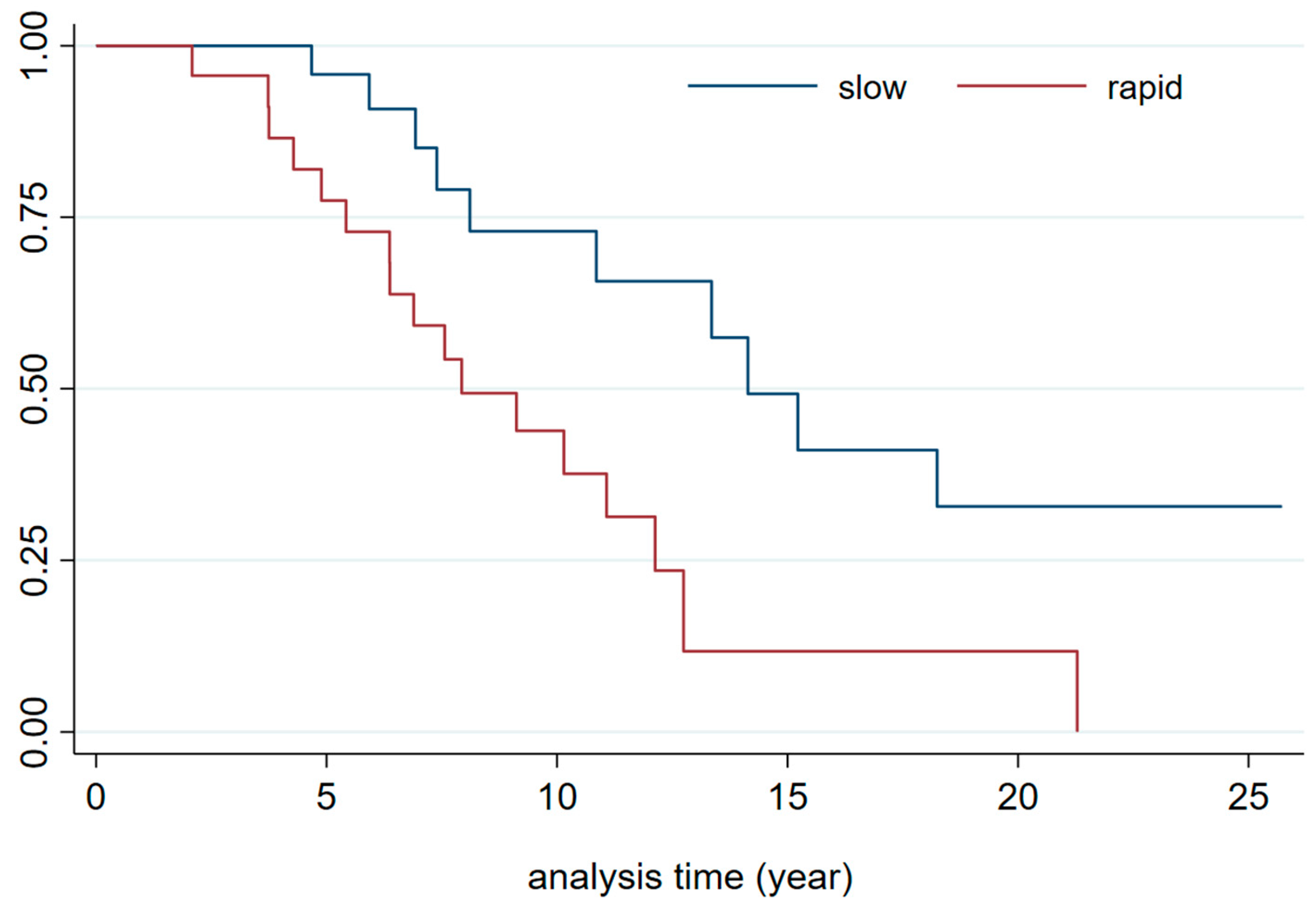
| CKD progression | ||||||||
| Slow progressors | 1.00 | 1.0 | ||||||
| medium | 1.22 | 0.66 | 0.49 | 3.05 | 1.21 | 0.70 | 0.46 | 3.20 |
| Rapid progressors | 3.40 | 0.002 | 1.57 | 7.39 | 4.29 | 0.001 | 1.89 | 9.75 |
| Albuminuria | ||||||||
| A1 (<30 mg/g) | 1.00 | |||||||
| A2 (30–300 mg/g) | 1.97 | 0.6 | 6.45 | |||||
| A3 (>300 mg/g) | 2.29 | 0.737 | 7.084 | |||||
Disclaimer/Publisher’s Note: The statements, opinions and data contained in all publications are solely those of the individual author(s) and contributor(s) and not of MDPI and/or the editor(s). MDPI and/or the editor(s) disclaim responsibility for any injury to people or property resulting from any ideas, methods, instructions or products referred to in the content. |
© 2025 by the authors. Licensee MDPI, Basel, Switzerland. This article is an open access article distributed under the terms and conditions of the Creative Commons Attribution (CC BY) license (https://creativecommons.org/licenses/by/4.0/).
Share and Cite
Serrano Salazar, M.L.; Almonacid, C.; Marques Vidas, M.; López-Sánchez, P.; Sánchez Sobrino, B.; Aguilar, M.; Rubio Arboli, L.; Martínez Morales, E.; Huerta, A.; Valdenebro Recio, M.; et al. Chronic Kidney Disease After Lung Transplantation in Spain: A Retrospective Single-Center Analysis. J. Clin. Med. 2025, 14, 2241. https://doi.org/10.3390/jcm14072241
Serrano Salazar ML, Almonacid C, Marques Vidas M, López-Sánchez P, Sánchez Sobrino B, Aguilar M, Rubio Arboli L, Martínez Morales E, Huerta A, Valdenebro Recio M, et al. Chronic Kidney Disease After Lung Transplantation in Spain: A Retrospective Single-Center Analysis. Journal of Clinical Medicine. 2025; 14(7):2241. https://doi.org/10.3390/jcm14072241
Chicago/Turabian StyleSerrano Salazar, Maria Luisa, Carlos Almonacid, Maria Marques Vidas, Paula López-Sánchez, Beatriz Sánchez Sobrino, Myriam Aguilar, Lucia Rubio Arboli, Eduardo Martínez Morales, Ana Huerta, Maria Valdenebro Recio, and et al. 2025. "Chronic Kidney Disease After Lung Transplantation in Spain: A Retrospective Single-Center Analysis" Journal of Clinical Medicine 14, no. 7: 2241. https://doi.org/10.3390/jcm14072241
APA StyleSerrano Salazar, M. L., Almonacid, C., Marques Vidas, M., López-Sánchez, P., Sánchez Sobrino, B., Aguilar, M., Rubio Arboli, L., Martínez Morales, E., Huerta, A., Valdenebro Recio, M., Ussetti, P., & Portoles, J. (2025). Chronic Kidney Disease After Lung Transplantation in Spain: A Retrospective Single-Center Analysis. Journal of Clinical Medicine, 14(7), 2241. https://doi.org/10.3390/jcm14072241