


Impaired Cardiovascular Hemodynamics in Patients Hospitalized with COVID-19 Pneumonia

 , , ,
, , ,  and
and 
Abstract
1. Introduction
2. Materials and Methods
2.1. Study Population
2.2. Clinical Examination
2.3. Impedance Cardiography
2.4. Echocardiography
- Heart chamber measurements: left ventricle end-diastolic dimension (LVDd), left ventricle end-systolic dimension (LVDs), right ventricle end-diastolic dimension (RVDd), left atrial dimension (LA), left atrial volume (LAV), and left atrial volume index (LAVi);
- Left ventricular systolic function parameters: left ventricle ejection fraction (LVEF, assessed using the Simpson method) and left ventricle global longitudinal strain (LV GLS);
- Left ventricular diastolic function parameters: septal annular e’ velocity (e’sept), lateral annular e’ velocity (e’lat), and left ventricular E/e’ average ratio (E/e’).
2.5. Statistical Methods
3. Results
3.1. Baseline Characteristics
3.2. Intergroup Comparison
4. Discussion
Study Limitations
5. Conclusions
Author Contributions
Funding
Institutional Review Board Statement
Informed Consent Statement
Data Availability Statement
Acknowledgments
Conflicts of Interest
References
- Dweck, M.; Bularga, A.; Hahn, R.; Bing, R.; Lee, K.; Chapman, A.; White, A.; Di Salvo, G.; Sade, L.; Pearce, K.; et al. Global evaluation of echocardiography in patients with COVID-19. Eur. Heart J.—Cardiovasc. Imaging 2020, 21, 949–958. [Google Scholar] [CrossRef] [PubMed]
- Guzik, T.; Mohiddin, S.; Dimarco, A.; Patel, V.; Savvatis, K.; Marelli-Berg, F.; Madhur, M.; Tomaszewski, M.; Maffia, P.; D’Acquisto, F.; et al. COVID-19 and the cardiovascular system: Implications for risk assessment, diagnosis, and treatment options. Cardiovasc. Res. 2020, 116, 1666–1687. [Google Scholar] [CrossRef]
- Scudiero, F.; Silverio, A.; Muraca, I.; Russo, V.; DiMaio, M.; Silvestro, A.; Personeni, D.; Citro, R.; Enrico, M.; Galasso, G.; et al. Long-term prognostic impact of right ventricular dysfunction in patients with COVID-19. J. Pers. Med. 2021, 12, 162. [Google Scholar] [CrossRef] [PubMed]
- Rav-Acha, M.; Orlev, A.; Itzhaki, I.; Zimmerman, S.F.; Fteiha, B.; Bohm, D.; Kurd, R.; Samuel, T.; Asher, E.; Helviz, Y.; et al. Cardiac arrhythmias amongst hospitalised Coronavirus 2019 (COVID-19) patients: Prevalence, characterisation, and clinical algorithm to classify arrhythmic risk. Int. J. Clin. Pract. 2021, 75, e13788. [Google Scholar] [CrossRef] [PubMed]
- Evans, P.; Rainger, G.; Mason, J.; Guzik, T.; Osto, E.; Stamataki, Z.; Neil, D.; Hoefer, I.; Fragiadaki, M.; Waltenberger, M.; et al. Endothelial dysfunction in COVID-19: A position paper of the ESCWorking Group for Atherosclerosis and Vascular Biology, and the ESC Council of Basic Cardiovascular Science. Cardiovasc. Res. 2020, 116, 2177–2184. [Google Scholar] [CrossRef]
- Pelisek, J.; Reutersberg, B.; Greber, U.F.; Zimmermann, A. Vascular dysfunction in COVID-19 patients: Update on SARS-CoV-2 infection of endothelial cells and the role of long non-coding RNAs. Clin. Sci. 2022, 136, 1571–1590. [Google Scholar] [CrossRef]
- Salamanna, F.; Maglio, M.; Landini, M.P.; Fini, M. Body Localization of ACE-2: On the Trail of the Keyhole of SARS-CoV-2. Front. Med. 2020, 7, 594495. [Google Scholar] [CrossRef]
- Ielapi, N.; Licastro, N.; Provenzano, M.; Andreucci, M.; de Franciscis, S.; Serra, R. Cardiovascular disease as a biomarker for an increased risk of COVID-19 infection and related poor prognosis. Biomark. Med. 2020, 14, 713–716. [Google Scholar] [CrossRef]
- Bansal, M. Cardiovascular disease and COVID-19. Diabetes Metab. Syndr. 2020, 14, 247–250. [Google Scholar] [CrossRef]
- Shi, S.; Qin, M.; Shen, B.; Cai, Y.; Liu, T.; Yang, F.; Gong, W.; Liu, X.; Liang, J.; Zhao, Q.; et al. Association of Cardiac Injury With Mortality in Hospitalized Patients with COVID-19 in Wuhan, China. JAMA Cardiol. 2020, 5, 802–810. [Google Scholar] [CrossRef]
- Guo, T.; Fan, Y.; Chen, M.; Wu, X.; Zhang, L.; He, T.; Wang, H.; Wan, J.; Wang, X.; Lu, Z. Cardiovascular Implications of Fatal Outcomes of Patients with Coronavirus Disease 2019 (COVID-19). JAMA Cardiol. 2020, 5, 811–818. [Google Scholar] [CrossRef] [PubMed]
- Ranard, L.S.; Fried, J.A.; Abdalla, M.; Anstey, D.E.; Givens, R.C.; Kumaraiah, D.; Kodali, S.K.; Takeda, K.; Karmpaliotis, D.; Rabbani, L.E.; et al. Approach to Acute Cardiovascular Complications in COVID-19 Infection. Circ. Heart Fail. 2020, 13, e007220. [Google Scholar] [CrossRef]
- Baldi, E.; Sechi, G.M.; Mare, C.; Canevari, F.; Brancaglione, A.; Primi, R.; Klersy, C.; Palo, A.; Contri, E.; Ronchi, V.; et al. Out-of-Hospital Cardiac Arrest during the COVID-19 Outbreak in Italy. N. Engl. J. Med. 2020, 383, 496–498. [Google Scholar] [CrossRef] [PubMed]
- Shao, F.; Xu, S.; Ma, X.; Xu, Z.; Lyu, J.; Ng, M.; Cui, H.; Yu, C.; Zhang, Q.; Sun, P.; et al. In-hospital cardiac arrest outcomes among patients with COVID-19 pneumonia in Wuhan, China. Resuscitation 2020, 151, 18–23. [Google Scholar] [CrossRef] [PubMed]
- Chen, D.; Li, X.; Song, Q.; Hu, C.; Su, F.; Dai, J.; Ye, Y.; Huang, J.; Zhang, X. Assessment of Hypokalemia and Clinical Characteristics in Patients with Coronavirus Disease 2019 in Wenzhou, China. JAMA Netw. Open 2020, 3, e2011122. [Google Scholar] [CrossRef]
- Packer, M.; Abraham, W.; Mehra, M.; Yancy, C.; Lawless, C.; Mitchell, J.; Smart, F.; Bijou, R.; O’Connor, C.; Massie, B.; et al. Utility of impedance cardiography for the identification of short-term risk of clinical decompensation in stable patients with chronic heart failure. J. Am. Coll. Cardiol. 2006, 47, 2245–2252. [Google Scholar] [CrossRef]
- Krzesiński, P.; Gielerak, G.; Kowal, J. Impedance cardiography—A modern tool for monitoring therapy of cardiovascular diseases. Kardiol. Pol. 2009, 67, 65–71. [Google Scholar]
- Galderisi, M.; Cosyns, B.; Edvardsen, T.; Cardim, N.; Delgado, V.; Di Salvo, G.; Donal, E.; Sade, L.E.; Ernande, L.; Garbi, M.; et al. Standardization of adult transthoracic echocardiography reporting in agreement with recent chamber quantification, diastolic function, and heart valve disease recommendations: An expert consensus document of the European Association of Cardiovascular Imaging. Eur. Heart J.-Cardiovasc. Imaging 2017, 18, 1301–1310. [Google Scholar] [CrossRef]
- Uziębło-Życzkowska, B.; Krzesiński, P.; Domino, B.; Chciałowski, A.; Maciorowska, M.; Gielerak, G. Echocardiographic assessment of cardiac function after mild coronavirus disease 2019: A preliminary report. J. Clin. Ultrasound 2022, 50, 17–24. [Google Scholar] [CrossRef]
- Butz, J.; Shan, Y.; Samayoa, A.; Kirton, O.; Vu, T. The utility of impedance cardiography in hemodynamic monitoring of patients with sepsis. Trauma Surg. Acute Care Open 2019, 4, e000349. [Google Scholar] [CrossRef]
- Napoli, A.; Machan, J.; Corl, K.; Forcada, A. The use of impedance cardiography in predicting mortality in emergency department patients with severe sepsis and septic shock. Acad. Emerg. Med. 2010, 17, 452–455. [Google Scholar] [CrossRef] [PubMed]
- Krzesiński, P.; Gielerak, G.; Stańczyk, A.; Uziębło-Życzkowska, B.; Smurzyński, P.; Piotrowicz, K.; Skrobowski, A. What does impedance cardiography add more to the assessment of left ventricular diastolic function in essential hypertension? Pol. Merkur. Lekarski 2015, 39, 352–358. [Google Scholar]
- Jurek, A.; Krzesiński, P.; Gielerak, G.; Witek, P.; Zieliński, G.; Kazimierczak, A.; Wierzbowski, R.; Banak, M.; Uziębło-Życzkowska, B. Cushing’s Disease: Assessment of Early Cardiovascular Hemodynamic Dysfunction with Impedance Cardiography. Front. Endocrinol. 2021, 12, 751743. [Google Scholar] [CrossRef] [PubMed] [PubMed Central]
- Cosyns, B.; Lochy, S.; Luchian, M.; Gimelli, A.; Pontone, G.; Allard, S.; Mey, J.; Rosseel, P.; Dweck, M.; Petersen, S.; et al. The role of cardiovascular imaging for myocardial injury in hospitalized COVID-19 patients. Eur. Heart J.—Cardiovasc. Imaging 2020, 21, 709–714. [Google Scholar] [CrossRef] [PubMed]
- Urban, S.; Fułek, M.; Błaziak, M.; Iwanek, G.; Jura, M.; Fułek, K.; Guzik, M.; Garus, M.; Gajewski, P.; Lewandowski, Ł.; et al. COVID-19 Related Myocarditis in Adults: A Systematic Review of Case Reports. J. Clin. Med. 2022, 11, 5519. [Google Scholar] [CrossRef] [PubMed]
- Bois, M.; Boire, N.; Layman, A.; Aubry, M.; Alexander, M.; Roden, A.; Hagen, C.; Quinton, R.; Larsen, C.; Erben, Y.; et al. COVID-19-associated Non-Occlusive Fibrin Microthrombi in the Heart. Circulation 2021, 143, 230–243. [Google Scholar] [CrossRef]
- Atri, L.; Morgan, M.; Harrell, S.; AlJaroudi, W.; Berman, A.E. Role of cardiac magnetic resonance imaging in the diagnosis and management of COVID 19 related myocarditis: Clinical and imaging considetions. World J. Radiol. 2021, 13, 283–293. [Google Scholar] [CrossRef]
- Puntmann, V.O.; Carerj, M.L.; Wieters, I.; Fahim, M.; Arendt, C.; Hoffmann, J.; Shchendrygina, A.; Escher, F.; Vasa-Nicotera, M.; Zeiher, A.M. Outcomes of cardiovascular magnetic resonance imaging in patients recently recovered from coronavirus disease 2019 (COVID-19). JAMA Cardiol. 2020, 5, 1265–1273. [Google Scholar] [CrossRef]

| VARIABLE | Mean ± SD; Median (Interquartile Range) or n (%) |
|---|---|
| Age [years] | 48.0 ± 8.8; 49 (12) |
| Male sex | 18 (60.0) |
| BMI [kg/m2] | 29.0 ± 4.1; 28.8 (6.2) |
| HR [bpm] | 88.2 ± 14.1; 85.5 (15) |
| SBP [mmHg] | 128.4 ± 13.3; 124.5 (20) |
| DBP [mmHg] | 87.2 ± 8.3; 85 (12) |
| AH | 8 (26.7) |
| LVEF [%] | 62.9 ± 2.6; 63.5 (4) |
| hs-troponin T (ng/L) | 7.0 ± 3.5; 6.4 (3.7) |
| NT-proBNP (pg/mL) | 64.9 ± 62.9; 40.0 (90.7) |
| VARIABLES | COVID Mean ± SD (Median; Interquartile Range) or n (%) | CG Mean ± SD (Median; Interquartile Range) or n (%) | p |
|---|---|---|---|
| Baseline clinical characteristics | |||
| Age [years] | 48.0 ± 8.8; 49 (12) | 46.7 ± 8.5; 44 (12) | 0.562 |
| Male sex | 18 (60.0) | 20 (66.7) | 0.592 |
| BMI [kg/m2] | 29.0 ± 4.1; 28.8 (6.2) | 27.8 ± 3.6; 28.0 (5.4) | 0.211 |
| AH, n [%] | 8 (26.7) | 9 (30) | 0.774 |
| LVEF [%] | 62.9 ± 2.6; 63.5 (4) | 67.3 ± 12.2; 67 (17) | <0.0001 |
| RVDd [mm] | 29.4 ± 3.1; 30 (4) | 30.0 ± 3.4; 30.5 (2.8) | 0.416 |
| LVDd [mm] | 47.7 ± 4.5; 48 (6) | 48.6 ± 3.8; 48.3 (5.5) | 0.375 |
| LA [mm] | 38.1 ± 4.6; 38.5 (4) | 36.4 ± 3.8; 35.9 (5.3) | 0.087 |
| Pharmacotherapy (discharge) | |||
| ACEI | 4 (13.3) | 8 (26.7) | 0.197 |
| ARB | 3 (10.0) | 1 (3.3) | 0.300 |
| Beta-blocker | 2 (6.7) | 0 (0.0) | 0.150 |
| Diuretic | 6 (20.0) | 3 (10.0) | 0.278 |
| Hemodynamics (Impedance Cardiography) | |||
| HR [bpm] | 88.2 ± 14.1; 85.5 (15) | 67.2 ± 12.2; 67 (17) | <0.0001 |
| SBP [mmHg] | 128.4 ± 13.3; 124.5 (20) | 119.0 ± 10.8; 118.5 (12) | 0.004 |
| DBP [mmHg] | 87.2 ± 8.3; 85 (12) | 75.2 ± 8.2; 76 (12) | <0.0001 |
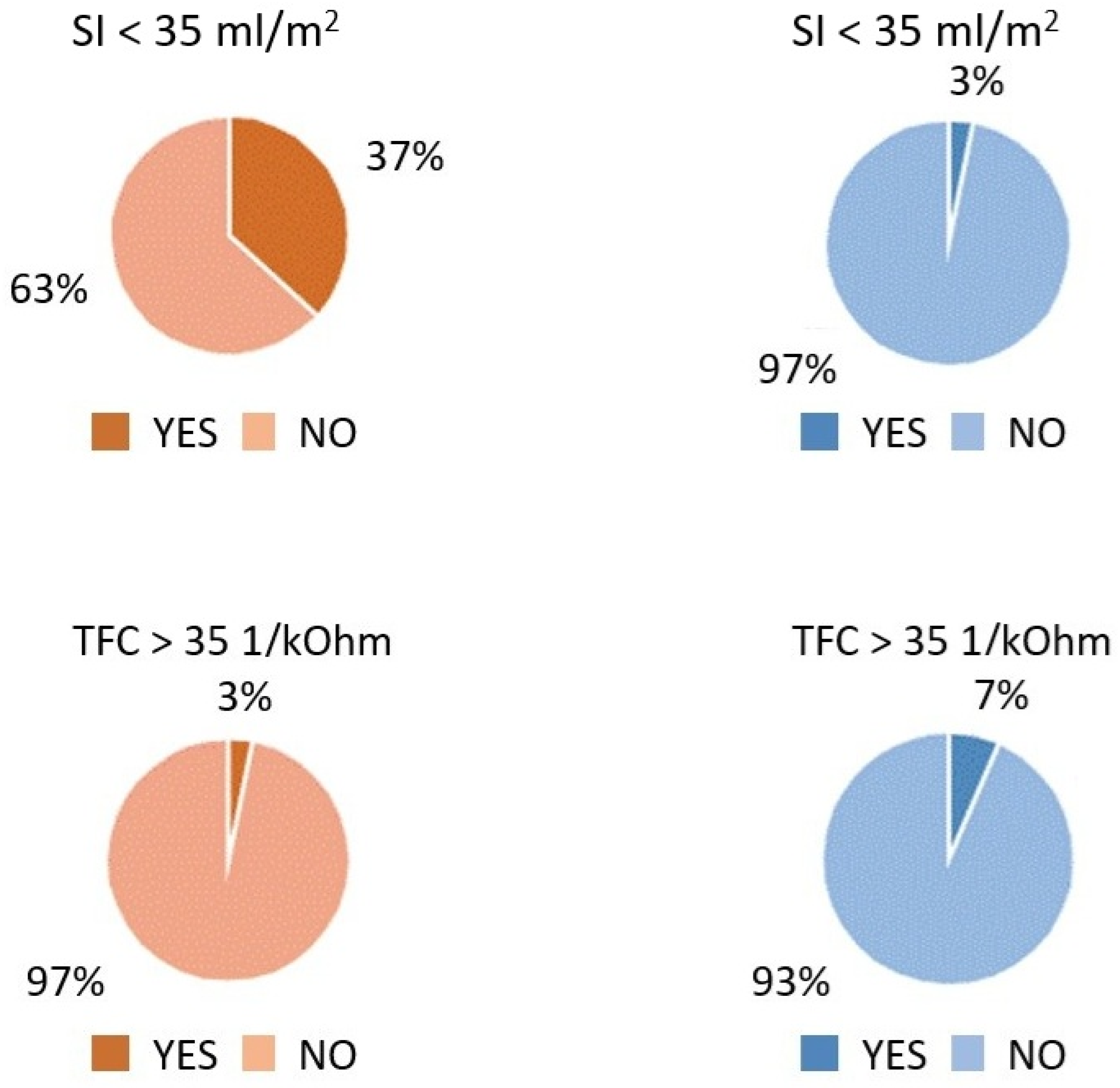
| SI [mL·m2] | 35.9 ± 9.3; 36.3 (13.3) | 50.2 ± 8.8; 50 (17) | <0.0001 |
| CI [mL·m2·min1] | 3.12 ± 0.56; 3.1 (0.8) | 3.34 ± 0.51; 3.15 (0.80) | 0.122 |
| SVRI [dyn·s·cm5·m2] | 2431 ± 546; 2359 (580) | 1945 ± 370; 1878 (452) | 0.0003 |
| VI [1 × 10001·s1] | 34.2 ± 10.4; 31.5 (16) | 47.5 ± 10.8; 49 (23) | <0.0001 |
| ACI [1 × 1001·s2] | 52.5 ± 19.2; 46 (28) | 70.8 ± 23.2; 76.5 (39) | 0.002 |
| HI [Ohm·s2] | 8.3 ± 3.0; 7.6 (4.2) | 12.9 ± 3.7; 12.8 (5.8) | <0.0001 |
| TFC [1·kOhm1] | 28.3 ± 3.9; 28.3 (6.6) | 29.1 ± 3.8; 29.0 (5.6) | 0.429 |
Disclaimer/Publisher’s Note: The statements, opinions and data contained in all publications are solely those of the individual author(s) and contributor(s) and not of MDPI and/or the editor(s). MDPI and/or the editor(s) disclaim responsibility for any injury to people or property resulting from any ideas, methods, instructions or products referred to in the content. |
© 2025 by the authors. Licensee MDPI, Basel, Switzerland. This article is an open access article distributed under the terms and conditions of the Creative Commons Attribution (CC BY) license (https://creativecommons.org/licenses/by/4.0/).
Share and Cite
Domino, B.; Włochacz, A.; Maciorowska, M.; Kłos, K.; Chciałowski, A.; Banak, M.; Uziębło-Życzkowska, B.; Krzesiński, P. Impaired Cardiovascular Hemodynamics in Patients Hospitalized with COVID-19 Pneumonia. J. Clin. Med. 2025, 14, 1806. https://doi.org/10.3390/jcm14061806
Domino B, Włochacz A, Maciorowska M, Kłos K, Chciałowski A, Banak M, Uziębło-Życzkowska B, Krzesiński P. Impaired Cardiovascular Hemodynamics in Patients Hospitalized with COVID-19 Pneumonia. Journal of Clinical Medicine. 2025; 14(6):1806. https://doi.org/10.3390/jcm14061806
Chicago/Turabian StyleDomino, Barbara, Agnieszka Włochacz, Małgorzata Maciorowska, Krzysztof Kłos, Andrzej Chciałowski, Małgorzata Banak, Beata Uziębło-Życzkowska, and Paweł Krzesiński. 2025. "Impaired Cardiovascular Hemodynamics in Patients Hospitalized with COVID-19 Pneumonia" Journal of Clinical Medicine 14, no. 6: 1806. https://doi.org/10.3390/jcm14061806
APA StyleDomino, B., Włochacz, A., Maciorowska, M., Kłos, K., Chciałowski, A., Banak, M., Uziębło-Życzkowska, B., & Krzesiński, P. (2025). Impaired Cardiovascular Hemodynamics in Patients Hospitalized with COVID-19 Pneumonia. Journal of Clinical Medicine, 14(6), 1806. https://doi.org/10.3390/jcm14061806