

Assessment of Autonomic Nervous System Function in Patients with Chronic Fatigue Syndrome and Post-COVID-19 Syndrome Presenting with Recurrent Syncope

 , , and
Abstract
1. Introduction
2. Materials and Methods
2.1. Study Protocol
2.2. Statistical Analysis
3. Results
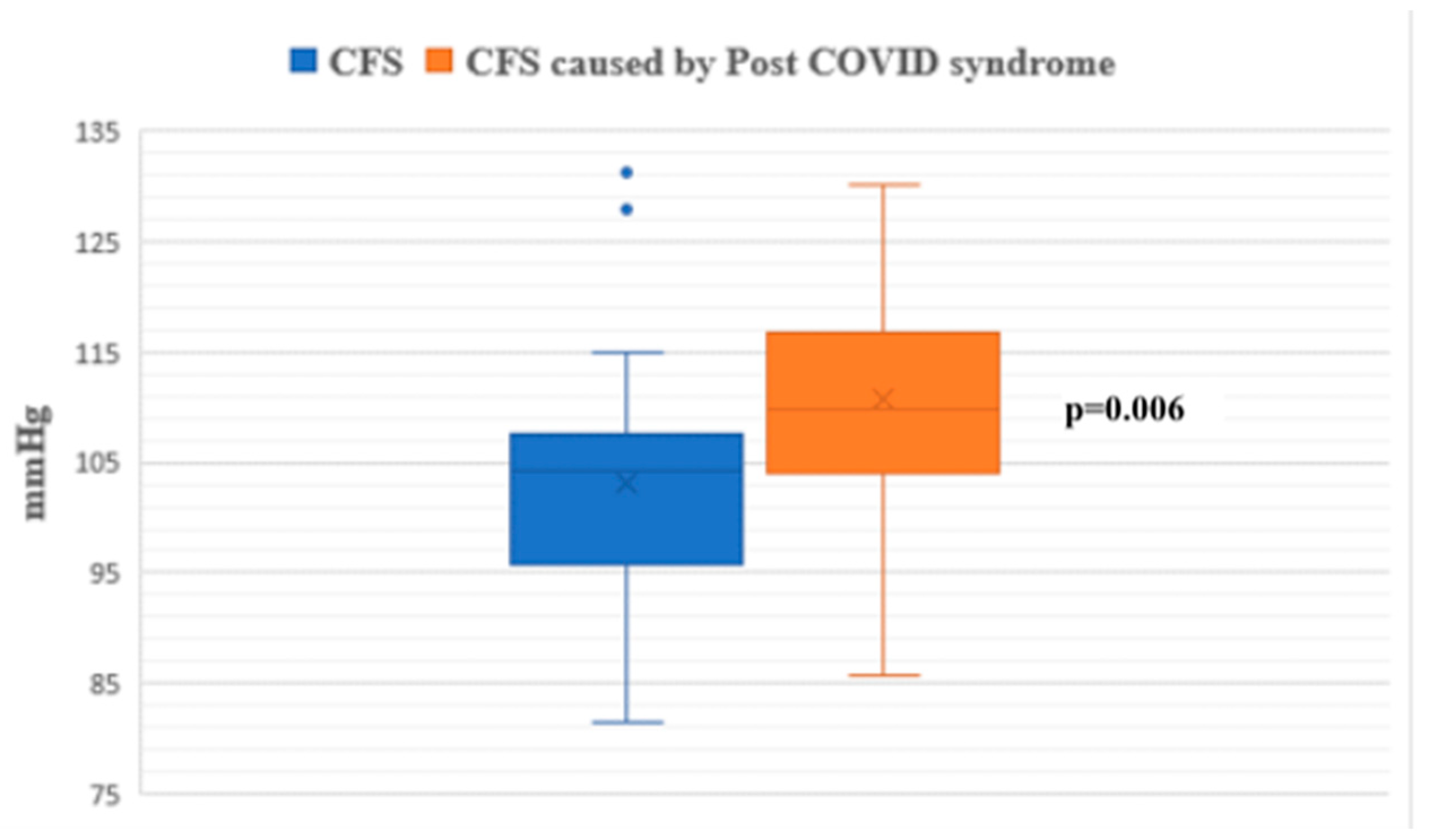
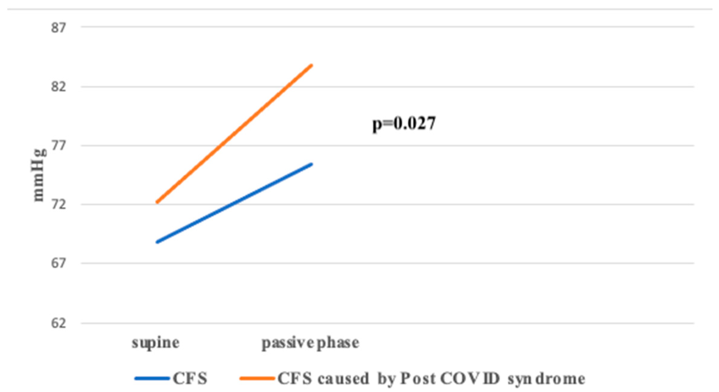
3.1. Head-Up Tilt Test (HUTT)
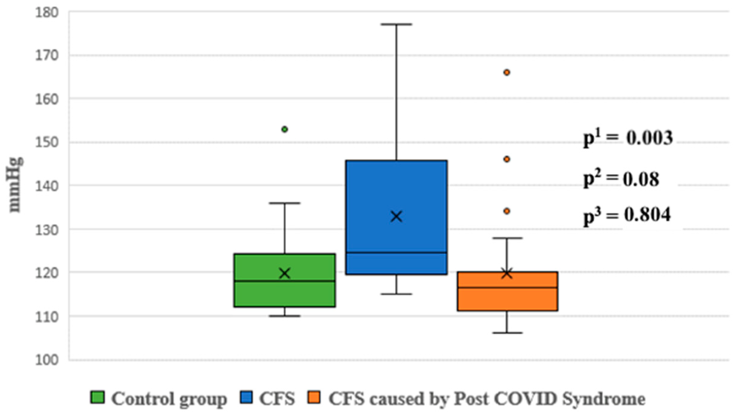
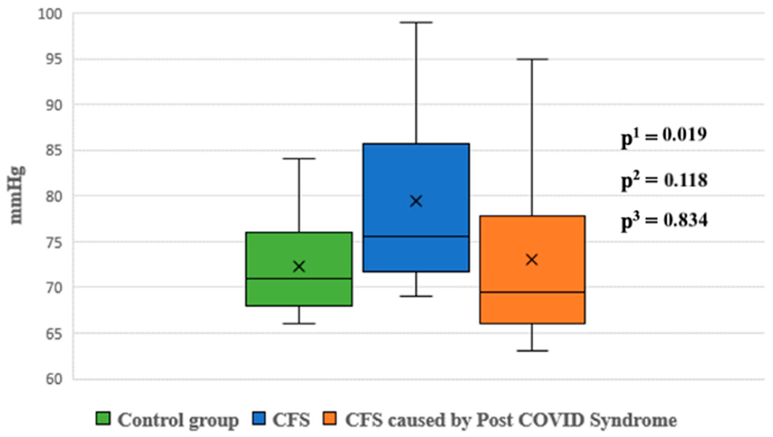
3.2. Cardiovascular Reflex Tests (CART)
3.3. Beat-to-Beat Analysis Using the Task Force Monitor
3.4. 24-Hour Holter ECG Monitoring
3.5. 24-Hour Ambulatory Blood Pressure Monitoring
4. Discussion
Study Limitations
5. Conclusions
Supplementary Materials
Author Contributions
Funding
Institutional Review Board Statement
Informed Consent Statement
Data Availability Statement
Conflicts of Interest
References
- Yuki, K.; Fujiogi, M.; Koutsogiannaki, S. COVID-19 pathophysiology: A review. Clin. Immunol. 2020, 215, 108427. [Google Scholar] [CrossRef]
- Tian, S.; Chang, Z.; Wang, Y.; Wu, M.; Zhang, W.; Zhou, G.; Zou, X.; Tian, H.; Xiao, T.; Xing, J.; et al. Clinical characteristics and reasons for differences in duration from symptom onset to release from quarantine among patients with COVID-19 in Liaocheng, China. Front. Med. 2020, 7, 210. [Google Scholar] [CrossRef] [PubMed]
- Poenaru, S.; Abdallah, S.J.; Corrales-Medina, V.; Cowan, J. COVID-19 and post-infectious myalgic encephalomyelitis/chronic fatigue syndrome: A narrative review. Ther. Adv. Infect. Dis. 2021, 8, 20499361211009385. [Google Scholar] [CrossRef] [PubMed]
- Komaroff, A.L.; Bateman, L. Will COVID-19 lead to myalgic encephalomyelitis/chronic fatigue syndrome? Front. Med. 2021, 7, 606824. [Google Scholar] [CrossRef] [PubMed]
- Vu, Q.M.; Fitzpatrick, A.L.; Cope, J.R.; Bertolli, J.; Sotoodehnia, N.; West, T.E. Estimates of incidence and predictors of fatiguing illness after SARS-CoV-2 infection. Emerg. Infect. Dis. 2024, 30, 539. [Google Scholar] [CrossRef] [PubMed]
- Townsend, L.; Dyer, A.H.; Jones, K.; Dunne, J.; Mooney, A.; Gaffney, F.; O’Connor, L.; Leavy, D.; O’Brien, K.; Dowds, J.; et al. Persistent fatigue following SARS-CoV-2 infection is common and independent of severity of initial infection. PLoS ONE. 2020, 15, e0240784. [Google Scholar] [CrossRef] [PubMed]
- Hickie, I.; Davenport, T.; Wakefield, D.; Vollmer-Conna, U.; Cameron, B.; Vernon, S.D.; Reeves, W.C.; Lloyd, A.; Dubbo Infection Outcomes Study Group. Post-infective and chronic fatigue syndromes precipitated by viral and non-viral pathogens: Prospective cohort study. BMJ 2006, 333, 575. [Google Scholar] [CrossRef] [PubMed]
- Fedorowski, A.; Fanciulli, A.; Raj, S.R.; Sheldon, R.; Shibao, C.A.; Sutton, R. Cardiovascular autonomic dysfunction in post-COVID-19 syndrome: A major health-care burden. Nat. Rev. Cardiol. 2024, 21, 379–395. [Google Scholar] [CrossRef] [PubMed]
- Słomko, J.; Estévez-López, F.; Kujawski, S.; Zawadka-Kunikowska, M.; Tafil-Klawe, M.; Klawe, J.J.; Morten, K.J.; Szrajda, J.; Murovska, M.; Newton, J.L.; et al. Autonomic phenotypes in Chronic Fatigue Syndrome (CFS) are associated with illness severity: A cluster analysis. J. Clin. Med. 2020, 9, 2531. [Google Scholar] [CrossRef]
- de Freitas, R.F.; Torres, S.C.; Martín-Sánchez, F.J.; Carbó, A.V.; Lauria, G.; Nunes, J.P.L. Syncope and COVID-19 disease—A systematic review. Auton. Neurosci. 2021, 235, 102872. [Google Scholar] [CrossRef]
- Haloot, J.; Bhavaraju-Sanka, R.; Pillarisetti, J.; Verduzco-Gutierrez, M. Autonomic dysfunction related to postacute SARS-CoV-2 syndrome. Phys. Med. Rehabil. Clin. N. Am. 2023, 34, 563–572. [Google Scholar] [CrossRef] [PubMed]
- Bou-Holaigah, I.; Rowe, P.C.; Kan, J.; Calkins, H. The relationship between neurally mediated hypotension and the chronic fatigue syndrome. JAMA 1995, 274, 961–967. [Google Scholar] [CrossRef]
- Kenny, R.A.; Graham, L.A. Chronic fatigue syndrome symptoms common in patients with vasovagal syncope. Am. J. Med. 2001, 110, 242–243. [Google Scholar] [CrossRef] [PubMed]
- Carod-Artal, F.J. Infectious diseases causing autonomic dysfunction. Clin. Auton. Res. 2017, 28, 67–81. [Google Scholar] [CrossRef] [PubMed]
- Price, R.W. Viral infections of the autonomic nervous system and its target organs: Pathogenetic mechanisms. Med. Hypotheses 1977, 3, 33–36. [Google Scholar] [CrossRef] [PubMed]
- Price, R.W.; Katz, B.J.; Notkins, A.L. Latent infection of the peripheral ANS with herpes simplex virus. Nature 1975, 257, 686–688. [Google Scholar] [CrossRef]
- Berkwits, M.; Flanagin, A.; Bauchner, H.; Fontanarosa, P.B. The COVID-19 pandemic and the JAMA network. JAMA 2020, 324, 1159–1160. [Google Scholar] [CrossRef]
- National Institute for Health and Care Excellence. Myalgic Encephalomyelitis (or Encephalopathy)/Chronic Fatigue Syndrome: Diagnosis and Management; National Institute for Health and Care Excellence (NICE): London, UK, 2021; Available online: https://www.nice.org.uk/guidance/ng206 (accessed on 25 September 2024).
- World Health Organization. Post COVID-19 Condition (Long COVID) [Internet]. www.who.int. 2022. Available online: https://www.who.int/europe/news-room/fact-sheets/item/post-covid-19-condition (accessed on 25 September 2024).
- Ewing, D.J.; Clarke, B.F. Diagnosis and management of diabetic autonomic neuropathy. Br. Med. J. (Clin. Res. Ed.) 1982, 285, 916–918. [Google Scholar] [CrossRef] [PubMed]
- Bellavere, F.; Bosello, G.; Fedele, D.; Cardone, C.; Ferri, M. Diagnosis and management of diabetic autonomic neuropathy. Br. Med. J. (Clin. Res. Ed.) 1983, 287, 61. [Google Scholar] [CrossRef] [PubMed]
- Fitzpatrick, A.P.; Theodorakis, G.; Vardas, P.; Sutton, R. Methodology of head-up tilt testing in patients with unexplained syncope. J. Am. Coll. Cardiol. 1991, 17, 125–130. [Google Scholar] [CrossRef] [PubMed]
- Gratze, G.; Fortin, J.; Holler, A.; Grasenick, K.; Pfurtscheller, G.; Wach, P.; Schönegger, J.; Kotanko, P.; Skrabal, F. A software package for non-invasive, real-time beat-to-beat monitoring of stroke volume, blood pressure, total peripheral resistance and for assessment of autonomic function. Comput. Biol. Med. 1998, 28, 121–142. [Google Scholar] [CrossRef] [PubMed]
- Parati, G.; Frattola, A.; Di Rienzo, M.; Castiglioni, P.; Pedotti, A.; Mancia, G. Effects of aging on 24-h dynamic baroreceptor control of heart rate in ambulant subjects. Am. J. Physiol. 1995, 268, H1606-12. [Google Scholar] [CrossRef] [PubMed]
- Schwalm, T. Modern Tilt Table Testing and Non—Invasive Monitoring, Traditional and Innovaritve Applications in Theory and Practice, 1st ed.; ABW Wissenschaftsverlag GmbH: Berlin, Germany, 2007. [Google Scholar]
- Hausenloy, D.J.; Arhi, C.; Chandra, N.; Franzen-McManus, A.-C.; Meyer, A.; Sutton, R. Blood pressure oscillations during tilt testing as a predictive marker of vasovagal syncope. Europace 2009, 11, 1696–1701. [Google Scholar] [CrossRef]
- van Campen, C.L.M.C.; Visser, F.C. Orthostatic intolerance in long-haul COVID after SARS-CoV-2: A case-control comparison with post-EBV and insidious-onset myalgic encephalomyelitis/chronic fatigue syndrome patients. Healthcare 2022, 10, 3058. [Google Scholar] [CrossRef]
- van Campen, C.M.C.; Verheugt, F.W.A.; Rowe, P.C.; Visser, F.C. Cerebral blood flow is reduced in ME/CFS during head-up tilt testing even in the absence of hypotension or tachycardia: A quantitative, controlled study using Doppler echography. Clin. Neurophysiol. Pract. 2020, 5, 50–58. [Google Scholar] [PubMed]
- Milovanovic, B.; Djajic, V.; Bajic, D.; Djokovic, A.; Krajnovic, T.; Jovanovic, S.; Verhaz, A.; Kovacevic, P.; Ostojic, M. Assessment of autonomic nervous system dysfunction in the early phase of infection with SARS-CoV-2 virus. Front. Neurosci. 2021, 15, 640835. [Google Scholar] [CrossRef] [PubMed]
- Ryabkova, V.A.; Rubinskiy, A.V.; Marchenko, V.N.; Trofimov, V.I.; Churilov, L.P. Similar patterns of dysautonomia in myalgic encephalomyelitis/chronic fatigue and post-COVID-19 syndromes. Pathophysiology 2024, 31, 1. [Google Scholar] [CrossRef] [PubMed]
- da Silva, F.S.; Bonifácio, L.P.; Bellissimo-Rodrigues, F.; Joaquim, L.F.; Martins Dias, D.P.; Dias Romano, M.M.; Romano, M.M.D.; Schmidt, A.; Crescêncio, J.C.; Buzinari, T.C.; et al. Investigating autonomic nervous system dysfunction among patients with post-COVID condition and prolonged cardiovascular symptoms. Front. Med. 2023, 10, 1216452. [Google Scholar] [CrossRef] [PubMed]
- Peckerman, A.; LaManca, J.J.; Qureishi, B.; Dahl, K.A.; Golfetti, R.; Yamamoto, Y.; Natelson, B.H. Baroreceptor reflex and integrative stress responses in chronic fatigue syndrome. Psychosom. Med. 2003, 65, 889–895. [Google Scholar] [CrossRef] [PubMed]
- Srivastava, P.; Nabeel, P.M.; Raj, K.V.; Soneja, M.; Chandran, D.S.; Joseph, J.; Wig, N.; Jaryal, A.K.; Thijssen, D.; Deepak, K.K. Baroreflex sensitivity is impaired in survivors of mild COVID-19 at 3–6 months of clinical recovery; association with carotid artery stiffness. Physiol. Rep. 2023, 11, 15845. [Google Scholar] [CrossRef]
- Tu, B.; Wu, L.; Hu, F.; Fan, S.; Liu, S.; Liu, L.; Ding, L.; Zheng, L.; Yao, Y. Cardiac deceleration capacity as an indicator for cardioneuroablation in patients with refractory vasovagal syncope. Heart Rhythm. 2022, 19, 562–569. [Google Scholar] [CrossRef] [PubMed]
- Walitt, B.; Singh, K.; LaMunion, S.R.; Hallett, M.; Jacobson, S.; Chen, K.; Enose-Akahata, Y.; Apps, R.; Barb, J.J.; Bedard, P.; et al. Deep phenotyping of post-infectious myalgic encephalomyelitis/chronic fatigue syndrome. Nat. Commun. 2024, 15, 907. [Google Scholar] [CrossRef]
- Bauer, A.; Malik, M.; Schmidt, G.; Barthel, P.; Bonnemeier, H.; Cygankiewicz, I.; Guzik, P.; Lombardi, F.; Dipl-Ing, A.M.; Oto, A.; et al. Heart rate turbulence: Standards of measurement, physiological interpretation, and clinical use. J. Am. Coll. Cardiol. 2008, 52, 1353–1365. [Google Scholar] [CrossRef] [PubMed]
- Newton, J.L.; Sheth, A.; Shin, J.; Pairman, J.; Wilton, K.; Burt, J.A.; Jones, D.E.J. Lower ambulatory blood pressure in chronic fatigue syndrome. Psychosom. Med. 2009, 71, 361–365. [Google Scholar] [CrossRef] [PubMed]
- Vojdani, A.; Vojdani, E.; Saidara, E.; Maes, M. Persistent SARS-CoV-2 infection, EBV, HHV-6 and other factors may contribute to inflammation and autoimmunity in long COVID. Viruses 2023, 15, 400. [Google Scholar] [CrossRef] [PubMed]
- Cameron, B.; Flamand, L.; Juwana, H.; Middeldorp, J.; Naing, Z.; Rawlinson, W.; Ablashi, D.; Lloyd, A. Serological and virological investigation of the role of the herpesviruses EBV, CMV and HHV-6 in post-infective fatigue syndrome. J. Med. Virol. 2010, 82, 1684–1688. [Google Scholar] [CrossRef]
- Endresen, G.K.M. Mycoplasma blood infection in chronic fatigue and fibromyalgia syndromes. Rheumatol. Int. 2003, 23, 211–215. [Google Scholar] [CrossRef]
- Chia, J.K.S.; Chia, L.Y. Chronic chlamydia pneumoniae infection: A treatable cause of chronic fatigue syndrome. Clin. Infect. Dis. 1999, 29, 452–453. [Google Scholar] [CrossRef] [PubMed]
- Chu, L.; Valencia, I.J.; Garvert, D.W.; Montoya, J.G. Onset patterns and course of myalgic encephalomyelitis/chronic fatigue syndrome. Front. Pediatr. 2019, 7, 12. [Google Scholar] [CrossRef] [PubMed]
- Lim, M.L.; Rickman, L.S. Brucellosis. Infect. Dis. Clin. Pract. 2004, 12, 7–14. [Google Scholar] [CrossRef]
- Sanjuan, N.A.; Lascano, E.F. Autonomic nervous system involvement in experimental genital infection by herpes simplex virus type 2. Arch. Virol. 1986, 91, 329–339. [Google Scholar] [CrossRef] [PubMed]
- Sakakibara, R.; Sawai, S.; Ogata, T. Varicella-zoster virus infection and autonomic dysfunction. Auton. Neurosci. 2022, 242, 103018. [Google Scholar] [CrossRef]
- Budzyński, J.; Kłopocka, M.; Bujak, R.; Swiatkowski, M.; Pulkowski, G.; Sinkiewicz, W. Autonomic nervous function in Helicobacter pylori-infected patients with atypical chest pain studied by analysis of heart rate variability. Eur. J. Gastroenterol. Hepatol. 2004, 16, 451–457. [Google Scholar] [CrossRef] [PubMed]
- Cohen, J.A.; Miller, L.; Polish, L. Orthostatic hypotension in human immunodeficiency virus infection may be the result of generalized autonomic nervous system dysfunction. JAIDS J. Acquir. Immune Defic. Syndr. 1991, 4, 31–33. [Google Scholar] [CrossRef]
- Goin, J.C.; Venera, G.; Biscoglio de Jiménez Bonino, M.; Sterin-Borda, L. Circulating antibodies against nicotinic acetylcholine receptors in chagasic patients. Clin. Exp. Immunol. 1997, 110, 219–225. [Google Scholar] [CrossRef] [PubMed]
- Rassi, A., Jr.; Rassi, A. Marcondes de Rezende, J. American trypanosomiasis (Chagas disease). Infect. Dis. Clin. N. Am. 2012, 26, 275–291. [Google Scholar] [CrossRef] [PubMed]
- Günlü, S.; Aktan, A. Cardiac arrhythmias, conduction system abnormalities, and autonomic tone in patients with brucellosis. Eur. Rev. Med. Pharmacol. Sci. 2022, 26, 9473–9479. [Google Scholar] [CrossRef] [PubMed]
- Kasmani, R.; Elkambergy, H.; Okoli, K. Postural orthostatic tachycardia syndrome associated with Mycoplasma pneumoniae. Infect. Dis. Clin. Pract. 2009, 17, 342–343. [Google Scholar] [CrossRef]
- Bennett, J.L.; Mahalingam, R.; Wellish, M.C.; Gilden, D.H. Epstein-Barr virus—Associated acute autonomic neuropathy. Ann. Neurol. 1996, 40, 453–455. [Google Scholar] [CrossRef]
- Nakao, K.; Namekawa, M.; Kondo, S.; Ono, S.; Nakano, I. Subacute autonomic and sensory neuropathy closely related to cytomegalovirus infection preceded by frequent syncopal attacks. Rinsho Shinkeigaku 2016, 56, 555–559. [Google Scholar] [CrossRef]
- Neville, B.G.; Sladen, G.E. Acute autonomic neuropathy following primary herpes simplex infection. J. Neurol. Neurosurg. Psychiatry 1984, 47, 648–650. [Google Scholar] [CrossRef]
- Hanai, S.; Komaki, H.; Sakuma, H.; Nakagawa, E.; Sugai, K.; Sasaki, M.; Oya, Y.; Higurashi, N.; Hamano, S. Acute autonomic sensory and motor neuropathy associated with parvovirus B19 infection. Brain Dev. 2011, 33, 161–165. [Google Scholar] [CrossRef] [PubMed]
- Pavesi, G.; Gemignani, F.; Macaluso, G.M.; Ventrua, P.; Magnani, G.; Fiocchi, A.; Medici, D.; Marbini, A.; Mancia, D. Acute sensory and autonomic neuropathy: Possible association with coxsackie B virus infection. J. Neurol. Neurosurg. Psychiatry 1992, 55, 613–615. [Google Scholar] [CrossRef] [PubMed]
- Adler, B.L.; Chung, T.; Rowe, P.C.; Aucott, J. Dysautonomia following Lyme disease: A key component of post-treatment Lyme disease syndrome? Front. Neurol. 2024, 15, 1344862. [Google Scholar] [CrossRef]
- Wang, H.; Siddharthan, V.; Hall, J.O.; Morrey, J.D. Autonomic nervous dysfunction in hamsters infected with West Nile virus. PLoS ONE 2011, 6, e19575. [Google Scholar] [CrossRef]
- Burch, G.E.; Chu, K.C.; Soike, K.F. Coxsackievirus B4 infection of sympathetic ganglia in squirrel monkeys. Angiology 1985, 36, 23–26. [Google Scholar] [CrossRef] [PubMed]
- Bellocchi, C.; Carandina, A.; Montinaro, B.; Targetti, E.; Furlan, L.; Rodrigues, G.D.; Tobaldini, E.; Montano, N. The interplay between autonomic nervous system and inflammation across systemic autoimmune diseases. Int. J. Mol. Sci. 2022, 23, 2449. [Google Scholar] [CrossRef]
- Globig, A.-M.; Zhao, S.; Roginsky, J.; Maltez, V.I.; Guiza, J.; Avina-Ochoa, N.; Heeg, M.; Hoffmann, F.A.; Chaudhary, O.; Wang, J. The β1-adrenergic receptor links sympathetic nerves to T cell exhaustion. Nature 2023, 622, 383–392. [Google Scholar] [CrossRef] [PubMed]
- Maya, J. Surveying the metabolic and dysfunctional profiles of T cells and NK cells in myalgic encephalomyelitis/chronic fatigue syndrome. Int. J. Mol. Sci. 2023, 24, 11937. [Google Scholar] [CrossRef]
- Loebel, M.; Grabowski, P.; Heidecke, H.; Bauer, S.; Hanitsch, L.G.; Wittke, K.; Meisel, C.; Reinke, P.; Volk, H.-D.; Fluge, Ø.; et al. Antibodies to β adrenergic and muscarinic cholinergic receptors in patients with Chronic Fatigue Syndrome. Brain Behav. Immun. 2016, 52, 32–39. [Google Scholar] [CrossRef]
- Gravelsina, S.; Vilmane, A.; Svirskis, S.; Rasa-Dzelzkaleja, S.; Nora-Krukle, Z.; Vecvagare, K.; Krumina, A.; Leineman, I.; Shoenfeld, Y.; Murovska, M. Biomarkers in the diagnostic algorithm of myalgic encephalomyelitis/chronic fatigue syndrome. Front. Immunol. 2022, 13, 928945. [Google Scholar] [CrossRef] [PubMed]
- Li, H.; Kem, D.C.; Reim, S.; Khan, M.; Vanderlinde-Wood, M.; Zillner, C.; Collier, D.; Liles, C.; Hill, M.A.; Cunningham, M.W.; et al. Agonistic autoantibodies as vasodilators in orthostatic hypotension: A new mechanism. Hypertension 2012, 59, 402–408. [Google Scholar] [CrossRef] [PubMed]
- Gunning, W.T., III; Kvale, H.; Kramer, P.M.; Karabin, B.L.; Grubb, B.P. Postural orthostatic tachycardia syndrome is associated with elevated G-protein coupled receptor autoantibodies. J. Am. Heart Assoc. 2019, 8, 013602. [Google Scholar] [CrossRef] [PubMed]
- Fedorowski, A.; Li, H.; Yu, X.; Koelsch, K.A.; Harris, V.M.; Liles, C.; Murphy, T.A.; Quadri, S.M.S.; Scofield, R.H.; Sutton, R.; et al. Antiadrenergic autoimmunity in postural tachycardia syndrome. Europace 2017, 19, 1211–1219. [Google Scholar] [CrossRef] [PubMed]


| CFS N = 210 | CFS After COVID-19 N = 137 | Control Groups N = 91 | |
|---|---|---|---|
| Years (mean ± SD) | 44.85 ± 12.67 | 45.02 ± 13.36 | 36.6 ± 11.43 |
| Male (n, %) | 43 (20.48%) | 33 (24.09%) | 37 (40.65%) |
| Female (n, %) | 167 (79.52%) | 104 (75.91%) | 54 (59.35%) |
| Sleep disturbances (n, %) | 210 (100%) | 137 (100%) | 0 (0%) |
| PEM (n, %) | 210 (100%) | 137 (100%) | 0 (0%) |
| Fibromylagia (n, %) | 154 (73.3%) | 86 (62.8%) | 0 (0%) |
| Joint pain (n, %) | 154 (73.3%) | 89 (65%) | 0 (0%) |
| Vertigo (n, %) | 94 (44.8%) | 58 (42.3%) | 0 (0%) |
| Disturbed regulation of temperature (n, %) | 98 (46.7%) | 18 (13.1%) | 0 (0%) |
| Tingling (n, %) | 113 (53.8%) | 50 (36.5%) | 0 (0%) |
| Burning sensation (n, %) | 128 (61%) | 36 (26.3%) | 0 (0%) |
| Groups | Control Group (n = 69) | CFS (n = 210) | CFS Caused by Post-COVID-19 Syndrome (n = 135) | Sig. |
|---|---|---|---|---|
| HGT * n (%) | 51 (73.9%) | 206 (96.1%) | 131 (97%) | p1 < 0.001 p2 < 0.001 p3 > 0.05 |
| OH n (%) | 0 (0%) | 73 (34.8%) | 34 (25.2%) | p1 < 0.001 p2 < 0.001 p3 > 0.05 |
| DS n (%) | 51 (73.9%) | 207 (98.6%) | 131 (97.0%) | p1 < 0.001 p2 < 0.001 p3 > 0.05 |
| VM n (%) | 17 (24.6%) | 68 (32.5%) | 51 (37.8%) | p1 > 0.05 p2 < 0.05 p3 > 0.05 |
| HRB n (%) | 11 (15.9%) | 168 (80.4%) | 95 (70.4%) | p1 < 0.001 p2 < 0.001 p3 < 0.05 |
| HRS n (%) | 36 (52.2%) | 195 (93.3%) | 118 (84.4%) | p1 < 0.001 p2 < 0.001 p3 > 0.05 |
| DP (definite) n (%) | 11 (15.9%) | 167 (79.9%) | 104 (77%) | p1 < 0.001 p2 < 0.001 p3 > 0.05 |
| CAN n (%) | 38 (55.1%) | 207 (97.6%) | 129 (95.6%) | p1 < 0.001 p2 < 0.001 p3 > 0.05 |
| Score | 4.33 (3.98–4.69) | 7.25 (7.04–7.47) | 6.96 (6.71–7.2) | p1 < 0.001 p2 < 0.001 p3 > 0.05 |
| Parameters | Control Group (n = 91) | CFS (n = 68) | CFS Caused by Post-COVID-19 (n = 60) | Sig. |
|---|---|---|---|---|
| HR (bpm) | 72.32 (70.14–74.5) | 77.46 (74.2–81.09) | 80.91 (76.82–85) | p1 < 0.05 p2 < 0.001 p3 > 0.05 |
| SBP (mmHg) | 115.9 (113.12–118.66) | 107.14 (103.39–110.88) | 111.84 (108.01–115.67) | p1 < 0.01 p2 > 0.05 p3 > 0.05 |
| DBP (mmHg) | 76.87 (74.44–79) | 68.88 (65.64–72.12) | 72.28 (69.3–75.25) | p1 < 0.001 p2 < 0.05 p3 > 0.05 |
| MAP (mmHg) | 88.86 (86.71–90.1) | 84.48 (81.18–87.77) | 88.15 (85.03–91.26) | p1 > 0.05 p2 > 0.05 p3 > 0.05 |
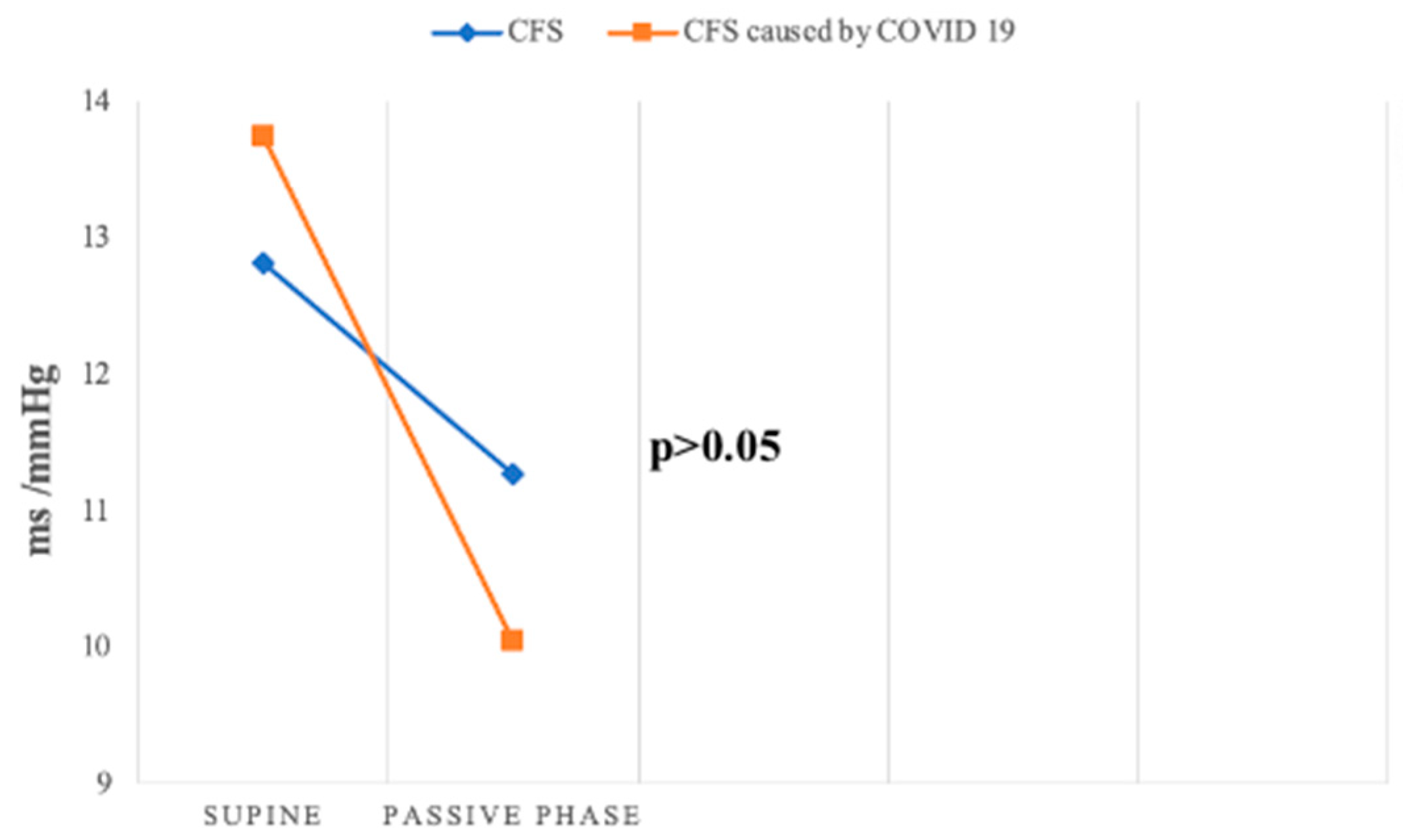
| BRS (ms/mmHg) | 17.9 (15.84–19.95) | 12.82 (10.17–15.46) | 13.75 (11.09–16.41) | p1 < 0.01 p2 > 0.05 p3 > 0.05 |
| BEI (%) | 121.12 (111.87–130.38) | 61.84 (54.10–69.57) | 64.41 (58.45–70.36) | p1 < 0.001 p2 < 0.001 p3 > 0.05 |
| Parameters | Control Group (n = 91) | CFS (n = 68) | CFS Caused by Post-COVID-19 (n = 60) | Sig. |
|---|---|---|---|---|
| LFnu-RRI (%) | 59.65 (56.25–63.05) | 62.95 (58.55–67.35) | 60.13 (55.18–65.08) | p1 > 0.05 p2 > 0.05 p3 > 0.05 |
| HFnu-RRI (%) | 40.02 (36.77–43.27) | 37.65 (33.16–42.15) | 39.87 (34.92–44.83) | p1 > 0.05 p2 > 0.05 p3 > 0.05 |
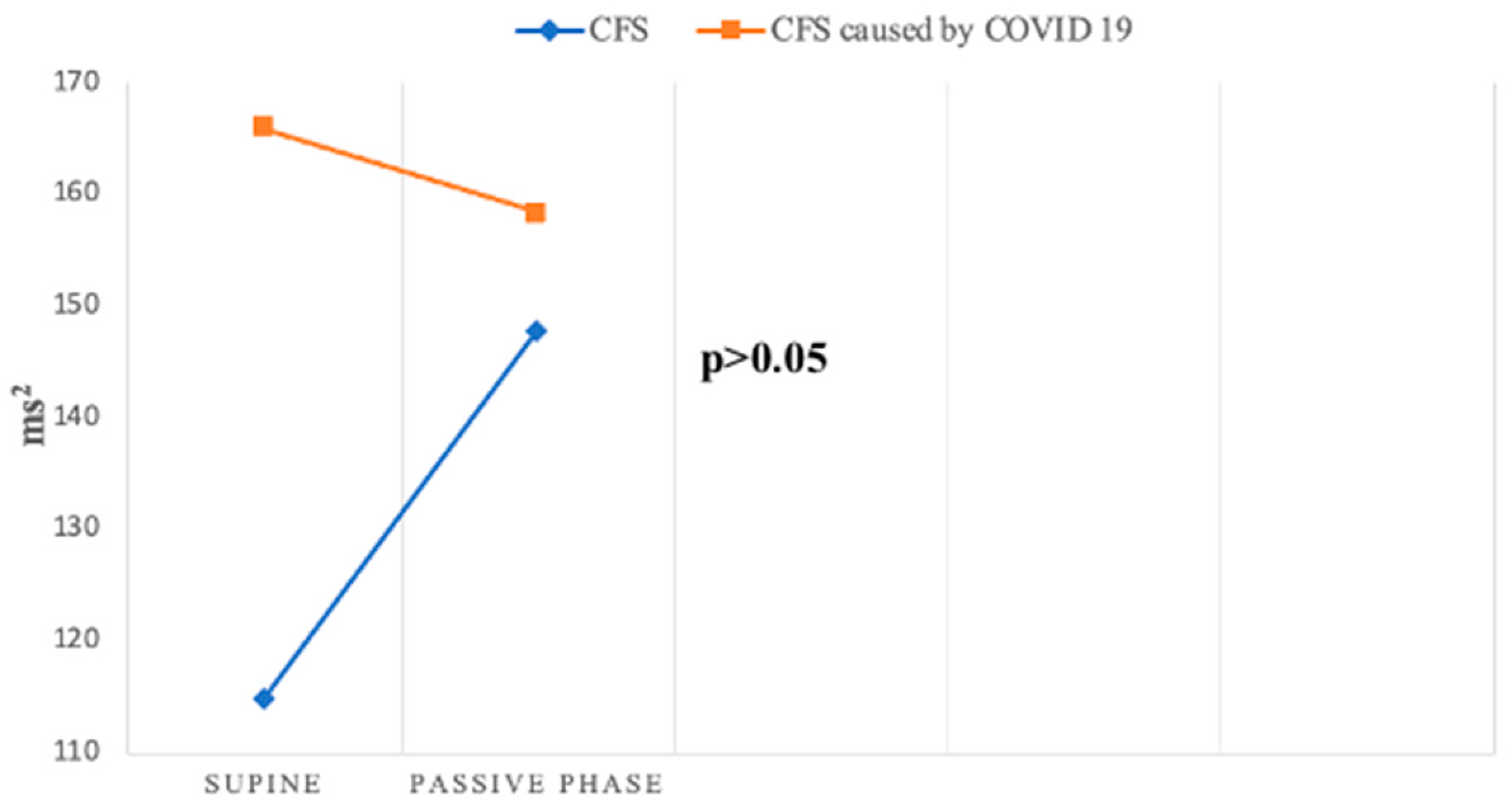
| PSD-RRI (ms2) | 1258.93 (977.24–1621.81) | 630.96 (467.74–851.14) | 645.65 (457.09–912.01) | p1 < 0.01 p2 < 0.01 p3 > 0.05 |
| VLF-RRI (ms2) | 257.04 (204.17–323.59) | 154.88 (112.20–213.80) | 134.90 (95.50–190.55) | p1 < 0.05 p2 < 0.01 p3 > 0.05 |
| LF-RRI (ms2) | 512.86 (407.38–645.65) | 223.87 (169.82–295.12) | 263.03 (190.55–363.08) | p1 < 0.001 p2 < 0.01 p3 > 0.05 |
| HF-RRI (ms2) | 316.23 (239.88–416.87) | 114.82 (79.43–165.96) | 165.96 (107.15–257.04) | p1 < 0.001 p2 < 0.05 p3 > 0.05 |
| LFnu/HFnu-RRI | 1.91 (1.58–2.29) | 2.14 (1.66–2.75) | 1.74 (1.38–2.19) | p1 > 0.05 p2 > 0.05 p3 > 0.05 |
| LF/HF-RRI | 1.55 (1.32–1.82) | 1.41 (1.12–1.78) | 1.29 (1.02–1.62) | p1 > 0.05 p2 > 0.05 p3 > 0.05 |
| Groups | Control Group (n = 86) | CFS (n = 64) | CFS Caused by Post-COVID-19 (n = 37) | Sig. |
|---|---|---|---|---|
| Heart rate (bpm) | 75.19 (73.37–77.00) | 73.83 (71.61–76.04) | 74.84 (71.95–77.26) | p1 > 0.05 p2 > 0.05 p3 > 0.05 |
| RR interval (ms) | 816.52 (794.33–829.09) | 811.32 (787.65–835) | 801.2 (770.28–832.13) | p1 > 0.05 p2 > 0.05 p3 > 0.05 |
| SDNN (ms) | 159.15 (151.44—166.85) | 145.94 (137.54–154.34) | 144.76 (132.01–157.51) | p1 > 0.05 p2 > 0.05 p3 > 0.05 |
| SDANN (ms) | 145.50 (137.41–153.59) | 130.33 (122.47–138.2) | 128.76 (115.85–141.67) | p1 < 0.05 p2 < 0.05 p3 > 0.05 |
| SDNNIN (ms) | 64.57 (60.26–69.18) | 54.95 (52.48–57.54) | 61.66 (52.48–72.44) | p1 < 0.05 p2 > 0.05 p3 > 0.05 |
| rMSSD (ms) | 38.65 (35.31–42.00) | 40.35 (35.15–45.54) | 37.3 (31.54–43.05) | p1 > 0.05 p2 > 0.05 p3 > 0.05 |
| pNN50 (%) | 11,75 (10.00–13.80) | 7.94 (6.46–9.77) | 6.61 (4.79–9.12) | p1 < 0.01 p2 < 0.05 p3 > 0.05 |
| Groups | Control Group (n = 86) | CFS (n = 64) | CFS Caused by Post-COVID-19 (n = 37) | Sig. |
|---|---|---|---|---|
| TP (ms2) | 4430.33 (3965.7–4894.96) | 3143.19 (2724.77–3561.61) | 3571.45 (2891.16–4251.75) | p1 < 0.01 p2 < 0.05 p3 > 0.05 |
| VLF (ms2) | 3012.07 (2659.27–3364.87) | 2160.95 (1874.38–2447.53) | 2408.60 (1925.15–2892.06) | p1 < 0.01 p2 > 0.05 p3 > 0.05 |
| LF (ms2) | 1053.97 (956.28–1151.66) | 707.31 (600.05–814.58) | 823.23 (650.67–995.78) | p1 < 0.001 p2 < 0.05 p3 > 0.05 |
| HF (ms2) | 331.13 (281.84–389.05) | 204.17 (169.82–245.47) | 229.09 (186.21–281.84) | p1 < 0.001 p2 < 0.05 p3 > 0.05 |
| LF/HF | 1.96 (1.89–2.03) | 3.37 (2.87–3.88) | 3.44 (2.87–4.0) | p1 < 0.01 p2 < 0.1 p3 > 0.05 |
| PVC (mean + 95% interval) | 59.16 (15.45–102.86) | 184.37 (105.85–262.9) | 247.38 (0–498.67) | p1 < 0.001 p2 < 0.01 p3 > 0.05 |
| PAC (mean + 95% interval) | 97.71 (0–195.70) | 68.69 (39.48–97.91) | 69.27 (24.7–113.84) | p1 < 0.001 p2 < 0.001 p3 > 0.05 |
| DC (ms) | / | 6.5 (6.4–7.3) | 7.54 (7.02–8.06) | p3 < 0.5 |
| AC (ms) | / | 7.51 (6.89–8.12) | 8.25 (7.59–8.92) | p3 > 0.05 |
| TO (PVC) (%) | / | −2 (3.83–−0.17) | 3.38 (−5.24—1.52) | p3 > 0.05 |
| TS (PVC) (ms/RR interval) | / | 13.3 (7.85–18.74) | 14.1 (9.86–18.34) | p3 > 0.05 |
| TO (PAC) (%) | / | 2.06 (0.34–3.77) | 0.78 (−1.54–3.1) | p3 > 0.05 |
| TS (PAC) (ms/RR interval) | / | 6.77 (4.75–8.8) | 7.57 (4.94–10.19) | p3 > 0.05 |
Disclaimer/Publisher’s Note: The statements, opinions and data contained in all publications are solely those of the individual author(s) and contributor(s) and not of MDPI and/or the editor(s). MDPI and/or the editor(s) disclaim responsibility for any injury to people or property resulting from any ideas, methods, instructions or products referred to in the content. |
© 2025 by the authors. Licensee MDPI, Basel, Switzerland. This article is an open access article distributed under the terms and conditions of the Creative Commons Attribution (CC BY) license (https://creativecommons.org/licenses/by/4.0/).
Share and Cite
Milovanovic, B.; Markovic, N.; Petrovic, M.; Zugic, V.; Ostojic, M.; Rankovic-Nicic, L.; Bojic, M. Assessment of Autonomic Nervous System Function in Patients with Chronic Fatigue Syndrome and Post-COVID-19 Syndrome Presenting with Recurrent Syncope. J. Clin. Med. 2025, 14, 811. https://doi.org/10.3390/jcm14030811
Milovanovic B, Markovic N, Petrovic M, Zugic V, Ostojic M, Rankovic-Nicic L, Bojic M. Assessment of Autonomic Nervous System Function in Patients with Chronic Fatigue Syndrome and Post-COVID-19 Syndrome Presenting with Recurrent Syncope. Journal of Clinical Medicine. 2025; 14(3):811. https://doi.org/10.3390/jcm14030811
Chicago/Turabian StyleMilovanovic, Branislav, Nikola Markovic, Masa Petrovic, Vasko Zugic, Milijana Ostojic, Ljiljana Rankovic-Nicic, and Milovan Bojic. 2025. "Assessment of Autonomic Nervous System Function in Patients with Chronic Fatigue Syndrome and Post-COVID-19 Syndrome Presenting with Recurrent Syncope" Journal of Clinical Medicine 14, no. 3: 811. https://doi.org/10.3390/jcm14030811
APA StyleMilovanovic, B., Markovic, N., Petrovic, M., Zugic, V., Ostojic, M., Rankovic-Nicic, L., & Bojic, M. (2025). Assessment of Autonomic Nervous System Function in Patients with Chronic Fatigue Syndrome and Post-COVID-19 Syndrome Presenting with Recurrent Syncope. Journal of Clinical Medicine, 14(3), 811. https://doi.org/10.3390/jcm14030811