Article

Short-Term Efficacy and Safety of Scleral Lenses in the Management of Severe Dry Eye in a Chinese Population

1
Department of Ophthalmology, Eye, Ear, Nose & Throat Hospital of Fudan University, Shanghai 200031, China
2
NHC Key laboratory of Myopia and Related Eye Diseases, Shanghai 200031, China
3
Shanghai Engineering Research Center of Synthetic Immunology, Shanghai 200032, China
4
Department of Ophthalmology, Children’s Hospital of Fudan University, National Pediatric Medical Center of China, Shanghai 201102, China
5
Centre for Ocular Research & Education, School of Optometry & Vision Science, University of Waterloo, Waterloo, ON N2L 3G1, Canada
*
Authors to whom correspondence should be addressed.
These authors contributed equally to this work.
J. Clin. Med. 2025, 14(3), 658; https://doi.org/10.3390/jcm14030658
Submission received: 13 November 2024 / Revised: 17 December 2024 / Accepted: 17 January 2025 / Published: 21 January 2025
(This article belongs to the Special Issue The Role of Scleral Lenses in the Management of Eye Disease)

Abstract

:
Background: Scleral lenses (SLs) are recommended in DEWS II to treat dry eye (DE) patients that do not respond well to conventional therapies. This study aimed to evaluate the short-term (one month) efficacy and safety of SLs in the management of severe DE. Methods: This single-center prospective study enrolled 15 patients (22 eyes) who were diagnosed with severe DE. The Ocular Surface Disease Index (OSDI), the Chinese version of the 25-item National Eye Institute Visual Function Questionnaire (CHI-VFQ-25), and LogMAR best-corrected visual acuity (BCVA) were evaluated at baseline and one month following SL fitting. DE-related parameters were obtained and analyzed before and after one month of SL treatment, including tear-film breakup time (TBUT), corneal fluorescein staining (CFS), non-invasive breakup time (NIBUT), tear meniscus height (TMH), Schirmer I test (SIT), and meibomian gland (MG) dropout. Complications and adverse events were monitored. Results: OSDI scores (53.9 ± 28.1 vs. 10.4 (4.2–25), p = 0.0001) and CFS scores (10.2 ± 3.9 vs. 7 (0–12), p = 0.001) decreased after one month of SL therapy, while CHI-VFQ-25 scores (74.4 (54.8–83.8) vs. 95 (78.7–98), p = 0.0001) and TBUT (0.6 ± 0.5 vs. 2.2 ± 1.0, p < 0.0001) increased significantly. LogMAR BCVA improved from 0 (0–0.1) to 0 (0–0) (p = 0.0147). The average types of medications per eye decreased from 2.82 ± 1.01 to 1.32 ± 0.64 (p = 0.025), and the proportion of eyes using glucocorticoids significantly decreased from 63.6% to 13.6% (p = 0.001). No severe SL-related adverse events were reported. Conclusions: SL treatment quickly alleviated subjective symptoms as well as clinical signs of DE with good safety and enhanced the visual function and vision-related quality of life, showing its usefulness in the management of severe DE.

1. Introduction

Dry eye (DE), a chronic multifactorial ocular surface disease, is usually characterized by a loss of homeostasis of the tear film, ocular surface inflammation, tissue damage, and neurological abnormalities, and leads to ocular discomfort symptoms, visual impairment, and impaired quality of life [1,2,3]. Its prevalence in China ranges from 4.29 to 50.33% in different areas [4]. According to the classification of DE severity, eyes with corneal fluorescein staining (CFS) ≥ two quadrants, a tear-film breakup time (TBUT) < 2 s, or a Schirmer I test (SIT) score of 0 were classified as severe DE [3].
The management of severe DE is a clinical challenge. According to the four-tiered strategy put forward by TFOS DEWS II guidelines [5], routine treatments for DE, which were defined as Level 1 and 2 in the guidelines, were not enough for these cases. More importantly, ocular discomfort symptoms usually impair the visual quality of severe DE patients and their vision-related quality of life (VR-QoL) [6,7], making it necessary and critical that the treatments alleviate the symptoms rapidly.
Scleral lenses (SLs) are large-diameter, rigid, gas-permeable contact lenses that vault over the cornea and rest on the sclera. Unlike soft contact lenses, SLs can be customized to fit individual ocular anatomy with or without a toric design, thus creating a tear reservoir between the lens and cornea. For over a decade, SLs have been used to correct corneal irregularities caused by keratoconus or corneal surgeries [8,9]. In recent years, their application has been extended to the treatment of refractory DE, particularly in patients who fail to respond well to conventional therapies [10,11,12,13]. They are recommended in the management of moderate to severe DE cases (Level 3 in TFOS DEWS II), especially when the ocular surface is compromised [5].
However, currently, most studies mainly focus on its mid- to long-term effects, with an average follow-up ranging from 6 to 20 months [10,11,14]. Nevertheless, short-term efficacy in severe DE patients deserves attention because they usually experience severe ocular pain and need quick relief of symptoms. Moreover, the efficacy of SLs in Chinese patients has not been extensively explored, and they are more susceptible to developing DE than other races [15]. Additionally, consensus is still lacking regarding the indications and contradictions of SL treatment for DE patients, as well as standardized fitting protocols and complication management.
Therefore, we conducted this prospective cohort study in a Chinese population to investigate the one-month short-term efficacy and safety of SL therapy in the management of severe DE and its impact on visual quality and VR-QoL so as to provide clinical evidence for its application as a non-pharmacological alternative in the management of severe DE.

2. Materials and Methods

2.1. Study Design and Subjects

This single-center prospective study, conducted at the Eye, Ear, Nose & Throat Hospital of Fudan University, was executed in accordance with the Declaration of Helsinki and with the approval of the Ethics Committee of the hospital (2024016). Written informed consents were obtained from all participants.
A total of 15 patients who met the diagnostic criteria for severe DE according to “Chinese expert consensus on dry eye: definition and classification (2020)” [3] were recruited from September 2023 to May 2024. In brief, the patient should meet all of the following criteria in at least one eye: (1) TBUT < 2 s; (2) corneal epithelial damage ≥ two quadrants; (3) CFS ≥ 30 points, or presented as fused coarse dot/bulk staining, or with filamentary formations. Due to low repeatability and large variation, SIT was not included in the criteria, only except that when the SIT value was 0 mm/5 min, the subject was considered eligible even though the above three criteria were not all met.
The exclusion criteria were listed as follows: (1) ocular conditions affecting corneal sensation or integrity (e.g., herpetic keratitis, diabetic keratopathy); (2) severe conjunctival abnormalities that affected SL fitting (e.g., pterygium, conjunctivochalasis); (3) active ocular inflammation, infection, or diseases potentially impacting SL fitting (e.g., severe allergic conjunctivitis, uveitis, scleritis); (4) history of ocular surgery that might impair corneal sensation, such as corneal refractive surgery; (5) severe visually impairing disorders (e.g., glaucoma, vitreous hemorrhage, central corneal scar); (6) compromised immune response; (7) pregnancy, breastfeeding, or planning to conceive; (8) allergies to fluorescein sodium or inability to cooperate with lens fitting; (9) inability to comply with follow-up visits.

2.2. Ocular Examinations

2.2.1. Order of Ocular Examinations

After the collection of demographic data including sex, age, concomitant systematic and ocular diseases, and the medications and DE therapies that had been used before planning treatment with SLs, all participants were asked to complete two validated questionnaires: the Ocular Surface Disease Index (OSDI) and a Chinese version of the 25-item National Eye Institute Visual Function Questionnaire (CHI-VFQ-25) [16,17]. The patients were given explanations if they could not understand the content of the questionnaires. All completed questionnaires were meticulously reviewed by the same researcher to confirm data completeness [6]. Then, each participant underwent the following examinations in sequence: best-corrected visual acuity (BCVA), slit-lamp biomicroscopy, anterior segment optical coherence tomography (AS-OCT), Oculus Pentacam, Corvis ST, Oculus Keratograph 5M (Oculus K5M), corneal sensitivity testing, and the Schirmer I test (SIT). TBUT and CFS were evaluated during slit-lamp biomicroscopy examination. All examinations were conducted by the same team composed of experienced ophthalmologists, optometrists, and trained technicians and were performed at the baseline and one month after regular SL wear.

2.2.2. OSDI Questionnaire

The OSDI questionnaire, which consisted of 12 items covering ocular symptoms, vision-related functions, and environmental triggers, evaluated the severity of DE symptoms. Each item was scored from 0 (never) to 4 (always) or not applicable (N/A) based on the frequency of the patient’s experiences over the past week, and the final score ranged from 0 to 100, which was calculated with the following formula: (25 × total scores of all items answered)/number of questions answered. A higher score indicated more severe DE symptoms [18].

2.2.3. CHI-VFQ-25 Questionnaire

The CHI-VFQ-25 questionnaire, which was validated [16] and used in previous studies [6,7], was applied to assess the VRQoL across 12 subscales: general health, general vision, near activity, distance activity, social function, color vision, peripheral vision, driving, role difficulties, ocular pain, dependency, and mental health. Each of the questions was scored from 0 (worst) to 100 (best), or N/A. The total scores were calculated following the guidelines provided by the National Eye Institute of the USA. A higher score equaled a better VR-QoL [19].

2.2.4. BCVA

Snellen BCVA was measured and then converted to the logarithm of the minimal angle of resolution (LogMAR) format. The lower the LogMAR value, the better the visual acuity.

2.2.5. Slit-Lamp Biomicroscopy

All patients underwent a slit-lamp examination to exclude any ocular abnormalities that could potentially affect SL fitting. TBUT and CFS were assessed under cobalt blue light, as previously described [6,7]. In brief, a sterile fluorescein strip (Jingmin, Tianjin, China), pre-moistened with sterile saline, was applied to the inferior conjunctival fornix. The participant was asked to blink 3–4 times naturally to ensure an even distribution of fluorescein on the ocular surface and to keep their eyes open as long as possible. The time from the last blink to the appearance of the first black spot or streak on the cornea was recorded as the TBUT. The test was performed three times. CFS was assessed within 3 min after fluorescein instillation. Based on the National Eye Institute grid [20], the cornea was divided into five parts: central, nasal, temporal, superior, and inferior. The fluorescence staining dots in each part were scored from 0 to 3 according to the following criteria: 0 for no staining; 1 for fewer than 15 dots; 2 for 16 to 30 dots; and 3 for more than 30 dots or strip/bulk staining or corneal filaments. The score for each part was added and the total ranged from 0 to 15.

2.2.6. AS-OCT

AS-OCT (CASIA2, Tomey Corporation, Nagoya, Japan) was performed to obtain central corneal thickness (CCT) and central epithelial thickness (CET). During the examination, the subjects, in a seated position, had their heads stabilized with a chin and brow rest and were instructed to maintain a forward gaze. High-resolution images of the anterior eye segment were captured along the horizontal meridian in single-scan mode. The outcomes of CCT and CET were automatically output by the customized software. The thickness of the precorneal tear film during the SL fitting procedure was also evaluated by AS-OCT.

2.2.7. Corneal Tomography

The corneal–scleral profile (CSP) mode of corneal tomography was performed to capture a series of CSPs using Pentacam (Oculus, Wetzlar, Germany), a high-precision non-contact Scheimpflug camera system. The participant was required to gaze forward, with superior, inferior, nasal, and temporal targets in a row to allow for the reconstruction of an 18 mm diameter corneal–scleral shape centered on the corneal vertex normal. The essential parameters such as sagittal height, horizontal visible iris diameter, and scleral angle were then derived for SL selection.

2.2.8. Corvis ST

Corvis ST, a non-contact tonometer that assesses corneal deformation with high-speed Scheimpflug imagery, was used to measure biomechanically corrected intraocular pressure (bIOP). Patients, who were seated with their heads stabilized, were asked to hold their eyes open after several normal blinks. The measurement was automatically taken and repeated three times. The average value was calculated for analysis.

2.2.9. Tear-Film Function

The Oculus K5M (Wetzlar, Germany) was used to evaluate the overall function of the tear film in a noncontact way. First, participants were told to blink twice in a dark environment and then to keep their eyes open until the system automatically recorded the first and average non-invasive breakup time (f-NIBUT, a-NIBUT). Then, the images of the lower tear meniscus were captured, and tear meniscus height (TMH) was measured using the built-in caliper. Finally, the upper and lower eyelids were gently everted to obtain images of the meibomian glands (MGs). The MG loss scores were assigned based on the proportion of dropout area: 0 for none, 1 for ≤1/3, 2 for 1/3 to 2/3, and 3 for >2/3. The lipid layer color (LLC) was qualitatively classified as normal (multicolor, red–green) or abnormal (blue–grey, hoary, achromatic), as previously reported [21].

2.2.10. Corneal Sensitivity

The Cochet–Bonnet aesthesiometer (Luneau Ophthalmologie, Paris, France), featuring a filament with a diameter of 0.12 mm and a maximum length of 60 mm, was used for corneal sensitivity testing. Patients were seated and asked to look straight ahead. The central cornea was gently touched with the tip of the filament from the temporal side in a tangential direction. The test commenced at the maximal length (60 mm) and was decremented by 5 mm with each application until the patient reported a foreign body sensation on the cornea and the visible deflection of the filament by the examiner. This procedure was repeated three times, and the average value was taken for analysis.

2.2.11. SIT

Without topical anesthesia, Schirmer paper strips (5 × 40 mm, Jingming, Tianjin, China) were carefully positioned into the outer one-third of the lower conjunctival sac cautiously to avoid irritative tearing. Patients were instructed to gently close their eyes for 5 min, after which the tear wetting length from the strip’s notch was measured and documented.

2.3. SL Fitting

SL fitting was conducted by one well-experienced ophthalmologist (ZC). Trial SLs were selected based on the parameters measured by Oculus Pentacam and AS-OCT. Before insertion, the SL was filled with non-buffered, non-preserved saline supplemented with fluorescein for enhanced visualization upon placement. When the SL was successfully settled, its position was immediately evaluated using slit-lamp biomicroscopy under white and cobalt blue light to ensure that its edge landed on the conjunctiva, neither contacting the cornea and the limbus nor exerting pressure on conjunctival vessels (Supplementary Figure S1). AS-OCT was then performed to evaluate the tear layer thickness beneath the lens, targeting a range of 200–300 µm for optimal moisture retention and minimal tear evaporation. These examinations were repeated 1 and 2 h after lens settlement for dynamic evaluation. Finally, trial SLs were removed, and rebound hyperemia or redness of the conjunctiva was checked [10,11]. Adjustments to the lens parameters, such as sagittal height, diameter, or edge design, were performed to achieve optimal fitting and ocular comfort. The fitting process was finalized with over-refraction with the SL in place to correct residual refractive errors and obtain the final prescription.
On the day of the SL delivery visit, the post-lens fluid reservoir and the lens alignment were evaluated again. After two hours of SL wear, the ocular surface was assessed after lens removal to check if any additional adjustments of lens parameters were needed to ensure the safety of lens wear. The patients were instructed to wear SLs for an average of 8–12 h per day, with overnight use explicitly avoided.

2.4. Statistical Analysis

Data at baseline and the one-month follow-up were analyzed using Stata 17.0 (StataCorp, College Station, TX, USA). The continuous data with normal distribution were described as mean ± standard deviation (SD) and analyzed with paired t-tests, otherwise they were presented as medians (P25–P75), and a Wilcoxon signed-rank test was performed. Categorical data were analyzed with Fisher’s exact test and the Chi-squared test. All statistical tests were conducted with a 95% confidence interval, and a two-tailed p-value < 0.05 was considered to indicate statistical significance.

3. Results

A total of 14 women and one man diagnosed with severe DE were enrolled in the study, with an average age of 48.3 ± 13.0 (14–73) years. The concomitant systematic diseases and ocular diseases related to severe DE included primary Sjögren’s syndrome (SS), graft-versus-host disease (GVHD), Stevens–Johnson syndrome (SJS), rheumatoid arthritis (RA), Vogt–Koyanagi–Harada (VKH) disease, thyroid eye disease (TED), and filamentary keratitis. After detailed examinations and careful evaluation, 22 eyes were prescribed SLs that were made of hexafocon A (13 eyes), Boston XO2 (eight eyes), and reflufocon D (one eye), respectively. The ranges of SAG, diameter (Dia), and base curves (BCs) were 3300~4500 um, 14.5~16.3 mm and 8.0~9.8 mm, respectively. The lenses were worn continuously for one month without overnight wear. The details of the demographic data and SLs are presented in Table 1.

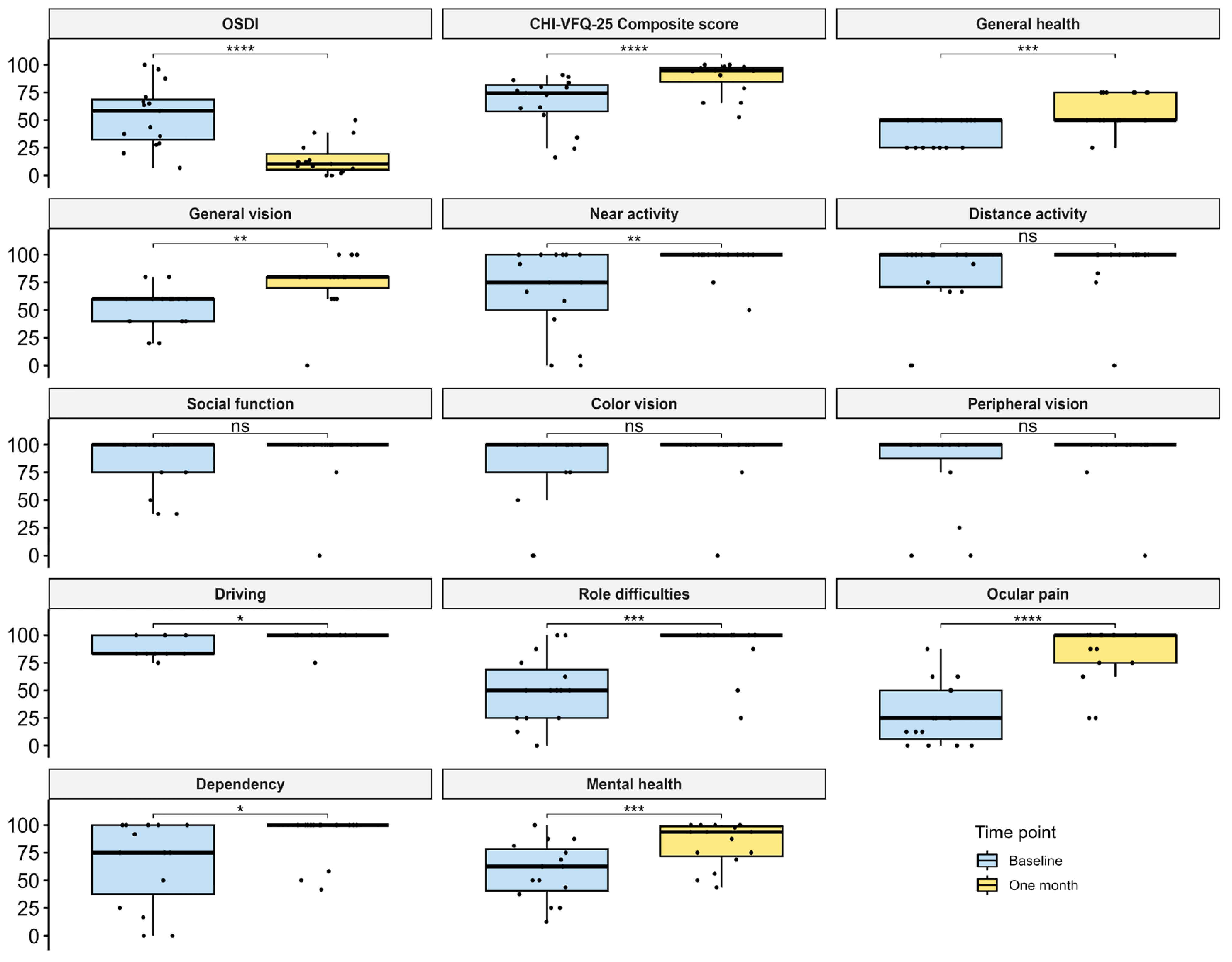
3.1. Improvement of DE Symptoms and VR-QoL

The OSDI score, CHI-VFQ-25 composite score, and scores for each subscale are presented in Figure 1. Compared to the baseline, a significant reduction in OSDI scores was found after one month of SL therapy (p = 0.0001). Meanwhile, the CHI-VFQ-25 composite scores significantly increased (p = 0.0001). Among the subscale scores of the CHI-VFQ-25, a notable improvement in the score for ocular pain was reported (median 28.3 vs. 100, p < 0.0001). Moreover, those for general health, general vision, near activity, driving, role difficulties, dependency, and mental health were also significantly increased (all p < 0.05).

3.2. Improvement of DE Signs

Compared to the baseline value, TBUT was remarkably prolonged after one month of SL therapy (p < 0.0001), along with reduced LogMAR BCVA (p = 0.0147) and decreased CFS scores (p = 0.001). Comparisons of the other parameters of DE did not show any statistical significance, except MG dropout scores, which were increased after one month (p = 0.039) (Table 2).

3.3. Dependency on Medication

The topical medications for DE were categorized into four types: artificial tears (with and without preservatives), serum extracts, immunosuppressants (e.g., cyclosporine), and glucocorticoids (e.g., fluorometholone). After one month of SL therapy, the average types of medications per eye significantly decreased from 2.82 ± 1.01 to 1.32 ± 0.64 (p = 0.025). Seventeen eyes (17/22, 77.3%) needed only one type of medication, and artificial tears were enough for 15 eyes (15/22, 68.2%) to control symptoms (Table 1). More importantly, the proportion of eyes using glucocorticoids dramatically decreased from 63.6% to 13.6% (p = 0.001).

3.4. Safety

During the one-month follow-up, one eye (4.6%) encountered subconjunctival hemorrhage with unknown etiology, which was spontaneously absorbed within 3 days. Additionally, 13 eyes (59.1%) reported mild midday fogging (MDF), which was resolved after removing, cleaning, and reapplying the lenses at noon or in the afternoon. It is notable that bIOP significantly decreased from 16.6 ± 3.3 mmHg to 15.3 ± 1.5 mmHg (p = 0.039). Importantly, neither severe adverse events such as microbial keratitis nor SL-induced conjunctival ischemia were found. After one month of SL therapy, corneal sensitivity, CCT, and CET did not have significant alterations compared to baseline values (Table 2).

4. Discussion

In the current study, remarkably decreased OSDI scores and CFS scores as well as improvement of TBUT were found after one month of SL therapy, indicating less ocular discomfort, better tear-film stability, and less corneal epithelial damage, which are crucial for ocular surface health. These findings revealed that SL treatment could alleviate the symptoms of ocular discomfort and improve clinical signs of severe DE within one month. SL wear not only kept ocular surface moisture stable and reduced tear evaporation [22] but also protected the ocular surface from environmental triggers such as smoke and dust during daily activities [23]. More importantly, SLs physically blocked direct contact between the eyelids and cornea, which avoided the shearing force from eyelids during blinking, thus facilitating corneal epithelial healing [24]. Notably, the patients were much less dependent on DE-related medications after SL therapy, especially the usage of corticosteroids, and benefited from decreased or discontinued use of glucocorticoids, as evidenced by decreased bIOP. All these findings supported that, as a non-pharmacological medical device, SL therapy might minimize the risks of long-term complications associated with chronic usage of various medications in the management of severe DE.
The current study is the first to report the impact of SLs on the VR-QoL of severe DE patients. Owing to fast epithelial recovery under the protection of SLs and the reestablishment of a smooth and regular optical interface [25,26], a great improvement in visual acuity was reported by patients after one month of treatment, along with a much better VR-QoL. It was notable that among all subscale scores, that for ocular pain presented the most significant improvement. Chronic ocular pain, which was quite common in severe DE patients, not only leading to anxiety [27,28] but also impairing their daily activities [6,29,30], was relieved by treatment with SLs, further contributing to improved general health and mental well-being.
The current study showed that one month of SL wear neither caused corneal edema nor affected corneal innervation. However, the incidence of MDF in our study was higher (59.1%) than that reported in previous studies (7%~46%) [31,32,33]. Abnormal alterations of lipid components (e.g., cholesteryl esters) and/or proportion (nonpolar lipid) were identified in eyes with MGD [34,35], which was one factor contributing to MDF [36]. Regarding the fact that the etiologies of severe DE, such as primary SS [37] and GVHD [38], were usually accompanied by MGD, the abnormalities of lipids in the tears of severe DE eyes made MDF more likely to occur during SL wear. Therefore, patient education on proper lens cleaning during the day is more crucial for these patients. Another unexpected safety issue finding was a slight increase in MG dropout scores after one month. Although one year of SL wear was reported to have no adverse effect on MG morphology in ametropic eyes [39], its impact on MGs in DE patients remained unclear. Given that MGD is quite common in severe DE eyes [37,38], it was reasonable to deduce that the MGs in these subjects were more fragile and more likely to be affected by the mechanical pressure exerted on eyelids by SLs. Further studies with longer follow-ups and more advanced MG imaging technology are needed to evaluate the long-term effect of SL wear on the structure and morphology of MGs in severe DE patients.
Several limitations should be acknowledged. First, the sample size is relatively small, and this is a single-center study, which might lead to selection bias. Second, the study was only performed in a Chinese population. It has been confirmed that compared to Caucasians, East Asians usually have smaller corneas, narrower limbi, tighter upper eyelids, and more congested anterior segments [40,41,42,43], making it difficult to compare the findings among different races. Third, the classification of DE, such as aqueous-deficient DE or evaporative DE, were not taken into consideration in the current study because the majority cases of severe DE, such as SS and GVHD, were mixed type, with both tear deficiency and over evaporation due to MGD being involved in the pathogenesis [37,38]. Further multicenter studies with larger sample sizes across ethnicities are needed to fully evaluate the efficacy and safety of SL therapy in the management of severe DE, and its long-term impacts on CCT, IOP, and MG morphology and function need more investigation.

5. Conclusions

In conclusion, SL therapy alleviates the symptoms and signs of severe DE within one month by providing continuous hydration and mechanical protection for the ocular surface. Meanwhile, it significantly improves the visual function and VR-QoL. No severe adverse events were found during one month of usage. SLs may serve as a potential alternative treatment for patients with refractory DE. Their long-term efficacy, safety, and potential use in various ethnic populations need further investigation.

Supplementary Materials

The following supporting information can be downloaded at https://www.mdpi.com/article/10.3390/jcm14030658/s1, Figure S1: Assessment of SL fitting under slit-lamp biomicroscopy.

Author Contributions

Writing—original draft, Methodology, Investigation, Formal analysis, Data curation, C.L.; Writing—review and editing, Methodology, Investigation, Formal analysis, Data curation, D.H.; Writing—review and editing, Methodology, Investigation, Data curation, L.Z.; Writing—review and editing, Software, Resources, Conceptualization, J.H.; Writing—review and editing, D.F.; Writing—review and editing, Data curation, Resources, X.Z.; Writing—review and editing, Validation, Supervision, Project administration, Funding acquisition, Conceptualization, Z.C. and Q.L. All authors have read and agreed to the published version of the manuscript.

Funding

This work was supported by the National Natural Science Foundation of China [82471042], the Natural Science Foundation of Shanghai [24ZR1409600], the Key Discipline Program of the Sixth Round of the Three-Year Public Health Action Plan (Year 2023–2025) of Shanghai, China: GWVI-11.1-32, and the Shanghai Commission of Science and Technology: 23Y11901100.

Institutional Review Board Statement

The study was conducted in accordance with the Declaration of Helsinki and approved by the Institutional Ethics Committee of the Eye, Ear, Nose, and Throat Hospital of Fudan University (protocol code 2024016, date of approval 31 July 2023).

Informed Consent Statement

Informed consent was obtained from all subjects involved in the study.

Data Availability Statement

Data are contained within the article/Supplementary Material.

Acknowledgments

The authors thank Zhuojia Ye and Kun Shan for their technical support, and Yan Shen and Xujiao Zhou for their contributions in data curation.

Conflicts of Interest

The authors declare no conflicts of interest.

References

  1. Craig, J.P.; Nichols, K.K.; Akpek, E.K.; Caffery, B.; Dua, H.S.; Joo, C.-K.; Liu, Z.; Nelson, J.D.; Nichols, J.J.; Tsubota, K.; et al. TFOS DEWS II Definition and Classification Report. Ocul. Surf. 2017, 15, 276–283. [Google Scholar] [CrossRef] [PubMed]
  2. Tsubota, K.; Yokoi, N.; Shimazaki, J.; Watanabe, H.; Dogru, M.; Yamada, M.; Kinoshita, S.; Kim, H.-M.; Tchah, H.-W.; Hyon, J.Y.; et al. New Perspectives on Dry Eye Definition and Diagnosis: A Consensus Report by the Asia Dry Eye Society. Ocul. Surf. 2017, 15, 65–76. [Google Scholar] [CrossRef] [PubMed]
  3. Asian Dry Eye Society China Branch; Cross-Strait Medical and Health Exchange Association Ophthalmology Professional Committee Ocular Surface and Lacrimal Disease Study Group; Chinese Medical Doctor Association Ophthalmology Division Ocular Surface and Dry Eye Study Group. Chinese Dry Eye Expert Consensus: Definition and Classification (2020). Chin. J. Ophthalmol. 2020, 56, 418–422. [Google Scholar]
  4. Song, P.; Xia, W.; Wang, M.; Chang, X.; Wang, J.; Jin, S.; Wang, J.; Wei, W.; Rudan, I. Variations of Dry Eye Disease Prevalence by Age, Sex and Geographic Characteristics in China: A Systematic Review and Meta-Analysis. J. Glob. Health 2018, 8, 020503. [Google Scholar] [CrossRef] [PubMed]
  5. Jones, L.; Downie, L.E.; Korb, D.; Benitez-Del-Castillo, J.M.; Dana, R.; Deng, S.X.; Dong, P.N.; Geerling, G.; Hida, R.Y.; Liu, Y.; et al. TFOS DEWS II Management and Therapy Report. Ocul. Surf. 2017, 15, 575–628. [Google Scholar] [PubMed]
  6. Le, Q.; Zhou, X.; Ge, L.; Wu, L.; Hong, J.; Xu, J. Impact of Dry Eye Syndrome on Vision-Related Quality of Life in a Non-Clinic-Based General Population. BMC Ophthalmol. 2012, 12, 22. [Google Scholar] [CrossRef] [PubMed]
  7. Le, Q.; Ge, L.; Li, M.; Wu, L.; Xu, J.; Hong, J.; Gong, L. Comparison on the Vision-related Quality of Life between Outpatients and General Population with Dry Eye Syndrome. Acta Ophthalmol. 2014, 92, e124–e132. [Google Scholar] [CrossRef] [PubMed]
  8. Rosenthal, P.; Croteau, A. Fluid-Ventilated, Gas-Permeable Scleral Contact Lens Is an Effective Option for Managing Severe Ocular Surface Disease and Many Corneal Disorders That Would Otherwise Require Penetrating Keratoplasty. Eye Contact Lens Sci. Clin. Pract. 2005, 31, 130–134. [Google Scholar] [CrossRef] [PubMed]
  9. Pecego, M.; Barnett, M.; Mannis, M.J.; Durbin-Johnson, B. Jupiter Scleral Lenses: The UC Davis Eye Center Experience. Eye Contact Lens Sci. Clin. Pract. 2012, 38, 179–182. [Google Scholar] [CrossRef]
  10. La Porta Weber, S.; Becco De Souza, R.; Gomes, J.Á.P.; Hofling-Lima, A.L. The Use of the Esclera Scleral Contact Lens in the Treatment of Moderate to Severe Dry Eye Disease. Am. J. Ophthalmol. 2016, 163, 167–173.e1. [Google Scholar] [CrossRef] [PubMed]
  11. Alipour, F.; Kheirkhah, A.; Jabarvand Behrouz, M. Use of Mini Scleral Contact Lenses in Moderate to Severe Dry Eye. Contact Lens Anterior Eye 2012, 35, 272–276. [Google Scholar] [CrossRef]
  12. Bavinger, J.C.; DeLoss, K.; Mian, S.I. Scleral Lens Use in Dry Eye Syndrome. Curr. Opin. Ophthalmol. 2015, 26, 319–324. [Google Scholar] [CrossRef] [PubMed]
  13. Mickles, C.V.; Harthan, J.S.; Barnett, M. Assessment of a Novel Lens Surface Treatment for Scleral Lens Wearers with Dry Eye. Eye Contact Lens Sci. Clin. Pract. 2021, 47, 308–313. [Google Scholar] [CrossRef]
  14. Tougeron-Brousseau, B.; Delcampe, A.; Gueudry, J.; Vera, L.; Doan, S.; Hoang-Xuan, T.; Muraine, M. Vision-Related Function After Scleral Lens Fitting in Ocular Complications of Stevens-Johnson Syndrome and Toxic Epidermal Necrolysis. Am. J. Ophthalmol. 2009, 148, 852–859.e2. [Google Scholar] [CrossRef] [PubMed]
  15. Stapleton, F.; Alves, M.; Bunya, V.Y.; Jalbert, I.; Lekhanont, K.; Malet, F.; Na, K.-S.; Schaumberg, D.; Uchino, M.; Vehof, J.; et al. TFOS DEWS II Epidemiology Report. Ocul. Surf. 2017, 15, 334–365. [Google Scholar] [CrossRef]
  16. Chan, C.W.S.; Wong, D.; Lam, C.L.K.; McGhee, S.; Lai, W.W. Development of a Chinese Version of the National Eye Institute Visual Function Questionnaire (CHI-VFQ-25) as a Tool to Study Patients with Eye Diseases in Hong Kong. Br. J. Ophthalmol. 2009, 93, 1431–1436. [Google Scholar] [CrossRef] [PubMed]
  17. Du, Y.; Mo, X.-H.; Li, X.-L.; Zeng, J.; Luo, W.; Huang, M.-L. Vision-Related Quality of Life and Depression in Rhegmatogenous Retinal Detachment Patients. Medicine 2019, 98, e14225. [Google Scholar] [CrossRef] [PubMed]
  18. Schiffman, R.M. Reliability and Validity of the Ocular Surface Disease Index. Arch. Ophthalmol. 2000, 118, 615. [Google Scholar] [CrossRef] [PubMed]
  19. CM, M. National Eye Institute Visual Function Questionnaire Field Test Investigators. Development of the 25-Item National Eye Institute Visual Function Questionnaire. Arch. Ophthalmol. 2001, 119, 1050–1058. [Google Scholar]
  20. Lemp, A. Report of the National Eye Institute/Industry Workshop on Clinical Trials in Dry Eyes. CLAO J. 1995, 21, 221–232. [Google Scholar] [PubMed]
  21. Shen, Y.; Wang, J.; Zhou, X.; Yu, Z.; Hong, J.; Le, Q. Impact of Dry Eye Disease on the Uncorrected Distance Visual Acuity after Small Incision Lenticule Extraction. J. Clin. Med. 2023, 12, 6179. [Google Scholar] [CrossRef]
  22. Qiu, S.X.; Fadel, D.; Hui, A. Scleral Lenses for Managing Dry Eye Disease in the Absence of Corneal Irregularities: What Is the Current Evidence? J. Clin. Med. 2024, 13, 3838. [Google Scholar] [CrossRef] [PubMed]
  23. Ruiz-Lozano, R.E.; Gomez-Elizondo, D.E.; Colorado-Zavala, M.F.; Loya-Garcia, D.; Rodriguez-Garcia, A. Update on Indications, Complications, and Outcomes of Scleral Contact Lenses. Med. Hypothesis Discov. Innov. Ophthalmol. 2022, 10, 165–178. [Google Scholar] [CrossRef] [PubMed]
  24. Lim, P.; Ridges, R.; Jacobs, D.S.; Rosenthal, P. Treatment of Persistent Corneal Epithelial Defect with Overnight Wear of a Prosthetic Device for the Ocular Surface. Am. J. Ophthalmol. 2013, 156, 1095–1101. [Google Scholar] [CrossRef] [PubMed]
  25. Van Der Worp, E.; Bornman, D.; Ferreira, D.L.; Faria-Ribeiro, M.; Garcia-Porta, N.; González-Meijome, J.M. Modern Scleral Contact Lenses: A Review. Contact Lens Anterior Eye 2014, 37, 240–250. [Google Scholar] [CrossRef]
  26. Schornack, M.M.; Pyle, J.; Patel, S.V. Scleral Lenses in the Management of Ocular Surface Disease. Ophthalmology 2014, 121, 1398–1405. [Google Scholar] [CrossRef] [PubMed]
  27. Li, M.; Gong, L.; Sun, X.; Chapin, W.J. Anxiety and Depression in Patients with Dry Eye Syndrome. Curr. Eye Res. 2011, 36, 1–7. [Google Scholar] [CrossRef]
  28. Kitazawa, M.; Sakamoto, C.; Yoshimura, M.; Kawashima, M.; Inoue, S.; Mimura, M.; Tsubota, K.; Negishi, K.; Kishimoto, T. The Relationship of Dry Eye Disease with Depression and Anxiety: A Naturalistic Observational Study. Transl. Vis. Sci. Technol. 2018, 7, 35. [Google Scholar] [CrossRef]
  29. Sayegh, R.R.; Yu, Y.; Farrar, J.T.; Kuklinski, E.J.; Shtein, R.M.; Asbell, P.A.; Maguire, M.G.; Dry Eye Assessment and Management (DREAM) Study Research Group. Ocular Discomfort and Quality of Life Among Patients in the Dry Eye Assessment and Management Study. Cornea 2021, 40, 869–876. [Google Scholar] [CrossRef]
  30. Mittal, R.; Patel, S.; Galor, A. Alternative Therapies for Dry Eye Disease. Curr. Opin. Ophthalmol. 2021, 32, 348–361. [Google Scholar] [CrossRef]
  31. Postnikoff, C.K.; Pucker, A.D.; Laurent, J.; Huisingh, C.; McGwin, G.; Nichols, J.J. Identification of Leukocytes Associated with Midday Fogging in the Post-Lens Tear Film of Scleral Contact Lens Wearers. Investig. Opthalmol. Vis. Sci. 2019, 60, 226. [Google Scholar] [CrossRef]
  32. Schornack, M.M.; Fogt, J.; Harthan, J.; Nau, C.B.; Nau, A.; Cao, D.; Shorter, E. Factors Associated with Patient-Reported Midday Fogging in Established Scleral Lens Wearers. Contact Lens Anterior Eye 2020, 43, 602–608. [Google Scholar] [CrossRef] [PubMed]
  33. Fuller, D.G.; Wang, Y. Safety and Efficacy of Scleral Lenses for Keratoconus. Optom. Vis. Sci. 2020, 97, 741–748. [Google Scholar] [CrossRef] [PubMed]
  34. Lam, S.M.; Tong, L.; Yong, S.S.; Li, B.; Chaurasia, S.S.; Shui, G.; Wenk, M.R. Meibum Lipid Composition in Asians with Dry Eye Disease. PLoS ONE 2011, 6, e24339. [Google Scholar] [CrossRef]
  35. Suzuki, T.; Kitazawa, K.; Cho, Y.; Yoshida, M.; Okumura, T.; Sato, A.; Kinoshita, S. Alteration in Meibum Lipid Composition and Subjective Symptoms Due to Aging and Meibomian Gland Dysfunction. Ocul. Surf. 2022, 26, 310–317. [Google Scholar] [CrossRef] [PubMed]
  36. Walker, M.K.; Bailey, L.S.; Basso, K.B.; Redfern, R.R. Nonpolar Lipids Contribute to Midday Fogging During Scleral Lens Wear. Investig. Opthalmol. Vis. Sci. 2023, 64, 7. [Google Scholar] [CrossRef] [PubMed]
  37. Wang, Y.; Qin, Q.; Liu, B.; Fu, Y.; Lin, L.; Huang, X.; Jin, X. Clinical Analysis: Aqueous-Deficient and Meibomian Gland Dysfunction in Patients with Primary Sjogren’s Syndrome. Front. Med. 2019, 6, 291. [Google Scholar] [CrossRef]
  38. Perez, V.L.; Mousa, H.M.; Soifer, M.; Beatty, C.; Sarantopoulos, S.; Saban, D.R.; Levy, R.B. Meibomian Gland Dysfunction: A Route of Ocular Graft-Versus-Host Disease Progression That Drives a Vicious Cycle of Ocular Surface Inflammatory Damage. Am. J. Ophthalmol. 2023, 247, 42–60. [Google Scholar] [CrossRef] [PubMed]
  39. García-Marqués, J.V.; Macedo-De-Araújo, R.J.; Cerviño, A.; García-Lázaro, S.; González-Méijome, J.M. Assessment of Meibomian Gland Drop-out and Visibility through a New Quantitative Method in Scleral Lens Wearers: A One-Year Follow-up Study. Contact Lens Anterior Eye 2023, 46, 101571. [Google Scholar] [CrossRef] [PubMed]
  40. Qin, B.; Tang, M.; Li, Y.; Zhang, X.; Chu, R.; Huang, D. Anterior Segment Dimensions in Asian and Caucasian Eyes Measured by Optical Coherence Tomography. Ophthalmic Surg. Lasers Imaging Retina 2012, 43, 135–142. [Google Scholar] [CrossRef] [PubMed]
  41. Hickson-curran, S.; Young, G.; Brennan, N.; Hunt, C. Chinese and Caucasian Ocular Topography and Soft Contact Lens Fit. Clin. Exp. Optom. 2016, 99, 149–156. [Google Scholar] [CrossRef] [PubMed]
  42. Le, Q.; Cordova, D.; Xu, J.; Deng, S.X. In Vivo Evaluation of the Limbus Using Anterior Segment Optical Coherence Tomography. Transl. Vis. Sci. Technol. 2018, 7, 12. [Google Scholar] [CrossRef] [PubMed]
  43. Vinciguerra, R.; Herber, R.; Wang, Y.; Zhang, F.; Zhou, X.; Bai, J.; Yu, K.; Chen, S.; Fang, X.; Raiskup, F.; et al. Corneal Biomechanics Differences Between Chinese and Caucasian Healthy Subjects. Front. Med. 2022, 9, 834663. [Google Scholar] [CrossRef] [PubMed]
Figure 1. Comparison of OSDI scores, CHI-VFQ-25 composite scores, and CHI-VFQ-25 subscale scores between the baseline and after one month of SL therapy. Note: * p < 0.05; ** p < 0.01; *** p < 0.001; **** p < 0.0001; ns: no significance.
Jcm 14 00658 g001
Table 1. Demographic data and SL parameters of all participants.
GenderAgeConcomitant Systematic and Ocular DiseasesPrevious Ocular Surface SurgeriesEyesPhysical Treatment and Medications Before SL TherapyMedications After SL TherapyBCVALens MaterialsSAG (um)Dia (mm)BC (mm)CT (mm)BVP (D)Cyl (D)Axis (°)Adverse Events
Midday FoggingOthers
Patient 1Female38SSNoODIPL;
Preserved AT (3); Preservative-free AT (1);
Cyclosporine;
Loteprednol
Preservative-free AT (1)0hexafocon A390016.008.050.30−5.25−0.7580YesNo
OSSame as ODSame as OD0hexafocon A390016.008.050.30−3.75−0.50100YesNo
Patient 2Female46SSNoODPreserved AT (1);
Serum extracts;
Cyclosporine;
Fluorometholone
Preserved AT (1); 0hexafocon A370015.608.250.30+3.50−0.2580NoNo
OSSame as ODPreservative-free AT (1)0hexafocon A370015.608.250.34+5.75−0.25100NoNo
Patient 3Female59TEDNoOSPreserved AT (1);
Serum extracts;
Cyclosporine
Preserved AT (1);
Cyclosporine
0hexafocon A340014.808.450.30+3.00−0.250YesNo
Patient 4Female58NoExcision of pingueculaODPreserved AT (1);
Fluorometholone;
Cyclosporine
Preservative-free AT (1);
Cyclosporine
0.22Boston XO2330014.509.100.42+1.00−0.36180NoNo
Patient 5Female51GVHDNoODIPL;
Preserved AT (1);
Serum extracts;
Fluorometholone
Preservative-free AT (1)0.05Boston XO2350014.508.200.42+3.00−0.36180YesNo
Patient 6Female46SSAmniotic membrane transplantationODPreserved AT (2); Preservative-free AT (1);
Serum extracts;
Loteprednol;
Fluorometholone
Preservative-free AT (1)0Boston XO2340014.508.600.420.50−0.36180Yes Subconjunctival hemorrhage of undetermined etiology
Patient 7Female47SSNoODIPL;
Preserved AT (1);
Loteprednol
Preserved AT (1)0Boston XO2360014.508.420.421.25−0.36180NoNo
Patient 8Female44SJSNoOSPreserved AT (2);
Serum extracts;
Cyclosporine;
Tacrolimus;
Fluorometholone
Preservative-free AT (1);
Fluorometholone;
Cyclosporine
0.70hexafocon A370015.608.250.30+1.25−1.00105YesNo
Patient 9Female54RANoODIPL;
Punctal Occlusion;
Preserved AT (1); Preservative-free AT (1);
Cyclosporine
Preserved AT (1)0Boston XO2340014.508.600.30−2.50−0.36180NoNo
OSSame as ODSame as OD0.05Boston XO2340014.508.600.30−1.00−0.36180NoNo
Patient 10Female57NoNoODPreserved AT (1)Preservative-free AT (1)0hexafocon A390015.608.050.30+3.50−0.250YesNo
OSSame as ODSame as OD0hexafocon A390015.608.050.30+4.25−0.5090YesNo
Patient 11Female45VKHNoODIPL;
Preserved AT (1); Preservative-free AT (1);
Serum extracts;
Fluorometholone
Preservative-free AT (1);
Fluorometholone
0Boston XO2390016.009.800.425.00−0.36180YesNo
OSSame as ODPreservative-free AT (1)0Boston XO23.9016.009.800.426.75−0.36180YesNo
Patient 12Female73SSNoODPreserved AT (1)
Serum extracts
Preserved AT (1); Preservative-free AT (1) 0.10hexafocon A340015.208.450.31+4.75−0.250YesNo
OSSame as ODSame as OD0.10hexafocon A340015.208.450.33+5.50−0.250YesNo
Patient 13Female14Filamentary keratitisNoODPreservative-free AT (2);
Serum extracts;
Cyclosporine;
Loteprednol;
Fluorometholone
Cyclosporine0hexafocon A400016.308.250.30−1.00−0.250NoNo
OSPreservative-free AT (2);
Serum extracts;
Tacrolimus;
Loteprednol; Fluorometholone
Tacrolimus0hexafocon A400016.308.250.30−2.00−0.50100NoNo
Patient 14Male39GVHDNoODPreserved AT (1);
Tacrolimus;
Fluorometholone
Preserved AT (1);
Tacrolimus;
Fluorometholone
0hexafocon A370015.608.250.30+1.75−1.5090NoNo
Patient 15Female54NoNoODSerum extractsPreservative-free AT (1)0.22reflufocon D451015.808.040.312.00−1.50145YesNo
Abbreviations: SAG, Sagittal depth; Dia, diameter; BC, base curve; CT, center thickness; BVP, back vertex power; Cyl, cylinder; AT, artificial tears; IPL, intense pulsed light; SS, Sjögren’s syndrome; VKH, Vogt–Koyanagi–Harada disease; TED, thyroid eye disease; GVHD, graft-versus-host disease; SJS, Stevens–Johnson syndrome; RA, rheumatoid arthritis; BCVA, best corrected visual acuity. Note “Preservative-free AT” refers to AT that do not contain preservatives, while “Preserved AT” refers to AT containing preservatives. The number in parentheses following “AT” indicates the number of AT types that the patient used at that time.
Table 2. Comparisons of the clinical signs and DE parameters between the baseline and after one month of SL treatment.
BaselineOne Monthp
LogMAR BCVA 0 (0–0.1)0 (0–0)0.015 *
ST (s)2.5 (0–9)2 (0–5)0.626
TBUT0.6 ± 0.52.2 ± 1.0<0.0001 *
CFS 10.2 ± 3.97 (0–12)0.001 *
First NIBUT3.7 (2.9–5.7)6.1 ± 3.30.109
Average NIBUT5.1 (3.4–7.6)8.1 ± 3.90.282
TMH0.2 (0.2–0.3)0.2 ± 0.10.445
MG dropout scores2 (1–3)2.7 ± 1.60.039 *
LLC 0.761
Normal10 (45.5%)9 (40.9%)
Abnormal12 (54.5%)13 (59.1%)
Corneal sensitivity31.6 ± 23.939.6 ± 20.00.078
CCT517.1 ± 49.9526.7 ± 52.60.055
CET53.4 (50.3–55.6)53.5 (50.2–56.1)0.429
bIOP16.6 ± 3.315.3 ± 1.50.039 *
* means a p-value less than 0.05. Abbreviations: ST, Schirmer test; TBUT, tear-film breakup time; CFS, corneal fluorescein staining; CCT, central corneal thickness; CET, corneal epithelial thickness; NIBUT, non-invasive break-up time; TMH, tear meniscus height, MG, meibomian gland; LLC, lipid layer color; bIOP, biometric intraocular pressure.
Disclaimer/Publisher’s Note: The statements, opinions and data contained in all publications are solely those of the individual author(s) and contributor(s) and not of MDPI and/or the editor(s). MDPI and/or the editor(s) disclaim responsibility for any injury to people or property resulting from any ideas, methods, instructions or products referred to in the content.

