Article

Imaging Findings of Human Papillomavirus-Positive and Human Papillomavirus-Negative Oropharyngeal Squamous Cell Carcinoma Associated with Recurrence

1
Department of Radiology, Gifu University, 1-1 Yanagido, Gifu 501-1194, Japan
2
Department of Otolaryngology, Gifu University, Gifu 501-1194, Japan
3
Center for One Medicine Innovative Translational Research (COMIT), Institute for Advanced Study, Gifu University, Gifu 501-1194, Japan
*
Author to whom correspondence should be addressed.
J. Clin. Med. 2025, 14(3), 1027; https://doi.org/10.3390/jcm14031027
Submission received: 9 January 2025 / Revised: 24 January 2025 / Accepted: 5 February 2025 / Published: 6 February 2025
(This article belongs to the Section Oncology)

Abstract

:
Objectives: This study aimed to compare the imaging findings associated with the recurrence of HPV-positive and HPV-negative oropharyngeal squamous cell carcinoma (OPSCC). Methods: In total, 68 patients (51 men; mean age, 64.4 years; age range, 41–86 years; 48 HPV-positive patients and 20 HPV-negative patients) with histopathologically proven OPSCC who underwent CT, MRI, and 18F-FDG-PET/CT before treatment between October 2014 and July 2022 were enrolled in this study. The imaging findings were retrospectively evaluated and statistically compared. Results: HPV-positive OPSCC had a significantly lower recurrence rate compared with that of HPV-negative OPSCC (p < 0.01). Among HPV-positive OPSCCs, patients with recurrence were considerably older than those without recurrence (p < 0.05); however, the T and N categories did not differ between the two groups. Meanwhile, among HPV-negative OPSCCs, the T and N categories were associated with recurrence (p < 0.05). Furthermore, the attenuation on contrast-enhanced CT (p < 0.05) and signal intensity on contrast-enhanced T1-weighted images (p < 0.05) of nodal metastases were significantly lower in recurrence patients compared to those in nonrecurrence patients. Cystic change in nodal metastases in HPV-positive and HPV-negative OPSCCs were similar in patients with and without recurrence. Conclusions: The T and N categories were associated with recurrence in HPV-negative OPSCC but not in HPV-positive OPSCC. Prognostic factors differed significantly between HPV-positive and HPV-negative OPSCC.

1. Introduction

Oropharyngeal squamous cell carcinoma (OPSCC) comprises cancers of the tonsils, base of the tongue, soft palate, and uvula. OPSCC is one of the most rapidly growing subtypes of head and neck cancers and has historically been linked to alcohol and tobacco consumption. Recently, the role of human papillomavirus (HPV) in carcinogenicity was confirmed, and HPV infection has become widely accepted as a cause of OPSCC [1,2,3].
OPSCC is classified into two disease entities with distinct clinical and molecular characteristics based on HPV involvement. HPV-positive OPSCC develops at a younger age when compared with HPV-negative OPSCC, regardless of smoking or drinking [4]. Because significant differences in clinical prognosis have been discovered between HPV-positive and HPV-negative OPSCC, making it challenging to accurately predict prognosis within the same category, the American Joint Committee on Cancer’s eighth edition of the TNM classification system was revised to include a new classification for HPV-positive and HPV-negative OPSCC [5].
Several previous studies have reported imaging findings of HPV status in OPSCC and prognostic factors for overall or HPV-positive OPSCC alone. Cystic lymph node metastasis was more common in HPV-positive OPSCC than in HPV-negative OPSCC [6]; therefore, OPSCC with cystic nodal metastasis had a favorable prognosis. Imaging predictors of poor prognosis in HPV-positive OPSCC included low-neck or retropharyngeal lymphadenopathy [7] and extranodal extension (ENE) [8,9,10]. Furthermore, CT-based radiomics could potentially aid prognostication for patients with OPSCC [11]. However, to the best of our knowledge, few studies have examined the prognostic value of imaging findings for HPV-negative OPSCC alone or HPV-positive and HPV-negative OPSCC separately. This study aimed to identify CT, MRI, and 18F-fluorodeoxyglucose (FDG)-PET/CT findings of HPV status in OPSCC to predict recurrence.

2. Materials and Methods

2.1. Patients

The Institutional Review Board of our hospital (Gifu University, 2020-101) approved this study, which follows the guidelines of the Health Insurance Portability and Accountability Act of 1996 and the Helsinki Declaration. Informed consent was waived because of the study’s retrospective nature. From October 2014 to July 2022, we searched our hospital’s electronic medical record system for OPSCC that had been pathologically diagnosed following CT, MRI, and 18F-FDG-PET/CT procedures. HPV status was determined by p16-INK4a immunohistochemistry staining. Positive p16 expression was defined as strong and diffuse nuclear and cytoplasmic staining in at least 70% of tumor specimens [12]. Exclusion criteria were as follows: (1) patients without CT, MRI, or 18F-FDG-PET/CT and (2) undetermined HPV status. Thus, the study identified and included 68 patients with OPSCC (51 men, 17 women; mean age, 64.4 years; age range, 41–86 years; 48 HPV-positive patients and 20 HPV-negative patients). Furthermore, we searched electronic medical records for medical and preference histories at the first visit, the therapeutic course, and post-treatment recurrence.

2.2. Imaging Technique

All patients had their MRI performed on 1.5T scanners (Intera Achieva 1.5T Pulsar or Ingenia Provida 1.5T; Philips Healthcare, Best, the Netherlands) or a 3T scanner (Ingenia 3.0T CX; Philips Healthcare, Best, the Netherlands). All transverse MRI images were obtained with a 3–4 mm section thickness and a 1 mm intersection gap. All patients had undergone T1-weighted fast spin-echo (TR/TE, 620–780/9–18 ms; field of view, 20 × 20 cm), T2-weighted spin-echo (TR/TE, 3200–5710/90 ms; field of view, 20 × 20 cm), and diffusion-weighted short-tau inversion recovery single-shot spin-echo echo-planar (TR/TE/TI, 4940–18600/65–72/170–240 ms; field of view, 24 × 24–40 × 40 cm; b value, 0 and 1000 s/mm2) images. Fat-suppressed gadolinium-enhanced T1-weighted spin-echo (TR/TE, 630–680/9–19 ms; field of view, 20 × 20 cm) images were obtained from all patients following intravenous injection of 0.1 mmol/kg gadopentetate dimeglumine (Magnevist, Bayer HealthCare, Leverkusen, Germany) or gadobutrol (Gadavist, Bayer HealthCare, Leverkusen, Germany).
CT imaging was performed on all patients using a 16-slice scanner (LightSpeed 16; GE Healthcare, Milwaukee, WI, USA), a 64-slice scanner (Brilliance CT 64 or Discovery CT750HD; GE Healthcare, Milwaukee, WI, USA), or a 512-slice scanner (Revolution CT; GE Healthcare). Axial unenhanced and enhanced CT images were obtained from all patients and reconstructed at a section thickness of 2.5 mm without overlap. Single-phase contrast-enhanced CT imaging was initiated 45 s after initiating an intravenous bolus injection of 100 mL of nonionic iodine contrast material (Optiray 240 [240 mg of iodine per ml]; Mallinckrodt Inc., Hazelwood, MO, USA) at an injection rate of 2 mL/s.
For all patients, whole-body PET/CT (Biograph Sensation 16; Siemens Medical Solutions or Discovery MI; GE Healthcare, Milwaukee, WI, USA) from the skull to the mid-thigh was performed. The patients received an intravenous injection of 18F-FDG following a minimum of 4 h of fasting. Approximately 60 min after 18F-FDG injection, CT and subsequent whole-body PET were performed. Transverse images were reconstructed using a 2 mm section thickness with no overlap. A 256 × 256 imaging matrix with a 50 × 50 cm field of view was used to obtain axial PET images.

2.3. Imaging Assessment

Two radiologists with 24 and 10 years of experience in head and neck imaging examined all images. The reviewers were blinded to all clinical information. Any disagreement among the reviewers was resolved via discussion to reach a consensus.
The reviewers qualitatively evaluated the primary lesions based on the margin (well-defined or ill-defined), necrosis (presence or absence), marginal invasion (presence or absence), and signal intensity relative to the cervical cord on T1-weighted, T2-weighted, contrast-enhanced T1-weighted, and diffusion-weighted images. Marginal invasion was defined according to the presence of moderately (T4a) or very advanced (T4b) local tumors. Subsequently, the reviewers assessed lymph node metastasis in terms of ENE (presence or absence), unenhanced area (presence or absence), and signal intensity relative to the cervical cord on T1-weighted, T2-weighted, contrast-enhanced T1-weighted, and diffusion-weighted images. Cervical lymph nodes that had a maximum diameter of 10 mm or more, contained central necrosis, or had indistinct margins were considered positive for metastasis. ENE was defined by poorly defined margins, thickening or enhancement of the nodal rim, irregularity of the nodal capsule, or evidence of infiltration into adjacent fat or other soft tissue [10,13]. The unenhanced area was categorized as follows: (1) pure cystic, a lesion with a cyst wall < 2 mm without any solid component; (2) pure cystic with a mural nodule, a pure cystic lesion with a mural nodule; and (3) others, a lesion with a solid component excluding the mural nodule and/or a cyst wall > 2 mm.
The attenuation (HU) on unenhanced and contrast-enhanced CT images and the signal intensity on T1-weighted, T2-weighted, contrast-enhanced T1-weighted, and diffusion-weighted images were measured for quantitative evaluation of the solid component of the primary lesions and lymph node metastases. The signal intensity of the primary lesion and lymph node metastasis excluding the necrotic area and that of the cervical cord at the same level were measured, and the signal intensity ratio (SIR) of the solid component to the cervical cord was computed. The apparent diffusion coefficient (ADC) value of the solid component was measured on ADC maps by placing regions of interest as broadly as possible over the lesion, excluding the necrotic area with reference to T2-weighted and contrast-enhanced T1-weighted images. The maximum standardized uptake value (SUVmax) of the lesion, defined as the highest voxel value among all tumor voxels, was measured.

2.4. Statistical Analysis

All statistical analyses were performed with EZR (Saitama Medical Center, Jichi Medical University, Saitama, Japan), which is a graphical user interface for R version 4.0 (The R Foundation for Statistical Computing, Vienna, Austria) [14]. The quantitative outcomes of OPSCC patients with and without recurrence were compared using the Mann–Whitney U test. The qualitative outcomes of patients with OPSCC with and without recurrence were compared using Fisher’s exact test. The statistical power between the 48 HPV-positive patients and 20 HPV-negative patients was 0.79. We set a significant level (α) at 0.05. If the p value falls below this threshold, the results are considered statistically significant. The interobserver variability of qualitative assessments was investigated using κ statistics.

3. Results

3.1. Patient Characteristics

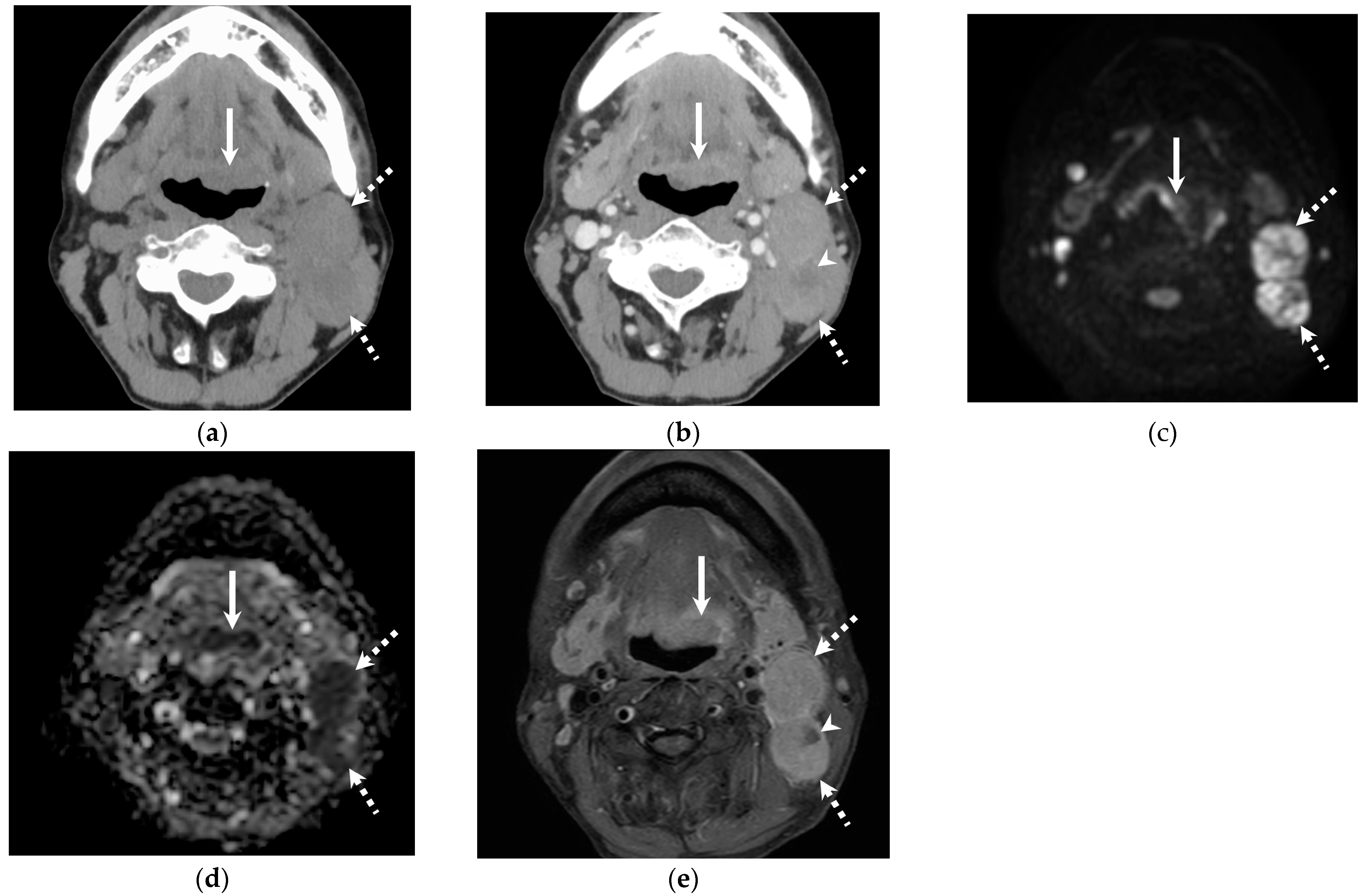
Table 1 shows patient characteristics. The recurrence rate of OPSCC in HPV-positive patients was significantly lower than in HPV-negative patients (15% vs. 50%; p < 0.01). Age was considerably higher in HPV-positive OPSCC patients with recurrence than in those without recurrence (73 years vs. 60 years, p < 0.05); however, there was no significant difference in the T (p = 0.52) and N stages (p > 0.99) based on the eighth edition of the staging system between patients with and without recurrence. Meanwhile, among HPV-negative OPSCC patients, patients with recurrence had higher T (p < 0.05) and N stages (p < 0.05) than those without recurrence (Figure 1 and Figure 2).

3.2. Quantitative and Qualitative Imaging Findings of the Primary Lesion

Table 2 summarizes the quantitative and qualitative imaging findings of the primary lesion. SIR on diffusion-weighted (1.66 vs. 1.32; p < 0.001) and contrast-enhanced T1-weighted images (2.05 vs. 1.82; p = 0.024) was significantly higher in HPV-positive OPSCC patients with recurrence than in those without recurrence; however, there was no significant difference in ADC value (p = 0.98) or attenuation on contrast-enhanced CT images (p = 0.88). In HPV-negative OPSCC patients, attenuation on contrast-enhanced CT images (42.1 HU vs. 49.4 HU; p < 0.01) was significantly lower in patients with recurrence than in those without recurrence; however, there was no significant difference in SIR on contrast-enhanced T1-weighted images (p = 0.91). There was no significant difference in qualitative imaging findings between the primary lesions.

3.3. Quantitative Imaging Findings of Cervical Lymph Node Metastasis

Table 3 shows the quantitative imaging findings of cervical lymph node metastasis. Lymph node metastasis was observed in 57 (84%) of the 68 patients with OPSCC, and 153 nodal metastases were confirmed. Attenuation on unenhanced CT images of the solid component within HPV-positive nodal metastases (44.1 HU vs. 47.1 HU; p = 0.010) was significantly lower in patients with recurrence in comparison to those without. In HPV-negative nodal metastases, attenuation on contrast-enhanced CT (79.4 HU vs. 87.9HU; p < 0.05) and SIR on contrast-enhanced T1-weighted images (1.79 vs. 2.41; p < 0.05) of the solid component were significantly lower in patients with recurrence than in those without. In HPV-negative nodal metastases, the ADC value was marginally lower in patients with recurrence than in those without (0.83 × 10⁻³ mm2/s vs. 1.13 × 10⁻³ mm2/s; p = 0.058), and SUVmax was marginally higher in patients with recurrence (7.86 vs. 4.31; p = 0.070) (Figure 1 and Figure 2).

3.4. Qualitative Imaging Findings of Cervical Lymph Node Metastasis

Table 4 shows the qualitative imaging findings of cervical lymph node metastasis. In HPV-positive nodal metastasis, there was no significant difference in findings between patients with and without recurrence. In HPV-negative nodal metastasis, the frequency of ENE on CT (4% vs. 44%; p < 0.05) and MRI (4% vs. 44%; p < 0.05) images was significantly lower in patients with recurrence than in those without. Regardless of HPV status, cystic degeneration of the lymph nodes was not associated with recurrence.
The κ values for the two observers revealed fair agreement regarding the margin of the primary lesion and marginal invasion (0.31–0.38); moderate agreement regarding necrosis, marginal invasion of the primary lesion, and ENE (0.48–0.50); and substantial agreement regarding cystic nodal metastasis and the unenhanced area of nodal metastasis (0.64–0.79).

4. Discussion

In this study, the recurrence rate was lower in HPV-positive OPSCC than in HPV-negative OPSCC. The T and N categories of HPV-negative OPSCCs were associated with recurrence, whereas those of HPV-positive OPSCCs did not differ between patients with and without recurrence. Regarding nodal metastases in HPV-negative OPSCC, attenuation on contrast-enhanced CT images and SIR on contrast-enhanced T1-weighted images were significantly lower in patients with recurrence than in those without. In HPV-positive and HPV-negative OPSCCs, cystic changes did not differ significantly between patients with and without recurrence.
In this study, the recurrence rate of HPV-positive OPSCC was lower than that of HPV-negative OPSCC. Many previous studies [15,16,17] have reported that HPV-positive OPSCC had a better prognosis when compared with HPV-negative OPSCC. Because of differing prognoses, the present eighth edition of the staging system establishes separate TNM classifications for HPV-positive and HPV-negative OPSCC.
This study observed no significant differences in TNM classification between HPV-positive OPSCC patients with and without recurrence. Meanwhile, HPV-negative OPSCC patients with and without recurrence showed significant differences in the T and N categories and the maximum diameter of the primary lesion. A previous study that assessed the prognosis of HPV-positive OPSCC using the seventh edition of the TNM classification system reported that TNM classification was not a prognostic factor [18]. Another study on OPSCC, which included 67 patients, 55 of whom were p16-positive, reported that the T category, according to the eighth edition of the TNM classification, was not a prognostic factor [19]. Chang et al. reported that lymph node metastasis in HPV-positive OPSCC patients had no significant effect on recurrence or mortality [20]. Conversely, several studies on HPV-negative OPSCC have reported that higher stages had a worse prognosis [20,21,22,23]. Although the prognosis of HPV-positive OPSCC patients may be unrelated to TNM classification, TNM classification is a critical prognostic factor for HPV-negative OPSCC patients.
The present study demonstrated that attenuation on contrast-enhanced CT images and SIR on contrast-enhanced T1-weighted images of nodal metastases were significantly lower in HPV-negative OPSCC patients with recurrence than in those without. Low attenuation and SIR on contrast-enhanced images may be caused by the histological heterogeneity of the keratinizing phenotype that is common in HPV-negative OPSCC [24]. Furthermore, ADC values for nodal metastases were lower in HPV-negative OPSCC patients with recurrence than in those without. Several studies analyzing OPSCC MRI results have reported that low ADC values were associated with HPV positivity and better prognosis [19,25]. However, among HPV-positive OPSCC patients, ADC values were not associated with overall survival [19]. Although no studies have reported a strong correlation between ADC values and prognosis in HPV-negative OPSCC patients, low ADC values are typically associated with high grade and poor prognosis. Low contrast enhancement and ADC values may predict the recurrence of HPV-negative OPSCC.
This study observed no significant difference in cystic change or necrosis between HPV-positive and HPV-negative patients with and without recurrence. Previous studies have reported that cystic nodal metastasis increases the risk of regional failure in OPSCC [26] and distant metastatic recurrence of nasopharyngeal carcinoma [27]. Another study concluded that cystic nodal metastasis indicated a low risk of recurrence compared with solid nodal metastasis in HPV-positive OPSCC [28]. Although cystic nodal metastasis helps in distinguishing between HPV-positive and HPV-negative OPSCC and suggests that HPV-positive OPSCC has a good prognosis, further study is required to determine the association between cystic nodal metastasis and prognosis.
In this study, ENE of nodal metastasis was associated with recurrence in HPV-negative OPSCC but not with recurrence in HPV-positive OPSCC. ENE was an independent predictor of survival in HPV-negative OPSCC patients [23]; thus, the eighth edition of the TNM classification system for OPSCC reflected ENE in only HPV-negative OPSCC. However, a recent meta-analysis reported that ENE in HPV-positive OPSCC patients is moderately associated with an increased risk of all-cause mortality and distant metastasis but not with locoregional recurrence [29]. The association between ENE and prognosis in HPV-positive OPSCC patients remains debatable.
This study has several limitations. First, the use of a single-center study resulted in a relatively small number of cases, especially among those with HPV-negative OPSCC and female patients. Gender distribution or unevenness in the number of HPV-positive and HPV-negative patients can affect the outcome. Second, CT and MRI images were obtained using various scanners. Finally, a survival analysis using the Kaplan–Meier curve was impossible because of the small number of cases.

5. Conclusions

In HPV-positive OPSCC, the T and N categories were not associated with recurrence, and no helpful imaging findings for predicting recurrence were found. However, T and N categories were associated with recurrence in HPV-negative OPSCC. In HPV-negative OPSCC, patients with recurrence had lower attenuation on contrast-enhanced CT images and SIR on contrast-enhanced T1-weighted images of nodal metastases than those without recurrence. Cystic lymph node metastasis was not associated with recurrence; however, ENE in HPV-negative OPSCC was associated with recurrence. Thus, prognostic factors differed considerably between HPV-positive and HPV-negative OPSCC.

Author Contributions

Conceptualization, M.K. and H.K.; methodology, T.S., M.K. and H.K.; software, T.S.; validation, T.S. and M.K.; formal analysis, H.K. and Y.N.; investigation, T.S. and M.K.; resources, M.K., H.S. and T.O.; data curation, H.S. and T.O.; writing—original draft preparation, T.S. and M.K.; writing—review and editing, H.K., T.A., Y.N. and F.H.; visualization, M.K.; supervision, M.M.; project administration, M.M. All authors have read and agreed to the published version of the manuscript.

Funding

This research received no external funding.

Institutional Review Board Statement

This study was conducted in accordance with the Declaration of Helsinki and approved by the Institutional Review Board of Gifu University Hospital (2020-101 and date of approval, 7 August 2020).

Informed Consent Statement

Patient consent was waived due to the retrospective nature of this study.

Data Availability Statement

The data presented in this study may be available on request from the corresponding author. The data are not publicly available due to data patient privacy concerns.

Conflicts of Interest

The authors declare no conflicts of interest.

References

  1. Lechner, M.; Liu, J.; Masterson, L.; Fenton, T.R. HPV-associated oropharyngeal cancer: Epidemiology, molecular biology and clinical management. Nat. Rev. Clin. Oncol. 2022, 19, 306–327. [Google Scholar] [CrossRef] [PubMed]
  2. Kobayashi, K.; Hisamatsu, K.; Suzui, N.; Hara, A.; Tomita, H.; Miyazaki, T. A Review of HPV-Related Head and Neck Cancer. J. Clin. Med. 2018, 7, 241. [Google Scholar] [CrossRef]
  3. Consalvo, F.; De Simone, M.; Scarpa, A.; Acerra, A.; Salzano, F.A.; Fineschi, V.; Santurro, A. Challenges and Complications in the Management of Advanced Oropharyngeal Carcinoma: Role of Post-Mortem Diagnosis and Future Perspectives. J. Clin. Med. 2024, 13, 5198. [Google Scholar] [CrossRef]
  4. D’Souza, G.; Kreimer, A.R.; Viscidi, R.; Pawlita, M.; Fakhry, C.; Koch, W.M.; Westra, W.H.; Gillison, M.L. Case-control study of human papillomavirus and oropharyngeal cancer. N. Engl. J. Med. 2007, 356, 1944–1956. [Google Scholar] [CrossRef]
  5. Lydiatt, W.M.; Patel, S.G.; O’Sullivan, B.; Brandwein, M.S.; Ridge, J.A.; Migliacci, J.C.; Loomis, A.M.; Shah, J.P. Head and Neck cancers-major changes in the American Joint Committee on cancer eighth edition cancer staging manual. CA Cancer J. Clin. 2017, 67, 122–137. [Google Scholar] [CrossRef]
  6. Cantrell, S.C.; Peck, B.W.; Li, G.; Wei, Q.; Sturgis, E.M.; Ginsberg, L.E. Differences in imaging characteristics of HPV-positive and HPV-Negative oropharyngeal cancers: A blinded matched-pair analysis. AJNR Am. J. Neuroradiol. 2013, 34, 2005–2009. [Google Scholar] [CrossRef] [PubMed]
  7. Bhattasali, O.; Thompson, L.D.R.; Schumacher, A.J.; Iganej, S. Radiographic nodal prognostic factors in stage I HPV-related oropharyngeal squamous cell carcinoma. Head Neck 2019, 41, 398–402. [Google Scholar] [CrossRef] [PubMed]
  8. Morey, T.; Hodge, J.C.; Stern, C.; Krishnan, S.; Foreman, A. Correlation between radiologic and pathologic extranodal extension in HPV-associated oropharyngeal cancer: Systematic review. Head Neck 2022, 44, 2875–2885. [Google Scholar] [CrossRef] [PubMed]
  9. Billfalk-Kelly, A.; Yu, E.; Su, J.; O’Sullivan, B.; Waldron, J.; Ringash, J.; Bartlett, E.; Perez-Ordonez, B.; Weinreb, I.; Bayley, A.; et al. Radiologic Extranodal Extension Portends Worse Outcome in cN+ TNM-8 Stage I Human Papillomavirus-Mediated Oropharyngeal Cancer. Int. J. Radiat. Oncol. Biol. Phys. 2019, 104, 1017–1027. [Google Scholar] [CrossRef] [PubMed]
  10. Huang, S.H.; O’Sullivan, B.; Su, J.; Bartlett, E.; Kim, J.; Waldron, J.N.; Ringash, J.; de Almeida, J.R.; Bratman, S.; Hansen, A.; et al. Prognostic importance of radiologic extranodal extension in HPV-positive oropharyngeal carcinoma and its potential role in refining TNM-8 cN-classification. Radiother. Oncol. 2020, 144, 13–22. [Google Scholar] [CrossRef]
  11. Choi, Y.; Nam, Y.; Jang, J.; Shin, N.Y.; Ahn, K.J.; Kim, B.S.; Lee, Y.S.; Kim, M.S. Prediction of Human Papillomavirus Status and Overall Survival in Patients with Untreated Oropharyngeal Squamous Cell Carcinoma: Development and Validation of CT-Based Radiomics. AJNR Am. J. Neuroradiol. 2020, 41, 1897–1904. [Google Scholar] [CrossRef]
  12. Kawaguchi, M.; Kato, H.; Tomita, H.; Hara, A.; Suzui, N.; Miyazaki, T.; Matsuo, M. Comparison of Imaging Findings between Human Papillomavirus-positive and -Negative Squamous Cell Carcinomas of the Maxillary Sinus. J. Clin. Imaging Sci. 2020, 10, 59. [Google Scholar] [CrossRef] [PubMed]
  13. Hiyama, T.; Kuno, H.; Nagaki, T.; Sekiya, K.; Oda, S.; Fujii, S.; Hayashi, R.; Kobayashi, T. Extra-nodal extension in head and neck cancer: How radiologists can help staging and treatment planning. Jpn. J. Radiol. 2020, 38, 489–506. [Google Scholar] [CrossRef] [PubMed]
  14. Kanda, Y. Investigation of the freely available easy-to-use software ’EZR’ for medical statistics. Bone Marrow Transplant. 2013, 48, 452–458. [Google Scholar] [CrossRef] [PubMed]
  15. Lassen, P.; Eriksen, J.G.; Hamilton-Dutoit, S.; Tramm, T.; Alsner, J.; Overgaard, J. Effect of HPV-associated p16INK4A expression on response to radiotherapy and survival in squamous cell carcinoma of the head and neck. J. Clin. Oncol. 2009, 27, 1992–1998. [Google Scholar] [CrossRef] [PubMed]
  16. Maxwell, J.H.; Kumar, B.; Feng, F.Y.; Worden, F.P.; Lee, J.S.; Eisbruch, A.; Wolf, G.T.; Prince, M.E.; Moyer, J.S.; Teknos, T.N.; et al. Tobacco use in human papillomavirus-positive advanced oropharynx cancer patients related to increased risk of distant metastases and tumor recurrence. Clin. Cancer Res. 2010, 16, 1226–1235. [Google Scholar] [CrossRef]
  17. Nomura, F.; Sugimoto, T.; Kitagaki, K.; Ito, T.; Kawachi, H.; Eishi, Y.; Watanabe, K.; Igaue, M.; Shimizu, N.; Tomita, M.; et al. Clinical characteristics of Japanese oropharyngeal squamous cell carcinoma positive for human papillomavirus infection. Acta Otolaryngol. 2014, 134, 1265–1274. [Google Scholar] [CrossRef] [PubMed]
  18. Mizumachi, T.; Kano, S.; Sakashita, T.; Hatakeyama, H.; Suzuki, S.; Homma, A.; Oridate, N.; Fukuda, S. Improved survival of Japanese patients with human papillomavirus-positive oropharyngeal squamous cell carcinoma. Int. J. Clin. Oncol. 2013, 18, 824–828. [Google Scholar] [CrossRef]
  19. Sistonen, H.J.; Aro, K.; Atula, T.; Jouhi, L.; Lindén, R.; Tapiovaara, L.; Loimu, V.; Markkola, A. Prognostic Value of Apparent Diffusion Coefficient in Oropharyngeal Carcinoma. Clin. Neuroradiol. 2021, 31, 1037–1048. [Google Scholar] [CrossRef] [PubMed]
  20. Chang, C.M.; Chen, M.M.; Bellile, E.L.; Rozek, L.S.; Carey, T.E.; Spector, M.E.; Wolf, G.T.; Taylor, J.M.; Chinn, S.B. Impact of Nodal Metastases in HPV-Negative Oropharyngeal Cancer. Cancer Epidemiol. Biomark. Prev. 2022, 31, 1554–1563. [Google Scholar] [CrossRef] [PubMed]
  21. Yin, L.X.; D’Souza, G.; Westra, W.H.; Wang, S.J.; van Zante, A.; Zhang, Y.; Rettig, E.M.; Ryan, W.R.; Ha, P.K.; Wentz, A.; et al. Prognostic factors for human papillomavirus-positive and negative oropharyngeal carcinomas. Laryngoscope 2018, 128, E287–E295. [Google Scholar] [CrossRef]
  22. Amin, M.B.; Greene, F.L.; Edge, S.B.; Compton, C.C.; Gershenwald, J.E.; Brookland, R.K.; Meyer, L.; Gress, D.M.; Byrd, D.R.; Winchester, D.P. The Eighth Edition AJCC Cancer Staging Manual: Continuing to build a bridge from a population-based to a more “personalized” approach to cancer staging. CA Cancer J. Clin. 2017, 67, 93–99. [Google Scholar] [CrossRef] [PubMed]
  23. Kumar, B.; Cipolla, M.J.; Old, M.O.; Brown, N.V.; Kang, S.Y.; Dziegielewski, P.T.; Durmus, K.; Ozer, E.; Agrawal, A.; Carrau, R.L.; et al. Surgical management of oropharyngeal squamous cell carcinoma: Survival and functional outcomes. Head Neck 2016, 38 (Suppl. 1), E1794–E1802. [Google Scholar] [CrossRef] [PubMed]
  24. Cai, C.; Chernock, R.D.; Pittman, M.E.; El-Mofty, S.K.; Thorstad, W.L.; Lewis, J.S., Jr. Keratinizing-type squamous cell carcinoma of the oropharynx: p16 overexpression is associated with positive high-risk HPV status and improved survival. Am. J. Surg. Pathol. 2014, 38, 809–815. [Google Scholar] [CrossRef] [PubMed]
  25. Ravanelli, M.; Grammatica, A.; Maddalo, M.; Ramanzin, M.; Agazzi, G.M.; Tononcelli, E.; Battocchio, S.; Bossi, P.; Vezzoli, M.; Maroldi, R.; et al. Pretreatment DWI with Histogram Analysis of the ADC in Predicting the Outcome of Advanced Oropharyngeal Cancer with Known Human Papillomavirus Status Treated with Chemoradiation. AJNR Am. J. Neuroradiol. 2020, 41, 1473–1479. [Google Scholar] [CrossRef]
  26. Huang, Y.H.; Yeh, C.H.; Cheng, N.M.; Lin, C.Y.; Wang, H.M.; Ko, S.F.; Toh, C.H.; Yen, T.C.; Liao, C.T.; Ng, S.H. Cystic nodal metastasis in patients with oropharyngeal squamous cell carcinoma receiving chemoradiotherapy: Relationship with human papillomavirus status and failure patterns. PLoS ONE 2017, 12, e0180779. [Google Scholar] [CrossRef] [PubMed]
  27. Ting, Y.; Chee, J.; Charn, T.C.; Loh, K.S.; Choong, C.C.; Ting, E.; Lim, C.M. Prognostic significance of cystic lymph nodal metastasis in nasopharyngeal carcinoma. Head Neck 2017, 39, 1832–1839. [Google Scholar] [CrossRef]
  28. Rath, T.J.; Narayanan, S.; Hughes, M.A.; Ferris, R.L.; Chiosea, S.I.; Branstetter, B.F.t. Solid Lymph Nodes as an Imaging Biomarker for Risk Stratification in Human Papillomavirus-Related Oropharyngeal Squamous Cell Carcinoma. AJNR Am. J. Neuroradiol. 2017, 38, 1405–1410. [Google Scholar] [CrossRef]
  29. Benchetrit, L.; Torabi, S.J.; Givi, B.; Haughey, B.; Judson, B.L. Prognostic Significance of Extranodal Extension in HPV-Mediated Oropharyngeal Carcinoma: A Systematic Review and Meta-analysis. Otolaryngol. Head Neck Surg. 2021, 164, 720–732. [Google Scholar] [CrossRef] [PubMed]
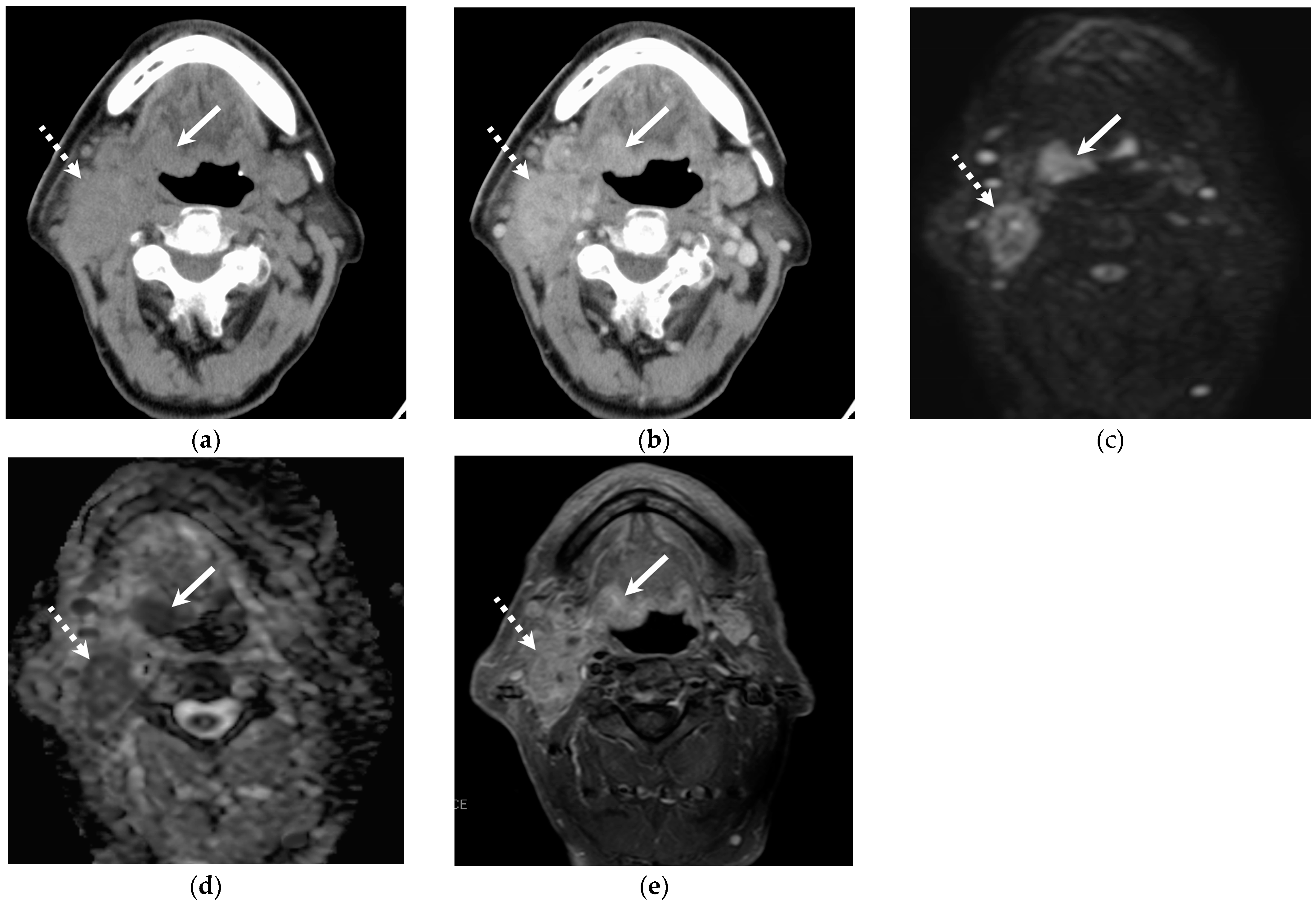
Figure 1. Representative images of an 86-year-old male with HPV-positive OPSCC in the base of the tongue. Local recurrence and pulmonary metastasis occurred. (a) Unenhanced CT shows soft tissue mass in the right base of the tongue (primary lesion, arrow) and right upper jugular lymph node metastasis (dotted arrow). CT attenuation of the nodal metastasis shows +42 HU. (b) Contrast-enhanced CT shows enhanced primary lesion (arrow) and nodal metastasis (dotted arrow). (c,d) Diffusion-weighted imaging and ADC map show high signal intensity and low ADC value in the primary lesion (arrow) and nodal metastasis (dotted arrow). (e) Fat-suppressed contrast-enhanced T1-weighted image shows primary lesion (arrow) and nodal metastasis (dotted arrow). The SIR of solid component is 2.15.
Jcm 14 01027 g001
Figure 2. Representative images of a 58-year-old male with HPV-negative OPSCC in the base of the tongue. Local recurrence and pulmonary metastasis occurred. (a) Unenhanced CT shows mass in the base of the tongue (primary lesion, arrow) and multiple left upper jugular lymph node metastases (dotted arrow). CT attenuation of the primary lesion shows +44 HU. (b) Contrast-enhanced CT shows primary lesion and nodal metastasis. CT attenuation of nodal metastasis demonstrated + 75 HU. (c,d) Diffusion-weighted imaging and ADC map show high signal intensity and low ADC value. ADC value of the nodal metastasis was 0.715 × 10⁻³mm2/s. (e) Fat-suppressed contrast-enhanced T1-weighted image shows primary lesion (arrow) and multiple nodal metastases (dotted arrow) with necrosis (arrowhead). The SIR of solid component of the lymph node is 1.80.
Jcm 14 01027 g002
Table 1. Clinical characteristics of patients with OPSCC.
All PatientsHPV-Positive OPSCCHPV-Negative OPSCC
RecurrencepRecurrencepRecurrencep
Yes
(n = 17)
No
(n = 51)
Yes
(n = 7)
No
(n = 41)
Yes
(n = 10)
No
(n = 10)
Age (years)
71
[67–75]
66
[52–71]
0.01373
[70–75]
60
[51–68]
0.04569
[67–74]
70
[52–76]
0.73
Male7 (41%)38 (75%)>0.995 (71%)30 (73%)>0.998 (80%)8 (80%)>0.99
Smoking13 (76%)34 (67%)0.555 (71%)27 (66%)>0.998 (80%)7 (70%)>0.99
Surgery8 (47%)37 (73%)0.084 (57%)12 (29%)0.205 (50%)8 (80%)0.35
Chemotherapy14 (82%)28 (55%)0.0495 (71%)23 (56%)0.689 (90%)5 (50%)0.14
Radiation15 (88%)35 (69%)0.206 (86%)29 (70%)0.669 (90%)6 (60%)0.30
T stage 0.02 0.52 0.04
T11 (6%)9 (18%) 1 (14%)6 (15%) 0 (0%)3 (30%)
T26 (35%)31 (61%) 4 (57%)26 (63%) 2 (20%)5 (50%)
T34 (24%)2 (4%) 1 (14%)1 (2%) 3 (30%)1 (10%)
T46 (35%)9 (18%) 1 (14%)8 (20%) 5 (50%)1 (10%)
N stage 0.35 >0.99 0.04
N01 (6%)10 (20%) 1 (14%)6 (15%) 0 (0%)4 (40%)
N18 (47%)27 (53%) 5 (71%)27 (66%) 3 (30%)0 (0%)
N27 (41%)12 (24%) 1 (14%)8 (20%) 6 (60%)4 (40%)
N31 (6%)2 (4%) 0 (0%)0 (0%) 1 (10%)2 (20%)
M stage NA NA NA
M10 (0%)0 (0%) 0 (0%)0 (0%) 0 (0%)0 (0%)
Note.: OPSCC = oropharyngeal squamous cell carcinoma, HPV = human papilloma virus. Fisher’s exact test and Mann–Whitney U test were used. Qualitative data are numbers of patients with percentages in parentheses. Quantitative data are expressed as medians with interquartile range in square brackets. TNM classification was based on the eighth edition of staging system.
Table 2. Quantitative and qualitative imaging findings of primary lesion.
Quantitative Imaging Findings of Primary Lesion
HPV-Positive OPSCCHPV-Negative OPSCC
RecurrencepRecurrencep
Yes (n = 7)No (n = 41)Yes (n = 10)No (n = 10)
Maximum diameter (mm)21.9 [18.2–26.6]28.4 [19.7–34.7]0.1832.7 [26.0–45.4]20.8 [19.3–33.9]0.04
CT
 Attenuation on UECT (HU)50.3 [41.6–55.7]50.4 [43.4–55.4]0.8142.1 [38.7–44.6]49.4 [46.6–50.6]<0.01
 Attenuation on CECT (HU)85.7 [82.3–93.7]85.0 [78.4–97.3]0.8890.2 [76.1–95.2]90.7 [83.8–97.8]0.87
MRI
 SIR on T1WI1.04 [0.98–1.06]0.98 [0.93–1.06]0.420.99 [0.92–1.03]0.94 [0.92–0.99]0.63
 SIR on T2WI1.12 [1.09–1.14]1.06 [1.00–1.15]0.371.02 [0.87–1.09]1.16 [1.06–1.22]0.089
 SIR on DWI1.66 [1.57–1.79]1.32 [1.08–1.46]<0.011.15 [0.99–1.36]1.23 [0.95–1.55]0.58
 SIR on CET1WI2.05 [1.92–2.21]1.82 [1.63–1.99]0.022.04 [1.76–2.19]1.97 [1.91–2.07]0.91
 ADC value (×10−3 mm2/s)0.82 [0.69–1.05]0.80 [0.73–0.89]0.980.91 [0.76–1.06]1.02 [0.91–1.08]0.51
18F-FDG-PET/CT
 SUVmax15.3 [10.6–18.3]12.61 [8.9–15.9]0.5816.5 [14.5–20.5]18.5 [12.0–20.7]0.96
Qualitative imaging findings of primary lesion
CT
 Well-defined margin5 (71%)23 (56%)0.685 (50%)4 (40%)>0.99
 Necrosis1 (14%)9 (22%)>0.992 (20%)1 (10%)>0.99
MRI
 Well-defined margin7 (100%)33 (80%)0.584 (40%)7 (70%)0.37
 Necrosis2 (29%)8 (20%)0.633 (30%)2 (20%)>0.99
 Marginal invasion1 (14%)6 (15%)>0.995 (50%)3 (30%)0.65
T1WIHyperintensity
Isointensity
Hypointensity
1 (14%)
2 (29%)
4 (57%)
2 (5%)
19 (46%)
20 (49%)
0.411 (10%)
3 (30%)
6 (60%)
0 (0%)
5 (50%)
5 (50%)
0.65
T2WIHyperintensity
Isointensity
Hypointensity
5 (71%)
2 (29%)
0 (0%)
32 (78%)
8 (20%)
1 (2%)
0.687 (70%)
2 (20%)
1 (10%)
9 (90%)
0 (0%)
1 (10%)
0.72
CET1WIHyperintensity7 (100%)41 (100%)NA10 (100%)10 (100%)NA
DWIHyperintensity
Isointensity
Hypointensity
6 (86%)
1 (14%)
0 (0%)
33 (80%)
5 (12%)
3 (7%)
>0.9910 (100%)
0 (0%)
0 (0%)
8 (80%)
1 (10%)
1 (10%)
0.47
Note: HPV = human papilloma virus, OPSCC = oropharyngeal squamous cell carcinoma, UECT = unenhanced CT, CECT = contrast-enhanced CT, SIR = signal intensity ratio, T1WI = T1-weighted images, T2WI = T2-weighted images, DWI = diffusion-weighted images, CET1WI = contrast-enhanced T1-weighted images, ADC = apparent diffusion coefficient, FDG = fluorodeoxyglucose, SUV = standardized uptake value. Fisher’s exact test and Mann–Whitney U test were used. Qualitative data are numbers of patients with percentages in parentheses. Quantitative data are expressed as medians with interquartile ranges in square brackets.
Table 3. Quantitative imaging findings of cervical lymph node metastasis.
Nodal Metastasis From
HPV-Positive OPSCC
Nodal Metastasis From
HPV-Negative OPSCC
RecurrencepRecurrencep
Yes (n = 24)No (n = 96)Yes (n = 24)No (n = 6)
Maximum diameter (mm)17.8 [14.0–22.5]16.8 [13.5–22.0]0.1518.5 [15.3–26.5]19.3 [16.5–22.3]0.83
CT (HU)
 UECT44.1 [38.6–47.9]47.1 [44.4–51.6]0.0145.6 [42.8–48.8]41.2 [34.3–47.1]0.27
 CECT82.2 [71.5–96.7]86.2 [75.4–98.4]0.5379.4 [68.2–84.3]87.9 [86.8–95.6]0.03
 Necrosis of NCCT38.3 [31.4–41.0]33.6 [27.2–44.2]0.4531.4 [25.8–40.2]28.3 [27.5–34.0]0.82
Necrosis of CECT49.0 [36.8–66.5]
(n = 13)
42.3 [33.2–55.5]
(n = 62)
0.2839.3 [29.8–47.0]
(n = 17)
33.9 [31.4–56.6]
(n = 5)
0.65
MRI
SIR on T1WI1.00 [0.93–1.04]0.97 [0.90–1.02]0.140.94 [0.89–1.02]0.89 [0.87–0.93]0.35
SIR on T2WI1.09 [0.96–1.36]1.12 [0.99–1.32]0.691.23 [0.95–1.37]1.31 [1.15–1.47]0.16
SIR on DWI1.43 [1.12–1.62]1.47 [1.19–1.83]0.671.49 [1.18–1.72]1.50 [1.45–2.11]0.44
SIR on CE-T1WI1.90 [1.74–2.04]1.79 [1.57–2.03]0.171.79 [1.64–2.08]2.41 [1.97–2.38]0.04
ADC value (×10−3 mm2/s)0.87 [0.68–0.99]0.86 [0.76–1.05]0.340.83 [0.72–1.02]1.13 [0.99–1.37]0.06
18F-FDG-PET/CT
SUVmax9.52
[5.91–12.7]
7.78
[4.51–11.0]
0.287.86
[4.40–15.5]
4.31
[3.78–4.78]
0.07
Note: HPV = human papilloma virus, OPSCC = oropharyngeal squamous cell carcinoma, UECT = unenhanced CT, CECT = contrast-enhanced CT, SIR = signal intensity ratio, T1WI = T1-weighted images, T2WI = T2-weighted images, DWI = diffusion-weighted images, CE-T1WI = contrast-enhanced T1-weighted images, ADC = apparent diffusion coefficient, FDG = fluorodeoxyglucose, SUV = standardized uptake value. Fisher’s exact test and Mann–Whitney U test were used. Quantitative data are expressed as medians with interquartile ranges in square brackets.
Table 4. Qualitative imaging findings of cervical lymph node metastasis.
Nodal Metastasis From HPV-Positive
OPSCC
Nodal Metastasis From HPV-Negative
OPSCC
RecurrencepRecurrencep
Yes (n = 24)No (n = 96)Yes (n = 24)No (n = 9)
CT
 ENE2 (8%)8 (8%)>0.991 (4%)4 (44%)0.01
Unenhanced area
- pure cystic
- pure cystic + mural nodule
- necrosis
12 (50%)
4 (17%)
1 (4%)
7 (29%)
62 (65%)
7 (7%)
14 (15%)
41 (43%)
0.24
 
0.15
 
17 (71%)
3 (13%)
4 (17%)
10 (42%)
6 (67%)
2 (22%)
0 (0%)
4 (44%)
>0.99
 
0.55
 
MRI
ENE2 (8%)8 (8%)>0.991 (4%)4 (44%)0.01
Unenhanced area
- pure cystic
- pure cystic + mural nodule
- necrosis
18 (75%)
5 (21%)
3 (13%)
10 (42%)
64 (67%)
5 (5%)
14 (15%)
46 (48%)
0.48
 
0.08
 
18 (75%)
4 (17%)
3 (13%)
10 (42%)
8 (89%)
1 (11%)
1 (11%)
6 (67%)
0.64
 
0.72
 
Solid component
T1WIHyperintensity
Isointensity
Hypointensity
0 (0%)
15 (63%)
9 (37%)
0 (0%)
40 (42%)
56 (58%)
0.113 (13%)
12 (50%)
9 (37%)
1 (11%)
2 (22%)
6 (67%)
0.35
T2WIHyperintensity
Isointensity
Hypointensity
12 (50%)
11 (46%)
1 (4%)
59 (61%)
30 (31%)
7 (7%)
0.5016 (67%)
7 (29%)
1 (4%)
5 (56%)
3 (33%)
1 (11%)
0.70
DWIHyperintensity
Isointensity
Hypointensity
20 (83%)
4 (17%)
0 (0%)
85 (89%)
9 (9%)
2 (2%)
0.5521 (88%)
2 (8%)
1 (4%)
7 (78%)
2 (22%)
0 (0%)
0.68
CE-T1WIHyperintensity
Isointensity
Hypointensity
24 (100%)
0 (0%)
0 (0%)
95 (99%)
1 (1%)
0 (0%)
>0.9924 (100%)
0 (0%)
0 (0%)
9 (100%)
0 (0%)
0 (0%)
NA
Note: HPV = human papilloma virus, OPSCC = oropharyngeal squamous cell carcinoma, ENE = extranodal extension, T1WI = T1-weighted images, T2WI = T2-weighted images, DWI = diffusion-weighted images, CE-T1WI = contrast-enhanced T1-weighted images. Fisher’s exact test and Mann–Whitney U test were used. Qualitative data are numbers of patients with percentages in parentheses.
Disclaimer/Publisher’s Note: The statements, opinions and data contained in all publications are solely those of the individual author(s) and contributor(s) and not of MDPI and/or the editor(s). MDPI and/or the editor(s) disclaim responsibility for any injury to people or property resulting from any ideas, methods, instructions or products referred to in the content.