Article

Accuracy of Robotic-Guided Pedicular Screw Insertion in Thoracolumbar Spinal Surgery

1
Department of Orthopaedics, Hospital Clínico San Carlos, 28040 Madrid, Spain
2
Department of Neurosurgery, Hospital Clínico San Carlos, 28040 Madrid, Spain
*
Author to whom correspondence should be addressed.
J. Clin. Med. 2025, 14(23), 8425; https://doi.org/10.3390/jcm14238425 (registering DOI)
Submission received: 23 October 2025 / Revised: 15 November 2025 / Accepted: 24 November 2025 / Published: 27 November 2025
(This article belongs to the Special Issue Advances in Spine Surgery: Current Innovations and Future Directions)

Abstract

Background/Objectives: Screw placement accuracy with robotic guidance shows better results than freehand techniques, yet there is scant data for region-specific outcomes, fusion rates, and complication profiles in different populations. The purpose of this study is to assess and describe screw-rod accuracy, fusion rates, and complications of robot-assisted pedicle screw placement in a Spanish cohort. Methods: Robotic-guided technique for thoracolumbar fusion in 115 patients (July 2020–January 2025) using ExcelsiusGPS® platform (Globus Medical Inc., Audubon, PA, USA). Inclusion criteria: adults (≥18 years) with postoperative CT to assess accuracy. Primary outcomes were screw accuracy (assessed on the Gertzbein–Robbins scale), fusion rates, and complications (infection, osteolysis, and subsidence). Jamovi 2.6 was employed for statistical analysis. Results: Of a total of 726 screws implanted, 590 screws (95 patients) were studied: 584 (99%) Grade A, 5 (0.8%) Grade B, and 1 (0.2%) Grade C. Fusion was achieved in 85 out of 95 patients (89.5%). Complications included superficial infections (3/115 patients, 2.6%) and asymptomatic osteolysis (8/95 patients, 8.4%, mostly at S1). There was no screw subsidence or removal. Conclusions: Robotic-guided pedicle screw placement demonstrated exceptional accuracy (99% Grade A), high fusion success (89.5%), and minimal complications in this Spanish cohort, supporting its clinical utility for spinal instrumentation.

1. Introduction

Spinal disorders that require surgery are becoming more widely acknowledged as a global health concern, mainly due to population aging and the increase in degenerative illnesses [1]. Over the past 15 years, spinal procedures have increased by 2.4 times [2]. Minimally invasive (MIS) techniques have been included in spinal surgery in many ways, including percutaneous pedicle screws fixation. Since screws are the main anchors in posterior spinal fusion procedures, it is essential to insert them precisely. Despite the relatively few clinical adverse events linked to malposition, misplaced screws can lead to dural tears [3], neurological/neurovascular injuries [4], sub-optimal biomechanics [5], and other visceral involvement [6]. Additionally, it is believed that up to 15.7% of cases with pedicle malposition go unreported [3]. In the meantime, the prevalence of pedicle screw problems varies greatly, from 1% to 54%, emphasizing the significance of anatomical features and patient-specific factors, including osteoporosis and deformities, in achieving successful screw insertion [7]. MIS modalities mainly include computed tomography navigation (CTnav), fluoroscopic guidance, and robot-guided surgery. Multiple systematic reviews have reported substantially growing evidence in all three fields [8,9]. While fluoroscopy alone has been reported to have a 91.3% accuracy [8], it has some drawbacks, like the obvious radiation exposure risk and ease-of-use limitations [10], as well as the level of surgeons’ experience, which introduces a degree of unpredictability. On the other hand, both CTnav and robotic-guided surgery have been reported to have higher predictability irrespective of the surgeons’ level of experience (7.0% [11] and 5.1% [12] vs. 15.7% [3], respectively) [3], and robotic-guided surgery is still reported to have higher accuracy compared to fluoroscopic guidance [11]. Furthermore, several recent meta-analyses and reviews have confirmed that robotic-assisted techniques achieve a far greater percentage of “perfect” (Grade A) and “clinically acceptable” (Grade A + B) pedicle screw insertions than freehand or fluoroscopic-guided methods. This advantage in radiographic accuracy is now well-established [13]. Less postoperative pain, faster recovery times, and less muscle damage have made robotic guidance more widely used [14]; however, this selection’s main drivers are accuracy, safety, and cost. Despite these advantages, significant barriers and confounding variables remain. The substantial capital investment and per-procedure costs are juxtaposed against an emerging body of literature arguing for its long-term cost-effectiveness by reducing revision rates and hospital stays [15,16]. Furthermore, the steep and highly variable learning curve, which one recent meta-analysis suggests may require 60 cases for many surgeons to overcome, acts as a significant clinical confounder [17]. Similarly, patient-specific factors, particularly poor bone quality, present a crucial challenge; a 2024 systematic review identified osteoporosis as a significant risk factor for mechanical complications like screw loosening and pseudarthrosis, which can mask the benefits of precise, robot-guided instrumentation [18]. These factors, along with case-specific challenges like high BMI or instrumentation in the technically demanding thoracic spine, complicate the interpretation of clinical outcome data [19].
These difficulties have created a crucial research gap. Numerous meta-analyses have demonstrated radiographic accuracy of robotic guiding, but there is still much disagreement about whether this translates into clearly better clinical outcomes. For example, recent systematic reviews and large-scale studies have found that patient-reported outcomes, complication rates, and revision rates do not significantly improve because of this precision [20]. Other recent meta-analyses, on the other hand, directly oppose this, stating that robotic aid does lead to decreased rates of surgical revision and postoperative complications [21,22]. This “accuracy-clinical outcome paradox” implies that the advantages of robotics might be strongly influenced by technology, surgical procedure, patient group, and handling of variables such as bone health and learning curve.
To explain this disparity, the field urgently needs high-quality and region-specific data. This study used robotic guidance to evaluate pedicle screw insertion accuracy, fusion rates, and complications in the Spanish population.

2. Materials and Methods

2.1. Study Design and Ethics

This is a consecutive, multicenter, observational, single-arm retrospective analysis study. During July 2020 to January 2025, adult patients who had robotically assisted pedicle screw implantation were included in this study. Due to its retrospective nature, the study was conducted in accordance with institutional ethical standards and applicable regulations.
All the data was anonymized and managed in compliance with the institutional data protection policies.

2.2. Patient Selection

Inclusion criteria were as follows:
  • Age ≥ 18 years.
  • Undergoing spinal fusion surgery involving thoracic, lumbar, or sacral pedicle screw placement.
  • Use of robotic guidance with the ExcelsiusGPS platform (Globus Medical Inc., Audubon, PA, USA) during screw placement.
  • Availability of postoperative CT imaging for accurate assessment.
Exclusion Criteria were as follows:
  • Patients younger than 18 years.
  • Procedures not involving pedicle screw instrumentation.
  • Cases in which robotic guidance was not utilized or was converted to freehand or navigation-assisted techniques intraoperatively.
  • Lack of postoperative CT imaging resulted in exclusion from the screw accuracy analysis only, but such patients were retained for all other clinical and radiological outcome analyses.
  • Incomplete or missing clinical or radiological data relevant to the analysis.
  • Revision surgeries.
Data collected included age, sex, clinical diagnosis, and presence of deformity, operative data including the treated anatomical levels, invasiveness, type of implants, preoperative/postoperative CT scans, fusion rates, and complications. Complications included infection, intervertebral cage subsidence, and osteolysis.

2.3. Robotic Workflows and Surgical Technique

The Excelsius GPS (Globus Medical Inc., Audubon, PA, USA) platform was used for all surgeries. Depending on the surgical needs, two workflows were used.
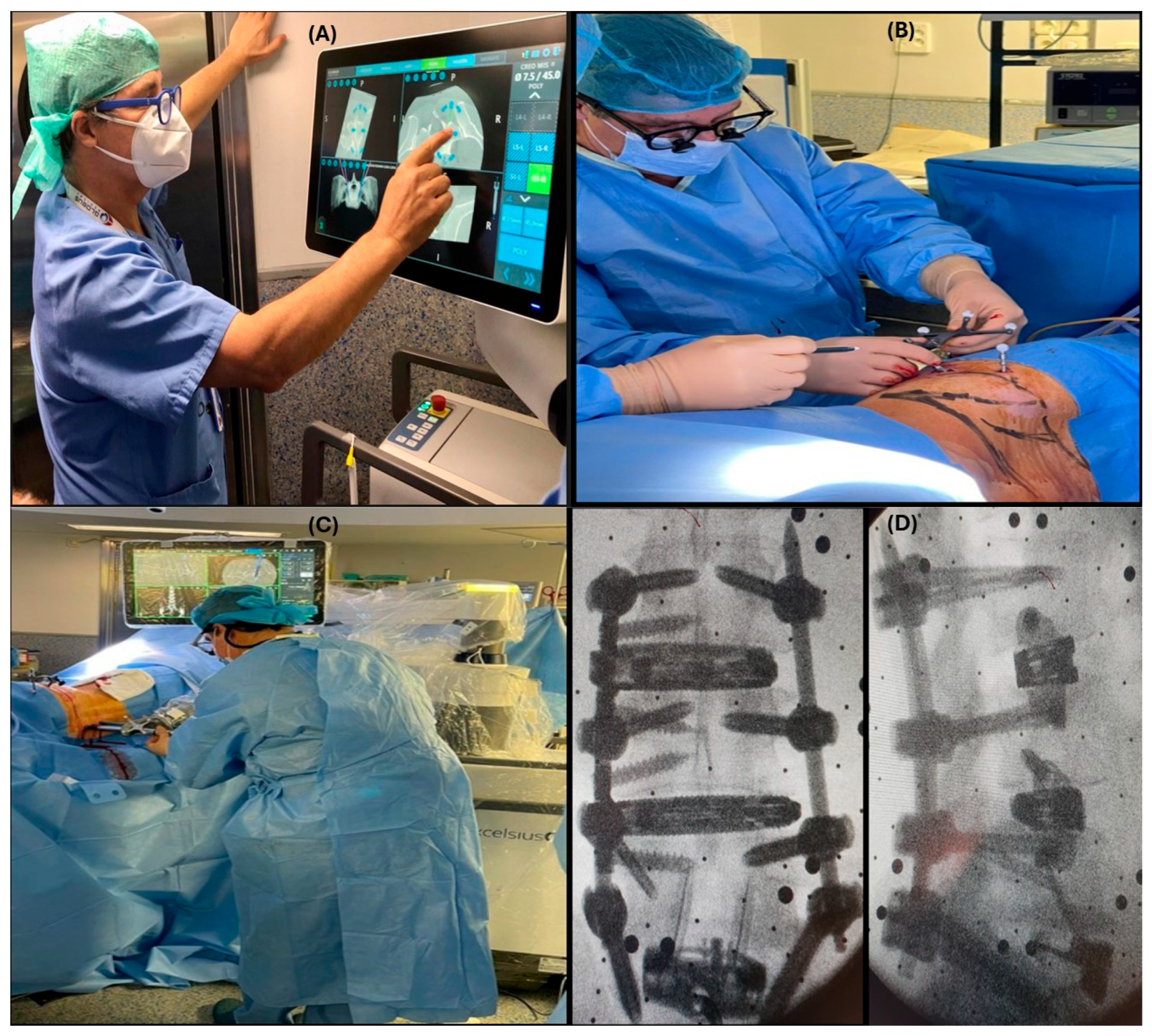
Preoperative workflow: A preoperative CT (Figure 1A) of the affected area is loaded by robotic computers. At this point, the surgeon plans the position of the implants and cages as well as trajectories and alignment. Once the patient is positioned on the operating table (prone or lateral) and after draping the surgical field, DRB (Dynamic Reference Base) and SM (Surveillance Marker) were positioned in both iliac crests (Figure 1B,C). A 2D X-ray is taken using a conventional C-arm. These images are merged with the preoperative ones (Figure 1D). As a result of this procedure, a virtual 3D image is available to start surgery.
Intraoperative workflow: In those cases, in which it is necessary to do any bone work that eventually changes anatomy, or when we are operating on specific anatomical areas such as the upper thoracic or cervical spine, it is mandatory to acquire intraoperative 3D images. In this study and all cases, this was performed using the Ziehmn Vision RFD 3D system (Ziehm Imaging, Nuremberg, Germany). After positioning, the DRB and SM were placed on the left iliac crest. An ICT is placed over the area to be intervened, and a 3D scan is performed. The surgeon plans the position and trajectories of implants, and the procedure is ready to start.
A final real image is taken before the patient leaves the operating room to confirm the correct position of the implants.

2.4. Screw Accuracy and Fusion Assessment

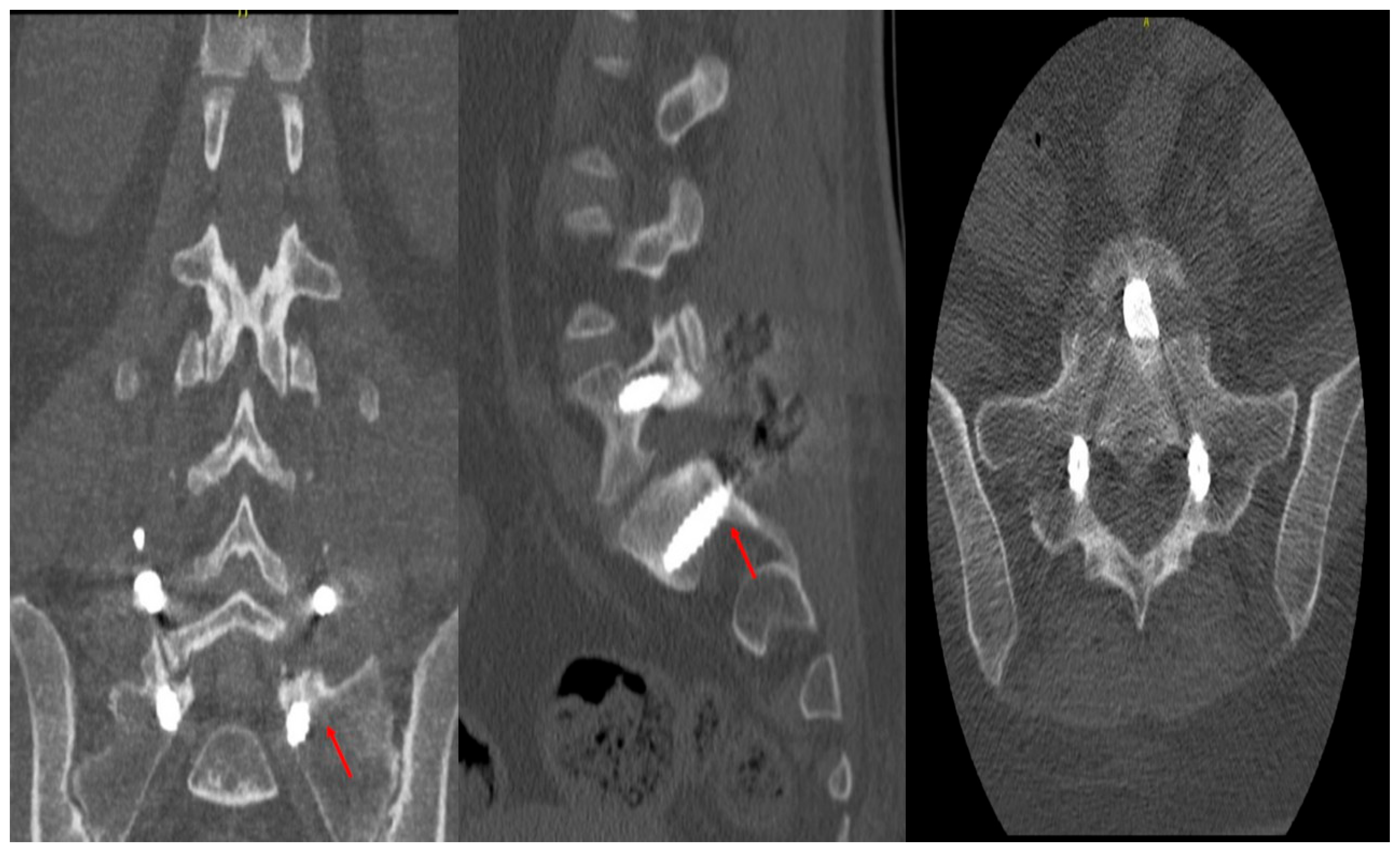
Accuracy of the pedicle screws was assessed using the classification of Gertzbein and Robins (CITA). A four-grade system (A to E) was created to determine the pedicular screws’ position in a postoperative CT scan of the instrumented area (Figure 2). Two fellowship-trained spinal surgeons who did not participate in the surgical procedures and were blind to all patient data and intraoperative results independently assessed postoperative CT scans. Axial and sagittal CT scans were used to assess screw accuracy using the Gertzbein–Robbins classification method. Consensus discussion was used to settle disagreements among reviewers to ensure consistent interpretation across all cases. Fusion was evaluated using postoperative CT scans obtained at approximately 6 months, defined using a bone trabecula passing through the affected area, consistent with our institutional follow-up protocol. We conducted a patient-level analysis, meaning that fusion status was assessed per patient rather than per individual spinal level.

2.5. Statistical Analysis

All statistical analysis in this study was performed using Jamovi software (Version 2.6). Categorical variables were presented as numbers and percentages, while continuous outcomes were reported as mean ± standard deviation if normally distributed and median (IQR) if the data were skewed. The normality of data was tested using the Shapiro–Wilk test. No inferential or regression analyses were performed, as the dataset lacked a control group and sufficient variability to support meaningful comparative or predictive modeling.

3. Results

3.1. Patient Population

In all, 115 patients were involved in this study. The median age was 64 years (IQR: 54, 64), with 52 (45.2%) being male. Most of the patients were diagnosed with degenerative discopathy (DDD), 56 (47.8%), or spondylolisthesis, 39 (33.9%). Seventeen patients (14.8%) had a deformity at diagnosis. See Table 1 for more data.

3.2. Surgical Data

In total, 726 screws were implanted, with 90 patients having interbody implants. The Altera banana expansion intervertebral cage was the most used, 48 (41.7%). All patients were operated on via a posterior approach, while 18 (15.7%) patients were operated on using an anterior approach simultaneously. A total of 89 (77.4%) underwent minimally invasive surgery (MIS), and 26 (22.6%) underwent open surgery. Table 2 summarizes the surgical data.

3.3. Screw Accuracy and Fusion Rate

Graded according to Gertzbein and Robbins system (GRS), pedicle screw accuracy and fusion rates were available for 95 (95/115) patients implanted with 590 (590/72631) screws. Overall, there were 584 (99%) screw Graded A and 5 (0.8%) graded B, only 1 (0.2%) graded C, and there were no screws graded D or E. GRS grades by level, diagnosis, type of anterior approach, type of posterior approach, and implant type are presented in Table 3, Table 4 and Table 5.
Fusion has been achieved in 85 (85/95) patients. And 10 patients showed no fusion diagnosis yet, types of posterior approach, anterior approach, and Implant are shown in Table 6.

3.4. Complications Resulting from the Procedures

Infection was reported in three (2.6%) patients. All of them were superficial, occurred in open procedures for Adult deformity, and were solved completely after debridement, lavage, and specific antibiotic therapy during four or six weeks, depending on the microbiologist’s choice. There was no screw revision or intervertebral cage subsidence. Osteolysis occurred in eight (8.4%) patients; most of them (five screws) were at the S1 level. All of them were asymptomatic. Those in S1 appeared in long fusions (above L2) with a TLIF technique, in which it was not feasible an ALIF due to vascular anatomy. No screws were removed from patients (Table 7).

4. Discussion

The primary goal of emerging robotic spinal surgeries is to enhance the accuracy of screw placement compared to conventional freehand techniques, as screw misplacement can lead to significant complications, including revision surgeries and neurological or vascular deficits [23]. Therefore, robotic surgeries aim to decrease postoperative complications and intraoperative errors, helping reduce revision surgeries and follow-up visits [24]. However, to create a measurement tool for screw placement accuracy, Gertzbein and Robbins developed a scale that starts with Grade A for optimal positioning with no cortical breach [25]. Grade B and C correspond to cortical breaches smaller and larger than 2 mm, respectively. Grade D indicates cortical breaches between 4 and 6 mm, while Grade E is for breaches larger than 6 mm.
On this scale, our study of 726 screws and 115 patients found that 584 (99%) screws were graded A, 5 screws (0.8%) were graded B, and 1 (0.2%) was graded C.
Our remarkable accuracy and fusion rates are validated by recent systematic reviews. For instance, a 2024 meta-analysis of 21 TLIF trials [26] revealed that robot-assisted surgery produced 74% fewer proximal facet joint violations and about 12% greater “perfect” (Grade A) screw accuracy than fluoroscopic freehand (RR≈1.12). Overall, surgical durations were similar to traditional methods, even though robotic cases in that analysis had lengthier setup and operating times in RCTs. As most series claim ≥95% Grade A placement, our 99% Grade A rate is in line with the top outcomes in the current literature. Our fusion rate (89.5%) is also consistent with recent publications; Chang et al., for example, reported approximately 87% interbody fusion at 2-year follow-up in a robot-assisted TLIF group [27]. These comparisons verify that our accuracy and fusion results are in line with existing standards.
Our findings also align with Khan et al., who studied 75 screws and 20 patients who underwent robotic screw placement for degenerative spinal pathologies, found that 74 (98.7%) of their screws were graded A, while only one screw was grade B [28]. Additionally, in a prospective analysis, Lonjon et al. found similar accuracy in a cohort of patients with degenerative spinal diseases who underwent robotic spinal surgeries, with 97.3% of screws placed at a grade A and B [29]. Similar results were also observed in a meta-analytical study of 6041 pedicle screw placements found that the robotic spinal surgery group doubled the rates of grade A optimal positioning compared to the freehand group, with an odds ratio of 2.43 (p < 0.001) [30]. This accuracy, attributed to detailed multi-dimensional imaging and continuous optical tracking of screw trajectories of robotic surgeries, reduces not only bony breaches but also damage to adjacent neural structures, such as leakage of cerebrospinal fluids or injury of nerve roots, whereas freehand surgeries, which depend on the surgeon’s tactile feedback and anatomical landmarks, introduce considerable precision variability, especially in complex and challenging areas such as thoracic spines [29,31,32].
In contrast to our results, Ringel et al., in a randomized controlled trial of 60 patients and 298 screw placements, of which 146 were performed using a robotic system, found that 85% of screws were graded A and B [33]. However, this lower accuracy in their study resulted from improper fixation of the robotic arm and the movement of the cannula during the screw placement [34]. Similarly, Wang et al. studied 61 patients with degenerative spinal pathologies who underwent minimally invasive, robot-assisted TLIF surgery and found that only 85.4% (234 screws) were inserted without any cortical breach [34]. They attributed this reduced accuracy to the movement of guide pins or violations of the lateral vertebral wall during the pedicle screws placement. However, these differences in accuracy are often attributed to variations in robotic systems that have distinct accuracy rates, preoperative imaging, Kirschner-wires requirement, or techniques for mounting the robotic arm (to bone, table, or floor) [35]. In general, reduced screw placement accuracy in robotic assisted spinal surgeries could be explained by various mechanical factors, such as shifting, which is a change in the position of the robotic arm relative to the patient’s position, or skiving, in which vertical force on the surgical instrumentation, such as cannula or drills, deviate from the pre-established bonny trajectories [36].
Our study found that 432 screws out of 436 screws in TLIF surgeries were placed with no cortical breaches, while only four screws had cortical breaches of less than 2 mm. These misplaced screws were scattered among different patients; no patient had more than one breach, and none of the impacted instances showed clinical signs or needed additional care. These accuracy rates were higher than those of Zhang et al., who studied 100 screws and 50 patients who underwent robotic TLIF surgery. Among these 100 screws, 85 were graded A, while 13 and 2 screws were graded B and C, respectively [37]. On the other hand, all our PLIF screws achieved an optimal placement of Grade A. These results were also higher than those of Kim et al., who found that only 91% of robotic-assisted TLIF screws received Grade A, while 8% and 1% were graded B and C, respectively [38].
Effective radiographic monitoring of fusion success rates is crucial for determining patients who may benefit from subsequent or revision surgery. A successful fusion is achieved when bone growth extends across at least 50% of the intended fusion area while maintaining the bone density achieved immediately after surgery [39]. From a biomechanical perspective, radiographic confirmation of solid fusion across one side of the total fusion area is sufficient to indicate stable fusion, regardless of any radiolucent features on the contralateral side [34]. In this context, our study demonstrated a successful fusion rate of 89.5% (85 out of 95 patients), with half of the fusion failures (5 out of 10) occurring in patients with degenerative spinal pathologies. Similarly, Chang et al. reported an 87.3% interbody fusion rate among 26 patients with degenerative spondylolisthesis who underwent robotic-assisted TLIF surgery [27].
Additionally, three patients in our cohort had postoperative infections, and eight showed signs of osteolysis. Osteolysis is a biological reaction that occurs at the bone–implant interface, where implant particles stimulate macrophages, leading to osteoclast activation and bone resorption, which appears as radiolucency around the implant and results in progressive bone loss, periprosthetic fracture, and implant loosening [40].
When it is non-symptomatic, it needs careful attention. If osteolysis progresses or symptoms appear, revision surgery is mandatory.
In our cohort, osteolysis was detected radiographically as progressively lucent zones around the pedicle screws or implants, a recognized sign of implant-associated bone resorption [41]. All osteolysis cases were asymptomatic, so we managed them nonoperatively with close imaging follow-up. Only if radiographic lucency had progressed or symptoms of loosening appeared would we recommend revision surgery.
This approach follows standard practice, since isolated radiolucent “clear zones” (pseudarthrosis signs) are monitored unless clinical or imaging deterioration mandates intervention.

4.1. Learning Curve, Operative Time, and Economic Considerations

Robotic spine surgery is expensive and has a steep learning curve. Setup and operation times are more prolonged at the initial stage of the learning curve, but efficiency quickly increases with familiarity. For instance, Paramasivam et al. reported that following the first ~20 cases, robotic setup time decreased from 24 to 17 min [42]. Similarly, following the initial learning period, operational time gaps between robot and traditional processes tend to diminish. Economically speaking, robotic platforms are expensive to build and maintain (typically >USD500K–USD1.2M) [43]. There are conflicting published cost studies; some predict net savings from shorter OR times and lower incidence of complications, while others report higher index hospitalization costs because of longer surgeries and higher equipment costs [43]. When assessing robotic technology, these pragmatic aspects (learning curve and cost) must be taken into account in addition to the clinical advantages.

4.2. Strengths and Limitations

To the best of our knowledge, this is the first study to evaluate the efficacy and safety of robotic spinal surgery in a Spanish population, improving applicability in local clinical practice. Moreover, using the established Gertzbein–Robbins scale allows for a direct comparison between our results and those from other centers. Additionally, the inclusion of diverse spinal pathologies (degenerative conditions, spondylolisthesis, deformities, and pathological/traumatic fractures) alongside both minimally invasive and open surgical approaches strengthens the generalizability of our results across different clinical scenarios. Despite these strengths, the lack of a randomized controlled design limits our ability to attribute our results to robotic surgeries alone. Also, the retrospective nature of our study introduces potential selection bias that could limit our results in other settings. Moreover, the diversity of implant types and surgical approaches, while reflecting real-world evidence, introduces confounding variables that further limit our ability to isolate the direct impact of robotic surgery on our population. Another drawback is that only 95 out of 115 patients (82.6%) had postoperative CT imaging available, which may have introduced some selection bias into the accuracy analysis. However, we think this had little effect on the validity of the accuracy results because CT imaging was carried out in accordance with standard institutional procedures rather than intraoperative concerns. The heterogeneity of surgical approaches (open vs. MIS; anterior vs. posterior) and patient pathologies further confounds our findings. These factors limit causal inferences and underscore the need for future prospective, controlled studies. Regression or correlation analyses were not performed to determine predictors of fusion rates or screw inaccuracy. Such connections should be investigated in future prospective multi-arm studies with larger and more varied cohorts.
5. Conclusions

Although robotic-assisted spinal surgery shows encouraging screw placement accuracy and good fusion rates, our study’s retrospective, single-arm methodology suggests these results should be interpreted cautiously. Potential advantages of robotic assistance are supported by the capacity to precisely implant longer and biomechanically favorable pedicle screws, especially in MIS operations where anatomical landmarks are scarce. To ascertain if the enhanced technical results are translated into long-term clinical and financial benefits, more high-quality evidence, such as randomized controlled trials and cost-effectiveness studies, should direct wider implementation.

Author Contributions

Conceptualization, I.D.; methodology, A.C. (Angela Carrascosa); software, A.C. (Alicia Collado); validation, A.C. (Angela Carrascosa) and P.A.L.; formal analysis, R.L. and A.C. (Alicia Collado); investigation, R.L. and J.P.C.; resources, P.A.L. and J.P.C.; data curation, R.L. and J.P.C.; writing—original draft preparation, I.D.; writing—review and editing, F.M.; visualization, A.C. (Angela Carrascosa) and A.C. (Alicia Collado); supervision, I.D., P.A.L. and F.M.; project administration, I.D.; funding acquisition, F.M. All authors have read and agreed to the published version of the manuscript.

Funding

This research received no external funding.

Institutional Review Board Statement

This retrospective observational study was conducted in accordance with the ethical standards of the institutional review board (IRB) at Hospital Clínico San Carlos (Madrid, Spain) and the 1964 Helsinki Declaration and its later amendments. Due to the retrospective and anonymized nature of the data analysis, the need for formal informed consent was waived by the approving IRB. All patient data were de-identified before analysis.

Informed Consent Statement

Not applicable. This retrospective study utilized anonymized patient data. No person’s data (including images or identifiers) is contained in this manuscript. All data were aggregated and presented in summary form only.

Data Availability Statement

All data relevant to the study are included in the article. Data may be available upon reasonable request from the corresponding author.

Conflicts of Interest

The authors declare no conflicts of interest.

Abbreviations

ALIFAnterior Lumbar Interbody Fusion
DDDDegenerative discopathy
DRBDynamic Reference Base
VRDynamic Reference (or Registration) Frame
FTPFull Transpedicular
GRSGertzbein and Robbins system
ICTIntraoperative Computed Tomography
LRISELateral RISE Cage System
MISMinimally Invasive Surgery
OLIFOblique Lumbar Interbody Fusion
PEEKPolyetheretherketone
PLIFPosterior Lumbar Interbody Fusion
SMSurface Marker
TLIFTransforaminal Lumbar Interbody Fusion
XLIFExtreme Lateral Interbody Fusion
ALIF + OLIFCombined Anterior and Oblique Lumbar Interbody Fusion
ALIF + OLIF (SP)Same-Patient Combined Anterior and Oblique Lumbar Interbody Fusion
ALIF + OLIF + XLIFCombined Anterior, Oblique, and Lateral Interbody Fusion
ALIF + OLIF + XLIF (SP)Same-Patient Combined Anterior, Oblique, and Lateral Interbody Fusion
OLIF + XLIFCombined Oblique and Extreme Lateral Lumbar Interbody Fusion
IQRInterquartile range

References

  1. Lee, B.H.; Moon, S.H.; Lee, H.M.; Kim, T.H.; Lee, S.J. Prevalence of hip pathology in patients over age 50 with spinal conditions requiring surgery. Indian J. Orthop. 2012, 46, 291–296. [Google Scholar] [CrossRef]
  2. Kobayashi, K.; Sato, K.; Kato, F.; Kanemura, T.; Yoshihara, H.; Sakai, Y.; Shinjo, R.; Ohara, T.; Yagi, H.; Matsubara, Y.; et al. Trends in the numbers of spine surgeries and spine surgeons over the past 15 years. Nagoya J. Med. Sci. 2022, 84, 155–162. [Google Scholar] [CrossRef]
  3. Hicks, J.M.; Singla, A.; Shen, F.H.; Arlet, V. Complications of pedicle screw fixation in scoliosis surgery: A systematic review. Spine 2010, 35, E465–E470. [Google Scholar] [CrossRef] [PubMed]
  4. Matsuzaki, H.; Tokuhashi, Y.; Matsumoto, F.; Hoshino, M.; Kiuchi, T.; Toriyama, S. Problems and solutions of pedicle screw plate fixation of lumbar spine. Spine 1990, 15, 1159–1165. [Google Scholar] [CrossRef] [PubMed]
  5. Açikbaş, S.C.; Arslan, F.Y.; Tuncer, M.R. The effect of transpedicular screw misplacement on late spinal stability. Acta Neurochir. 2003, 145, 949–954. [Google Scholar] [CrossRef] [PubMed]
  6. O’Brien, J.R.; Krushinski, E.; Zarro, C.M.; Sciadini, M.; Gelb, D.; Ludwig, S. Esophageal injury from thoracic pedicle screw placement in a polytrauma patient: A case report and literature review. J. Orthop. Trauma. 2006, 20, 431–434. [Google Scholar] [CrossRef]
  7. Hu, X.; Ohnmeiss, D.D.; Lieberman, I.H. Robotic-assisted pedicle screw placement: Lessons learned from the first 102 patients. Eur. Spine J. 2013, 22, 661–666. [Google Scholar] [CrossRef]
  8. Kosmopoulos, V.; Schizas, C. Pedicle screw placement accuracy: A meta-analysis. Spine 2007, 32, E111–E120. [Google Scholar] [CrossRef]
  9. Tian, N.F.; Huang, Q.S.; Zhou, P.; Zhou, Y.; Wu, R.K.; Lou, Y.; Xu, H.Z. Pedicle screw insertion accuracy with different assisted methods: A systematic review and meta-analysis of comparative studies. Eur. Spine J. 2011, 20, 846–859. [Google Scholar] [CrossRef]
  10. Kantelhardt, S.R.; Martinez, R.; Baerwinkel, S.; Burger, R.; Giese, A.; Rohde, V. Perioperative course and accuracy of screw positioning in conventional, open robotic-guided and percutaneous robotic-guided, pedicle screw placement. Eur. Spine J. 2011, 20, 860–868. [Google Scholar] [CrossRef]
  11. Schatlo, B.; Martinez, R.; Alaid, A.; von Eckardstein, K.; Akhavan-Sigari, R.; Hahn, A.; Stockhammer, F.; Rohde, V. Unskilled unawareness and the learning curve in robotic spine surgery. Acta Neurochir. 2015, 157, 1819–1823. [Google Scholar] [CrossRef]
  12. Wood, M.J.; McMillen, J. The surgical learning curve and accuracy of minimally invasive lumbar pedicle screw placement using CT based computer-assisted navigation plus continuous electromyography monitoring-a retrospective review of 627 screws in 150 patients. Int. J. Spine Surg. 2014, 8, 27. [Google Scholar] [CrossRef] [PubMed]
  13. Wang, Y.; Xiao, Y.; Wang, J.; Rexiti, P.; Wang, Y. Spine-specific robots may offer superior screw placement accuracy in spinal surgery procedures. A systematic review and network and proportional meta-analysis. J. Orthop. Surg. Res. 2025, 20, 920. [Google Scholar] [CrossRef]
  14. OpenPR. Global Surgical Robots for the Spine Industry Trend, Growth Shares, Strategy and Forecasts 2016 to 2022; OpenPR: Niedersachsen, Germany, 2016. [Google Scholar]
  15. Sturgill, D.; How, J.; Blajda, T.; Davis, Z.; Ali, M.; O’Malley, G.; Patel, N.V.; Khan, M.F.; Goldstein, I. Are the Clinical Outcomes and Cost-Effectiveness of Robot-Assisted Pedicle Screw Placement in Lumbar Fusion Surgery Superior to Computed Tomography Navigation and Freehand Fluoroscopy-Guided Techniques? A Systematic Review and Network Meta-Analysis. World Neurosurg. 2024, 191, 81–90. [Google Scholar] [CrossRef]
  16. Soliman, M.A.R.; Pollina, J.; Poelstra, K.; Chaudhary, S.; Foley, K. Can a Spine Robot Be More Efficient and Less Expensive While Maintaining Accuracy? Int. J. Spine Surg. 2022, 16, S50–S54. [Google Scholar] [CrossRef] [PubMed]
  17. McNamee, C.; Kelly, D.; McDonnell, J.M.; Sowa, A.M.; Menon, A.; Darwish, S.; Butler, J.S. The learning curve of robotic assisted pedicle screw placement: Individual patient data meta-analysis. Spine J. Off. J. N. Am. Spine Soc. 2025, S1529-9430(25)00343-2, Advance online publication. [Google Scholar] [CrossRef]
  18. Filley, A.; Baldwin, A.; Ben-Natan, A.R.; Hansen, K.; Arora, A.; Xiao, A.; Hammond, D.; Chen, C.; Tweedt IRohde, J.; Link, T.; et al. The influence of osteoporosis on mechanical complications in lumbar fusion surgery: A systematic review. N. Am. Spine Soc. J. 2024, 18, 100327. [Google Scholar] [CrossRef] [PubMed]
  19. Zhao, X.; Wang, Q.; Wang, P.; Kong, C.; Lu, S. Exploring the impact of body mass index on the accuracy of vertebral bone quality in determining bone mineral density in patients undergoing lumbar fusion surgery. J. Orthop. Surg. Res. 2024, 19, 763. [Google Scholar] [CrossRef]
  20. Sun, W.X.; Huang, W.Q.; Li, H.Y.; Wang, H.S.; Guo, S.L.; Dong, J.; Chen, B.L.; Lin, Y.P. Clinical efficacy of robotic spine surgery: An updated systematic review of 20 randomized controlled trials. EFORT Open Rev. 2023, 8, 841–853. [Google Scholar] [CrossRef]
  21. Morello, A.; Colonna, S.; Lo Bue, E.; Chiari, G.; Mai, G.; Pesaresi, A.; Garbossa, D.; Cofano, F. Accuracy and Safety Between Robot-Assisted and Conventional Freehand Fluoroscope-Assisted Placement of Pedicle Screws in Thoracolumbar Spine: Meta-Analysis. Medicina 2025, 61, 690. [Google Scholar] [CrossRef]
  22. Li, Y.; Wang, Y.; Ma, X.; Ma, J.; Dong, B.; Yang, P.; Sun, Y.; Zhou, L.; Shen, J. Comparison of short-term clinical outcomes between robot-assisted and freehand pedicle screw placement in spine surgery: A meta-analysis and systematic review. J. Orthop. Surg. Res. 2023, 18, 359. [Google Scholar] [CrossRef]
  23. Davidar, A.D.; Jiang, K.; Weber-Levine, C.; Bhimreddy, M.; Theodore, N. Advancements in Robotic-Assisted Spine Surgery. Neurosurg. Clin. N. Am. 2024, 35, 263–272. [Google Scholar] [CrossRef]
  24. Zhang, Q.; Han, X.G.; Xu, Y.F.; Liu, Y.J.; Liu, B.; He, D.; Sun, Y.Q.; Tian, W. Robot-Assisted Versus Fluoroscopy-Guided Pedicle Screw Placement in Transforaminal Lumbar Interbody Fusion for Lumbar Degenerative Disease. World Neurosurg. 2019, 125, e429–e434. [Google Scholar] [CrossRef] [PubMed]
  25. Gertzbein, S.D.; Robbins, S.E. Accuracy of pedicular screw placement in vivo. Spine 1990, 15, 11–14. [Google Scholar] [CrossRef] [PubMed]
  26. Guan, J.; Feng, N.; Yu, X.; Yang, K. Comparison of robot-assisted versus fluoroscopy-guided transforaminal lumbar interbody fusion (TLIF) for lumbar degenerative diseases: A systematic review and meta-analysis of randomized controlled trails and cohort studies. Syst. Rev. 2024, 13, 170. [Google Scholar] [CrossRef]
  27. Chang, M.; Wang, L.; Yuan, S.; Tian, Y.; Zhao, Y.; Liu, X. Percutaneous Endoscopic Robot-Assisted Transforaminal Lumbar Interbody Fusion (PE RA-TLIF) for Lumbar Spondylolisthesis: A Technical Note and Two Years Clinical Results. Pain Physician 2022, 25, E73–E86. [Google Scholar]
  28. Khan, A.; Meyers, J.E.; Siasios, I.; Pollina, J. Next-Generation Robotic Spine Surgery: First Report on Feasibility, Safety, and Learning Curve. Oper. Neurosurg. 2019, 17, 61–69. [Google Scholar] [CrossRef]
  29. Lonjon, N.; Chan-Seng, E.; Costalat, V.; Bonnafoux, B.; Vassal, M.; Boetto, J. Robot-assisted spine surgery: Feasibility study through a prospective case-matched analysis. Eur. Spine J. 2016, 25, 947–955. [Google Scholar] [CrossRef]
  30. Fu, W.; Tong, J.; Liu, G.; Zheng, Y.; Wang, S.; Abdelrahim, M.E.A.; Gong, S. Robot-assisted technique vs conventional freehand technique in spine surgery: A meta-analysis. Int. J. Clin. Pract. 2021, 75, e13964. [Google Scholar] [CrossRef] [PubMed]
  31. Kolin, D.A.; Mackie, A.T.; Heath, M.R.; Uppstrom, T.J.; Green, D.W.; Fabricant, P.D. No difference in patient reported outcomes between cohorts undergoing lesion-specific surgery for osteochondritis dissecans of the knee. J. Orthop. 2023, 37, 22–26. [Google Scholar] [CrossRef]
  32. Al-Naseem, A.O.; Al-Muhannadi, A.; Ramadhan, M.; Alfadhli, A.; Marwan, Y.; Shafafy, R.; Abd-El-Barr, M.M. Robot-assisted pedicle screw insertion versus navigation-based and freehand techniques for posterior spinal fusion in scoliosis: A systematic review and meta-analysis. Spine Deform. 2024, 12, 1203–1215. [Google Scholar] [CrossRef]
  33. Ringel, F.; Stüer, C.; Reinke, A.; Preuss, A.; Behr, M.; Auer, F.; Stoffel, M.; Meyer, B. Accuracy of robot-assisted placement of lumbar and sacral pedicle screws: A prospective randomized comparison to conventional freehand screw implantation. Spine 2012, 37, E496–E501. [Google Scholar] [CrossRef]
  34. Wang, L.; Li, C.; Wang, Z.; Li, D.; Tian, Y.; Yuan, S.; Liu, X. Comparison of robot-assisted versus fluoroscopy-assisted minimally invasive transforaminal lumbar interbody fusion for degenerative lumbar spinal diseases: 2-year follow-up. J. Robot. Surg. 2023, 17, 473–485. [Google Scholar] [CrossRef] [PubMed]
  35. Lee, Y.S.; Cho, D.C.; Kim, K.T. Navigation-Guided/Robot-Assisted Spinal Surgery: A Review Article. Neurospine 2024, 21, 8–17. [Google Scholar] [CrossRef] [PubMed]
  36. Buza, J.A.; 3rd Good, C.R.; Lehman, R.A.; Jr Pollina, J.; Chua, R.V.; Buchholz, A.L.; Gum, J.L. Robotic-assisted cortical bone trajectory (CBT) screws using the Mazor X Stealth Edition (MXSE) system: Workflow and technical tips for safe and efficient use. J. Robot. Surg. 2021, 15, 13–23. [Google Scholar] [CrossRef] [PubMed]
  37. Zhang, Q.; Xu, Y.F.; Tian, W.; Le, X.F.; Liu, B.; Liu, Y.J.; He, D.; Sun, Y.Q.; Yuan, Q.; Lang, Z.; et al. Comparison of Superior-Level Facet Joint Violations Between Robot-Assisted Percutaneous Pedicle Screw Placement and Conventional Open Fluoroscopic-Guided Pedicle Screw Placement. Orthop. Surg. 2019, 11, 850–856. [Google Scholar] [CrossRef]
  38. Kim, H.J.; Lee, S.H.; Chang, B.S.; Lee, C.K.; Lim, T.O.; Hoo, L.P.; Yi, J.M.; Yeom, J.S. Monitoring the quality of robot-assisted pedicle screw fixation in the lumbar spine by using a cumulative summation test. Spine 2015, 40, 87–94. [Google Scholar] [CrossRef]
  39. Fogel, G.R.; Toohey, J.S.; Neidre, A.; Brantigan, J.W. Fusion assessment of posterior lumbar interbody fusion using radiolucent cages: X-ray films and helical computed tomography scans compared with surgical exploration of fusion. Spine J. 2008, 8, 570–577. [Google Scholar] [CrossRef]
  40. Saadi, S.B.; Ranjbarzadeh, R.; Ozeir, K.; Amirabadi, A.; Ghoushchi, S.J.; Kazemi, O.; Azadikhah, S.; Bendechache, M. Osteolysis: A Literature Review of Basic Science and Potential Computer-Based Image Processing Detection Methods. Comput. Intell. Neurosci. 2021, 2021, 4196241. [Google Scholar] [CrossRef]
  41. Lehr, A.; Duits, A.A.A.; Reijnders, M.R.L.; Nutzinger, D.; Castelein, R.M.; Oner, F.C.; Kruyt, M.C. Assessment of Posterolateral Lumbar Fusion: A Systematic Review of Imaging-Based Fusion Criteria. JBJS Rev. 2022, 10, e22.00129. [Google Scholar] [CrossRef]
  42. Paramasivam Meenakshi Sundaram, P.; Lai, M.C.; Kaliya-Perumal, A.-K.; Oh, J.Y.-L. Overcoming the Learning Curve in Robot-Assisted Spinal Surgery—How Does It Compare to O-Arm Navigation? Surgeries 2024, 5, 896–907. [Google Scholar] [CrossRef]
  43. Antonacci, C.L.; Zeng, F.; Block, A.; Davey, A.; Makanji, H. Robotic-assisted spine surgery-a narrative review. J. Spine Surg. 2024, 10, 305–312. [Google Scholar] [CrossRef] [PubMed]
Figure 1. Preoperative workflow: (A) Scan and plan, (B) single lateral position DRB and SM in the left iliac crest, (C) percutaneous pedicular screws in lateral position, (D) intraoperative X-rays. Single-position surgery. L3-S1 percutaneous pedicular screws. ALIF, OLIF, and XLIF.
Jcm 14 08425 g001
Figure 2. A four-grade system (A to E) was created to determine the pedicular screws’ position in a postoperative CT scan of the instrumented area. In this images, it is possible to see a Grade B medial breach antero-posterior, lateral, and axial views in the CT scan.
Jcm 14 08425 g002
Table 1. Demographics and baseline.
VariableData
Number of patients 115
Age (year), Median (Q1, Q3) (Range)64 (54, 64) (16–91)
Sex
Male, n (%) 52 (45.2)
Female, n (%)63 (54.8)
Diagnosis, n (%)
Degenerative discopathy56 (47.8)
Spondylolisthesis39 (33.9)
Adjacent Segment Degeneration2 (1.7)
Adult deformity11 (9.6)
Adolescent deformity1 (0.9)
Pseudoarthrosis1 (0.9)
Traumatic fracture4 (3.5)
Pathological fracture1 (0.9)
Deformity, n (%)17 (14.8)
Levels treated, n of patients (%)
T31 (0.9)
T43 (2.6)
T54 (3.5)
T63 (2.6)
T74 (3.5)
T83 (2.6)
T93 (2.6)
T105 (4.3)
T112 (8.7)
T1210 (8.7)
L112 (10.4)
L217 (14.8)
L326 (22.6)
L483 (72.2)
L5106 (92.2)
S171 (61.7)
Iliac7 (6.19)
Table 2. Surgical data of the 90 patients.
Variablen (%)
Total number of implanted screws 726
Invasiveness
MIS89 (77.4)
Open26 (22.6)
Posterior approach types, n = 115
TLIF87 (75.7)
FTP24 (20.9)
PLIF4 (3.5)
Anterior approach types, n = 18
ALIF1 (0.9)
OLIF1 (0.9)
XLIF2 (1.7)
ALIF + OLIF2 (1.7)
ALIF + OLIF (SP)1 (0.9)
ALIF + OLIF + XLIF6 (5.2)
ALIF + OLIF + XLIF (SP)1 (0.9)
ALIF + XLIF2 (1.7)
OLIF + XLIF2 (1.7)
Types of Implants, n = 90
Altera banana expansion cage48 (41.7)
LRISE expansion cage37 (32.2)
Static cage5 (4.3)
PEEK1 (0.9)
PEEK + Altera banana expansion cage1 (0.9)
Total number of implanted screws 731
Invasiveness
MIS89 (77.4)
Open26 (22.6)
Anterior approach types, n = 115
TLIF87 (75.7)
FTP24 (20.9)
PLIF4 (3.5)
Posterior approach types, n = 18
ALIF1 (0.9)
OLIF1 (0.9)
XLIF2 (1.7)
ALIF + OLIF2 (1.7)
ALIF + OLIF (SP)1 (0.9)
ALIF + OLIF + XLIF6 (5.2)
ALIF + OLIF + XLIF (SP)1 (0.9)
ALIF + XLIF2 (1.7)
OLIF + XLIF2 (1.7)
Types of Implants, n = 90
Altera banana expansion cage48 (41.7)
LRISE expansion cage37 (32.2)
Static cage5 (4.3)
PEEK1 (0.9)
PEEK + Altera banana expansion cage1 (0.9)
Table 3. GRS grading according to level.
Level TreatedGrade AGrade BGrade CGrade DGrade E
Overall, n = 590584 (99%)5 (0.8%)1 (0.2%)0 (0%)0 (0%)
T44 (0.7%)0 (0%)0 (0%)0 (0%)0 (0%)
T54 (0.7%)0 (0%)0 (0%)0 (0%)0 (0%)
T62 (0.3%)0 (0%)0 (0%)0 (0%)0 (0%)
T73 (0.5%)0 (0%)0 (0%)0 (0%)0 (0%)
T83 (0.5%)0 (0%)0 (0%)0 (0%)0 (0%)
T92 (0.3%)0 (0%)0 (0%)0 (0%)0 (0%)
T103 (0.5%)0 (0%)0 (0%)0 (0%)0 (0%)
T1114 (2.4%)0 (0%)0 (0%)0 (0%)0 (0%)
T1215 (2.5%)1 (0.2%)0 (0%)0 (0%)0 (0%)
L121 (3.6%)0 (0%)0 (0%)0 (0%)0 (0%)
L224 (4.1%)0 (0%)1 (0.2%)0 (0%)0 (0%)
L345 (7.6%)1 (0.2%)0 (0%)0 (0%)0 (0%)
L4139 (23.6%)3 (0.5%)0 (0%)0 (0%)0 (0%)
L5137 (30%)0 (0%)0 (0%)0 (0%)0 (0%)
S1120 (20.3%)0 (0%)0 (0%)0 (0%)0 (0%)
Iliac8 (1.4%)0 (0%)0 (0%)0 (0%)0 (0%)
Table 4. GRS grading by diagnosis.
DiagnosisGrade AGrade BGrade CGrade DGrade E
Degenerative discopathy276 (46.9%)4 (0.7%)1 (0.2%)0 (0%)0 (0%)
Spondylolisthesis160 (27.2)1 (0.2%)0 (0%)0 (0%)0 (0%)
Adjacent Segment Degeneration5 (0.8%)0 (0%)0 (0%)0 (0%)0 (0%)
Pseudoarthrosis10 (1.7%)0 (0%)0 (0%)0 (0%)0 (0%)
Adult deformity118 (20%)0 (0%)0 (0%)0 (0%)0 (0%)
Traumatic fracture8 (1.4%)0 (0%)0 (0%)0 (0%)0 (0%)
Pathological fracture6 (1%)0 (0%)0 (0%)0 (0%)0 (0%)
Table 5. GRS grading by approach type and implant.
Grade AGrade BGrade CGrade DGrade E
Posterior approach
TLIF432 (73.2%)4 (0.7%)0 (%)0 (%)0 (%)
FTP120 (23.7%)1 (0.2%)1 (0.2%)0 (%)0 (%)
PLIF12 (2%)0 (0%)0 (%)0 (%)0 (%)
Anterior Approach
Not approached anteriorly434 (73.6%)4 (0.7%)0 (0%)0 (0%)0 (0%)
OLIF5 (0.8%)0 (0%)0 (0%)0 (%)0 (%)
XLIF27 (4.6%)0 (0%)0 (0%)0 (%)0 (%)
ALIF + OLIF18 (3.1%)0 (0%)0 (0%)0 (%)0 (%)
ALIF + OLIF (SP)6 (1%)0 (0%)0 (0%)0 (%)0 (%)
ALIF + OLIF + XLIF62 (10.5%)1 (0.2%)1 (0.2%)0 (%)0 (%)
ALIF + OLIF + XLIF (SP)8 (1.4%)0 (0%)0 (0%)0 (%)0 (%)
OLIF + XLIF24 (4.1%)0 (0%)0 (0%)0 (%)0 (%)
Implant type
No implant 133 (22%)1 (0.2%)1 (0.2%)0 (%)0 (%)
Altera banana expansion cage
253 (42.9%)3 (0.5%)0 (0%)0 (%)0 (%)
LRISE expansion cage128 (28.5%)0 (0%)0 (0%)0 (%)0 (%)
Static cage21 (3.6%)1 (0.2%)0 (0%)0 (%)0 (%)
PEEK6 (1%) 0 (0%)0 (%)0 (%)
PEEK + Altera banana expansion cage6 (1%)0 (0%)0 (0%)0 (%)0 (%)
0 (0%)
Table 6. Fusion failure data.
Variable
Number10 (10/95)
Diagnosis
Degenerative discopathy5 (5.3%)
Spondylolisthesis2 (2.1%)
Adult deformity 1 (1.1%)
Traumatic fracture1 (1.1%)
Pathological fracture 1 (1.1%)
Posterior approach
TLIF4 (4.2%)
FTP6 (6.3%)
Anterior approach
Not approached anteriorly9 (9.5%)
OLIF + XLIF1 (1.1%)
Implant
Not implanted6 (6.3%)
Altera banana expansion cage3 (3.2%)
LRISE expansion cage1 (1.1%)
Table 7. Type of Complications.
Complicationn (%)
Infection 3 (2.6%)
Screw or cage subsidence 0 (0)
Osteolysis, Patients = 8 (8.4%)
S15 (0.69%)
L12 (0.28)
L32 (0.28)
L41 (0.14)
L52 (0.22)
Screw removal 0 (0)
Disclaimer/Publisher’s Note: The statements, opinions and data contained in all publications are solely those of the individual author(s) and contributor(s) and not of MDPI and/or the editor(s). MDPI and/or the editor(s) disclaim responsibility for any injury to people or property resulting from any ideas, methods, instructions or products referred to in the content.