Article

Analysis of Potential Subtypes of SAPHO Syndrome

Beijing Jishuitan Hospital, Capital Medical University, Beijing 100035, China
*
Author to whom correspondence should be addressed.
J. Clin. Med. 2025, 14(22), 8116; https://doi.org/10.3390/jcm14228116 (registering DOI)
Submission received: 27 September 2025 / Revised: 8 November 2025 / Accepted: 11 November 2025 / Published: 17 November 2025
(This article belongs to the Section Orthopedics)

Abstract

Objectives: The purpose of this study is to identify clinical subtypes of SAPHO syndrome using cluster analysis, and to systematically investigate the associated clinical characteristics, therapeutic approaches, and short-term prognostic outcomes in order to enhance patient management. Methods: We recruited patients who had been diagnosed with SAPHO syndrome at Beijing Jishuitan Hospital. Bone lesions were assessed using a 99Tc bone scan. Based on bone lesions and clinical features, patients were categorized using a hierarchical clustering algorithm. Laboratory test results and prognostic differences were compared among clusters. Results: Overall, 135 patients were included. Cluster analysis identified three distinct clusters. Eighty-seven patients were assigned to cluster 1, characterized by anterior chest wall involvement; 74.7% also had skin involvement. Nineteen patients were assigned to cluster 2, characterized by spinal involvement, and 10.5% showed skin manifestations. Twenty-nine patients were assigned to cluster 3, characterized by peripheral bone involvement, with 24.1% exhibiting skin manifestations. Patients in cluster 3 were younger at disease onset (36.92 ± 16.62 years); their BASDAI and BASFI scores were lower (2.51 ± 1.18 and 0.89 ± 1.37, respectively). In cluster 1, significant reductions in Visual Analog Scale scores were observed at 1 and 6 months after treatment compared with baseline (7.08 vs. 2.59, p < 0.001, n = 63; 7.09 vs. 1.93, p < 0.001, n = 45). Similar improvements were noted in clusters 2 and 3 (7.18 vs. 3.00, p < 0.001, n = 11; 7.00 vs. 3.00, p < 0.001, n = 7; 6.70 vs. 2.65, p < 0.001, n = 20; 6.60 vs. 2.47, p < 0.001, n = 15). Conclusions: SAPHO syndrome may be classified into three subtypes: typical, axial, and peripheral. All subtypes show rapid improvement with timely treatment. Defining the clinical characteristics of these three subtypes can aid diagnosis and provide pathogenesis insights regarding this heterogeneous syndrome.

1. Introduction

SAPHO syndrome is a rare inflammatory disorder characterized by synovitis, acne, pustulosis, hyperostosis, and osteitis, from which its acronym is derived. The syndrome was first described in 1987 [1]. Diagnosis is primarily based on clinical manifestations because there currently is no universally accepted standard for the diagnosis, treatment, or prognosis of SAPHO syndrome. Commonly referenced diagnostic criteria include those proposed by Benhamou and colleagues in 1988 [2] and by Kahn in 1994, later revised in 2003 [3]. No specific clinical features or laboratory markers have been established as diagnostic indicators for SAPHO syndrome.
The clinical presentation of SAPHO syndrome is diverse, with variable effects on patients’ quality of life. Chronic nonbacterial osteomyelitis (CNO), its severe form chronic recurrent multifocal osteomyelitis (CRMO), and pustular arthro-osteitis (PAO) are considered closely related entities; they may represent limited forms of SAPHO syndrome [3,4,5,6]. Atypical manifestations and incomplete understanding of the disease can lead to substantial diagnostic delays [7,8]. Osteitis and hyperostosis—core features of SAPHO syndrome—usually involve multiple skeletal regions [9]. The most commonly affected site is the anterior chest wall (ACW), followed by the axial skeleton (including spine and sacroiliac joints) and the long bones of the limbs [10]. Most patients exhibit skin involvement, typically manifesting as palmoplantar pustulosis (PPP) or severe acne (SA) [9]. Over 70% of patients develop both skin and osteoarticular manifestations within 2 years of disease onset, although longer intervals have also been reported [11]. Many drugs have been used to manage SAPHO syndrome [12,13,14], but treatment responses considerably vary among patients.
These variations in clinical phenotype and therapeutic response complicate the diagnosis, treatment, and prognostic evaluation of SAPHO syndrome. This study aimed to classify the clinical characteristics of patients with SAPHO syndrome, identify distinct subtypes, and evaluate their associations with treatment response and prognosis. Such findings may contribute to more precise disease management. Cluster analysis offers an important approach to improving understanding of SAPHO syndrome and may aid its clinical diagnosis.

2. Materials and Methods

2.1. Patients

All patients were recruited from Beijing Jishuitan Hospital. The inclusion criteria were as follows: (1) fulfillment of the diagnostic criteria for SAPHO syndrome proposed by Kahn, with minor modifications [3]; (2) completion of a 99Tc bone scan for the assessment of bone and joint lesions; and (3) confirmation of lesions by computed tomography and magnetic resonance imaging, as evaluated by the same rheumatologist and radiologist. Exclusion criteria were infectious osteitis, bone tumors, infectious PPP, and noninflammatory bone-condensing lesions. This study was approved by the Ethics Committee of Beijing Jishuitan Hospital (approval number: K2023-134-02). Written informed consent to participate was obtained from each patient.

2.2. Clinical Evaluation

The following clinical data were collected: sex, age, disease duration, bone and joint manifestations, skin manifestations, erythrocyte sedimentation rate (ESR), C-reactive protein (CRP), cytokines, bone metabolism markers, imaging findings, and pathological results (bone biopsy performed when deemed necessary by the clinician and the patient). Disease activity was assessed using the Visual Analog Scale (VAS), Bath Ankylosing Spondylitis Disease Activity Index (BASDAI), Bath Ankylosing Spondylitis Functional Index (BASFI), and Health Assessment Questionnaire (HAQ). All patients received treatment with iguratimod and alendronate. Follow-up evaluations were conducted at 1 month and 6 months after treatment (Figure 1).

2.3. Statistical Analysis

All data analyses were performed using R software (version 4.5.0). Categorical variables were analyzed using a hierarchical clustering algorithm with the Average Linkage method and Gower’s similarity coefficient. Variables included in the cluster analysis comprised the most commonly affected bone and joint locations, lesion distribution, bone lesion type, and skin involvement.
Group comparisons were performed as follows: normality was assessed using the Kolmogorov–Smirnov test. Descriptive data were expressed as mean (range) or number (%). The χ2 test or Fisher’s exact test was used for categorical variables. Normally distributed continuous variables were tested using the t-test. Non-normally distributed continuous variables were tested using the Mann-Whitney U test. Paired t-tests assessed changes in individual parameters across time points. All statistical tests were two-tailed, and p-values < 0.05 were considered statistically significant.

3. Results

3.1. Demographic and Clinical Characteristics

In total, 135 patients (95 women and 40 men) were included in this study, with a mean age of 44.49 ± 13.89 years. Bone lesions were predominantly symmetrical (85/135, 63.0%), and most presented as osteosclerosis (102/135, 75.6%). All patients exhibited osteoarticular symptoms. The frequencies of ACW, spinal, and peripheral bone or joint involvement were 65.2% (88/135), 40.7% (55/135), and 27.4% (37/135), respectively. Concurrent skin manifestations were present in 54.8% (74/135) of patients. The most common skin presentations were PPP in 28.1% (38/135), SA in 8.1% (11/135), and psoriasis vulgaris (PV) in 3.7% (5/135). Elevated ESR was observed in 41.5% (56/108), and elevated CRP in 24.4% (33/106). Bone biopsy was performed in 21 patients, and low-virulence bacteria were identified in 10 cases through bacterial culture or next-generation sequencing. The detailed demographic and clinical characteristics are summarized in Table 1.

3.2. Cluster Analysis

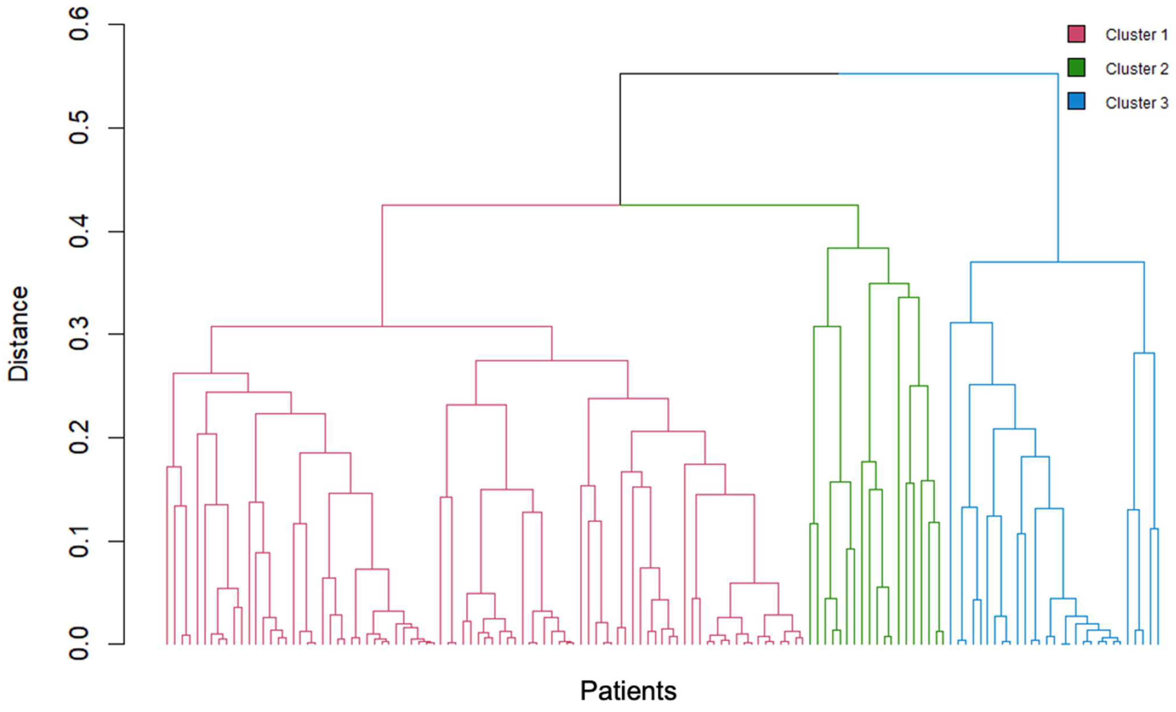
Cluster analysis classified patients with SAPHO syndrome into three distinct clusters based on the distribution of bone and joint involvement, lesion type, and presence of skin manifestations. Each cluster demonstrated unique clinical characteristics (Figure 2).
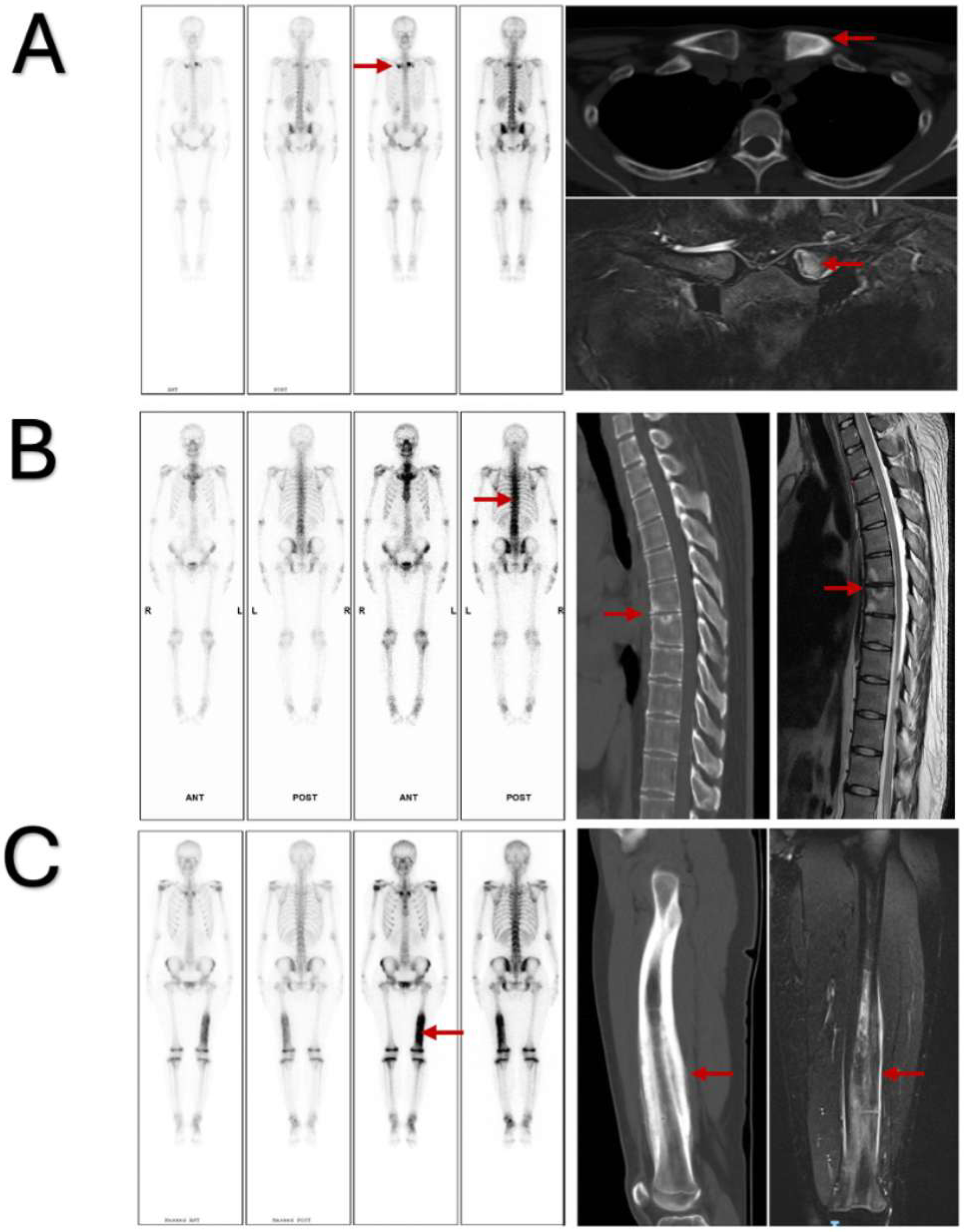
Cluster 1, characterized by ACW and skin involvement, included 87 patients (Figure 3). The disease in this cluster could be defined as typical SAPHO syndrome. Bone lesions were primarily located in the ACW (88.5%) and spine (51.7%); osteosclerosis was the predominant feature (80.5%). Lesions were usually symmetrical (88.5%), and most patients presented with skin involvement (74.7%). This cluster represents the disease form most easily recognized in clinical practice.
Cluster 2, characterized by spinal involvement, included 19 patients (Figure 3). The disease in this cluster could be defined as axial SAPHO syndrome. Bone lesions were mainly located in the spine, without prominent osteosclerosis or bone destruction. Lesions tended to be asymmetrical, and skin manifestations were infrequent (10.5%).
Cluster 3, characterized by peripheral bone and joint involvement, included 29 patients (Figure 3). The disease in this cluster could be defined as peripheral SAPHO syndrome. Lesions were mainly found in peripheral bones and joints (75.9%); they predominantly exhibited osteosclerosis (96.6%). Most lesions involved a single site (86.2%), and skin manifestations were relatively uncommon (24.1%). This cluster represents the disease form most challenging to diagnose in clinical settings.
Patients in cluster 3 were younger at disease onset (36.92 ± 16.62 years). BASDAI and BASFI scores were lower in cluster 3 (2.51 ± 1.18 and 0.89 ± 1.37, respectively). Among the clusters, cluster 3 showed the highest proportion of osteosclerosis (96.6%). No significant differences were evident in ESR, CRP, cytokines, or bone metabolism markers among the clusters. Detailed comparisons are presented in Table 2.
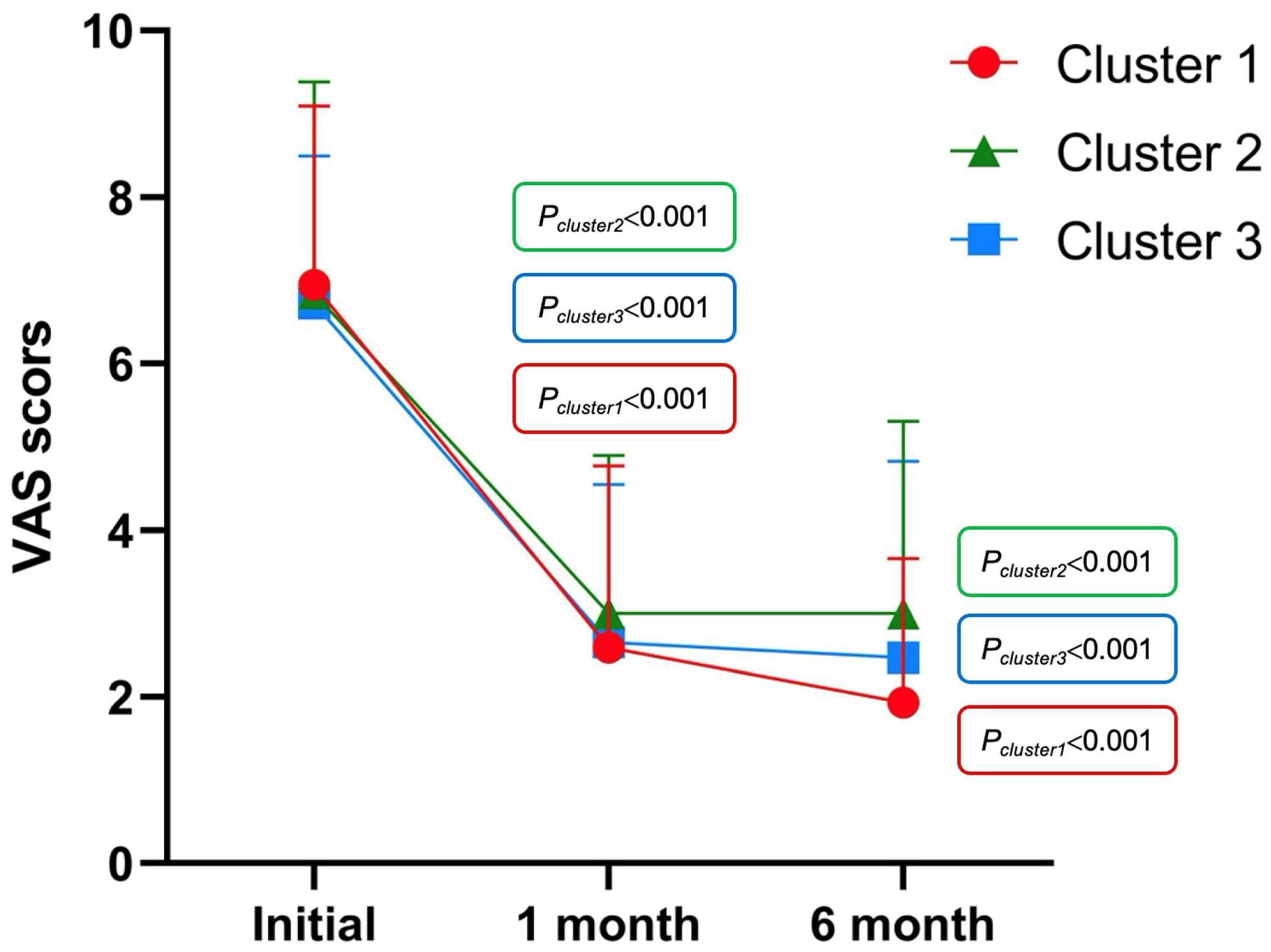
In cluster 1, significant reductions in VAS scores were observed at 1 and 6 months after treatment compared with baseline (7.08 vs. 2.59, p < 0.001, n = 63; 7.09 vs. 1.93, p < 0.001, n = 45). Similar improvements were noted in Clusters 2 and 3 (7.18 vs. 3.00, p < 0.001, n = 11; 7.00 vs. 3.00, p < 0.001, n = 7; 6.70 vs. 2.65, p < 0.001, n = 20; 6.60 vs. 2.47, p < 0.001, n = 15) (Figure 4).

4. Discussion

In this study, new subtypes of SAPHO syndrome were identified through cluster analysis based on clinical manifestations and radiologic findings.
Previous research has identified ACW involvement in 65–90% of patients, a characteristic feature of SAPHO syndrome [15,16]. Typically affected structures include the sternoclavicular joint, sternocostal joint, and clavicular ligament. Axial involvement occurs in 32–52% of cases [15]. In the present study, the typical SAPHO syndrome group (cluster 1) showed similar findings: ACW and spinal involvement were observed in 88.5% and 51.7% of patients, respectively. On bone scintigraphy, increased uptake in the sternoclavicular region, known as the “bullhead sign,” is considered a characteristic imaging feature of SAPHO syndrome [17]. The progression of bone and joint changes in this area can be divided into three stages: soft tissue swelling, bone erosion, and new bone formation with hyperostosis and osteosclerosis [15]. In our study, cluster 1 mainly involved the ACW and spine; osteosclerosis was the predominant manifestation (80.5%). Therefore, bone scintigraphy is recommended for patients with ACW involvement to prevent missed diagnoses of concomitant spinal lesions. Skin manifestations are also common; characteristic skin changes may appear before the onset of arthritis (25%), concurrently with joint symptoms (63.5%), or after arthritis develops (5.8%) [18]. In our study, most patients in cluster 1 exhibited skin involvement (74.7%). Therefore, this subtype is more readily recognized by rheumatologists in clinical settings. Patients in the typical SAPHO syndrome group (cluster 1) generally had longer disease duration, with more severe pain and functional impairment. However, follow-up results showed substantial symptom relief within 1 month after treatment initiation, with sustained improvement for at least 6 months. At present, it remains uncertain whether early aggressive treatment is warranted for this subtype.
Axial skeleton involvement may occur in several forms and combinations, including sclerosis of one or more vertebral bodies with osteosclerosis, osteolytic lesions with varying degrees of vertebral collapse, and spondylitis with or without discitis [15]. In the present study, patients with axial SAPHO syndrome (cluster 2) exhibited bone lesions characterized by osteosclerosis and bone destruction. Lesions were typically multifocal (94.7%), and only a small proportion of patients presented with skin manifestations (10.5%). Axial SAPHO syndrome (cluster 2) should be distinguished from axial spondyloarthritis (axSpA) in clinical practice. Compared with the findings in axSpA, magnetic resonance imaging findings in SAPHO syndrome more commonly show bone marrow edema at the anterior corners of vertebrae, bone erosion, and swelling of the intervertebral disks and endplates [19]. In axSpA, elevated levels of ESR and tumor necrosis factor (TNF) are frequently observed; however, we found that ESR was elevated in some patients with axial SAPHO syndrome (66.7%), but TNF was not increased. Previous studies have shown that patients with spinal involvement often experience more severe clinical manifestations [20]. Similarly, we noted that axial SAPHO syndrome (cluster 2) had a greater impact on spinal function and overall quality of life. Nonetheless, symptoms in most patients improved rapidly after treatment.
Long bone involvement is uncommon in adults; such sites are frequently affected in children, typically involving the distal femur and proximal or distal tibia. Single-bone involvement has also been reported, and the mandible may be affected, revealed diffuse unilateral sclerosis [21]. In the present study, peripheral SAPHO syndrome (cluster 3) was mainly characterized by peripheral bone and joint involvement. Affected patients often lacked typical skin manifestations and experienced frequent diagnostic delays. Previous cohort studies have shown diagnostic delays of up to 9 years in some SAPHO cases, resulting in irreversible structural damage, increased disease burden, and disability [8,22]. Therefore, improvements to diagnostic accuracy for peripheral SAPHO syndrome (cluster 3) are particularly important. Patients in this cluster presented at a younger age, had a shorter disease duration, and predominantly exhibited osteosclerosis as the primary bone lesion. Pain intensity and functional impairment were milder than in other clusters. Bone biopsy is often required in these patients to exclude alternative diagnoses.
At present, there is no standardized treatment protocol for SAPHO syndrome worldwide, and its exact pathogenesis remains unclear. Several cytokines are believed to contribute to disease development. Previous studies have shown that proinflammatory cytokines—such as TNF-α, interleukin (IL)-1β, IL-6, and IL-8—are strongly expressed in patients with SAPHO syndrome, promoting an amplified inflammatory response. These cytokines facilitate disease progression by enhancing inflammation, inducing acute-phase reactions, and promoting chemotaxis. In contrast, the anti-inflammatory cytokine IL-10 and transforming growth factor-β exert protective effects by regulating immune responses and suppressing excessive inflammation [23]. A systematic review suggested that TNF inhibitors are the preferred treatment for patients who do not respond to conventional therapy. For patients who show insufficient response to TNF blockade, IL-1 inhibitors and biologic agents targeting the IL-17/IL-23 axis may be considered [12]. Moreover, the interaction among the transcription factor forkhead box O1 (FoxO1), Propionibacterium acnes, NLR family pyrin domain-containing protein 3 (NLRP3) inflammasomes, and IL-1β may contribute to the pathogenesis of osteitis [24]. Currently, no single therapeutic regimen can address all manifestations of SAPHO syndrome [14]. In the present study, the therapeutic responses of patients across the three identified clusters were followed for a short duration. Treatment decisions were made jointly by physicians and patients without external intervention. All three clusters showed rapid symptomatic improvement after treatment. However, the potential for different subtypes to require distinct therapeutic strategies warrants further investigation.
This study had some limitations. The sample size was relatively small, and the follow-up period was short. Future studies should include larger cohorts and longer observation periods. Further research is also needed to elucidate pathogenic mechanisms underlying differences among the subtypes.

5. Conclusions

This study is the first to use cluster analysis to classify SAPHO syndrome based on clinical characteristics. The results suggest that SAPHO syndrome can be divided into three subtypes: typical, axial, and peripheral. All subtypes demonstrated rapid improvement after timely treatment. Thus, early and accurate diagnosis is particularly important for optimal management.

Author Contributions

The authors’ contributions are as follows: Conceptualization, X.D.; methodology, H.D. and X.D.; software, H.D.; validation, W.T.; formal analysis, H.D.; resources, X.D.; data curation, H.D. and W.T.; writing—original draft, H.D.; writing—review and editing, X.D.; visualization, H.D.; supervision, X.D.; project administration, X.D.; funding acquisition, X.D. All authors have read and agreed to the published version of the manuscript.

Funding

This research received no external funding.

Institutional Review Board Statement

This study was approved by the Ethics Committee of Beijing Jishuitan Hospital (approval numbers: K2023-134-00, 9 May 2023 and K2023-134-02, 30 May 2025).

Informed Consent Statement

Written informed consent was obtained from all participants regarding publication of this paper.

Data Availability Statement

The original data supporting the findings of this study are included in the article. Further inquiries can be directed to the corresponding author.

Acknowledgments

The authors thank the Department of Radiology, Beijing Jishuitan Hospital, for assistance; they acknowledge Hong Zhao, Xueya Lv, and Lingge Wu for valuable contributions to this study. The authors also thank Ryan Chastain-Gross for editing the English text of a draft of this manuscript.

Conflicts of Interest

The authors declare no conflicts of interest.

Abbreviations

The following abbreviations are used in this manuscript:
SAPHOsynovitis, acne, pustulosis, hyperostosis, and osteitis
CRMOchronic recurrent multifocal osteomyelitis
CNOchronic nonbacterial osteomyelitis
PAOpustular osteoarthritis
ACWanterior chest wall
PPPpalmoplantar pustulosis
SAsevere acne
ESRerythrocyte sedimentation rate
CRPC-reactive protein
VASVisual Analog Scale
BASDAIBath Ankylosing Spondylitis Disease Activity Index
BASFIBath Ankylosing Spondylitis Functional Index
HAQHealth Assessment Questionnaire
PVpsoriasis vulgaris
axSpAaxial spondyloarthritis

References

  1. Chamot, A.M.; Benhamou, C.L.; Kahn, M.F.; Beraneck, L.; Kaplan, G.; Prost, A. Acne-pustulosis-hyperostosis-osteitis syndrome. Results of a national survey. 85 cases. Rev. Rhum. Mal. Osteoartic. 1987, 54, 187–196. [Google Scholar] [PubMed]
  2. Benhamou, C.L.; Chamot, A.M.; Kahn, M.F. Synovitis-acne-pustulosis hyperostosis-osteomyelitis syndrome (SAPHO). A new syndrome among the spondyloarthropathies? Clin. Exp. Rheumatol. 1988, 6, 109–112. [Google Scholar]
  3. Kahn, M.F.; Khan, M.A. The SAPHO syndrome. Baillieres Clin. Rheumatol. 1994, 8, 333–362. [Google Scholar] [CrossRef]
  4. Wu, N.; Shao, Y.; Huo, J.; Zhang, Y.; Cao, Y.; Jing, H.; Zhang, F.; Yu, C.; Yu, Y.; Li, C.; et al. Clinical characteristics of pediatric synovitis, acne, pustulosis, hyperostosis, and osteitis (SAPHO) syndrome: The first Chinese case series from a single center. Clin. Rheumatol. 2021, 40, 1487–1495. [Google Scholar] [CrossRef] [PubMed]
  5. Kerrison, C.; Davidson, J.E.; Cleary, A.G.; Beresford, M.W. Pamidronate in the treatment of childhood SAPHO syndrome. Rheumatology 2004, 43, 1246–1251. [Google Scholar] [CrossRef]
  6. Maccora, I.; Marrani, E.; Maniscalco, V.; Mastrolia, M.V.; Pagnini, I.; Simonini, G. Diagnostic challenge of synovitis, acne, pustulosis, hyperostosis, and osteitis (SAPHO) syndrome in pediatric age: A monocentric case series. Mod. Rheumatol. 2021, 31, 1228–1231. [Google Scholar] [CrossRef] [PubMed]
  7. Colina, M.; Govoni, M.; Orzincolo, C.; Trotta, F. Clinical and radiologic evolution of synovitis, acne, pustulosis, hyperostosis, and osteitis syndrome: A single center study of a cohort of 71 subjects. Arthritis Rheum. 2009, 61, 813–821. [Google Scholar] [CrossRef]
  8. Witt, M.; Meier, J.; Hammitzsch, A.; Proft, F.; Schulze-Koops, H.; Grunke, M. Disease burden, disease manifestations and current treatment regimen of the SAPHO syndrome in Germany: Results from a nationwide patient survey. Semin. Arthritis Rheum. 2014, 43, 745–750. [Google Scholar] [CrossRef]
  9. Li, C.; Cao, Y.; Zhang, W. Clinical heterogeneity of SAPHO syndrome: Challenge of diagnosis. Mod. Rheumatol. 2018, 28, 432–434. [Google Scholar] [CrossRef]
  10. Boutin, R.D.; Resnick, D. The SAPHO syndrome: An evolving concept for unifying several idiopathic disorders of bone and skin. Am. J. Roentgenol. 1998, 170, 585–591. [Google Scholar] [CrossRef]
  11. Hayem, G.; Bouchaud-Chabot, A.; Benali, K.; Roux, S.; Palazzo, E.; Silbermann-Hoffman, O.; Kahn, M.F.; Meyer, O. SAPHO syndrome: A long-term follow-up study of 120 cases. Semin. Arthritis Rheum. 1999, 29, 159–171. [Google Scholar] [CrossRef]
  12. Daoussis, D.; Konstantopoulou, G.; Kraniotis, P.; Sakkas, L.; Liossis, S.N. Biologics in SAPHO syndrome: A systematic review. Semin. Arthritis Rheum. 2019, 48, 618–625. [Google Scholar] [CrossRef]
  13. Slouma, M.; Bettaieb, H.; Rahmouni, S.; Litaiem, N.; Dhahri, R.; Gharsallah, I.; Metoui, L.; Louzir, B. Pharmacological Management of Synovitis, Acne, Pustulosis, Hyperostosis, and Osteitis Syndrome Syndrome: A Proposal of a Treatment Algorithm. J. Clin. Rheumatol. 2022, 28, e545–e551. [Google Scholar] [CrossRef] [PubMed]
  14. Li, S.W.S.; Roberts, E.; Hedrich, C. Treatment and monitoring of SAPHO syndrome: A systematic review. RMD Open 2023, 9, e003688. [Google Scholar] [CrossRef]
  15. Depasquale, R.; Kumar, N.; Lalam, R.K.; Tins, B.J.; Tyrrell, P.N.; Singh, J.; Cassar-Pullicino, V.N. SAPHO: What radiologists should know. Clin. Radiol. 2012, 67, 195–206. [Google Scholar] [CrossRef]
  16. Dihlmann, W.; Dihlmann, S.W. Acquired hyperostosis syndrome: Spectrum of manifestations at the sternocostoclavicular region. Radiologic evaluation of 34 cases. Clin. Rheumatol. 1991, 10, 250–263. [Google Scholar] [CrossRef] [PubMed]
  17. Freyschmidt, J.; Sternberg, A. The bullhead sign: Scintigraphic pattern of sternocostoclavicular hyperostosis and pustulotic arthroosteitis. Eur. Radiol. 1998, 8, 807–812. [Google Scholar] [CrossRef] [PubMed]
  18. Przepiera-Bedzak, H.; Brzosko, M. Clinical symptoms, imaging, and treatment of SAPHO syndrome: A single-center study of 52 cases. Pol. Arch. Intern. Med. 2018, 128, 396–399. [Google Scholar] [CrossRef]
  19. Zhang, L.H.; Han, S.B.; Song, L.; Gao, S.; Zhao, Q.; Deng, X.L.; Yuan, H.S. Comparative analysis and differentiation between SAPHO syndrome and spondyloarthropathies using whole-spine MRI. Clin. Radiol. 2021, 76, 394.e9–394.e14. [Google Scholar] [CrossRef]
  20. Cao, Y.; Li, C.; Xu, W.; Wu, X.; Sun, X.; Zhang, W.; Jing, H.; Gu, Z.; Yuan, S.; Li, L.; et al. Spinal and sacroiliac involvement in SAPHO syndrome: A single center study of a cohort of 354 patients. Semin. Arthritis Rheum. 2019, 48, 990–996. [Google Scholar] [CrossRef]
  21. Earwaker, J.W.; Cotten, A. SAPHO: Syndrome or concept? Imaging findings. Skeletal Radiol. 2003, 32, 311–327. [Google Scholar] [CrossRef]
  22. Leerling, A.T.; Dekkers, O.M.; Appelman-Dijkstra, N.M.; Winter, E.M. Clinical and therapeutic diversity in adult chronic nonbacterial osteomyelitis (CNO) of the sternocostoclavicular region: A meta-analysis. Rheumatology 2023, 62, 512–522. [Google Scholar] [CrossRef] [PubMed]
  23. Yang, Y.; Chen, Q.; Zhong, W. The role of cytokines in the pathogenesis of SAPHO syndrome. Front. Immunol. 2024, 15, 1427784. [Google Scholar] [CrossRef] [PubMed]
  24. Berthelot, J.M.; Corvec, S.; Hayem, G. SAPHO, autophagy, IL-1, FoxO1, and Propionibacterium (Cutibacterium) acnes. Jt. Bone Spine 2018, 85, 171–176. [Google Scholar] [CrossRef] [PubMed]
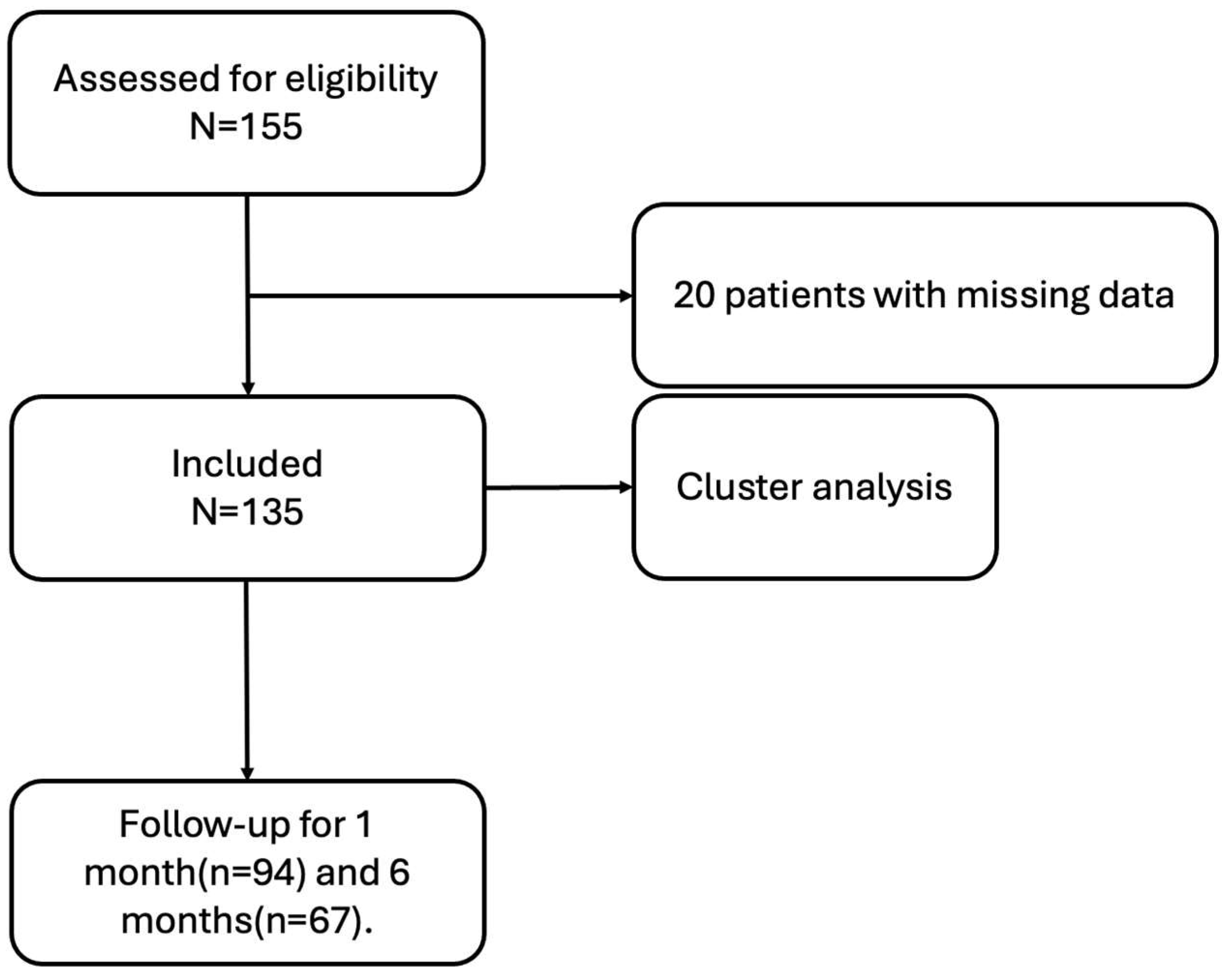
Figure 1. Flow diagram of the study.
Jcm 14 08116 g001
Figure 2. Three clusters of patients with SAPHO syndrome identified by cluster analysis. Colors indicate cluster membership.
Jcm 14 08116 g002
Figure 3. Representative whole-body bone scintigraphy, computed tomography, and magnetic resonance imaging findings in patients with SAPHO syndrome. (A): Cluster 1 (typical SAPHO syndrome); (B): Cluster 2 (axial SAPHO syndrome); (C): Cluster 3 (peripheral SAPHO syndrome).
Jcm 14 08116 g003
Figure 4. Improvement in VAS scores across the three clusters after treatment.
Jcm 14 08116 g004
Table 1. Baseline demographic and clinical characteristics of patients with SAPHO syndrome.
Demographic and Clinical Characteristics
Demographic characteristics
Sex, female/male95/40
Age, mean (S.D.), years44.49 (13.89)
Disease duration, mean (S.D.), months59.10 (81.50)
Smoking, n (%)26 (19.3%)
Alcohol intake, n (%)30 (22.2%)
Clinical characteristics
Osteoarticular symptoms
Single site involvement, n (%)26 (19.3%)
Symmetry of lesion site, n (%)85 (63.0%)
Bone destruction, n (%)12 (8.9%)
Osteosclerosis, n (%)102 (75.6%)
Anterior chest wall, n (%)88 (65.2%)
Spine, n (%)55 (40.7%)
Peripheral bones and joints, n (%)37 (27.4%)
Skin manifestations
PPP, n (%)38 (28.1%)
PV, n (%)5 (3.7%)
SA, n (%)11 (8.1%)
Nonspecific rash, n (%)22 (16.7%)
None61 (45.2%)
Disease assessment index
VAS, mean (S.D.)6.88 (2.12)
BASDAI, mean (S.D.)3.46 (1.92)
BASFI, mean (S.D.)1.79 (2.02)
HAQ, mean (S.D.)0.41 (0.48)
Laboratory tests
Elevated ESR (n = 108), n (%)56 (41.5%)
Elevated CRP (n = 106), n (%)33 (24.4%)
TNF-α (n = 81), mean (S.D.)2.50 (1.41)
IL-6 (n = 82), mean (S.D.)8.82 (7.57)
IL-8 (n = 82), mean (S.D.)10.10 (12.07)
tP1NP (n = 71), mean (S.D.)74.28 (97.57)
β-CTX (n = 36), mean (S.D.)4.69 (11.91)
PTH (n = 99), mean (S.D.)35.31 (13.11)
25-(OH)VD3 (n = 108), mean (S.D.)22.88 (9.33)
Pathogenic bacteria (n = 21), n (%)10 (47.6%)
PPP, palmoplantar pustulosis; PV, psoriasis vulgaris; SA, severe acne; VAS, Visual Analog Scale; BASDAI, Bath Ankylosing Spondylitis Disease Activity Index; BASFI, Bath Ankylosing Spondylitis Functional Index; HAQ, Health Assessment Questionnaire.
Table 2. Comparison of clinical and laboratory features among clusters.
Demographic
Characteristics
Cluster 1 (n = 87)Cluster 2 (n = 19)Cluster 3 (n = 29)p Values *p Values **p Values ***
Sex, female64 (73.6%)11 (57.9%)20 (69.0%)0.2640.6380.541
Age46.75 (12.83)45.68 (10.09)36.92 (16.62)0.5470.0030.047
Disease duration60.68 (76.77)54.90 (70.45)57.14 (102.34)0.4010.0120.374
Smoking18 (20.7%)4 (21.1%)4 (13.8%)0.9720.5860.509
Alcohol intake23 (26.4%)6 (31.6%)1 (3.4%)0.7770.0070.007
Osteoarticular
symptoms
Single site involvement01 (5.3%)25 (86.2%)0.032<0.001<0.001
Symmetry of lesion site77 (88.5%)7 (36.6%)1 (3.4%)<0.001<0.0010.002
Bone destruction6 (6.9%)1 (5.3%)5 (17.2%)0.7950.1000.220
osteosclerosis70 (80.5%)4 (21.1%)28 (96.6%)<0.0010.038<0.001
Anterior chest wall77 (88.5%)7 (36.8%)4 (13.8%)<0.001<0.0010.063
Spine45 (51.7%)9 (47.4%)1 (3.4%)0.803<0.001<0.001
Peripheral bones and joints12 (13.8%)3 (15.8%)22 (75.9%)0.821<0.001<0.001
Skin manifestations65 (74.7%)2 (10.5%)7 (24.1%)<0.001<0.0010.237
VAS6.94 (2.15)6.84 (2.54)6.72 (1.77)0.9500.4570.482
BASDAI3.79 (2.09)3.42 (1.57)2.51 (1.18)0.5780.0030.033
BASFI1.99 (2.12)2.26 (2.01)0.89 (1.37)0.3960.0010.002
HAQ0.42 (0.51)0.57 (0.46)0.25 (0.33)0.1030.0600.008
Laboratory tests
Elevated ESR (n = 92)33 (50.0%)10 (66.7%)13 (48.1%)0.2691.0000.337
Elevated CRP (n = 90)20 (30.8%)4 (30.8%)9 (32.1%)1.0001.0000.930
*: cluster 1 vs. cluster 2; **: cluster 1 vs. cluster 3; ***: cluster 2 vs. cluster 3.
Disclaimer/Publisher’s Note: The statements, opinions and data contained in all publications are solely those of the individual author(s) and contributor(s) and not of MDPI and/or the editor(s). MDPI and/or the editor(s) disclaim responsibility for any injury to people or property resulting from any ideas, methods, instructions or products referred to in the content.