Review

Radiolabeled LHRH and FSH Analogues as Cancer Theranostic Agents: A Systematic Review

Nuclear Medicine Unit, Department of Medical-Surgical Sciences and of Translational Medicine, Faculty of Medicine and Psychology, “Sapienza” University of Rome, 00189 Rome, Italy
*
Author to whom correspondence should be addressed.
J. Clin. Med. 2025, 14(21), 7811; https://doi.org/10.3390/jcm14217811
Submission received: 30 September 2025 / Revised: 21 October 2025 / Accepted: 1 November 2025 / Published: 3 November 2025

Abstract

Background: G-protein-coupled receptors (GPCRs) play pivotal roles in tumor growth and progression. Among these, the luteinizing hormone-releasing hormone receptor (LHRH-R) and follicle-stimulating hormone receptor (FSH-R) represent promising translational targets, unlike luteinizing hormone receptors (LH-Rs). Indeed, both LHRH-R and FSH-R are selectively expressed in various cancers and their vasculature, offering opportunities for receptor-mediated imaging and therapy. Objectives: This systematic review aims to evaluate radiolabeled LHRH- and FSH-derivative biomolecules, including peptides, monoclonal antibodies and nanocarriers, for their applications in cancer diagnosis and treatment. Methods: The systematic review was conducted following PRISMA 2020 guidelines. A systematic search of PubMed, Scopus and Web of Science was conducted for studies published between 2005 and 2025. A total of 248 records were identified, and 156 articles were screened after removing duplicate records. Two authors independently selected eligible studies. Quality of evidence was assessed by the Quality Assessment of Diagnostic Accuracy Studies (QUADAS) approach. Results: A total of 25 studies met the inclusion criteria and were included in the final review. Radiolabeled LHRH and FSH derivatives showed receptor-specific tumor localization in both preclinical and clinical applications. FSH-R expression in tumor blood vessels supports its potential as a biomarker for early cancer diagnosis. FSHβ-derived peptides exhibit improved pharmacokinetics compared to monoclonal antibodies in PET imaging. LHRH analogues, particularly D-Lys6-modified peptides, proved effective for SPECT, PET and therapeutic applications, particularly in breast and prostate cancer. The integration of radiolabeled LHRH and FSH derivatives with nanocarriers further enhanced probe stability and tumor targeting, increasing tumor accumulation and image contrast compared to free peptide. Conclusions: Radiopharmaceuticals targeting LHRH-R and FSH-R are promising tools for cancer imaging and treatment. Advances in nanotechnology enhance delivery precision and reduce systemic toxicity, thereby increasing its translational promise in oncology.

1. Introduction

G-protein-coupled receptors (GPCRs) are the largest and most common class of cell surface signaling receptors known to play essential roles in physiological activities, including tumor growth and metastasis. A broad range of ligands, including hormones, lipids, peptides and neurotransmitters, activate GPCRs, enabling these receptors to interact with G-proteins and activate multiple downstream signaling pathways. Integration of these complexes’ signaling networks produces numerous biochemical responses that drive diverse pathophysiological processes, including cancer development. In the GPCR family, the luteinizing hormone-releasing hormone receptor (LHRH-R) and follicle-stimulating hormone receptor (FSH-R) offer strong translational potential [1].
LHRH, also known as gonadotropin-releasing hormone (GnRH), is a hypothalamic decapeptide (sequence: pGlu-His-Trp-Ser-Tyr-Gly-Leu-Arg-Pro-Gly-NH2) that plays a pivotal role in the regulation of reproductive physiology. It stimulates the pituitary gland to release LH and FSH, which control the production of gonadal sex steroids in both males and females [2,3]. In addition to its well-established role in the hypothalamic–pituitary–gonadal axis, expression of extra-pituitary LHRH-R (or GnRH-R) has been observed in several tissues and malignancies, including cancer cells [4]. Although its biological role in tumors remains unclear, evidence suggests that LHRH peptides may act as local regulators of tumor growth [5]. Early studies primarily linked LHRH-R overexpression to hormone-dependent cancers [6], such as breast [7], endometrial [8], ovarian [9] and prostate cancers [10] (Figure 1). However, subsequent findings have demonstrated variable receptor expression in several hormone-independent cancers, including pancreatic [11], renal [12] and lung carcinomas [13], as well as melanoma [14] and glioblastoma [15] (Figure 1).
Notably, LHRH-R expression in these cancers is often significantly higher than in corresponding normal tissues, including those of reproductive origin. Clinical investigations have also shown that treatment with LHRH agonists does not exert substantial therapeutic effects in cancers characterized by LHRH-R overexpression [16]. Nonetheless, the selective expression of these receptors in tumors highlights the potential of LHRH targeting peptides and their derivatives as drug-delivery systems, enabling targeted therapy while sparing healthy tissues. As highlighted by Li and colleagues, various LHRH-targeted drug delivery approaches are currently under investigation for oncological applications [17,18,19].
Similarly, FSH-R may also be considered a promising biomarker for targeted therapy and diagnosis in aggressive tumors [20]. FSH-Rs are usually expressed by cells of the gonads and are absent in other normal tissues. They are mainly overexpressed by ovarian and prostate cancer cells [20,21].
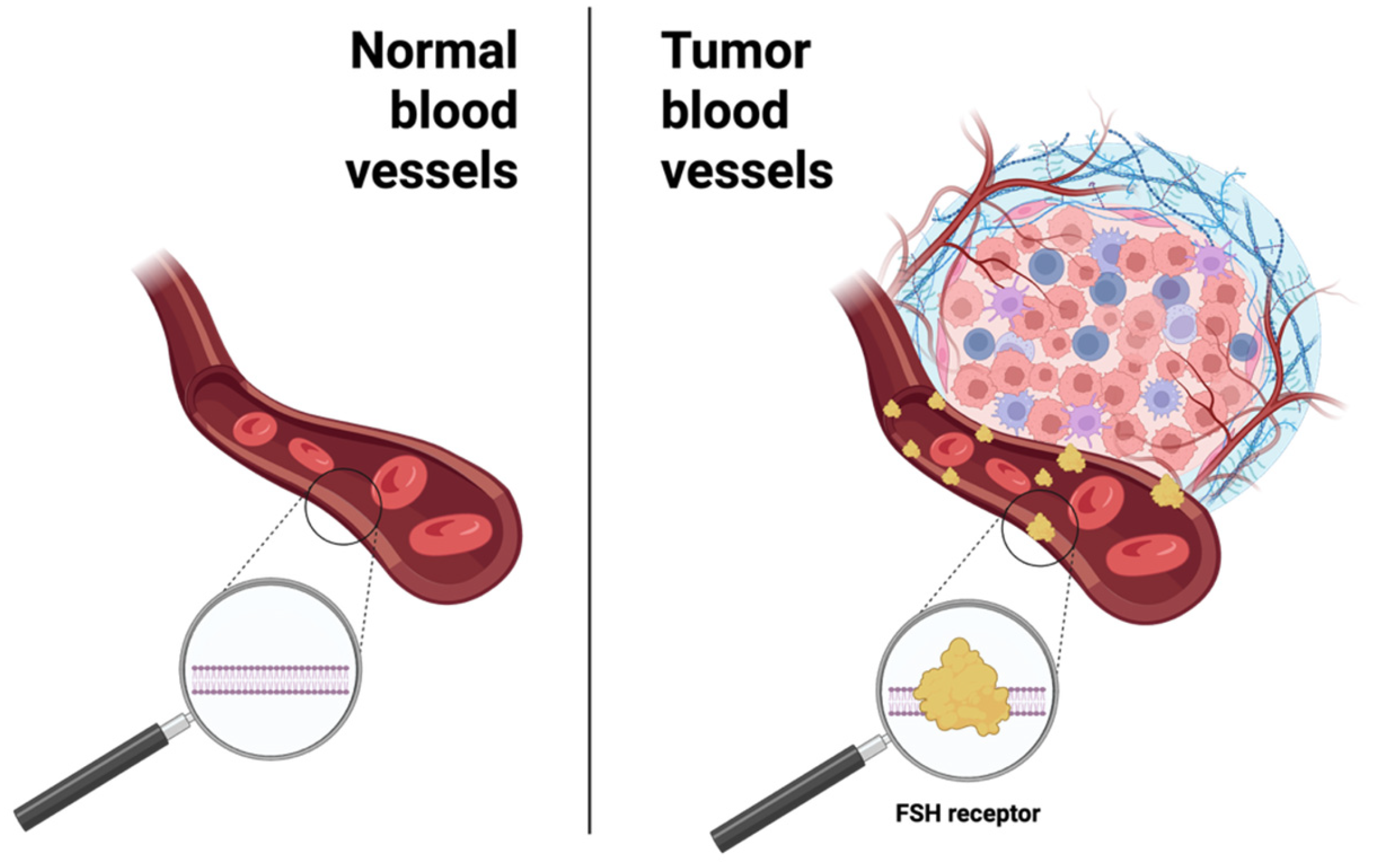
In the 2010s, FSH-R raised renewed interest as a highly selective marker for tumor blood vessels (TBVs) in primary and metastatic solid cancers, detectable at very early stages of the disease (Figure 2).
Adjacent normal tissues and blood vessels in inflammatory lesions and in normal wound healing were uniformly negative for FSH-R [22,23]. Notably, FSH-R-positive (FSH-R+) TBVs are located at the boundary between tumoral and normal tissues, suggesting that FSH-R contributes to tumor initiation, progression and metastasis, consistent with its established role in regulating ovarian angiogenesis [21]. Additionally, FSH-R expression is not restricted to gonadal tumors [20]. Recently, it has been demonstrated that a wide range of human and murine cancer cells heterogeneously express FSH-R. To this end, many efforts have already been made to design new tools for noninvasive imaging of FSH-R.
Since LHRH-R and FSH-R are overexpressed only in cancer cells [16,17,24,25] and absent in normal tissues, they can be considered a selective target useful to minimize off-target radiation exposure both in imaging and therapy.
Recent advances in the development of radiolabeled LHRH and FSH biomolecules, such as antibodies and peptides and integration with nano-platforms, have been widely used for single-photon emission computed tomography (SPECT, using 99mTc and 111In) and positron emission tomography (PET, using 68Ga and 18F) imaging, with a smaller number evaluated also for therapeutic use (177Lu).
Radiolabeled monoclonal antibodies (mAbs) are evaluated for their ability to bind with high specificity and affinity to defined molecular structures, which makes them particularly suitable for both therapeutic and diagnostic applications. The use of monoclonal antibodies in nuclear medicine offers several advantages but also presents limitations. Radiolabeled mAbs offer excellent target selectivity and can provide insights into tumor heterogeneity, receptor density and disease progression over time. This precision enables personalized diagnostic strategies and potentially aids in treatment planning. However, the relatively large size of mAbs leads to slower pharmacokinetics, prolonged circulation times and sometimes suboptimal tumor penetration. Additionally, the use of long-lived radionuclides may increase radiation burden, while antibody production itself can be costly and technically demanding.
Radiolabeled peptides have emerged as a versatile class of molecular probes in nuclear medicine, with applications in both diagnostic imaging and targeted radionuclide therapy. Their relatively small size, compared to monoclonal antibodies, facilitates rapid tissue penetration and favorable pharmacokinetics, resulting in a high target-to-background contrast within a shorter time frame. Moreover, their small size also ensures rapid clearance from the bloodstream.
Peptides can be chemically modified with relative ease, allowing optimization of pharmacokinetics, enzymatic stability and receptor binding. However, sometimes their rapid renal clearance may lead to insufficient tumor retention, reducing sensitivity in imaging and efficacy in therapy. Peptides generally exhibit lower absolute binding specificity compared to whole antibodies, which may increase background signal in specific settings. Their relatively short in vivo half-life may necessitate the use of radionuclides with matching physical properties, limiting flexibility in probe design.
Furthermore, nanotechnology is increasingly applied in nuclear medicine to enhance the delivery and efficacy of radiopharmaceuticals. Gold nanoparticles, BSA nanoclusters and also solid-lipid nanocarriers are just some of the examples of nanotechnologies currently in use which have been identified for applications in targeted imaging of tumors overexpressing specific receptors (Figure 3).
This systematic review compiles the studies reported over the past 20 years on radiolabeled LHRH and FSH biomolecules for cancer management. We include radiolabeling chemistry and characterization of peptide and antibody conjugates, in vitro and in vivo biodistribution and pharmacokinetics, clinical investigations and integrations with nanotechnology platforms.
Our objective is to integrate the reported studies to evaluate LHRH/FSH targeting for consistent radiolabeling and receptor-specific localization in tumors and their vasculature, thereby supporting present and future theranostic applications.

2. Materials and Methods

2.1. Search Strategy

The published studies were systematically collected using databases such as PubMed, Scopus and Web of Science. A search algorithm based on the combined terms “((“Follicle stimulating hormone” OR FSH) AND “radiolabeling” AND “cancer”)”, “((“Luteinizing hormone releasing hormone” OR LHRH) AND “radiolabeling” AND “cancer”)”, “((“Gonadotropin releasing hormone” OR GnRH) AND “radiolabeling” AND “cancer”)” was employed. Reference lists of included studies and relevant reviews were also screened to identify additional records.
Other filters were applied such as “Research Articles” and “Publication year from 2005 to 2025”. In PubMed a database-specific date filter was used, selecting papers from “1 January 2005” to “30 September 2025”.
Only original research papers published in English were considered, following the PRISMA 2020 guidelines [26,27].
Quality control was ensured by implementing the PRISMA checklist (Supplementary Table S1). Two authors independently critically assessed the overall quality of the selected studies. Only studies using radionuclides for nuclear medicine applications were included.

2.2. Inclusion and Exclusion Criteria

Literature studies published up to September 2025 were assessed. The bibliography of the research articles was manually searched to assess further relevant articles. Full-text articles were evaluated, and preclinical results were considered for inclusion and analysis. Letters to the editor, supplementary commentaries, book chapters, review articles and non-English publications were excluded from the analysis.

2.3. Data Extraction and Management

Two authors independently screened full-text manuscripts for eligibility, summarizing essential data. Risk assessment of any potential bias and data collection were performed using a standardized questionnaire reported in Appendix A (Table A1). The questionnaire was adapted to the studies included in this systematic review.

2.4. Assessment of Risk Bias in Included Studies

All included studies were evaluated by using the Quality Assessment of Diagnostic Accuracy Studies (QUADAS) approach for any potential source of bias and variation [27]. To obtain all this information, each study was analyzed individually.
The following variables were analyzed: (1) animal selection bias (animal origin, animal model and sex distribution), (2) animal variation (sex distribution and disease severity), (3) index test bias (bacterium origin, bacterium number, conjugation chelator-biomolecule, radiochemical purity and extra in vitro/in vivo/ex-vivo studies), (4) index test variation (observer variation, availability of experimental information, radiopharmaceutical purity and specific activity and test execution), (5) reference standard bias (inappropriate or inconsistent reference standard and incorporation bias), (6) reference standard variation (definition of a control model) and (7) flow and timing bias (radiopharmaceutical administration time, imaging time bias, uninterpretable results and study flow).

3. Results and Discussion

3.1. Data Synthesis

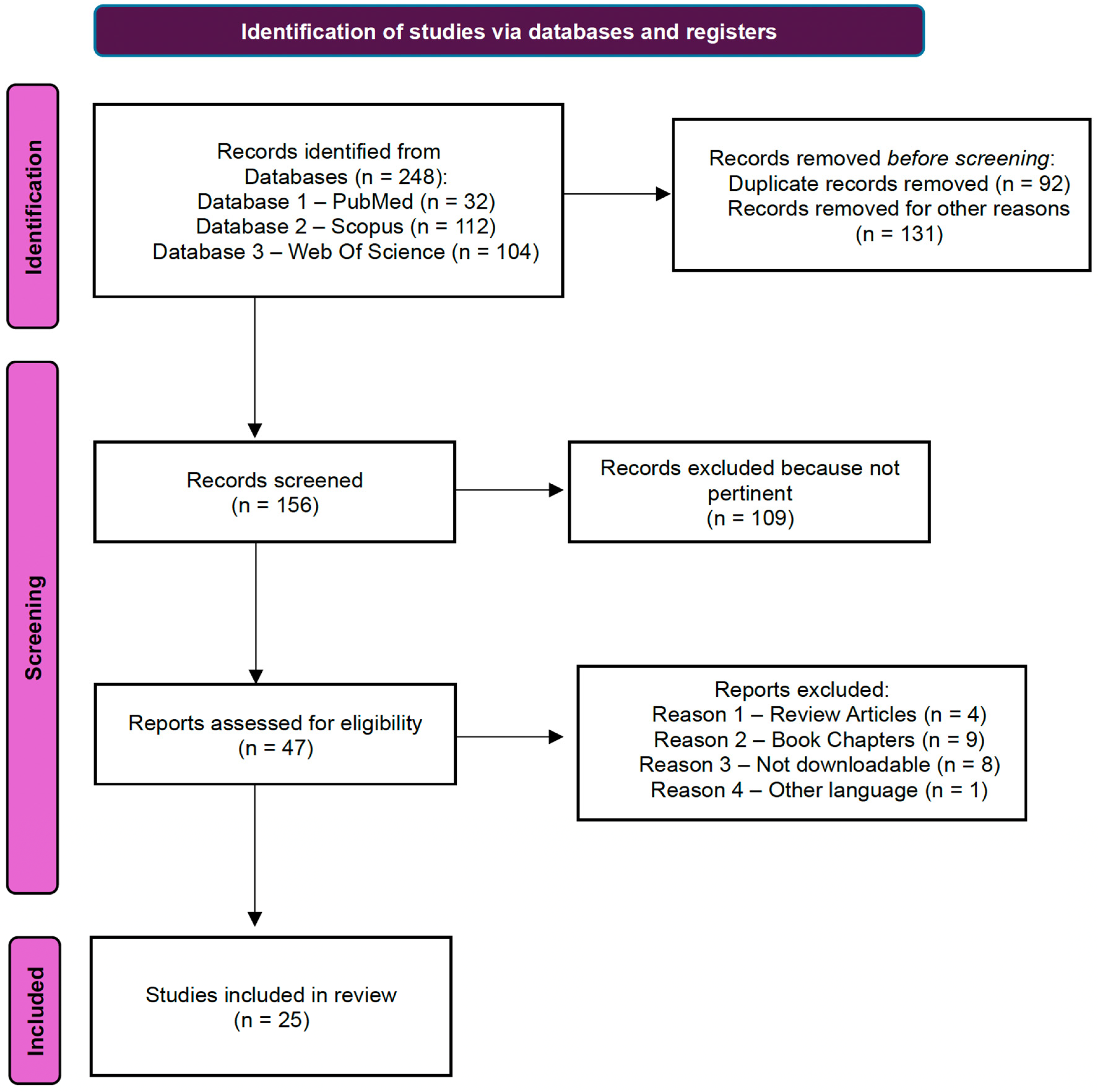
Studies for this systematic review were selected by two authors. In Figure 4 a flowchart illustrating the search for eligible studies is reported. Starting from 248 papers identified on three different databases (32 on PubMed, 112 on Scopus and 104 on Web of Science), duplicate records were removed (n = 92). Of the 156 remaining works, 109 were excluded because they were not pertinent to our criteria (research articles regarding FSH- and LHRH-derivative radiopharmaceuticals useful in nuclear medicine). Following this procedure, 47 potential studies were identified. After excluding non-review articles (n = 4), book chapters (n = 9), non-downloadable (n = 8) and other language (n = 1) articles, only 25 published articles were selected for our study. A summary of each paper’s results is reported in the following paragraphs.
In Table 1 and Figure 5, QUADAS analysis results are reported. To facilitate the analysis, the studies were divided into analyses of radiopharmaceuticals derived from FSH and LHRH, and for the latter, they were further divided into SPECT, PET, therapy and other applications. Selected papers appear to be heterogeneous as they involve only in vitro and in vivo studies.

3.2. Radiolabeled Biomolecule Targeting FSH-R

FSH-R has the potential to serve as a valuable biomarker in oncology, facilitating early cancer detection and contributing predictive value to personalized medicine. In 2005, Fan and Hendrickson solved the first crystallographic structure of the FSH hormone in a complex with the extracellular domain of the FSH-R [53]. FSH is a heterodimeric glycoprotein consisting of two distinct subunits, α and β, with the latter conferring biological activity and FSH-R specificity [54]. The interaction between the glycoprotein hormones and their corresponding receptors is highly selective: the FSH-FSH-R structure allows us to understand in detail the molecular requirements underlying the interaction specificity [53] and paves the way for the design of highly specific and selective FSH derivatives. Some of them are based on monoclonal antibodies (mAbs) or peptides derived from the human FSHβ portion. Molecular imaging with radionuclides, such as PET, provides a powerful tool to visualize and monitor dynamic biochemical processes and the distribution of specific molecular targets in vivo. To improve the tumor localization and the sustained detection of FSH-R expression, researchers have applied PET imaging using different radiopharmaceuticals using both monoclonal antibodies and peptides.

3.2.1. Radiolabeled Monoclonal Antibodies Against FSH-R

In this context, a monoclonal antibody (FSH-R mAb) against FSH-R has already been used in a nuclear medicine (Table 2).
In 2015, Hong and coworkers performed functionalization with a chelator agent and radiolabeling with 64Cu for PET imaging of FSH-R in various tumor models [28]. They developed 64Cu-NOTA−FSH-R mAbs, an immune-PET tracer for FSH-R. Since FSH-R is selectively expressed in the vasculature of a wide range of solid tumors but largely absent from most normal tissues, the authors aimed to establish its broad applicability as a molecular imaging target. In this study, in vivo PET/CT in nude mice bearing CAOV-3 (FSH-R+) ovarian, SKOV-3 (FSH-R-) ovarian, MDA-MB-231 breast and PC-3 prostate xenografts showed robust, time-dependent uptake. Histology studies confirmed widespread microvascular FSH-R across models and notable tumor-cell FSH-R in CAOV-3, PC-3 and MDA-MB-231; the authors note that endothelial versus tumor-cell contributions could not be fully disentangled, and in vivo blocking was not performed due to limited antibodies. Overall, 64Cu-NOTA-FSH-R mAbs enabled specific, quantitative imaging of FSH-R across multiple tumor types, supporting FSH-R as a broadly applicable target for non-invasive cancer imaging and potential theranostics.
One year later, Yang et al. engineered a tumor-vasculature-targeted nano-graphene oxide (GO) platform [29]. GO nanosheets were initially functionalized with branched PEG, and after they were attached to NOTA and FSH-R mAbs and radiolabeled with 64Cu. Graphene derivatives are considered attractive drug-delivery systems. In this study, the authors demonstrated efficient metastatic tumor targeting of GO conjugates in vivo, in a murine model of lung metastasis for breast cancer. In vitro, fluorescein-GO-FSH-R-mAbs bound MDA-MB-231 cells (FSH-R+) far more strongly than non-targeted GO, with negligible uptake in SKOV-3 cells (FSH-R-); antibody denaturation abrogated binding, confirming specificity. In an experimental lung-metastasis model (female nude mice injected intravenous with cbgLuc-MDA-MB-231), serial PET at increasing times post-injection showed rapid and sustained uptake of 64Cu-NOTA-GO-FSH-R-mAbs in metastatic nodules, significantly exceeding both non-targeted 64Cu-NOTA-GO and a denatured-antibody control. Biodistribution indicated predominant hepatobiliary clearance and low muscle background, yielding high-contrast detection of micro-metastases; histology localized GO-FSH-R mAbs to tumor vasculature and confirmed liver/spleen capture. As a drug carrier, GO-FSH-R mAbs demonstrated a high doxorubicin loading capacity and enhanced delivery to metastatic sites, as confirmed by fluorescence imaging, supporting this system as a dual diagnostic–therapeutic platform for early metastasis detection and targeted delivery.
So, as can be understood from these studies, antibodies represent an excellent alternative in PET imaging. Despite these results, there are limits due to their nature, such as long circulating half-life (t1/2) and nonspecific accumulation, with consequent poor tumor-to-blood and tumor-to-organ ratios. Conversely, different peptides have been tested with promising results, and the most successful turned out to be FSHβ33-53, a linear peptide deriving from the human FSHβ portion [54].
In the QUADAS analysis, no sources of bias were reported for these two studies.

3.2.2. Radiolabeled FSHβ-Derivative Peptides in Cancer Imaging

FSHβ33-53 has been investigated since the early 90s by Santa Coloma and coworkers for ovarian cancer targeting [54,55]. The amino acid residues of FSHβ that interact with the receptor are found both in the loop containing the sequence FSHβ 95–100 and in the region of the loop comprising residues 38–48, as reported by Jiang and colleagues [56].
FSHβ33–53 has recently gained interest as a development for FSH-R-selective drugs and radiopharmaceuticals for cancer imaging [30,31,32,33] and therapy [28,57]. It demonstrates excellent in vivo stability and FSH-R specificity but shows suboptimal pharmacokinetics and tumor-to-kidney ratios [30,31,32]. These limitations are attributed to dimerization at physiological pH via intermolecular disulfide bonds between cysteine residues on the monomers [55]. In general, peptide dimerization may reduce its biological efficacy by altering conformation and solubility, decreasing receptor specificity and bioavailability, limiting tumor penetration and leading to suboptimal tumor-to-kidney ratios.
In this context, four different research articles reporting radiolabeled FSHβ-derivative peptides have been published (Table 3). The first one was published in 2014 by Xu and coworkers, who synthesized and characterized 18F-Al-NOTA-MAL-FSH1, a novel 18F-labeled FSH-R probe for tumor PET imaging [30]. In vivo evaluation was performed in nude mice bearing FSH-R-positive and FSH-R-negative PC-3 xenograft tumors. PET imaging demonstrated clear visualization of FSH-R-positive tumors, with significantly higher tracer uptake compared to controls and favorable tumor-to-muscle ratios. The probe showed rapid tumor accumulation evident within 30 min post-injection and relatively low background activity, except for renal clearance. Receptor-blocking experiments further validated the specificity of tumor uptake. This peptide has favorable pharmacokinetics, specific tumor targeting, rapid blood clearance and predominantly renal excretion. 18F-Al-NOTA-MAL-FSH1 represents a potential and promising radiotracer for the non-invasive visualization of FSH-R-positive tumors in vivo.
In 2016, Zhu and coworkers modified the FSH-R antagonist peptide sequence with a hydrophilic linker (18F-Al-NOTA-MAL-FSH2) to improve the pharmacokinetic properties and reduce nonspecific uptake in non-target tissue [31]. This radiolabeled peptide remains stable in PBS and human serum. In vitro assays confirmed high binding affinity and specificity for FSH-R. PET imaging of nude mice bearing FSH-R-positive PC-3 xenografts demonstrated pronounced tumor uptake and clear visualization against a low background. In conclusion, 18F-Al-NOTA-MAL-FSH2 showed better pharmacokinetics than the previously reported 18F-Al-NOTA-MAL-FSH1. A favorable preclinical study revealed that the incorporation of a hydrophilic linker enhances the imaging performance. 18F-Al-NOTA-MAL-FSH2 appears to be a promising candidate for FSH-R-positive tumor imaging.
In addition to 18F, 68Ga is currently a promising PET radionuclide due to its availability, nearly quantitative reaction and short physical half-life. In 2017, Pan and coworkers developed a 68Ga labeled FSH1 peptide for FSH-R imaging [32]. In vitro studies and microPET imaging were performed in the PC-3 prostate tumor model. [68Ga]Ga-NOTA-MAL-FSH1 was stable in PBS and human serum for at least 2 h. PET imaging revealed that the PC-3 xenografts were clearly visualized, with specific tumor uptakes and good tumor-to-background contrast, with rapid renal excretion and minimal nonspecific accumulation in other organs. Biodistribution studies confirmed these findings, demonstrating high tumor-to-muscle ratios and receptor-mediated specificity confirmed by blocking experiments. Preclinical data obtained in this paper indicate that this radiopharmaceutical is promising for noninvasive visualization of FSH-R expression in vivo, even in other cancer types.
In the QUADAS analysis, the main source of bias was related to the animal sex distribution and inconsistent reference standards and definitions of control models being applied.
In 2022, Pan and colleagues optimized the performance of [68Ga]Ga-NOTAMAL-FSH1 in a prostate cancer model by co-administration of aprotinin [33]. Aprotinin is a competitive, reversible, heat- and acid-stable inhibitor of proteolytic and esterolytic activities. To improve the in vivo stability and tumor uptake of peptide-radiopharmaceuticals, the authors modified the amino acid sequence, resulting in [68Ga]Ga-NOTAMAL-FSH4, and evaluated it with aprotinin. In vivo studies in nude mice bearing PC-3 prostate cancer xenografts demonstrated that co-injection with aprotinin further increased tumor uptake and tumor-to-background contrast while maintaining low nonspecific accumulation in non-target tissues. Biodistribution analyses confirmed enhanced tumor retention and favorable tumor-to-muscle ratios without affecting renal clearance. All these findings indicate that the sequence modification combined with protease inhibition effectively improves the performance of the 68Ga labeled peptide to benefit future clinical applications.
Molecular PET imaging of FSH-R is beneficial for cancer prognosis, and the findings related to the FSHβ33–53 peptide confirmed its receptor selectivity and affirmed its relevance in nuclear medicine imaging applications.

3.3. Radiolabeled LHRH Derivative Peptides

A significant number of tumors overexpresses the gonadotropin-releasing hormone type 1 receptor (GnRH1 receptor), which offers a promising molecular target for both diagnostic imaging by PET or SPECT and radionuclide therapy. More generally, GnRH receptors are overexpressed in various hormone-dependent (and independent) malignancies, including prostate, breast and ovarian cancers, making them attractive targets in nuclear medicine. Several preclinical studies have explored radiolabeled analogues of GnRH (or LHRH) peptides as agents for receptor-targeted nuclear medicine applications, and the following works provide critical insights into their development and translational potential.
In the following paragraphs, a wide variety of radiolabeled biomolecules suitable for overexpressing LHRH cancer imaging, specifically for PET (68Ga, 18F), SPECT (111In, 99mTc) and therapy (177Lu) applications, are reported. In addition, we also describe these biomolecules conjugated with novel nanotechnology, like nanoparticles and macroaggregates.

3.3.1. Radiolabeled LHRH in SPECT Imaging Applications

Several LHRH derivative peptides radiolabeled with 99mTc and 111In have been published (Table 4).
In 2019 Zoghi and coworkers investigated the synthesis, radiolabeling and preclinical evaluation of a novel 111In-labeled GnRH-I peptide-based radiopharmaceutical ([111In]-DOTA-TRP) designed for SPECT imaging of tumors overexpressing GnRH1 receptors [34]. The radiolabeled compound demonstrated excellent stability in vitro both in PBS and in human serum, with minimal degradation observed over 24 h. In vitro receptor binding assays confirmed that the radiolabeled peptide retained its affinity for GnRH-I receptors, indicating that the DOTA conjugation and radioisotope incorporation did not compromise its biological function. In biodistribution studies performed in normal and tumor-bearing mice (4T1-bearing mice), the tracer exhibited favorable pharmacokinetics with rapid clearance from the bloodstream, primarily through the renal pathway and low non-specific uptake in non-target tissues. Most importantly, the radiopharmaceuticals showed target-specific accumulation in tumors, with significantly higher uptake in receptor-positive tumors compared to background organs and muscle, leading to promising tumor-to-nontumor contrast ratios. Distinct ovarian uptake was also observed. In the end, authors presented [111In]-DOTA-TRP as a promising radiotracer candidate for GnRH receptor SPECT imaging. The results support further preclinical validation and provide a solid basis for future clinical translation, particularly in the context of ovarian and other GnRH receptor-rich tumors.
In 2011, Guo and coworkers developed three novel DOTA-conjugated peptides for breast cancer imaging [35]. The chelator was conjugated in different positions: to the epsilon or alpha amino group of D-lysine or the epsilon amino group of L-lysine via an Ahx linker, obtaining DOTA-Ahx-(D-Lys6-GnRH1), DOTA-Ahx-(D-Lys6-GnRH2) and DOTA-Ahx-(L-Lys6-GnRH3). Only the DOTA-Ahx-(D-Lys6-GnRH1) peptide maintained the nanomolar GnRH receptor binding affinity. Moreover, the biodistribution and tumor imaging properties of 111In-DOTA-Ahx-(D-Lys6-GnRH1) were evaluated in MDA-MB-231 human breast cancer-xenografted nude mice. These radiopharmaceuticals exhibited specific GnRH receptor binding, rapid tumor uptake and fast clearance through the urinary system. Tumor lesions were clearly visualized 1 h post-injection by SPECT/CT. Ultimately, authors demonstrated the impact of chelator position on the binding affinity for the receptor and the potential of the radiopharmaceuticals as a novel imaging probe for human breast cancer imaging.
Based on previous results, Guo and coworkers designed a new study in 2011 by radiolabeling (DOTA)-Ahx-(D-Lys6-GnRH1) with 111In [36]. The authors evaluated the tumor targeting and imaging properties of the novel radiopharmaceuticals for human prostate cancer, both in vitro and in vivo, in DU145 human prostate cancer-xenografted nude mice. The radiopharmaceuticals exhibited high binding affinity and specific internalization in GnRH receptor–positive cells, and in vivo biodistribution studies demonstrated rapid tumor uptake, fast clearance from blood and non-target tissues and clear tumor visualization on SPECT/CT images within 30 min of administration. Renal retention was observed due to the pharmacokinetic profile of the small peptide, but overall tumor-to-background ratios were favorable. The ability of 111In-DOTA-Ahx-(D-Lys6)-GnRH1 demonstrated its promise as a novel probe for the imaging of human prostate tumors.
In 2017, Xu and coworkers reported the design, synthesis and biological evaluation of novel 111In-labeled GnRH peptide analogues for prostate cancer imaging [37]. Three GnRH peptides were synthesized with different hydrocarbon linkers: 111In-DOTA-Aoc-D-Phe-(D-Lys6-GnRH), 111In-DOTA-βAla-D-Phe-(D-Lys6-GnRH) and 111In-DOTA-Aun-D-Phe-(D-Lys6-GnRH). The authors demonstrated that Aoc is better than βAla and Aun linkers in retaining strong receptor binding affinity. 111In-DOTA-Aoc-D-Phe-(D-Lys6-GnRH) shows faster tumor uptake and urinary clearance in DU145 human prostate cancer-xenografted nude mice. These findings highlighted the potential of this 111In-labeled GnRH peptide as a promising molecular probe for prostate cancer imaging by SPECT, supporting further optimization and translational research in this area.
Farahani et al. conducted a series of preclinical studies to evaluate a 99mTc-labeled-DLys6-GnRH analogue for prostate cancer imaging [38,39]. In one report, the peptide was modified with aminobutyric acid (GABA) as linker (99mTc-HYNIC-GABA-D-Lys6-GnRH) [38], while, in another report, amino hexanoic acid (Ahx) was used as a hydrocarbon linker to generate 99mTc-HYNIC-Ahx-DLys6-GnRH [39]. Both the analogues showed the highest affinity for LN-CaP cells. Reported Kd values were ~89 nM (GABA-linker analogue) and ~42 nM (Ahx-linker analogue), indicating stronger binding affinity for the Ahx-containing derivative. In vivo biodistribution studies in LN-CaP xenografted nude mice revealed similar pharmacokinetic profiles for both tracers, but tumor uptake and imaging contrast differed. The GABA analogue achieved a tumor uptake of 1.72 %ID/g at one hour with a tumor-to-muscle ratio of 2.3. In contrast, the Ahx analogue reached a higher uptake of 3.67 %ID/g at one hour and a high tumor-to-muscle ratio of 4.14. SPECT imaging confirmed clear visualization of LN-CaP tumors within 1–2 h for both agents, with the Ahx variant producing sharper contrast. Collectively, these studies demonstrate that radiolabeled GnRH analogues reliably target GnRH receptor-positive prostate cancer.
In 2017, Hao and colleagues performed direct radiolabeling of 99mTc with LHRH using a pre-thinning approach with sodium gluconate to suppress colloid formation [40]. The serum stability results showed a drop of RCP from ~90% at two hours to ~75% by three hours, indicating a narrow window for optimal image acquisition. In vitro receptor binding assay on rat pituitary membrane showed high-affinity, saturable binding (KD ≈ 0.435 nM; RT ≈ 23.2 pmol). In vivo biodistribution of 125I-LHRH in mice showed rapid blood clearance (<1 %ID/g by four hours) with renal uptake (~9.24 %ID/g at 15 min), yielding high target-to-background kinetics for receptor imaging.
Calderon et al. functionalized LHRH with a tridentate Acdien chelator and radiolabeled with 99mTc (99mTc-Acdien-LHRH) [41]. The radiolabeled analogue achieved 99% RCP. In breast cancer cells (MDA-MB-231 cells), the radiolabeled analogue showed time-dependent uptake (15–120 min) that was significantly higher than fac-[99mTc(CO)3(H2O)3]+. Excess LHRH competitively reduced uptake, confirming LHRH-receptor–mediated targeting. These findings indicate a co-ligand-free LHRH-carbonyl-Tc design with clean radiochemistry and selective in vitro targeting appropriate for in vivo testing.
Overall, these studies highlight the potential of LHRH derivative peptides radiolabeled with 111In or 99mTc. The receptor-targeted imaging approach offers an advantage by increasing the chance of related tumor detection, decreasing false negative and nonspecific accumulations. Nevertheless, 111In is burdened by its longer half-life, poorer image quality and less favorable dosimetry and higher costs compared to 99mTc.
In the QUADAS analysis, the main source of bias was related to the unspecified animal origin and to the animal sex distribution.

3.3.2. Radiolabeled LHRH in PET Imaging Applications

In this section all LHRH-derived radiopharmaceuticals useful for PET imaging are mentioned.
In two studies, Zoghi and coworkers reported the design, radiolabeling and preclinical evaluation of two novel 68Ga-labeled analogues of Triptorelin, [68Ga]-DOTA-Hyd-TRP [42] and [68Ga]-DOTA-TRP [43] peptides, useful in PET imaging of GnRH receptor-positive tumors.
In the first study, Zoghi and coworkers [42] optimized synthesis, radiolabeling and quality controls of [68Ga]-DOTA-Hyd-TRP. The biodistribution of the radiopharmaceuticals demonstrated high uptake of the tracer in the kidney (In few minutes) and also in testes. Block studies using Triptorelin demonstrated significant specific uptake in GnRH-rich organs.
In a second study, Zoghi and colleagues reported the development and evaluation of [68Ga]-DOTA-TRP [43]. The biodistribution of these radiopharmaceuticals demonstrates significant uptake in the kidney, stomach and testes. Significant tumor uptake was observed in 4T1 tumor-bearing female mice 30–120 min post-injection with tumor-to-blood and tumor-to-muscle ratios of 28 and >50 in 1 h, respectively.
In 2008, Schottelius and coworkers described the design, synthesis and preclinical evaluation of new radiolabeled GnRH-I analogues engineered for PET targeting of GnRH-R [44]. Utilizing the D-Lys6-GnRH-I peptide, they developed two radiotracers labeled with 18F and 68Ga. For 68Ga-labeling, the peptide was coupled with DOTA on the side chain of D-Lys6. To allow 18F-labeling via chemo-selective oxime formation, D-Lys6-GnRH-I was conjugated with a spacer, like aminohexanoic acid (Ahx) or β-Ala. Three analogues were synthesized based on a D-Lys6-GnRH-I scaffold: D-Lys6-[68Ga]DOTA-GnRH-I, D-Lys6-Ahx([18F]FBOA)-GnRH-I or D-Lys6-β-Ala([18F]FBOA)-GnRH-I (FBOA = fluorobenzyloxime acetyl). The 18F-labeled Ahx compound showed the most promising results, with high receptor affinity and internalization levels approaching those of the reference ligand [125I]Triptorelin. In contrast, the βAla analogue exhibited lower affinity and uptake, while the 68Ga-labeled compound showed almost complete loss of binding affinity and ligand internalization. Despite the in vitro success of D-Lys6-Ahx([18F]FBOA)-GnRH-I, a biodistribution study using [125I]Triptorelin in OVCAR-3 tumor-bearing mice revealed poor tumor uptake, with activity in tumors significantly lower than in blood. These findings suggest that the inherently low surface expression of GnRH-R in vivo may limit the utility of these radiopharmaceuticals for imaging applications. In conclusion, while the study identifies D-Lys6-Ahx([18F]FBOA)-GnRH-I as a high-affinity ligand suitable for receptor targeting, it also highlights the biological limitations of GnRH-R as a viable in vivo imaging target. Further optimization or alternative strategies may be required to overcome the low receptor expression levels observed in tumor models. Ovarian cancer is a highly lethal gynecologic malignancy, often diagnosed at an advanced stage due to the lack of early detection strategies.
Huang et al. synthesized an 18F-labeled GnRH agonist by coupling with a small prosthetic group of 4-nitrophenyl-2-[18F]fluoropropionate ([18F]FP-D-Lys6-GnRH) to improve binding affinity and stability [45]. The dynamic micro-PET studies were performed in nude mice bearing PC-3 and SKBR-3 xenografts. The radiolabeled-peptide showed higher tumor uptake in the PC-3 prostate tumor model than in the SKBR-3 breast tumor model. In PC-3-bearing mice, blocking studies indicated that unblocked controls had quicker and more distinct tumor visibility. Co-injection of unlabeled D-Lys6-GnRH demonstrated receptor specificity by reducing tumor uptake and contrast (at ~one hour, tumor-to-muscle 3.55 and tumor-to-heart 2.01 in controls versus 1.14 and 0.78 with blocking). The biodistribution showed high accumulation in the gallbladder and abdomen, which restricted the application of this particular 18F-labeled GnRH probe in a clinical study.
In 2020, Huang and coworkers redesigned an 18F-labeled GnRH probe by incorporating a hydrophilic pegylated linker between the NOTA and a D-Lys6-GnRH [46]. For this radiolabeling method the Al18F-chelation route enables automated 18F labeling with optimal yields and high molar activity (≈35 ± 10% in ~35 min; 20–80 GBq/µmol). Dynamic micro-PET studies were performed in nude mice bearing PC-3 and MDA-MB-231 xenografts. The Al [18F]F-NOTA-PEG3-D-Lys6-GnRH tracer led to earlier tumor visualization (≤10 min) in PC-3 and MDA-MB-231, with renal clearance and minimal liver uptake, resulting in continuously improved contrast and clearer abdominal backgrounds. Ex vivo micro-PET of dissected rats showed high uptake of radiolabeled peptide in the hypophysis but low uptake in the muscle. These data were consistent with the expression of GnRH receptors in these tissues. Overall, micro-PET investigations in xenograft tumor mouse models revealed considerable tumor uptake and trapping inside tumor tissue in two GnRH-positive models.
As well as for SPECT studies, radiolabeled LHRH-derived peptides were used for PET applications, mainly in breast, prostate and ovarian cancer. D-Lys6-GnRH is the most frequently used peptide. 68Ga and 18F labeled probes showed good results both in vitro and in vivo.
Table 5 provides an overview of the radiolabeled LHRH-derived peptides for PET imaging applications.
In the QUADAS analysis, the main source of bias was related to the unspecified animal origin and to the animal sex distribution. Moreover, sometimes experimental information is not clearly explained.

3.3.3. Nanoradiopharmaceuticals: Applications in Nuclear Medicine Imaging

Nanomaterials have been mainly used as carriers for high drug loads. Recently, they have been labeled with radioisotopes to investigate their pharmacokinetics, pharmacodynamics and in vivo biodistribution [58,59,60]. Nanoradiopharmaceuticals provide several advantages over peptides. They have a high surface area, high radionuclide payload capacity, efficient labeling and control of physicochemical parameters. In addition, they can be used as nano-theranostics by introducing diagnostic or therapeutic isotopes and engineered with ligands for multiple tumor-overexpressed receptors.
Over time, these radiolabeled platforms have shown growing promise for cancer diagnosis and treatment.
Radiolabeled solid-lipid nanocarriers (SLNs) are being developed as theranostic platforms, which combine targeted delivery and nuclear imaging or therapy. They can encapsulate anticancer payloads or receptor-directed ligands, which are subsequently radiolabeled for diagnosis or with therapeutic nuclides for targeted tumor cell apoptosis [61]. De and coworkers conjugated LHRH to doxorubicin (DX)-loaded SLN (FDX-SLN) [47]. Both conjugated and nonconjugated nanoparticles (NPs) were radiolabeled with 99mTc. In PC3-bearing nude mice at three hours post-injection, 99mTc-F-DX-SLN achieved greater tumor accumulation than 99mTc-DX-SLN (7.89 ± 0.38 vs. 6.50 ± 0.19 %ID/g; p < 0.05), with a tumor-to-muscle ratio of ~19.4. Both formulations showed significantly high urine excretion (40–47%) and significant hepatic accumulation (22–20%). Planar scintigraphy revealed a strong signal in the xenograft tumor region. However, in mice given DX-SLN, the tumor area was less visible. Overall, LHRH functionalization increased tumor accumulation and imaging contrast without compromising radiochemical stability.
Metal nanoclusters gained attention due to their distinct characteristics, which differ significantly from those of the corresponding atoms and bulk materials. Metal nanoclusters are a promising material for biomedical applications due to their tiny size, biocompatibility, stability, robust processing and luminescence. Gao et al. developed ultrasmall chelator-free radioactive [64Cu]Cu nanoclusters using LHRH-conjugated bovine serum albumin (BSA) ([64Cu]CuNC@BSA-LHRH) as a scaffold for PET imaging in an orthotopic lung cancer model [48]. Gamma-counting confirmed high renal uptake (>30 %ID/g) and higher tumor uptake with LHRH-targeted nanoclusters (12 %ID/g) compared to non-targeted (3 %ID/g; ~4x) in A549 subcutaneous tumors. In the orthotopic A549 lung model (left lung), the LHRH-targeted nanoclusters provided clear lesion visualization from 0.5 to 4 h post-injection. In vivo near-infrared fluorescence (NIRF) imaging failed to detect the deep orthotopic tumor. However, ex-vivo NIRF and histology revealed preferential tumor/kidney localization and tumor marker CD326 (EpCAM) colocalization for the targeted probe. These results indicated that the limited penetration depth of photons for NIRF imaging hindered non-invasive imaging of deep-seated tumors. Given that most clinical tumors are located in deep positions in the body, PET imaging using [64Cu]Cu nanoclusters as tracers would be more appropriate for translating into the clinical setting.
Gold nanoclusters (AuNCs) are gaining attention due to their high fluorescence, nontoxicity, favorable biocompatibility and water solubility. Owing to the remarkable biological features of gold nanoclusters, several synthesis and modification procedures have been widely explored for usage in biomedical applications. Simulations indicate that AuNCs mimic thioredoxin and enhance interactions with target proteins. The peptide may provide AuNCs with additional bioactive properties. Han and coworkers developed 124I-labeled LHRH-modified human-serum-albumin-stabilized AuNCs (124I-HSALHRH AuNCs) for early diagnosis in a lung cancer model [49]. The nanoconjugate demonstrated excellent in vitro stability (>90% after 24 h) and a modestly prolonged circulation half-life (~1.03 h), compared with BSA-AuNCs (~0.75 h).
Dynamic PET analysis in normal female Sprague Dawley rats showed predominant hepatobiliary clearance (~16.2% ID in the liver at 1 h post-injection), accompanied by gradual thyroid uptake, suggesting partial in vivo de-iodination. These data imply that while the formulation remains stable in circulation, a fraction undergoes metabolic degradation, releasing free iodine. A549 subcutaneous xenografts showed a discernible lesion signal on PET and concordant ex vivo NIRF. At the same time, in an A549 orthotopic lung model, tumors in the left lung were clearly visualized between 2 and 5 h post-injection and exhibited higher radioactivity than the contralateral (non-tumor) lung (n = 3, p < 0.05). The dual-modality imaging capability provides complementary spatial and functional information for early diagnosis of lung lesions. However, the moderate hepatic accumulation indicates potential off-target exposure, which warrants further optimization. Future strategies such as PEGylation or surface modification could mitigate non-specific hepatic uptake, improve blood circulation and enhance the in vivo safety profile before clinical translation.
Collectively, 124I-LHRH-HSA AuNCs exhibited high radiochemical integrity, dual PET/NIRF imaging capability and tumor-specific accumulation, representing a promising platform for the non-invasive detection of deep-seated lung malignancies with further scope for refinement in biocompatibility (Table 6).
In the QUADAS analysis, no potential source of bias was reported for these three studies.

3.3.4. Radiolabeled LHRH for Therapeutic Applications

A novel radiolabeled GnRH receptor targeting peptide was also evaluated for prostate cancer therapy. In particular, the following two studies aim to explore the therapeutic potential of receptor-mediated radiotherapy using a radiolabeled peptide analogue. The same authors labeled the D-Trp6-GnRH-I peptide with 111In or 177Lu.
In 2018, Zoghi and coworkers also reported the design and evaluation of a GnRH-I analogue, conjugated with DOTA and radiolabeled with 111In, but as a potential antiproliferative agent for cancer therapy [50]. The radiopharmaceuticals retained high binding affinity for GnRH receptors, which are overexpressed in several tumor types and demonstrated significant cytotoxicity against cancer cell lines in proliferation assays. Preliminary in vivo experiments indicated favorable pharmacokinetics and receptor-mediated tumor uptake, suggesting selective delivery of therapeutic radiation to GnRH receptor–positive tumors. The authors concluded that 111In-DOTA-GnRH-I could serve as a promising candidate for targeted radionuclide therapy, combining receptor specificity with direct antiproliferative effects of the peptide analogue.
Moreover, one year later Zoghi and coworkers presented a preclinical study including the development and biological assessment of 177Lu-labeled peptide tracer targeting GnRH1 receptors for use in radionuclide therapy of hormone-sensitive tumors [51].
The researchers synthesized a DOTA-conjugated GnRH-I analogue, [177Lu]-DOTA-Triptorelin Hydrazide ([177Lu]-DOTA-TRPHYD), which enables stable chelation of the therapeutic β-emitting radionuclide 177Lu. This radiolabeled peptide presents high stability, suitable for therapeutic use. In vitro evaluation demonstrated that [177Lu]-DOTA-TRPHYD retained strong receptor-binding affinity, confirming that neither DOTA conjugation nor radiolabeling compromised the peptide’s ability to recognize GnRH-I receptors, and demonstrated receptor-mediated internalization in GnRH receptor-positive cancer cells. Stability assays showed that the compound remained intact in both PBS and human serum over long periods, supporting its suitability for in vivo use. In vivo biodistribution experiments were conducted in mice bearing GnRH1 receptor-positive tumors (4T1), revealing efficient tumor localization and favorable pharmacokinetics. The tracer exhibited predominantly renal clearance, low background uptake and high tumor-to-nontumor ratios, indicating selective receptor-mediated accumulation in the target tissues. Notably, significant tumor uptake persisted over time, suggesting effective tumor retention of the therapeutic dose. Moreover, the authors carried out a preliminary therapeutic efficacy assessment, observing notable tumor growth inhibition in treated animals compared to controls. These findings suggest the therapeutic potential of [177Lu]-DOTA-TRPHYD for targeted treatment of GnRH-expressing tumors. The radiotracer shows promise for theranostic applications, particularly when paired with diagnostic analogues labeled with PET or SPECT radionuclides (68Ga or 111In) in the same peptide platform. In conclusion, this study introduces 177Lu-DOTA-GnRH-I as a promising candidate for targeted radionuclide therapy, combining receptor specificity, favorable in vivo kinetics and preliminary therapeutic efficacy.
All results reported in these two studies provide a strong foundation for further investigations and potential clinical translation of this radiopeptide in the treatment of advanced GnRH receptor-positive cancers, including ovarian carcinoma. Its favorable therapeutic index and receptor specificity also make it a promising candidate for receptor-targeted internal radiotherapy, especially in advanced or metastatic ovarian cancer, where systemic treatment options are limited (Table 7).
In the QUADAS analysis, no potential source of bias was reported for these two studies.

3.3.5. Study of an LHRH-Derived Vaccine by SPECT

In 2007, Chang and colleagues investigated the in vivo biodistribution and pharmacokinetics of a synthetic LHRH vaccine labeled with iodine-131 using longitudinal micro-SPECT/CT imaging in rats [52].
The aim was to noninvasively monitor how the radiolabeled vaccine is retained, distributed and cleared over time, in order to assess its potential as an anticancer immunotherapeutic strategy.
The imaging data revealed prolonged retention of 131I-LHRH at the injection site, accumulation in lymphoid tissues and a gradual clearance pattern, which together support its ability to stimulate immune responses.
Pharmacokinetic analysis confirmed these findings, providing a quantitative framework for dosing considerations. Overall, the study demonstrates the utility of micro-SPECT/CT for tracking vaccine dynamics and highlights the therapeutic promise of 131I-labeled LHRH vaccines in cancer research (Table 8).
In the QUADAS analysis, generally unclear information is reported, and the insufficiently specified index test and reference standard might have led to the presence of bias in QUADAS analysis.

3.4. Final Summary of Results

A table containing all FSH- and LHRH-derivative radiopharmaceuticals identified and explored in this systematic review is reported below (Table 9).

4. Conclusions and Future Perspectives

To conclude, our study shows how radiopharmaceuticals targeting LHRH and FSH receptors are an excellent tool for the diagnosis and treatment of tumors such as breast, prostate and ovarian cancers (Table 9).
It is well-known that FSH-R is heterogeneously expressed by a wide range of human and murine cancer cells and in TBVs. Therefore, FSH-R can be considered a good biomarker for primary and metastatic solid cancers, detectable at very early stages of the disease. To this end, radiolabeled monoclonal antibodies and FSHβ-derived peptides were actually studied for PET imaging applications. For our purposes it would appear that FSHβ-derived peptides performances are superior to monoclonal antibodies.
The LHRH receptor can also be considered a great target for cancer detection and personalized treatment. In this case, LHRH derivative peptides for SPECT (111In and 99mTc) and PET (18F and 68Ga) imaging applications and also for therapy were investigated. Currently, applications have been mainly for the diagnosis of breast and prostate cancer. In order to facilitate the functionalization of the peptide with chelating agents, the most used peptide has been the one that sees the replacement of Gly6 with a D-Lys6, in order to exploit the NH2 of the side chain for the functionalization.
Moreover, nanotechnology offers innovative strategies for enhancing the delivery and efficacy of radiopharmaceuticals in nuclear medicine. Gold nanoparticles, owing to their biocompatibility, surface versatility and easy radiolabeling, provide valuable platforms for both diagnostic and therapeutic applications. BSA nanoclusters have recently gained attention as multifunctional nanoplatforms for biomedical applications, including nuclear medicine.
Their intrinsic biocompatibility, low immunogenicity and well-characterized structural properties make them suitable carriers for radionuclides. Likewise, solid-lipid nanocarriers combine stability with controlled release properties, reducing systemic toxicity while enabling targeted delivery. Collectively, these nanostructured systems represent promising tools to advance precision and safety in nuclear medicine.
Overall, all these results suggest that radiolabeled peptides, both standalone and combined with nanotechnology, represent the best compromise. Compared to current commercially available radiopharmaceuticals, the presence of the receptor-selective peptide reduces the possibility of nonspecific uptake (e.g., compared to [18F]FDG) and accelerates the biodistribution of the radiopharmaceutical (e.g., compared to 18F-PSMA).
Based on the results obtained so far with these radiolabeled compounds, future perspectives may include the chemical modification of peptides to generate more effective radiopharmaceuticals. In addition, novel strategies could involve the replacement of the conventional amino acids with one or more D-amino acids and peptoid monomers within the peptide sequence, in order to stabilize potential enzymatic cleavage sites [62], or adding PEG linkers to extend half-life without compromising receptor binding [46]. Another promising approach might be the development of bifunctional systems combining LHRH and FSH peptides, aimed at enhancing probe uptake in imaging applications or improving therapeutic efficacy [63].
In conclusion, despite the promising applications, none of these radiopharmaceuticals are yet being tested on humans. All candidates remain confined in a pre-clinical setting, probably due to the presence of different biases that limit their impact and reproducibility. The most frequent sources of bias found in selected articles were related to animal selection and reference standards. In order to advance studies and select radiopharmaceuticals for clinical testing, it may be useful to standardize the applied protocols and therefore use common guidelines for those who wish to further study these systems.

Supplementary Materials

The following supporting information can be downloaded at: https://www.mdpi.com/article/10.3390/jcm14217811/s1, Table S1: PRISMA checklist.

Author Contributions

For conceptualization, A.G. and P.N.; writing draft, A.G., V.B. and M.V.; editing, A.G., C.L. and P.N.; figures and tables, P.N. and M.V. All authors have read and agreed to the published version of the manuscript.

Funding

This research was funded by “Associazione Italiana Ricerca Cancro” (AIRC), grant number IG-Grant 30717.

Informed Consent Statement

Not applicable.

Acknowledgments

“Sapienza” University of Rome, “Associazione Italiana Ricerca Cancro” (AIRC) for generous financial support and Alberto Signore, Gabriela Capriotti, Daniela Prosperi and Luciano Carideo for providing useful suggestions for this review.

Conflicts of Interest

The authors declare no conflicts of interest.

Appendix A

Table A1. QUADAS questionnaire for preclinical studies.
DomainAnimal SelectionIndex TestReference StandardFlow and Timing
Signaling question (yes, no or unclear)Does the origin of animals come from company?(1) Is the origin of bacterial cells certified ATCC?
(2) Can the chelator-peptide synthesis be a source of bias?
(3) Can the radiolabelling be a source of bias?
(4) Were additional in vitro, in vivo, and ex vivo tests performed to support the main results?
Is the reference standard used appropriate for the study?Is the imaging time appropriated for the study?
Risk of bias (high, low or unclear)Could the selection of animals have introduced bias?Could the methodology of experiments have introduced bias?Could the reference standard or its interpretation have introduced bias?Could the Radiopharmaceuticals injection administration time be a source of bias?
Concerns about applicability (high, low or unclear)Are there concerns that the included animals do not match the review questions?Are there concerns that the index test or its interpretation differ from the review question?Are there concerns that the target condition as defined by the reference standard does not match the review question?Could the study flow have introduced bias?

References

  1. Chaudhary, P.K.; Kim, S. An Insight into GPCR and G-Proteins as Cancer Drivers. Cells 2021, 10, 3288. [Google Scholar] [CrossRef] [PubMed]
  2. Conn, P.M.; Huckle, W.R.; Andrews, W.V.; Mcardle, C.A. The Molecular Mechanism of Action of Gonadotropin Releasing Hormone (GnRH) in the Pituitary. In Proceedings of the 1986 Laurentian Hormone Conference; Clark, J.H., Ed.; Recent Progress in Hormone Research; Academic Press: Boston, MA, USA, 1987; Volume 43, pp. 29–68. ISBN 978-0-12-571143-2. [Google Scholar]
  3. Conn, P.M.; Crowley, W.F., Jr. Gonadotropin-releasing hormone and its analogs. Annu. Rev. Med. 1994, 45, 391–405. [Google Scholar] [CrossRef] [PubMed]
  4. Wickramasuriya, N.; Hawkins, R.; Atwood, C.; Butler, T. The Roles of GnRH in the Human Central Nervous System. Horm. Behav. 2022, 145, 105230. [Google Scholar] [CrossRef] [PubMed]
  5. Emons, G.; Weiss, S.; Ortmann, O.; Grundker, C.; Schulz, K.D. LHRH Might Act as a Negative Autocrine Regulator of Proliferation of Human Ovarian Cancer. Eur. J. Endocrinol. 2000, 142, 665–670. [Google Scholar] [CrossRef]
  6. Dharap, S.S.; Wang, Y.; Chandna, P.; Khandare, J.J.; Qiu, B.; Gunaseelan, S.; Sinko, P.J.; Stein, S.; Farmanfarmaian, A.; Minko, T. Tumor-Specific Targeting of an Anticancer Drug Delivery System by LHRH Peptide. Proc. Natl. Acad. Sci. USA 2005, 102, 12962–12967. [Google Scholar] [CrossRef]
  7. Buchholz, S.; Seitz, S.; Schally, A.V.; Engel, J.B.; Rick, F.G.; Szalontay, L.; Hohla, F.; Krishan, A.; Papadia, A.; Gaiser, T.; et al. Triple-Negative Breast Cancers Express Receptors for Luteinizing Hormone-Releasing Hormone (LHRH) and Respond to LHRH Antagonist Cetrorelix with Growth Inhibition. Int. J. Oncol. 2009, 35, 789–796. [Google Scholar] [CrossRef]
  8. Völker, P.; Gründker, C.; Schmidt, O.; Schulz, K.-D.; Emons, G. Expression of Receptors for Luteinizing Hormone-Releasing Hormone in Human Ovarian and Endometrial Cancers: Frequency, Autoregulation, and Correlation with Direct Antiproliferative Activity of Luteinizing Hormone-Releasing Hormone Analogues. Am. J. Obstet. Gynecol. 2002, 186, 171–179. [Google Scholar] [CrossRef]
  9. Gründker, C.; Emons, G. Role of Gonadotropin-Releasing Hormone (GnRH) in Ovarian Cancer. Cells 2021, 10, 437. [Google Scholar] [CrossRef]
  10. Halmos, G.; Arencibia, J.M.; Schally, A.V.; Davis, R.; Bostwick, D.G. High Incidence of Receptors for Luteinizing Hormone-Releasing Hormone (LHRH) and LHRH Receptor Gene Expression in Human Prostate Cancers. J. Urol. 2000, 163, 623–629. [Google Scholar] [CrossRef]
  11. Friess, H.; Büchler, M.; Kiesel, L.; Krüger, M.; Beger, H.G. LH-RH Receptors in the Human Pancreas. Int. J. Pancreatol. 1991, 10, 151–159. [Google Scholar] [CrossRef]
  12. Keller, G.; Schally, A.V.; Gaiser, T.; Nagy, A.; Baker, B.; Halmos, G.; Engel, J.B. Receptors for Luteinizing Hormone Releasing Hormone Expressed on Human Renal Cell Carcinomas Can Be Used for Targeted Chemotherapy with Cytotoxic Luteinizing Hormone Releasing Hormone Analogues. Clin. Cancer Res. 2005, 11, 5549–5557. [Google Scholar] [CrossRef]
  13. Koushik, K.; Bandi, N.; Sundaram, S.; Kompella, U.B. Evidence for LHRH-Receptor Expression in Human Airway Epithelial (Calu-3) Cells and Its Role in the Transport of an LHRH Agonist. Pharm. Res. 2004, 21, 1034–1046. [Google Scholar] [CrossRef]
  14. Moretti, R.M.; Montagnani Marelli, M.; Van Groeninghen, J.C.; Limonta, P. Locally Expressed LHRH Receptors Mediate the Oncostatic and Antimetastatic Activity of LHRH Agonists on Melanoma Cells. J. Clin. Endocrinol. Metab. 2002, 87, 3791–3797. [Google Scholar] [CrossRef]
  15. Marelli, M.M.; Moretti, R.M.; Mai, S.; Müller, O.; Van Groeninghen, J.C.; Limonta, P. Novel Insights into GnRH Receptor Activity: Role in the Control of Human Glioblastoma Cell Proliferation. Oncol. Rep. 2009, 21, 1277–1282. [Google Scholar] [CrossRef] [PubMed]
  16. Liu, S.V.; Schally, A.V.; Hawes, D.; Xiong, S.; Fazli, L.; Gleave, M.; Cai, J.; Groshen, S.; Brands, F.; Engel, J.; et al. Expression of Receptors for Luteinizing Hormone-Releasing Hormone (LH-RH) in Prostate Cancers Following Therapy with LH-RH Agonists. Clin. Cancer Res. 2010, 16, 4675–4680. [Google Scholar] [CrossRef] [PubMed]
  17. Li, X.; Taratula, O.; Taratula, O.; Schumann, C.; Minko, T. LHRH-Targeted Drug Delivery Systems for Cancer Therapy. Mini Rev. Med. Chem. 2017, 17, 258–267. [Google Scholar] [CrossRef] [PubMed]
  18. Nagy, A.; Schally, A.V. Targeting of Cytotoxic Luteinizing Hormone-Releasing Hormone Analogs to Breast, Ovarian, Endometrial, and Prostate Cancers. Biol. Reprod. 2005, 73, 851–859. [Google Scholar] [CrossRef]
  19. Engel, J.; Emons, G.; Pinski, J.; Schally, A.V. AEZS-108: A Targeted Cytotoxic Analog of LHRH for the Treatment of Cancers Positive for LHRH Receptors. Expert. Opin. Investig. Drugs 2012, 21, 891–899. [Google Scholar] [CrossRef]
  20. Wang, H.-Q.; Zhang, W.-D.; Yuan, B.; Zhang, J.-B. Advances in the Regulation of Mammalian Follicle-Stimulating Hormone Secretion. Animals 2021, 11, 1134. [Google Scholar] [CrossRef]
  21. Sardella, C.; Russo, D.; Raggi, F.; Lombardi, M.; Urbani, C.; Brogioni, S.; Boggi, U.; Funel, N.; Chifenti, B.; Campani, D.; et al. Ectopic Expression of FSH Receptor Isoforms in Neoplastic but Not in Endothelial Cells from Pancreatic Neuroendocrine Tumors. J. Endocrinol. Invest. 2013, 36, 174–179. [Google Scholar] [CrossRef]
  22. Radu, A.; Pichon, C.; Camparo, P.; Antoine, M.; Allory, Y.; Couvelard, A.; Fromont, G.; Hai, M.T.V.; Ghinea, N. Expression of Follicle-Stimulating Hormone Receptor in Tumor Blood Vessels. N. Engl. J. Med. 2010, 363, 1621–1630. [Google Scholar] [CrossRef] [PubMed]
  23. Siraj, A.; Desestret, V.; Antoine, M.; Fromont, G.; Huerre, M.; Sanson, M.; Camparo, P.; Pichon, C.; Planeix, F.; Gonin, J.; et al. Expression of Follicle-Stimulating Hormone Receptor by the Vascular Endothelium in Tumor Metastases. BMC Cancer 2013, 13, 246. [Google Scholar] [CrossRef] [PubMed]
  24. Papadimitriou, K.; Kountourakis, P.; Kottorou, A.E.; Antonacopoulou, A.G.; Rolfo, C.; Peeters, M.; Kalofonos, H.P. Follicle-Stimulating Hormone Receptor (FSHR): A Promising Tool in Oncology? Mol. Diagn. Ther. 2016, 20, 523–530. [Google Scholar] [CrossRef] [PubMed]
  25. Zhang, Z.; Jia, L.; Feng, Y.; Zheng, W. Overexpression of Follicle-Stimulating Hormone Receptor Facilitates the Development of Ovarian Epithelial Cancer. Cancer Lett. 2009, 278, 56–64. [Google Scholar] [CrossRef]
  26. Page, M.J.; McKenzie, J.E.; Bossuyt, P.M.; Boutron, I.; Hoffmann, T.C.; Mulrow, C.D.; Shamseer, L.; Tetzlaff, J.M.; Akl, E.A.; Brennan, S.E.; et al. The PRISMA 2020 Statement: An Updated Guideline for Reporting Systematic Reviews. BMJ 2021, 372, n71. [Google Scholar] [CrossRef]
  27. Whiting, P.F.; Rutjes, A.W.S.; Westwood, M.E.; Mallett, S.; Deeks, J.J.; Reitsma, J.B.; Leeflang, M.M.G.; Sterne, J.A.C.; Bossuyt, P.M.M.; QUADAS-2 Group. QUADAS-2: A Revised Tool for the Quality Assessment of Diagnostic Accuracy Studies. Ann. Intern. Med. 2011, 155, 529–536. [Google Scholar] [CrossRef]
  28. Hong, H.; Yan, Y.; Shi, S.; Graves, S.A.; Krasteva, L.K.; Nickles, R.J.; Yang, M.; Cai, W. PET of Follicle-Stimulating Hormone Receptor: Broad Applicability to Cancer Imaging. Mol. Pharm. 2015, 12, 403–410. [Google Scholar] [CrossRef]
  29. Yang, D.; Feng, L.; Dougherty, C.A.; Luker, K.E.; Chen, D.; Cauble, M.A.; Banaszak Holl, M.M.; Luker, G.D.; Ross, B.D.; Liu, Z.; et al. In Vivo Targeting of Metastatic Breast Cancer via Tumor Vasculature-Specific Nano-Graphene Oxide. Biomaterials 2016, 104, 361–371. [Google Scholar] [CrossRef]
  30. Xu, Y.; Pan, D.; Zhu, C.; Xu, Q.; Wang, L.; Chen, F.; Yang, R.; Luo, S.; Yang, M.; Yan, Y. Pilot Study of a Novel 18F-Labeled FSHR Probe for Tumor Imaging. Mol. Imaging Biol. 2014, 16, 578–585. [Google Scholar] [CrossRef]
  31. Zhu, C.; Xu, Q.; Pan, D.; Xu, Y.; Liu, P.; Yang, R.; Wang, L.; Sun, X.; Luo, S.; Yang, M. Prostate Cancer Imaging of FSHR Antagonist Modified with a Hydrophilic Linker. Contrast Media Mol. 2016, 11, 99–105. [Google Scholar] [CrossRef]
  32. Pan, D.; Liu, G.; Xu, Y.; Wang, Y.; Yue, Y.; Wang, L.; Yan, J.; Wang, X.; Yang, R.; Yang, M. PET Imaging of FSHR Expression in Tumors with 68Ga-Labeled FSH1 Peptide. Contrast Media Mol. Imaging 2017, 2017, 2674502. [Google Scholar] [CrossRef] [PubMed]
  33. Pan, D.; Wang, L.; Wang, X.; Yan, J.; Xu, Y.; Yang, M. Optimizing the Performance of 68 Ga Labeled FSHR Ligand in Prostate Cancer Model by Co-Administration of Aprotinin. Int. J. Radiat. Biol. 2022, 98, 1571–1580. [Google Scholar] [CrossRef] [PubMed]
  34. Zoghi, M.; Attar Nosrati, S.; Rogni, F.; Mahdiyani, B. Evaluation of 111In-Labeled GnRH-I Tracer for SPECT Tumor Imaging. Radiochemistry 2019, 61, 226–232. [Google Scholar] [CrossRef]
  35. Guo, H.; Lu, J.; Hathaway, H.; Royce, M.E.; Prossnitz, E.R.; Miao, Y. Synthesis and Evaluation of Novel Gonadotropin-Releasing Hormone Receptor-Targeting Peptides. Bioconjug. Chem. 2011, 22, 1682–1689. [Google Scholar] [CrossRef] [PubMed]
  36. Guo, H.; Gallazzi, F.; Sklar, L.A.; Miao, Y. A Novel Indium-111-Labeled Gonadotropin-Releasing Hormone Peptide for Human Prostate Cancer Imaging. Bioorg. Med. Chem. Lett. 2011, 21, 5184–5187. [Google Scholar] [CrossRef]
  37. Xu, J.; Feng, C.; Miao, Y. Evaluation of Novel 111In-Labeled Gonadotropin-Releasing Hormone Peptides for Human Prostate Cancer Imaging. Bioorg. Med. Chem. Lett. 2017, 27, 4647–4651. [Google Scholar] [CrossRef]
  38. Farahani, A.M.; Maleki, F.; Sadeghzadeh, N.; Abediankenari, S.; Abedi, S.M.; Erfani, M. Evaluation of a New 99mTc-Labeled GnRH Analogue as a Possible Imaging Agent for Prostate Cancer Detection. ACAMC 2020, 20, 1695–1703. [Google Scholar] [CrossRef]
  39. Masteri Farahani, A.; Maleki, F.; Sadeghzadeh, N.; Abediankenari, S.; Abedi, S.M.; Erfani, M. 99mTc-(EDDA/Tricine)-HYNIC-GnRH Analogue as a Potential Imaging Probe for Diagnosis of Prostate Cancer. Chem. Biol. Drug Des. 2020, 96, 850–860. [Google Scholar] [CrossRef]
  40. Hao, D.; Sun, L.; Hu, X.; Hao, X. 99mTc-LHRH in Tumor Receptor Imaging. Oncol. Lett. 2017, 14, 569–578. [Google Scholar] [CrossRef]
  41. Calderon, L.E.; Black, C.A.; Rollins, J.D.; Overbay, B.; Shiferawe, S.; Elliott, A.; Reitz, S.; Liu, S.; Li, J.; Ng, C.K.; et al. Synthesis of Radiolabeled Technetium- and Rhenium-Luteinizing Hormone-Releasing Hormone (99mTc/Re-Acdien-LHRH) Conjugates for Targeted Detection of Breast Cancer Cells Overexpressing the LHRH Receptor. ACS Omega 2021, 6, 1846–1856. [Google Scholar] [CrossRef]
  42. Zoghi, M.; Niazi, A.; Jalilian, A.R.; Johari-daha, F.; Alirezapour, B.; Ramezanpour, S. Development of a Ga-68 Labeled Triptorelin Analog for GnRH Receptor Imaging. Radiochim. Acta 2016, 104, 247–255. [Google Scholar] [CrossRef]
  43. Zoghi, M.; Jalilian, A.R.; Niazi, A.; Johari-daha, F.; Alirezapour, B.; Ramezanpour, S. Development of a 68Ga-Peptide Tracer for PET GnRH1-Imaging. Ann. Nucl. Med. 2016, 30, 400–408. [Google Scholar] [CrossRef] [PubMed]
  44. Schottelius, M.; Berger, S.; Poethko, T.; Schwaiger, M.; Wester, H.-J. Development of Novel 68Ga- and 18F-Labeled GnRH-I Analogues with High GnRHR-Targeting Efficiency. Bioconjug. Chem. 2008, 19, 1256–1268. [Google Scholar] [CrossRef]
  45. Huang, S.; Li, H.; Han, Y.; Fu, L.; Ren, Y.; Zhang, Y.; Li, Y.; Sun, P.; Wang, M.; Wu, H.; et al. Synthesis and Evaluation of 18F-Labeled Peptide for Gonadotropin-Releasing Hormone Receptor Imaging. Contrast Media Mol. Imaging 2019, 2019, 5635269. [Google Scholar] [CrossRef]
  46. Huang, S.; Wu, H.; Li, B.; Fu, L.; Sun, P.; Wang, M.; Hu, K. Automated Radiosynthesis and Preclinical Evaluation of Al[18F]F-NOTA-P-GnRH for PET Imaging of GnRH Receptor-Positive Tumors. Nucl. Med. Biol. 2020, 82–83, 64–71. [Google Scholar] [CrossRef]
  47. De, K.; Tanbir, S.K.E.; Sinha, S.; Mukhopadhyay, S. Lipid-Based Nanocarrier by Targeting with LHRH Peptide: A Promising Approach for Prostate Cancer Radio-Imaging and Therapy. Mol. Pharm. 2024, 21, 4128–4146. [Google Scholar] [CrossRef]
  48. Gao, F.; Cai, P.; Yang, W.; Xue, J.; Gao, L.; Liu, R.; Wang, Y.; Zhao, Y.; He, X.; Zhao, L.; et al. Ultrasmall [64Cu]Cu Nanoclusters for Targeting Orthotopic Lung Tumors Using Accurate Positron Emission Tomography Imaging. ACS Nano 2015, 9, 4976–4986. [Google Scholar] [CrossRef]
  49. Han, W.; Yang, W.; Gao, F.; Cai, P.; Wang, J.; Wang, S.; Xue, J.; Gao, X.; Liu, Y. Iodine-124 Labeled Gold Nanoclusters for Positron Emission Tomography Imaging in Lung Cancer Model. J. Nanosci. Nanotechnol. 2020, 20, 1375–1382. [Google Scholar] [CrossRef]
  50. Zoghi, M.; Nosrati, S.A.; Rogni, F.; Rajabifar, S. Preparation of a Radiolabeled GnRH-I Analogue Derivative with 111In as a New Anti-Proliferative Agent. J. Label. Compd. Radiopharm. 2018, 61, 903–911. [Google Scholar] [CrossRef]
  51. Zoghi, M.; Attar Nosrati, S.; Rogni, F.; Shirvani, G.; Johari Daha, F. Preclinical Evaluation of New GnRH-I Receptor Radionuclide Therapy with 177Lu-Peptide Tracer. J. Label. Compd. Radiopharm. 2019, 62, 310–320. [Google Scholar] [CrossRef]
  52. Chang, C.-H.; Hsu, W.-C.; Wang, C.-Y.; Jan, M.-L.; Tsai, T.-H.; Lee, T.-W.; Lynn, S.-G.; Yeh, C.-H.; Chang, T.-J. Longitudinal MicroSPECT/CT Imaging and Pharmacokinetics of Synthetic Luteinizing Hormone-Releasing Hormone (LHRH) Vaccine in Rats. Anticancer Res. 2007, 27, 3251–3257. [Google Scholar]
  53. Fan, Q.R.; Hendrickson, W.A. Structure of Human Follicle-Stimulating Hormone in Complex with Its Receptor. Nature 2005, 433, 269–277. [Google Scholar] [CrossRef]
  54. Santa Coloma, T.A.; Dattatreyamurty, B.; Reichert, L.E. A Synthetic Peptide Corresponding to Human FSH Beta-Subunit 33-53 Binds to FSH Receptor, Stimulates Basal Estradiol Biosynthesis, and Is a Partial Antagonist of FSH. Biochemistry 1990, 29, 1194–1200. [Google Scholar] [CrossRef] [PubMed]
  55. Santa-Coloma, T.A.; Crabb, J.W.; Reichert, L.E. Serine Analogues of hFSH-Beta-(33–53) and hFSH-Beta-(81–95) Inhibit hFSH Binding to Receptor. Biochem. Biophys. Res. Commun. 1992, 184, 1273–1279. [Google Scholar] [CrossRef] [PubMed]
  56. Jiang, X.; Dias, J.A.; He, X. Structural Biology of Glycoprotein Hormones and Their Receptors: Insights to Signaling. Mol. Cell. Endocrinol. 2014, 382, 424–451. [Google Scholar] [CrossRef] [PubMed]
  57. Yang, R.; Liu, P.; Pan, D.; Zhang, P.; Bai, Z.; Xu, Y.; Wang, L.; Yan, J.; Yan, Y.; Liu, X.; et al. An Investigation on a Novel Anti-Tumor Fusion Peptide of FSH33-53-IIKK. J. Cancer 2016, 7, 1010–1019. [Google Scholar] [CrossRef]
  58. Varani, M.; Bentivoglio, V.; Lauri, C.; Ranieri, D.; Signore, A. Methods for Radiolabelling Nanoparticles: SPECT Use (Part 1). Biomolecules 2022, 12, 1522. [Google Scholar] [CrossRef]
  59. Bentivoglio, V.; Varani, M.; Lauri, C.; Ranieri, D.; Signore, A. Methods for Radiolabelling Nanoparticles: PET Use (Part 2). Biomolecules 2022, 12, 1517. [Google Scholar] [CrossRef]
  60. Bentivoglio, V.; Nayak, P.; Varani, M.; Lauri, C.; Signore, A. Methods for Radiolabeling Nanoparticles (Part 3): Therapeutic Use. Biomolecules 2023, 13, 1241. [Google Scholar] [CrossRef]
  61. Ozgenc, E.; Karpuz, M.; Arzuk, E.; Gonzalez-Alvarez, M.; Sanz, M.B.; Gundogdu, E.; Gonzalez-Alvarez, I. Radiolabeled Trastuzumab Solid Lipid Nanoparticles for Breast Cancer Cell: In Vitro and in Vivo Studies. ACS Omega 2022, 7, 30015–30027. [Google Scholar] [CrossRef]
  62. Giorgio, A.; Del Gatto, A.; Pennacchio, S.; Saviano, M.; Zaccaro, L. Peptoids: Smart and Emerging Candidates for the Diagnosis of Cancer, Neurological and Autoimmune Disorders. Int. J. Mol. Sci. 2023, 24, 16333. [Google Scholar] [CrossRef]
  63. Bentivoglio, V.; D’Ippolito, E.; Nayak, P.; Giorgio, A.; Lauri, C. Bispecific Radioligands (BRLs): Two Is Better Than One. J. Clin. Med. 2025, 14, 5628. [Google Scholar] [CrossRef]
Figure 1. LHRH overexpression in hormone-dependent/independent cancers (created using BioRender.com).
Jcm 14 07811 g001
Figure 2. FSH-R is a highly selective TBV marker for primary and metastatic solid tumors (created using BioRender.com).
Jcm 14 07811 g002
Figure 3. Different radiopharmaceuticals useful in nuclear medicine: radiolabeled small molecules, peptides, monoclonal antibodies and nanotechnologies (created using BioRender.com).
Jcm 14 07811 g003
Figure 4. A flowchart illustrating the search for eligible studies.
Jcm 14 07811 g004
Figure 5. Graphical representation of frequencies of biases in analyzed papers by QUADAS 2.
Jcm 14 07811 g005
Table 1. Summary of QUADAS analysis.
First Name and Ref.Risk of BiasApplicability Concerns
Animal
Selection
Index TestReference StandardFlow and
Timing
Animal
Selection
Index TestReference Standard
Radiolabeled biomolecules targeting FSH-R
Hong [28]Jcm 14 07811 i001Jcm 14 07811 i001Jcm 14 07811 i001Jcm 14 07811 i001Jcm 14 07811 i001Jcm 14 07811 i001Jcm 14 07811 i001
Yang [29]Jcm 14 07811 i001Jcm 14 07811 i001Jcm 14 07811 i001Jcm 14 07811 i001Jcm 14 07811 i001Jcm 14 07811 i001Jcm 14 07811 i001
Xu [30]Jcm 14 07811 i001Jcm 14 07811 i002Jcm 14 07811 i002Jcm 14 07811 i001Jcm 14 07811 i001Jcm 14 07811 i002Jcm 14 07811 i002
Zhu [31]Jcm 14 07811 i001Jcm 14 07811 i001Jcm 14 07811 i002Jcm 14 07811 i001Jcm 14 07811 i002Jcm 14 07811 i001Jcm 14 07811 i002
Pan [32]Jcm 14 07811 i001Jcm 14 07811 i001Jcm 14 07811 i003Jcm 14 07811 i001Jcm 14 07811 i002Jcm 14 07811 i001Jcm 14 07811 i003
Pan [33]Jcm 14 07811 i001Jcm 14 07811 i001Jcm 14 07811 i003Jcm 14 07811 i001Jcm 14 07811 i002Jcm 14 07811 i001Jcm 14 07811 i003
Radiolabeled biomolecules targeting LHRH-R
SPECT applications
Zoghi [34]Jcm 14 07811 i003Jcm 14 07811 i001Jcm 14 07811 i002Jcm 14 07811 i002Jcm 14 07811 i003Jcm 14 07811 i001Jcm 14 07811 i002
Guo [35]Jcm 14 07811 i001Jcm 14 07811 i001Jcm 14 07811 i001Jcm 14 07811 i001Jcm 14 07811 i001Jcm 14 07811 i001Jcm 14 07811 i001
Guo [36]Jcm 14 07811 i002Jcm 14 07811 i002Jcm 14 07811 i001Jcm 14 07811 i001Jcm 14 07811 i003Jcm 14 07811 i001Jcm 14 07811 i001
Xu [37]Jcm 14 07811 i001Jcm 14 07811 i001Jcm 14 07811 i001Jcm 14 07811 i001Jcm 14 07811 i001Jcm 14 07811 i001Jcm 14 07811 i001
Farahani [38]Jcm 14 07811 i001Jcm 14 07811 i001Jcm 14 07811 i001Jcm 14 07811 i001Jcm 14 07811 i001Jcm 14 07811 i001Jcm 14 07811 i001
Masteri Farahani [39]Jcm 14 07811 i001Jcm 14 07811 i001Jcm 14 07811 i001Jcm 14 07811 i001Jcm 14 07811 i001Jcm 14 07811 i001Jcm 14 07811 i001
Hao [40]- **Jcm 14 07811 i002Jcm 14 07811 i002Jcm 14 07811 i002- **Jcm 14 07811 i002Jcm 14 07811 i003
Calderon [41]- **Jcm 14 07811 i001Jcm 14 07811 i001Jcm 14 07811 i001- **Jcm 14 07811 i001Jcm 14 07811 i002
PET applications
Zoghi [42]Jcm 14 07811 i003Jcm 14 07811 i001Jcm 14 07811 i003Jcm 14 07811 i002Jcm 14 07811 i003Jcm 14 07811 i002Jcm 14 07811 i003
Zoghi [43]Jcm 14 07811 i003Jcm 14 07811 i001Jcm 14 07811 i003Jcm 14 07811 i002Jcm 14 07811 i003Jcm 14 07811 i002Jcm 14 07811 i003
Schottelius [44]Jcm 14 07811 i001Jcm 14 07811 i001Jcm 14 07811 i001Jcm 14 07811 i002Jcm 14 07811 i002Jcm 14 07811 i002Jcm 14 07811 i002
Huang [45]Jcm 14 07811 i003Jcm 14 07811 i001Jcm 14 07811 i001Jcm 14 07811 i001Jcm 14 07811 i002Jcm 14 07811 i002Jcm 14 07811 i001
Huang [46]Jcm 14 07811 i001Jcm 14 07811 i001Jcm 14 07811 i002Jcm 14 07811 i001Jcm 14 07811 i001Jcm 14 07811 i001Jcm 14 07811 i002
Nanotechnologies
De [47]Jcm 14 07811 i001Jcm 14 07811 i001Jcm 14 07811 i001Jcm 14 07811 i001Jcm 14 07811 i001Jcm 14 07811 i001Jcm 14 07811 i001
Gao [48]Jcm 14 07811 i001Jcm 14 07811 i001Jcm 14 07811 i001Jcm 14 07811 i001Jcm 14 07811 i001Jcm 14 07811 i001Jcm 14 07811 i001
Han [49]Jcm 14 07811 i001Jcm 14 07811 i002Jcm 14 07811 i001Jcm 14 07811 i001Jcm 14 07811 i001Jcm 14 07811 i002Jcm 14 07811 i002
Therapeutic applications
Zoghi [50]Jcm 14 07811 i001Jcm 14 07811 i001Jcm 14 07811 i001Jcm 14 07811 i001Jcm 14 07811 i002Jcm 14 07811 i001Jcm 14 07811 i001
Zoghi [51]Jcm 14 07811 i001Jcm 14 07811 i001Jcm 14 07811 i001Jcm 14 07811 i001Jcm 14 07811 i002Jcm 14 07811 i001Jcm 14 07811 i001
Other applications
Chang [52]Jcm 14 07811 i001Jcm 14 07811 i003Jcm 14 07811 i003Jcm 14 07811 i002Jcm 14 07811 i002Jcm 14 07811 i002Jcm 14 07811 i003
In this table Jcm 14 07811 i001 is “low risk of bias”, Jcm 14 07811 i003 is “high risk of bias”, Jcm 14 07811 i002 is “unclear risk of bias”; ** missing data because studies are not uniform or missing.
Table 2. Characteristics of radiolabeled FSH mAbs useful for PET imaging.
RadiopharmaceuticalmAbDelivery SystemRadionuclideChelatorCancer ModelRef.
64Cu-NOTA-FSH-R-mAbFSH-R mAbNo drug delivery system64Cup-SCN-Bn-NOTACAOV-3, SKOV-3, MDA-MB-231, PC-3 [28]
64Cu-NOTA-GO-FSH-R-mAbFSH-R mAbNano-graphene oxide (GO) platform64Cup-SCN-Bn-NOTAMDA-MB-231, cbgLuc-MDA-MB-231, SKOV-3 [29]
Table 3. Characteristics of radiolabeled FSH-derivative peptides useful for PET imaging.
RadiopharmaceuticalPeptideRadionuclideChelatorRCP %Cancer ModelRef.
18F-Al-NOTA-MAL-FSH1FSH1 (FSHβ33–53 *)18FAl-NOTA-MAL>98%PC-3 [30]
18F-Al-NOTA-MAL-FSH2FSH2 (GGGRDN-FSHβ33–53 *)18FAl-NOTA-MAL>95%PC-3 [31]
[68Ga]Ga-NOTA-MAL-FSH1FSH1 (FSHβ33–53 *)68GaNOTA-MAL>95%PC-3 [32]
[68Ga]Ga-NOTA-MAL-FSH4FSH4 (FSHβ33–53 *-NDRGGG)68GaNOTA-MAL>95%PC-3 [33]
* FSHβ33–53 peptide sequence is YTRDLVYKDPARPKIQKTCTF.
Table 4. Characteristics of radiolabeled LHRH-derivative peptides useful for SPECT imaging.
RadiopharmaceuticalPeptideRadionuclideChelatorRCP%Cancer ModelRef.
In VitroIn Vivo
[111In]-DOTA-TRPTRP *111InDOTA-NCS>95%- **4T1 tumor-bearing female mice [34]
111In-DOTA-Ahx-(D-Lys6-GnRH1)D-Lys6-GnRH1111InDOTA>95%MDA-MB-231 MDA-MB-231 cancer-xenografted nude mice [35]
111In-DOTA-Ahx-(D-Lys6-GnRH2)D-Lys6-GnRH2
111In-DOTA-Ahx-(L-Lys6-GnRH3)L-Lys6-GnRH3
111In-DOTA-Ahx-(D-Lys6-GnRH1)D-Lys6-GnRH1111InDOTA>95%- **DU145 cancer-xenografted nude mice [36]
111In-DOTA-Aoc-D-Phe-(D-Lys6-GnRH)D-Lys6-GnRH111InDOTA>98%- **DU145 cancer-xenografted nude mice [37]
111In-DOTA-βAla-D-Phe-(D-Lys6-GnRH)
111In-DOTA-Aun-D-Phe-(D-Lys6-GnRH)
99mTc-HYNIC-GABA-D-Lys6-GnRHD-Lys6-GnRH99mTcHYNIC~97%LN-CaP, DU-145, PC-3 LN-CaP xenografted mice [38]
99mTc-HYNIC-Ahx-DLys6-GnRHD-Lys6-GnRH99mTcHYNIC~97%LN-CaP, DU-145 LN-CaP xenografted mice [39]
99mTc-LHRHGnRH99mTcDirect labeling93.9–96.4%- **- ** [40]
99mTc-Acdien-LHRHD-Lys6-GnRH99mTcAcdien>99%MDA-MB-231- ** [41]
* TRP: Triptorelin; ** missing data because studies are not uniform or missing.
Table 5. Characteristics of radiolabeled LHRH-derivative peptides useful for PET imaging.
RadiopharmaceuticalPeptideRadionuclideChelatorRCP%Cancer ModelRef.
In VitroIn Vivo
[68Ga]-DOTA-Hyd-TRPTriptorelin68GapSCN-Bn-DOTA>99%- *4T1 tumor-bearing female mice [42]
[68Ga]-DOTA-TRPTriptorelin68GapSCN-Bn-DOTA91–95%- *4T1 tumor-bearing female mice [43]
D-Lys6-[68Ga]DOTA-GnRH-ID-Lys6-GnRH-I68GaDOTA>99%EFO-27, SKOV-3, LNCaP, DU-145, MDA-MB-231, SKBR-3OVCAR-3 tumor-bearing female mice[44]
D-Lys6-Ahx([18F]FBOA)-GnRH-ID-Lys6-GnRH-I18F4-[18F] fluorobenzaldehyde>99%
D-Lys6-β-Ala([18F]FBOA)-GnRH-I
[18F]FP-D-Lys6-GnRHD-Lys6-GnRH18F4-nitrophenyl-2-[18F]fluoropropionate>95%PC-3PC-3 and SKBR-3 xenografts [45]
Al [18F]F-NOTA-PEG3-D-Lys6-GnRHD-Lys6-GnRH18FNOTA + PEG3 linker≥98%- *MDA-MB-231; rat hypophysis (ex vivo) [46]
* Missing data because studies are not uniform or missing.
Table 6. Characteristics of LHRH-R targeted radiolabeled nanotechnologies.
NPsPeptideRadionuclideChelator/LinkerPhysicochemical CharacteristicsRCP%Cancer ModelRef.
In VitroIn Vivo
SLNPsLHRHDX + 99mTcHYNIC, PEG spacer to DPPESize: 245 ± 54 nm (F-DX-SLN); PDI 0.23 ± 0.07; Zeta +33.6 ± 3.4 mV~92%PC3 and SKBR3PC3 xenograft nude mice [47]
NanoclustersLHRH64CuChelator-free [64Cu]Cu; LHRH–BSA via EDC/NHS amideSize: 3.8 ± 0.5 nm (CuNC@BSA-LHRH); Zeta -15.8 mV97.8%A549 (LHRH-R+) and MRC-5 (LHRH-R)A549 subcutaneous xenografted
tumors
[48]
AuNCsLHRH124IChelator-free radioiodination of Tyr residues using chloramine-TSize: 6 ± 0.5 nm (LHRH-HSA AuNCs)98%- *A549
xenografted and A549 orthotopic lung cancer model
[49]
* Missing data because studies are not uniform or missing.
Table 7. Characteristics of radiolabeled LHRH-derivative peptides useful for cancer therapy.
RadiopharmaceuticalPeptideRadionuclideRCP%ChelatorCancer ModelRef.
[111In]-DOTA-TRP-HYDD-Trp6-GnRH-I111In>95%p-SCN-Bn-DOTA4T1 tumor-bearing female mice [50]
[177Lu]-DOTA-TRPHYDD-Trp6-GnRH-I177Lu>98%p-SCN-Bn-DOTA4T1 tumor-bearing female mice [51]
Table 8. Characteristics of radiolabeled LHRH-derivative peptides useful as vaccine.
RadiopharmaceuticalPeptideRadionuclideRCP%Ref.
131I-labeled LHRH immunogensLHRH131I>95% [52]
Table 9. Characteristics of all radiolabeled FSH- and LHRH-derivative peptides.
RadiopharmaceuticalBiomoleculeCancerRef.
Radiolabeled biomolecule targeting FSH-R
64Cu-NOTA-FSH-R-mAbFSH-R mAbOvarian, breast and prostate [28]
64Cu-NOTA-GO-FSH-R-mAbFSH-R mAbOvarian and breast [29]
18F-Al-NOTA-MAL-FSH1FSH1 (FSHβ33–53)Prostate [30]
18F-Al-NOTA-MAL-FSH2FSH2 (GGGRDN-FSHβ33–53)Prostate [31]
[68Ga]Ga-NOTA-MAL-FSH1FSH1 (FSHβ33–53)Prostate [32]
[68Ga]Ga-NOTA-MAL-FSH4FSH4 (FSHβ33–53-NDRGGG)Prostate [33]
Radiolabeled biomolecule targeting LHRH-R
SPECT applications
[111In]-DOTA-TRPTRPBreast [34]
111In-DOTA-Ahx-(D-Lys6-GnRH1)
111In-DOTA-Ahx-(D-Lys6-GnRH2)
111In-DOTA-Ahx-(L-Lys6-GnRH3)
D-Lys6-GnRH1
D-Lys6-GnRH2
D-Lys6-GnRH3
Breast [35]
111In-DOTA-Ahx-(D-Lys6-GnRH1)D-Lys6-GnRHProstate [36]
111In-DOTA-Aoc-D-Phe-(D-Lys6-GnRH)
111In-DOTA-βAla-D-Phe-(D-Lys6-GnRH)
111In-DOTA-Aun-D-Phe-(D-Lys6-GnRH)
D-Lys6-GnRHProstate [37]
99mTc-HYNIC-GABA-D-Lys6-GnRHD-Lys6-GnRHProstate [38]
99mTc-HYNIC-Ahx-DLys6-GnRHD-Lys6-GnRHProstate [39]
99mTc-LHRHGnRH- [40]
99mTc-Acdien-LHRHD-Lys6-GnRHBreast [41]
PET applications
[68Ga]-DOTA-Hyd-TRPTriptorelinBreast [42]
[68Ga]-DOTA-TRPTriptorelinBreast [43]
D-Lys6-[68Ga]DOTA-GnRH-I
D-Lys6-Ahx([18F]FBOA)-GnRH-I
D-Lys6-β-Ala([18F]FBOA)-GnRH-I
D-Lys6-GnRH-IOvarian [44]
[18F]FP-D-Lys6-GnRHD-Lys6-GnRHProstate and breast [45]
Al [18F]F-NOTA-PEG3-D-Lys6-GnRHD-Lys6-GnRHBreast [46]
Nanotechnologies
99mTc-DX-SLNLHRHProstate and breast [47]
[64Cu]CuNC@BSA-LHRHLHRHLung [48]
124I-LHRH-HSA AuNCsLHRHLung [49]
Therapeutic applications
[111In]-DOTA-TRP-HYDD-Trp6-GnRH-IBreast [50]
[177Lu]-DOTA-TRPHYDD-Trp6-GnRH-IBreast [51]
Other applications
131I-labeled LHRH immunogensLHRH- [52]
Disclaimer/Publisher’s Note: The statements, opinions and data contained in all publications are solely those of the individual author(s) and contributor(s) and not of MDPI and/or the editor(s). MDPI and/or the editor(s) disclaim responsibility for any injury to people or property resulting from any ideas, methods, instructions or products referred to in the content.