Abstract
Background: Simultaneous pancreas–kidney transplantation (SPKT) is an effective treatment for patients with type 1 diabetes (T1D) and end-stage kidney disease (ESKD). However, its long-term impact on diabetic retinopathy (DR) stability is not fully understood. This study evaluated DR severity, visual outcomes, and health-related quality of life (HRQoL) in patients with T1D post-SPKT. Methods: This quantitative longitudional study included 24 patients with T1D and ESKD who underwent SPKT between 2013 and 2020. Data included HbA1cand creatinine levels, comprehensive ophthalmic evaluations with fundus imaging, and HRQoL assessment using the 15D instrument. Results: Eighteen patients completed follow-up. The mean age at SPKT was 39 ± 7 years, with 67% male. Post-SPKT, HbA1c and creatinine improved significantly among all participants. The mean ETDRS letter gain was 5.2 letters (95% CI 0.03 to 10.29; p = 0.049). Cataract progression occurred in 39% of phakic eyes (p < 0.001), and seven patients had previous cataract surgery. Seventeen (89%) patients had proliferative DR (PDR) pre-SPKT, with 28% progressing to sight-threatening DR post-SPKT (p = 0.037). One patient (6%) was visually impaired. HRQoL scores were comparable to controls, though patients with PDR had lower vision-related scores (p = 0.023). Conclusions: Despite metabolic improvements after SPKT, 28% of patients experienced DR progression to severe PDR, highlighting the need for long-term ophthalmic follow-up.
1. Introduction
The global diabetes epidemic is affecting an increasing number of individuals across the world. The International Diabetes Federation project has estimated that the population with diabetes will rise to 783 million by the year 2045 [1]. Diabetic retinopathy (DR) is the most common microvascular end-organ complication of diabetes, occurring in 30% to 40% of all individuals with diabetes, and the rise in diabetes prevalence is clearly paralleled in DR [2]. DR is a leading cause of irreversible visual loss, which can be prevented through early detection by effective fundus-photography-based screening programs and timely intervention of sight-threatening DR with laser treatment, intravitreal anti-VEGF therapy or surgical interventions [3]. Diabetes duration and glycemic control are associated with the development and progression of DR, although the multifactorial pathogenesis of DR is not fully understood. It is well accepted that management of systemic disease with strict regulation and treatment of hyperglycemia, hypertension, and hyperlipidemia is the first step in delaying onset and progression of DR.
As diabetes continues to be a worldwide epidemic, diabetic chronic kidney disease (CKD) remains the single most important cause of end-stage kidney disease (ESKD). CKD in individuals with diabetes is diagnosed by the persistent elevation of urinary albumin excretion (albuminuria), low estimated glomerular filtration rate (eGFR), or other manifestations of kidney damage in the absence of signs or symptoms of other primary causes of kidney damage. Approximately one-third of individuals with type 1 diabetes (T1D) develop CKD, and the prevalence of ESKD in T1D increases with long-standing duration of diabetes, similarly to the risk of DR [4]. Substantial increases in morbidity and mortality along with marked rise in treatment costs and marked reduction in quality of life (QoL) are the usual consequences of onset of CKD and progression to ESKD in patients with T1D. Simultaneous pancreas–kidney transplantation (SPKT) is a promising treatment option for patients with both T1D and advanced ESKD. By restoring euglycemia and improving kidney function, SPKT has the potential to influence diabetic-related complications, including DR. However, despite the proven metabolic benefits of SPKT, there is a risk of DR progression in some patients.
Previous studies have shown some controversial results of the impact of SPKT on DR [5,6,7,8,9]. While SPKT has been shown to stabilize and even improve the course of DR in many patients [7,10,11,12], including reduced active vascular proliferation, decreased need for retinal laser photocoagulation, and vitrectomy procedures [13,14], most studies have reported the SPKT outcomes on DR after a relatively short follow-up periods. Consequently, the long-term influence of SPKT on DR stability is not fully understood.
Both T1D and DR have been shown to exert emotional and social impacts, potentially altering individuals’ perceptions of their health-related quality of life (HRQoL) [15,16]. Severe hypoglycemia remains a persistent challenge for individuals with T1D throughout their lifespan, while DR progression further impairs daily functioning and has been associated with poor glycemic control [17]. There is substantial evidence that maintaining excellent glycemic control over three decades significantly reduces the incidence of diabetes-related complications and mortality, while also improving HRQoL [18]. Although there has been considerable progress in the development of insulin delivery devices, newer insulin molecules, and glucose monitoring systems in recent years, there is still a significant negative impact on the QoL in relation to disease management among patients with T1D. SPKT offers a means to restore euglycemia in patients with diabetes. However, the high risk of surgical complications and the lifelong requirement for immunosuppressive therapy may influence HRQoL post-transplant, despite evidence of overall HRQoL improvement following SPKT [19,20]. Lack of wide availability, high cost, infections, and immunological problems following SPKT have long been the challenges precluding its widespread use. However, advanced surgical techniques and newer immunosuppressive drug regimens in recent years have resulted in dramatic improvement in treatment outcomes of SPKT.
The aim of this prospective-retrospective longitudional study was to evaluate the long-term effects of SPKT on DR and its progression, visual acuity, ocular comorbidities, blood glucose and serum creatinine levels, and HRQoL in patients with T1D.
2. Materials and Methods
All SPKT procedures in Finland are performed at Helsinki University Hospital [21], however, patient selection and follow-up are carried out at local hospitals. This consecutive case series included all patients who underwent SPKT by December 2020 within the Northern Ostrobothnia Healthcare District. As previously described, pre-operative workup consisted of cardio-pulmonary evaluation with spirometry, echocardiogram, and coronary angiography or myocardial perfusion scan. Electroneuromyography, computed tomography of abdomen, and ultrasound of carotid arteries were performed to assess possible diabetic neuropathy and evaluate status of abdominal/carotid calcifications. In addition, comprehensive pre-transplant laboratory work-up included coagulation status (thrombin time, activated partial thrombin time, antithrombin 3, fibrinogen, D-dimer, and factor VIII) [21]. All patients had ophthalmic control visits within 6 months before transplantation, at 6 months after surgery, and then annually. Ophthalmologist evaluated the severity of DR prior to entry and in cases of active proliferative diabetic retinopathy (PDR) panretinal photocoagulation (PRP) was performed. The participants of the current study were followed for at least two years and participated in the study visit at Oulu University Hospital. The study was conducted according to the declaration of Helsinki and was given approval by the Ethical Research Committee of Oulu University Hospital in 2018. Informed consent was obtained from all participants. Some patients had previously undergone SPKT as early as 2013, and data pertaining to their surgical procedures were retrospectively extracted from medical records after the study approval. Nevertheless, all patients included in the study underwent prospective ophthalmologic assessments at Oulu University Hospital between 2018 and 2020, subsequent to the study’s approval. A comprehensive ophthalmologic evaluation included measurements of best-corrected visual acuity (BCVA) and intraocular pressure (IOP), optical coherence tomography (OCT), fundus photography and slit-lamp examination in mydriasis, performed both pre-SPKT and at the study visit. The severity scale of DR was defined as non-DR, mild nonproliferative diabetic retinopathy (NPDR; presence of microaneurysms; no other signs of retinopathy), moderate NPDR (more severe retinal changes, including cotton wool spots and intraretinal hemorrhages, but no signs of neovascularization), severe NPDR (at least one of the following is present: hemorrhages in four quadrants, venous beading in two or more quadrants, intraretinal microvascular abnormalities in one or more quadrants), and PDR (defined by the presence of neovascularization on the optic disc or elsewhere in the retina, which can lead to serious complications such as vitreous hemorrhage or retinal detachment). Glycemic control (HbA1c) and creatinine were measured before SPKT, one month after and every 6 months thereafter. Low-density lipoprotein (LDL) cholesterol was measured before SPKT and then at the study visit. HRQoL was evaluated using the 15D instrument [22], and scores were compared with healthy age- and sex- matched controls. The 15D is a generic, comprehensive, 15-dimensional, standardized, self-administered measure of HRQoL that can be used both as a profile and single index score measure. It is a tool designed to assess health-related quality of life across 15 different dimensions. These dimensions include physical health, mental well-being, social functioning, emotional well-being, and other aspects of daily life and health. Participants respond to questions about their health in various domains, using a scale from 0 to 1. The single index (15D score) on a 0–1 scale, representing the overall HRQoL (0 = being dead, 0.0162 = being unconscious or comatose, 1 = no problems on any dimension = ‘full’ HRQoL) is calculated from the health state descriptive system by using a set of population-based preference or utility weights. Paired t-tests were used for continuous pre- and post-SPKT comparisons, and χ2-tests for categorical outcomes. Longitudinal changes in HbA1c and serum creatinine were assessed with linear mixed-effects models. A two-sided p < 0.05 was considered statistically significant. Ophthalmic outcomes were analyzed from both eyes of a single patient, when interocular differences were presented, data from the worse eye was used. The criteria for significant DR progression were defined as a change in more than one class in DR severity scales or a change from stable DR to sight-threatening DR.
3. Results
Of the 24 patients who underwent SPKT, 18 (75%) completed the follow-up examinations, while six (25%) declined to participate. No patients were excluded due to graft failure or death. A total of 24 patients with T1D underwent SPKT, of whom 18 (75%) completed follow-up examinations. The follow-up duration was at least 2 years, with a mean follow-up time of 4.6 ± 2.6 years and a maximum of 10 years. The mean age at SPKT was 39 ± 7 years, and 12 (67%) patients were male. The mean duration of preceding dialysis was 1.5 ± 1.2 years. Clinical characteristics of the participants are presented in Table 1.

Table 1.
Clinical characteristics of participants after SPKT.
All patients were diagnosed with DR pre-SPKT, with 16 (89%) having PDR. Following SPKT, significant DR progression was observed in 7 patients. Among them, 5 patients (28%) progressed from mild NPDR or inactive PDR to active PDR (χ2-test, comparing pre- vs. post-SPKT DR severity, p = 0.037). An additional 2 patients experienced less severe DR progression but still required treatment for DR. Treatments included macular laser in one patient (6%), panretinal photocoagulation in 2 (11%), and intravitreal anti-VEGF injection alone or in combination with laser in 4 patients (22%). None of the study participants developed vitreous hemorrhages or required pars plana vitrectomy. The mean time from SPKT to DR progression was 18 months, ranging from 3 months to almost 6 years. DR was stable or improved without further ophthalmic treatment in 11 out of 18 patients (61%) (Table 2).

Table 2.
Ophthalmological features and outcomes.
At the study visit, mean BCVA in the worse eye was 69 ± 24 ETDRS letters, with only one patient (6%) classified as visually impaired according to WHO criteria (BCVA < 0.3 or ETDRS less than 59 letters in a better eye). Mean ETDRS letter gain post-SPKT was 5.2 letters (95% CI 0.03 to 10.29; paired t-test, p = 0.049). No significant change in central retinal thickness (CRT) was observed post-SPKT (mean difference 11.5 µm; 95% CI −4.7 to 27.7; paired t-test, p = 0.137). Prior to SPKT, 7 (39%) patients had undergone cataract surgery with intraocular lens (IOL) implantation, and a significant cataract progression was noted in 7 (39%) phakic eyes post-SPKT (χ2-test, p < 0.001). Ophthalmic outcomes are presented in Table 2.
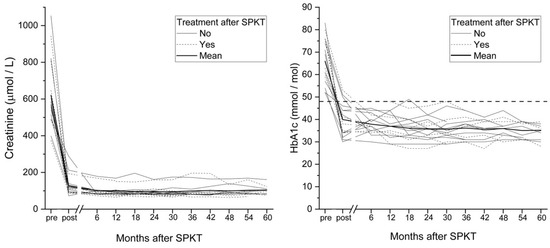
All patients had successful SPKT, with significant and sustained improvements in HbA1c and creatinine, including patients who experienced DR progression (Figure 1). Standard immunosuppressive therapy included mycophenolate mofetil (MMF) and tacrolimus for all patients (Table 1). One patient (6%) experienced gastrointestinal adverse effects, necessitating a switch to azathioprine as the antiproliferative agent. Additionally, 3 patients (16%) required adjuvant prednisolone therapy. Graft dysfunction was observed in 27.8% of patients. However, the frequency of graft dysfunction did not differ between patients who required post-SPKT treatment for progression of DR and those who did not (χ2-test, p = 0.952).

Figure 1.
HbA1c and serum creatinine levels in patients with or without ophthalmic treatment following simultaneous pancreas–kidney transplantation.
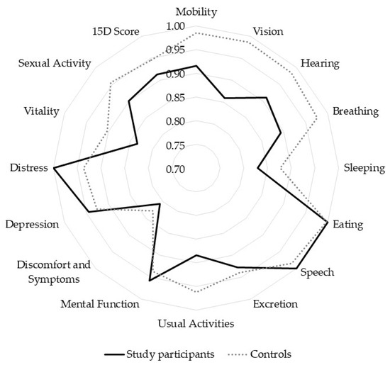
HRQoL, assessed by the 15D instrument (Figure 2), was comparable to healthy age- and sex-matched controls, with a total 15D score of 0.914 versus 0.946 (mean difference —0.033, 95% CI −0.072 to 0.007, p = 0.1, paired t-test). However, patients with PDR had a significantly lower vision-related dimension score compared to controls (0.86 vs. 0.99, mean difference —0.13, p = 0.023, paired t-test). The study participants received slightly higher scores for the 15D-dimensions for mental function and distress compared to age-matched, healthy controls.

Figure 2.
15D health-related quality of life measures in patients with type 1 diabetes after simultaneous pancreas–kidney transplantation and age- and sex-matched controls.
4. Discussion
Significant improvement in BCVA and stability of DR was observed in most patients after SPKT during a long follow-up period of 2 to 10 years. On average, a significant 5.2-letter improvement in BCVA was achieved, regardless of DR stability. Two patients were visually impaired before SPKT, but only one met the criteria for visual impairment after transplantation. The improvement in vision is likely associated with the positive effects of stabilization of retinal glucose balance metabolism. Cataract progression was observed in 39% of phakic eyes, consistent with previous studies [7]. This finding is not unexpected, as the use of post-transplant immunosuppressants is known to increase the risk for cataract development.
Another key finding was the DR progression post-SPKT. While normalization of dysglycemia following SPKT generally slows microvascular complications, including DR, the extent of improvement may depend on DR severity at the time of transplantation. This may explain some differences between our results and previous studies [7,19]. Most of our study participants, 89%, had undergone panretinal photocoagulation for PDR, whereas in a previously published study, only half had any DR pre-SPKT [19]. Despite stable graft function and metabolic improvement, 28% of patients experienced DR progression from mild DR or inactive PDR to active PDR. In addition, irreversible capillary damage associated with PDR or inflammatory mechanisms that may persist independently of glucose levels could have contributed to the progression of DR among the study participants. Unlike some earlier studies, none of these study patients with progression to active PDR required vitrectomy [8,12]. Pre-transplant condition of the retina, particularly the level of metabolic control, likely influences post-transplant outcomes, and inadequate pre-transplant control may predispose to worse retinal outcomes even after achieving euglycemia.
The median time to DR progression post-SPKT was 9 months but ranged from 3 months to almost 6 years. One previous study revealed progression in more than one-third of patients within the first postoperative year [8]. In contrast, another study observed 5% DR progression during the same period [7]. These discrepancies may reflect differences in pre-transplant DR stability and metabolic control. In our study, metabolic control and kidney function improved and sustained throughout the follow-up period, confirming the long-term efficacy of SPKT in managing T1D. However, our results highlight that DR progression can still occur years after transplantation, underscoring the need for continuous ophthalmic follow-up even with stable graft function and metabolic control.
SPKT offers considerable benefits in terms of metabolic control and visual outcomes for patients with T1D and DR, as well as a positive impact on HRQoL [19,20]. Our results demonstrate that HRQoL scores of patients with T1D post-SPKT were comparable to healthy controls, although patients with PDR had lower vision-related scores, likely reflecting ongoing concerns requiring active monitoring and management. Improved vision contributes to maintaining proper functionality and HRQoL, regardless of the stability of DR after SPKT, as a decline in visual acuity has been shown to significantly impair HRQoL [23]. Interestingly, among the study patients, the 15D questionnaire results concerning mental well-being and distress were higher than those of the control group. This may be explained by satisfaction with the relief in everyday life after the procedure, without the need for constant insulin injections, blood glucose monitoring, and fear of the consequences of hypoglycemia.
Hyperglycemia plays a key role in DR development leading to damage of retinal blood vessels, but much remains to be further explored about the multifactorial pathogenesis of DR. Several mechanisms related to diabetes are known to involve in the pathogenesis of DR, primarily characterized by microvascular damage to the retina and resulting in vision-threatening damage if untreated. Other pathophysiological mechanisms in the development and progression of DR include oxidative stress, inflammation, and changes in retinal blood flow. Pericyte dysfunction may lead to capillary dropouts and subsequent retinal ischemia. Understanding the molecular mechanisms involved is crucial for developing new therapeutic strategies for DR. There is strong evidence that the course and severity of DR are related to the long-term metabolic control in patients with T1D, and improvements in metabolic control are crucial in prevention of complications [24]. The current therapeutic modalities of diabetes, including continuous glucose monitoring and advanced insulin delivery systems, enable tighter diabetes management [25,26]. However, recent study has shown that only one-fifth of the patients with T1D achieve the recommended HbA1c target of 53 mmol/mol [27]. Pancreas and islet transplantation remain the only approaches capable of reliably achieving near-physiological and robust glucose control, while SPKT specifically benefits patients with ESKD [28,29]. Despite the considerable specific risks, transplantation represents a lifesaving and life-enhancing option for carefully selected patients, although limited availability of the organs for transplantation, the need for long-term immunosuppression to prevent rejection, peri- and post-operative complications of SPKT, lack of resources and the expertise for the procedure in many centers, and the cost implications related to the surgery and postoperative care of these patients are major issues faced by clinicians worldwide.
Several limitations should be acknowledged, including the small sample size and single-center design, as well as variability in pre-SPKT DR treatment history, which could have influenced outcomes. Nevertheless, this study provides valuable long-term data for up to 10 years on visual outcomes, DR progression, and HRQoL following SPKT in patients with advanced DR, a population rarely described in previous research.
5. Conclusions
In conclusion, SPKT improves metabolic control, stabilizes or improves DR, enhances visual acuity, and restores HRQoL to levels comparable with healthy, age-matched controls in most patients with T1D. The results of the current study indicate that DR progression may still occur years after achieving euglycemia, emphasizing the importance of long-term ophthalmic follow-up and monitoring after SPKT.
Author Contributions
J.W.: Conceptualization, Data Curation, Formal analysis, Investigation, Methodology, Validation, Visualization, Writing—review & editing; R.I.: Conceptualization, Methodology, Writing—review & editing; A.-M.K.: Conceptualization, Data Curation, Investigation, Methodology, Writing—review & editing; P.O.: Conceptualization, Data Curation, Formal analysis, Investigation, Methodology, Validation, Visualization, Writing—review & editing; N.H.: Conceptualization, Data curation, Formal analysis, Investigation, Methodology, Project administration, Resources, Supervision, Validation, Visualization, Writing—original draft, Writing—review & editing. All authors have read and agreed to the published version of the manuscript.
Funding
This research received no external funding.
Institutional Review Board Statement
The study was conducted in accordance with the Declaration of Helsinki and approved by the Institutional Review Board of Oulu University Hospital (protocol code 1312018, date 20 February 2018).
Informed Consent Statement
Informed consent was obtained from all subjects involved in the study.
Data Availability Statement
The data presented in this study are available on request from the corresponding author.
Acknowledgments
We thank Kaire Piir, nephrologist at Oulu University Hospital, for kindly providing part of the laboratory data used in this study.
Conflicts of Interest
The authors declare no conflicts of interest.
References
- Sun, H.; Saeedi, P.; Karuranga, S.; Pinkepank, M.; Ogurtsova, K.; Duncan, B.B.; Stein, C.; Basit, A.; Chan, J.C.; Mbanya, J.C.; et al. IDF Diabetes Atlas: Global, regional and country-level diabetes prevalence estimates for 2021 and projections for 2045. Diabetes Res. Clin. Pract. 2022, 183, 109119. [Google Scholar] [CrossRef] [PubMed]
- Yau, J.W.Y.; Rogers, S.L.; Kawasaki, R.; Lamoureux, E.L.; Kowalski, J.W.; Bek, T.; Chen, S.-J.; Dekker, J.M.; Fletcher, A.; Grauslund, J.; et al. Global Prevalence and Major Risk Factors of Diabetic Retinopathy. Diabetes Care 2012, 35, 556–564. [Google Scholar] [CrossRef] [PubMed]
- Hautala, N.; Aikkila, R.; Korpelainen, J.; Keskitalo, A.; Kurikka, A.; Falck, A.; Bloigu, R.; Alanko, H. Marked reductions in visual impairment due to diabetic retinopathy achieved by efficient screening and timely treatment. Acta Ophthalmol. 2014, 92, 582–587. [Google Scholar] [CrossRef]
- Bakris, G.L.; Molitch, M. Are All Patients with Type 1 Diabetes Destined for Dialysis if They Live Long Enough? Probably Not. Diabetes Care 2018, 41, 389–390. [Google Scholar] [CrossRef]
- Ramsay, R.C.; Goetz, F.C.; Sutherland, D.E.; Mauer, S.M.; Robison, L.L.; Cantrill, H.L.; Knobloch, W.H.; Najarian, J.S. Progression of Diabetic Retinopathy after Pancreas Transplantation for Insulin-Dependent Diabetes Mellitus. N. Engl. J. Med. 1988, 318, 208–214. [Google Scholar] [CrossRef]
- Shipman, K.E.; Patel, C.K. The effect of combined renal and pancreatic transplantation on diabetic retinopathy. Clin. Ophthalmol. 2009, 3, 531–535. [Google Scholar] [CrossRef]
- Pearce, I.A.; Ilango, B.; Sells, R.A.; Wong, D. Stabilisation of diabetic retinopathy following simultaneous pancreas and kidney transplant. Br. J. Ophthalmol. 2000, 84, 736–740. [Google Scholar] [CrossRef]
- Voglová, B.; Hladíková, Z.; Nemétová, L.; Zahradnická, M.; Kesslerová, K.; Sosna, T.; Lipár, K.; Kožnarová, R.; Girman, P.; Saudek, F. Early worsening of diabetic retinopathy after simultaneous pancreas and kidney transplantation—Myth or reality? Am. J. Transplant. 2020, 20, 2832–2841. [Google Scholar] [CrossRef]
- Wang, Q.; Klein, R.; Moss, S.E.; Klein, B.E.; Hoyer, C.; Burke, K.; Sollinger, H.W. The Influence of Combined Kidney-pancreas Transplantation on the Progression of Diabetic Retinopathy—A case series. Ophthalmology 1994, 101, 1071–1076. [Google Scholar] [CrossRef]
- KožNarová, R.; Saudek, F.; Sosna, T.; Adamec, M.; Jedináková, T.; BoučEk, P.; Bartoš, V.; Lánská, V. Beneficial Effect of Pancreas and Kidney Transplantation on Advanced Diabetic Retinopathy. Cell Transplant. 2000, 9, 903–908. [Google Scholar] [CrossRef] [PubMed]
- Zech, J.C.; Trepsat, D.; Gain-Gueugnon, M.; Lefrancois, N.; Martin, X.; Dubernard, J.M. Ophthalmological follow-up of Type 1 (insulin-dependent) diabetic patients after kidney and pancreas transplantation. Diabetologia 1991, 34 (Suppl. 1), S89–S91. [Google Scholar] [CrossRef][Green Version]
- Chow, V.C.; Pai, R.P.; Chapman, J.R.; O’Connell, P.J.; Allen, R.D.; Mitchell, P.; Nankivell, B.J. Diabetic retinopathy after combined kidney-pancreas transplantation. Clin. Transplant. 1999, 13, 356–362. [Google Scholar] [CrossRef] [PubMed]
- Abendroth, D.; Schmand, J.; Landgraf, R.; Illner, W.D.; Land, W. Diabetic microangiopathy in Type 1 (insulin-dependent) diabetic patients after successful pancreatic and kidney or solitary kidney transplantation. Diabetologia 1991, 34, S131–S134. [Google Scholar] [CrossRef]
- Ulbig, M.; Kampik, A.; Thurau, S.; Land, W.; Landgraf, R. Long-term follow-up of diabetic retinopathy for up to 71 months after combined renal and pancreatic transplantation. Graefe’s Arch. Clin. Exp. Ophthalmol. 1991, 229, 242–245. [Google Scholar] [CrossRef] [PubMed]
- Graham-Rowe, E.; Lorencatto, F.; Lawrenson, J.G.; Burr, J.M.; Grimshaw, J.M.; Ivers, N.M.; Presseau, J.; Vale, L.; Peto, T.; Bunce, C.; et al. Barriers to and enablers of diabetic retinopathy screening attendance: A systematic review of published and grey literature. Diabet. Med. 2018, 35, 1308–1319. [Google Scholar] [CrossRef] [PubMed]
- Fenwick, E.K.; Cheng, G.H.-L.; Man, R.E.K.; Khadka, J.; Rees, G.; Wong, T.Y.; Pesudovs, K.; Lamoureux, E.L. Inter-relationship between visual symptoms, activity limitation and psychological functioning in patients with diabetic retinopathy. Br. J. Ophthalmol. 2018, 102, 948–953. [Google Scholar] [CrossRef]
- Rassart, J.; Luyckx, K.; Oris, L.; Goethals, E.; Moons, P.; Weets, I. Coping with type 1 diabetes through emerging adulthood: Longitudinal associations with perceived control and haemoglobin A1c. Psychol. Health 2016, 31, 622–635. [Google Scholar] [CrossRef]
- Herman, W.H.; Braffett, B.H.; Kuo, S.; Lee, J.M.; Brandle, M.; Jacobson, A.M.; Prosser, L.A.; Lachin, J.M. What are the clinical, quality-of-life, and cost consequences of 30 years of excellent vs. poor glycemic control in type 1 diabetes? J. Diabetes Complicat. 2018, 32, 911–915. [Google Scholar] [CrossRef]
- Kwiatkowski, A.; Michalak, G.; Czerwinski, J.; Wszola, M.; Nosek, R.; Ostrowski, K.; Chmura, A.; Danielewicz, R.; Lisik, W.; Adadynski, L.; et al. Quality of Life After Simultaneous Pancreas–Kidney Transplantation. Transplant. Proc. 2005, 37, 3558–3559. [Google Scholar] [CrossRef]
- López-Sánchez, J.; Esteban, C.; Iglesias, M.J.; González, L.M.; Quiñones, J.E.; González-Muñoz, J.I.; Tabernero, G.; Iglesias, R.A.; Fraile, P.; Muñoz-González, J.I.; et al. Factors affecting diabetic patient’s long-term quality of life after simultaneous pancreas-kidney transplantation: A single-center analysis. Langenbeck’s Arch. Surg. 2021, 406, 873–882. [Google Scholar] [CrossRef]
- Ahopelto, K.; Sallinen, V.; Helanterä, I.; Bonsdorff, A.; Grasberger, J.; Beilmann-Lehtonen, I.; Mäkisalo, H.; Nordin, A.; Ortiz, F.; Savikko, J.; et al. The first 10 years of simultaneous pancreas-kidney transplantation in Finland. Clin. Transplant. 2023, 37, e14992. [Google Scholar] [CrossRef]
- Sintonen, H. The 15D instrument of health-related quality of life: Properties and applications. Ann. Med. 2001, 33, 328–336. [Google Scholar] [CrossRef]
- Taipale, J.; Mikhailova, A.; Ojamo, M.; Nättinen, J.; Väätäinen, S.; Gissler, M.; Koskinen, S.; Rissanen, H.; Sainio, P.; Uusitalo, H. Low vision status and declining vision decrease Health-Related Quality of Life: Results from a nationwide 11-year follow-up study. Qual. Life Res. 2019, 28, 3225–3236. [Google Scholar] [CrossRef]
- Braffett, B.H.; Bebu, I.; Lorenzi, G.M.; Martin, C.L.; Perkins, B.A.; Gubitosi-Klug, R.; Nathan, D.M.; DCCT/EDIC Research Group. The NIDDK Takes on the Complications of Type 1 Diabetes: The Diabetes Control and Complications Trial/Epidemiology of Diabetes Interventions and Complications (DCCT/EDIC) Study. Diabetes Care 2025, 48, 1089–1100. [Google Scholar] [CrossRef]
- Beck, R.W.; Bergenstal, R.M.; Laffel, L.M.; Pickup, J.C. Advances in technology for management of type 1 diabetes. Lancet 2019, 394, 1265–1273. [Google Scholar] [CrossRef] [PubMed]
- Battelino, T.; Alexander, C.M.; A Amiel, S.; Arreaza-Rubin, G.; Beck, R.W.; Bergenstal, R.M.; Buckingham, B.A.; Carroll, J.; Ceriello, A.; Chow, E.; et al. Continuous glucose monitoring and metrics for clinical trials: An international consensus statement. Lancet Diabetes Endocrinol. 2023, 11, 42–57. [Google Scholar] [CrossRef] [PubMed]
- Foster, N.C.; Beck, R.W.; Miller, K.M.; Clements, M.A.; Rickels, M.R.; DiMeglio, L.A.; Maahs, D.M.; Tamborlane, W.V.; Bergenstal, R.; Smith, E.; et al. State of Type 1 Diabetes Management and Outcomes from the T1D Exchange in 2016–2018. Diabetes Technol. Ther. 2019, 21, 66–72. [Google Scholar] [CrossRef] [PubMed]
- Choudhary, P.; Rickels, M.R.; Senior, P.A.; Vantyghem, M.-C.; Maffi, P.; Kay, T.W.; Keymeulen, B.; Inagaki, N.; Saudek, F.; Lehmann, R.; et al. Evidence-Informed Clinical Practice Recommendations for Treatment of Type 1 Diabetes Complicated by Problematic Hypoglycemia. Diabetes Care 2015, 38, 1016–1029. [Google Scholar] [CrossRef]
- Choksi, H.B.; Pleass, H.M.; Robertson, P.; Au, E.M.; Rogers, N.M. Long-term Metabolic Outcomes Post–Simultaneous Pancreas-Kidney Transplantation in Recipients with Type 1 Diabetes. Transplantation 2025, 109, 1222–1229. [Google Scholar] [CrossRef]
Disclaimer/Publisher’s Note: The statements, opinions and data contained in all publications are solely those of the individual author(s) and contributor(s) and not of MDPI and/or the editor(s). MDPI and/or the editor(s) disclaim responsibility for any injury to people or property resulting from any ideas, methods, instructions or products referred to in the content. |
© 2025 by the authors. Licensee MDPI, Basel, Switzerland. This article is an open access article distributed under the terms and conditions of the Creative Commons Attribution (CC BY) license (https://creativecommons.org/licenses/by/4.0/).