Article

FEV1/FEV6 Cutoff Points to Avoid False Negatives When Using Portable Devices, PICO-6® and COPD-6®, in COPD Detection in Primary Healthcare Services

by
Miguel A. Hernandez-Mezquita
1,2,3,*,
Idania De los Santos-Ventura
1,
Vanesa Hidalgo-Sierra
4,
Alfonso Pérez-Trullen
5,
Ruth García Garcia
1,
Tamara Clavero-Sánchez
1 and
Enrique Barrueco-Otero
6
1
Pneumology Department, Salamanca University Hospital, 37007 Salamanca, Spain
2
Department of Medicine, University of Salamanca (USAL), 37007 Salamanca, Spain
3
Institute of Biomedical Research of Salamanca (IBSAL), 37007 Salamanca, Spain
4
Primary and Community Care, Tejares, Salamanca, 37007 Salamanca, Spain
5
Pneumology Service, Zaragoza University Hospital, 50009 Zaragoza, Spain
6
Primary and Community Care, Ciudad Rodrigo, 37007 Salamanca, Spain
*
Author to whom correspondence should be addressed.
J. Clin. Med. 2025, 14(2), 576; https://doi.org/10.3390/jcm14020576
Submission received: 16 September 2024 / Revised: 5 November 2024 / Accepted: 5 January 2025 / Published: 17 January 2025
(This article belongs to the Section Respiratory Medicine)

Abstract

:
Background: Chronic obstructive pulmonary disease (COPD) is a frequent but underdiagnosed disease, primarily due to the lack of access to forced spirometry (FS) in primary care. Portable, easy-to-use expiratory flow meters like Piko-6® and COPD-6® that measure FEV1, FEV6, and FEV1/FEV6 ratio provide an alternative. Given that Piko-6® and COPD-6® devices measure FEV6 but not FVC, the aim of the study is to determine the optimal cutoff value for the FEV1/FEV6 ratio of each device to avoid false negatives when these devices are used for COPD screening in primary care (PC). Methods: A total of 664 patients of 35 years of age or older with a cumulative tobacco consumption of 10 or more packs/year were recruited at two university hospitals. FS (gold standard) was performed and FEV1, FVC, and FEV1/FVC measurements were compared with FEV1, FEV6, and FEV1/FEV6 measurements acquired using Piko-6® and COPD-6® devices. The devices were compared using statistical methods including Pearson correlation coefficients, the Youden index (YI), kappa coefficient, Bland–Altman plots, and ROC curves analysis. Results: Correlations between FEV1/FEV6 using Piko-6® and COPD-6® and FEV1/FVC with FS were 0.79 and 0.73, respectively. Piko-6® achieved the best YI in FEV1/FEV6 (0.73), whereas for COPD-6®, it was 0.80. Concordance between Piko-6® and FS was 83.9% (kappa 0.67 ± 0.028) and for COPD-6®, it was 68.7% (kappa 0.42 ± 0.02). Conclusions: This is the first study that compares two hand-held expiratory flow meters with FS. Piko-6® and COPD-6® devices are effective tools for COPD detection, as their measurements provide a good correlation with FS. In order to avoid false negative results, the FEV1/FEV6 cutoff point needs to be increased to 0.73 and 0.80 with Piko-6® and COPD-6®, respectively.

1. Introduction

COPD is a prevalent and progressive disease for which early diagnosis is essential. Forced spirometry (FS) is recognized as the gold standard diagnostic test; however, it is frequently underutilized or unavailable in clinical practice [1,2,3,4]. FS is only useful as long as the quality criteria are met; however, in primary care (PC), accessibility is often limited and guidelines are often not correctly followed [5,6,7]. Underdiagnosis is largely attributed to the population’s limited awareness of the disease and the low availability of FS [8,9]. According to EPISCAN (Epidemiologic Study of COPD in Spain), approximately 1,595,000 Spanish people suffer from COPD without being aware of it, which represents a serious problem [10]. FS may be technically difficult to perform. Multiple studies have shown that forced expiratory volume at six seconds of exhalation (FEV6) may serve as an appropriate substitute for forced vital capacity (FVC) [11,12,13,14,15]; execution is simpler and there is less variability because exhalatory time is reduced. This is the reason why more simple devices are accepted for evaluating FEV6 instead of FVC [16,17,18,19,20,21]. If we assume the accuracy of FEV6 and FEV1/FEV6 to determine obstruction, COPD screening could be performed in PC using hand-held expiratory flow meters that are easy to manipulate. Their use may help decrease underdiagnosis of the disease. The European Respiratory Society (ERS) decided in 2015 to investigate the role of these devices in the early diagnosis of COPD [22]. GesEPOC (Spanish COPD Guideline) also recommends their use in COPD screening for the at-risk patient population [23].
Significant regional disparities and methodological variations complicate the interpretation of prevalence data [24]. In Spain, 10% of adults over 40 years of age are affected by COPD [25] and this is expected to rise in the coming years. It represents the third cause of mortality in Spain and surrounding countries [26] and is responsible for a large proportion of health care costs [10,27].
The rate of underdiagnosis and undertreatment is over 70%. FS is recognized as the gold standard diagnostic test, which is set with FEV1/FVC < 70% post bronchodilator use (or less than the lower limit of normal) [28]. Nevertheless, it is underutilized in clinical practice both in PC and specialized care [29,30]. FVC is highly dependent on the duration of expiration, especially in elderly patients or those with airflow obstruction. Using FE has been suggested because it is more repeatable and has good concordance with FVC [13,14,15,16,17,31]. This is the reason why the National Lung Health Education Program North American Administration recommends the use of FEV1/FEV6 to detect COPD in PC clinical practice [12].
A complement to FS in the diagnostic process is hand-held expiratory flow meters, which measure FEV1, FEV6, and the FEV1/FEV6 ratio. They are inexpensive, easy to use, and do not need periodic calibrations, resulting in them being quite useful for COPD screening [4,12,32]. A meta-analysis of 11 studies showed that FEV1/FEV6 provides a sensitivity of 89% and a specificity of 98% when compared to FEV1/FVC. However, there is no agreement on the cutoff point for FEV1/FEV6 [13].
The proposed FEV1/FEV6 cutoff point with the COPD-6® device is 0.70. According to Represas et al., the best cutoff point to diagnose obstruction is 0.75–0.80 [33]. In the studies by Sichletidis et al. [34] and Hidalgo et al. [35], the correlation between Piko-6® and FS was excellent. Since FS is not always available in primary care, there is a need for research into other alternatives for analyzing lung function beyond the strict quality control in laboratories. In a recent meta-analysis, Zhou et al. [36] concluded that microrespirometers are “user-friendly, patient-friendly, inexpensive, and portable, making them suitable for primary care use and providing a feasible pathway for early diagnosis of COPD”.
The objective of our study was to assess the validity of COPD-6® and Piko-6® for COPD screening and to determine the best FEV1/FEV6 cutoff point for each device, i.e., the point that best allows airway obstruction to be confidently ruled out and, also, to determine the most accurate device for COPD screening. COPD screening results are important. According to the GOLD guidelines for COPD diagnosis, a diagnosis of COPD should only be made following lung function tests with FEV1/FVC. If obstruction is suspected using devices that measure FEV1/FEV6, the patient should be referred for appropriate spirometry. However, in the current context of limited resources in primary care, it is not always possible to perform spirometry, which is why Zhou’s recommendation is especially important. Zhou et al. [36] concluded that portable spirometers are highly accurate for diagnosing COPD.

2. Methods

2.1. Sample Size and Inclusion Criteria

The required sample size was established by the Epidat 4.2 program to be 569 patients. A total of 689 outpatients were recruited from the pneumology and tobacco cessation departments at the University Hospital of Salamanca and Clinic Hospital of Zaragoza (Spain) between March 2015 and January 2017. All patients participating in the study signed an informed consent form approved by the clinical research ethics committee. The inclusion criteria encompassed subjects aged over 35, smokers or ex-smokers of over 10 packs/year, with or without respiratory symptoms. The exclusion criteria were the inability to undertake valid and repeatable spirometry measurements (quality A, B, and C) and presenting any kind of medical condition or absolute contraindications to perform the test.

2.2. Development of Study Procedures

All spirometry tests were conducted following the ERS/ATS standards for spirometry 2005 by qualified operators at lung function laboratories (trained doctors, nurses, or physician assistants). The variables collected included: anthropometric data, packs/year, symptoms, FEV1, FVC, and FEV1/FVC ratio obtained via FS (Jaeger 920 Masterlab, Würzburg, Germany) measured in milliliters and in percentage with respect to the reference values for FEV1, FEV6, and FEV1/FEV6 ratio obtained by the hand-held expiratory flow meters Vitalograph COPD-6® and Piko-6®. In addition, to evaluate the subjective difficulty of performing the three tests (FS, COPD-6®, and Piko-6®), the results were measured on a visual score from 0 to 10. All subjects followed the same test sequence: (1) FS, administration of Albuterol 400 mcg through a spacer chamber and 15 min later BDT; (2) a test with COPD-6®; and (3) a test with Piko-6®. Once completed, participants were required to evaluate the subjective difficulty of the tests using a visual score from 0 to 10. The staff who administered the tests ensured that patients understood and correctly performed the procedure for each test and rested adequately between each test.

2.3. Statistical Analysis

Different statistical tests were applied depending on the specific variable analyzed. A significance level of 0.05 was applied across all analyses. The statistical analyses used to compare the devices were Pearson correlations, Youden index (YI), kappa coefficient, Bland–Altman plots, and ROC curves, and the analyses were conducted using IBM SPSS 23 version software.

3. Results

Sample Studied and Main Results

A total number of 664 patients from the total of 689 recruited subjects met the criteria to be taken into account: 491 males (74%) and 173 females (26%). The average age was 61.5 ± 11 years; 338 are ex-smokers (51%) and 326 are current smokers (49%); and the average was 41.2 ± 22 packs/year of smoking (range 10–150). Table 1 shows individuals classified as healthy or ill according to both hand-held expiratory flow meters and FS. The alternative devices classified more healthy patients than FS by using FEV1/FEV6 0.7. FS detected 411 patients with COPD (62%), Piko-6® detected 340 (51%) and COPD-6® detected 209 (31.5%).
Table 2 includes measurements in percentages and milliliters for each variable. We observed that absolute measurements and percentages for FEV1 and FEV1/FEV6 recorded by the portable expiratory flow meters were lower. A contingency table with Piko-6®, COPD-6®, and FS was created. Obstruction was defined as FEV1/FVC < 70%. Piko-6® showed a sensitivity of 78.35% and a specificity of 93%, with a positive predictive value of 94.7% and a negative predictive value of 72.5%. The YI was 0.71. Moreover, COPD-6® obtained a sensitivity of 50.12%, a specificity of 98.8%, a positive predictive value of 98.5%, a negative predictive value of 54.9%, and a YI of 0.49. Intra-cluster correlation was used to analyze concordance between the parameters, as shown in Table 3 (adapted from Hernández-Mezquita et al. [37]).
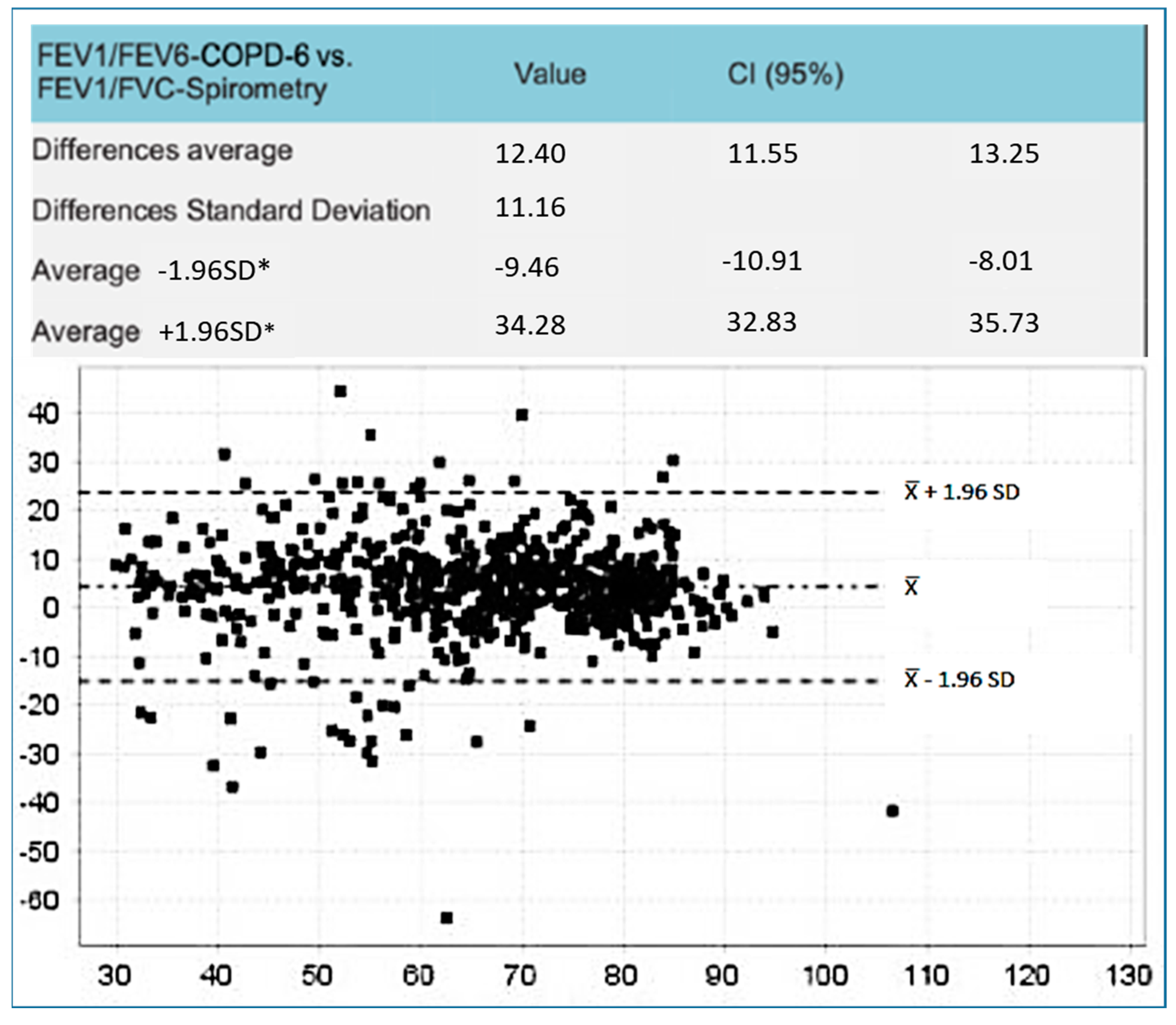
The Bland–Altman plots represent the differences observed (Figure 1 and Figure 2). The difference between FEV1/FEV6 and FEV1/FVC was smaller with Piko-6®. The biggest differences were observed between FEV6 by using both devices and FVC with FS (4.01; CI 95% 3.26–4.7 vs. 12.4; IC 95% 11.5–13.25).
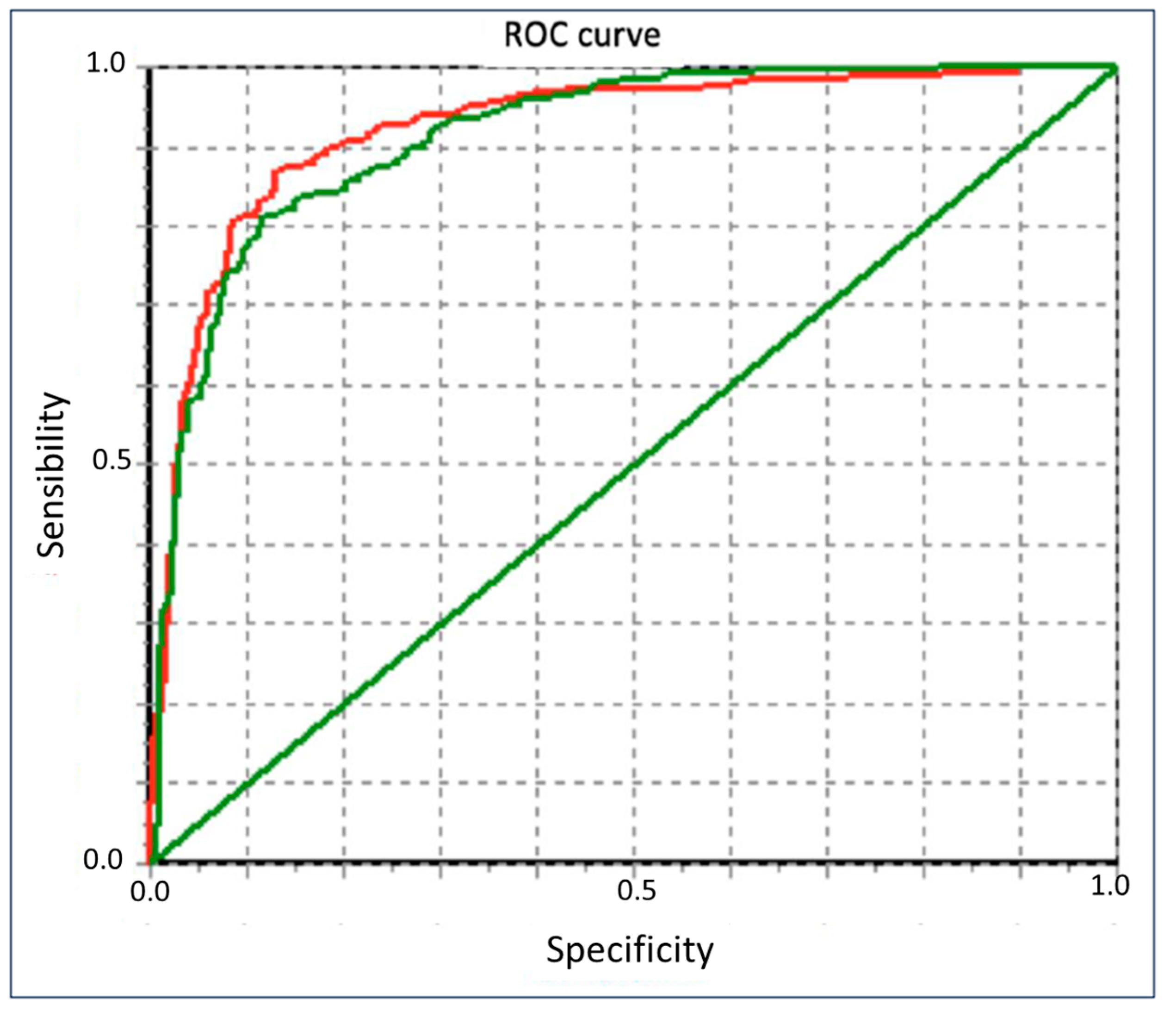
The Pearson correlation index of FEV1 between FS and Piko-6® and between FS and COPD-6® was 0.94 and 0.97, respectively (Table 4). Correlations between FEV1/FEV6 by using Piko-6® and COPD-6® were 0.79 and 0.73, respectively (Figure 3). The ROC curves graphically show the FEV1/FEV6 sensitivity and specificity of Piko-6® and COPD-6®, using FEV1/FVC < 70% as a reference. The area under the curve (UAC) was 0.922 for Piko-6® and 0.913 for COPD-6® (Chi-squared test p = 0.438) (Figure 4).
Table 5 shows the sensitivity, specificity, NPV, PPV, and YI for both devices at the different cutoff points of the FEV1/FEV6 ratio. The YI of Piko-6® (greater relation between sensitivity and specificity) was better at the cutoff of <73% (0.74). When using COPD-6®, the sensitivity value was lower, so the cutoff point was 80% (the cutoff point of the FEV1/FEV6 ratio at which a better Youden index is obtained with each device has been marked in bold). The concordance observed between Piko-6® and FS was 83.9%, with a kappa value of 0.67 ± 0.028. Moreover, COPD-6®‘s concordance was 68.7% and the kappa value was 0.42 ± 0.02. With regard to the visual scale of subjective difficulty of the tests, Piko-6® showed an average of 2.55, COPD-6® showed an average of 2.78, and FS showed an average of 3.87. More than 75% of patients reported difficulty ≤ 3 and less than 2% ≥ 7 with Piko-6®. With COPD-6®, more than 30% of patients showed a difficulty level ≤ 3, and with FS, this was less than 50%. Almost 10% of patients reported a ≥ 7 level of difficulty.

4. Discussion

COPD is underdiagnosed, so screening tools are needed to improve disease management. FS undertaken in PC is often unreliable due to the challenges of managing the procedure. Artificial intelligence can help improve outcomes in the diagnosis and management of COPD by facilitating the analysis of spirometry data, optimizing early detection, and reducing errors in the evaluation process in primary care [38]. The European Respiratory Society (ERS) has proposed investigating the utility of hand-held expiratory flow meters as an alternative [22]. Several studies have examined the utility of FEV1/FEV6 as an alternative to FEV1/FVC [13,32,39]. Recently, our research team also published a scientific paper [36] about the accuracy of PIKO-6® and COPD-6® devices in COPD screening, complementary to this article, that focuses on the analysis of cutoff points to avoid false negatives with both devices.
This research was conducted to assess the validity of Piko-6® and COPD-6® in the diagnosis of airway obstruction and to identify which is the most reliable. To achieve this aim, FEV1, FEV6, and FEV1/FEV6 measurements obtained by Piko-6® and COPD-6® were compared with FEV1, FVC, and FEV1/FVC obtained via FS.
Jing et al.’s [13] meta-analyses established that FEV1/FEV6 has a sensitivity of 89% (IC 95%: 83–93%) and a specificity of 98% (IC 95%: 95–99%) in relation to FEV1/FVC. The FEV1/FEV6 AUC ROC to detect airway obstruction was excellent at 0.97 and the authors concluded that FEV1/FEV6 could substitute FEV1/FVC. Frith et al. [32], Van den Bemt et al. [38], and Kauffman et al. [40] showed good results with Piko-6®, although they used different methods and targets in their studies. There are two studies with similar objectives and designs to our project: Hidalgo et al. [35], who studied Piko-6®, and Represas et al. [33], who studied COPD-6®. In our study, FEV1 and FEV6 obtained by COPD-6® and Piko-6®, both in milliliters and percentages were lower than FEV1 and FVC collected via FS (p < 0.001). Our COPD-6® results did not substantially differ from those of Represas et al. [33]. In both studies, FEV1/FEV6 was higher than FEV1/FVC (p < 0.001), which was expected because FEV6 was lower than FVC. COPD-6® FEV1 measurement was also lower than that obtained via FS. Based on this, the cutoff point for diagnosing obstruction should be reconsidered from 0.7, since COPD-6® did not detect obstruction in almost half of patients. In our study, we observed less dispersion of FEV1 with respect to FS than Represas, an average difference of 144 mL (IC 95%: 126–162) vs. 167 mL (IC 95%: 144–190). In contrast, in relation to the FEV6 value, we found slightly greater dispersion. Represas observed a good correlation between FEV1, FEV6, and FS, which was similar to our study, and a higher correlation between FEV1/FEV6 and FS. In summary, we observed a good correlation between COPD-6® and FS, particularly with FEV1 (r = 0.97) measurement, with consistent results similar to those published by Represas [33].
In both Hidalgo’s study and our present project in relation to Piko-6®, FEV1 and FEV6 values were also lower than FEV1 and FVC determined via FS (p < 0.001); however, no significant differences were observed between FEV1/FEV6 and FEV1/FVC. When we analyzed the FEV1 dispersion of Piko-6® versus FS by using Bland–Altman plots, the average dispersion (8%; IC 95%: 5–12) was higher than in Hidalgo et al.’s study (4%; IC 95%: 2–6) [35]. The average dispersion of the FEV1/FEV6 ratio was 4% (IC 95%: 3–4) and in Hidalgo et al.’s study, it was 0.7% (IC 95%: 0.1–1.5). Hidalgo et al. [35] reported a good correlation between FEV1, FEV6, and FEV1/FEV6. In contrast, we observed the best correlation with FEV1 (r = 0.94 versus r = 0.87) and slightly worse with the ratio (r = 0.79 versus 0.94) [35]. This correlation could be considered excellent based on linear regression lines. We tried to find out the best FEV1/FEV6 cutoff point in terms of sensitivity and specificity to detect obstruction (defined as FEV1/FVC < 0.7 post bronchodilator test according to clinical guidelines). However, the FEV1/FEV6 acquired by the hand-held expiratory flow meter was greater than FEV1/FVC. This is the reason why a higher cutoff should be considered. To achieve this aim, we used the YI. The cutoff point in the studies by Represas et al. [33], Fritz et al. [32], Hidalgo et al. [35], and Van de Bemt et al. [38] varied between 0.70 (Hidalgo) and 0.78 (Represas). In their research, Fritz and Van de Bemt obtained intermediate values of 0.75 and 0.73, respectively. We noticed different cutoff points given that the sensitivity and specificity of each device were different. The Piko-6® cutoff point with the best YI was 0.73, and with COPD-6®, it was 0.8 and had considerably lower sensitivity. Represas et al. [33] concluded that when using COPD-6®, a cutoff point of 0.7 was not valid for COPD screening and that a cutoff point of 0.75–0.80 was needed, which was in accordance with our findings. Nevertheless, Hidalgo et al. [35] observed that FEV1/FEV6 with the best YI was 0.70 when using Piko-6®, which was slightly inferior to our YI of 0.73. Nonetheless, this is not a limitation for using this device for COPD screening. To sum up, the best YI to FEV1/FEV6 ratios were 0.73 for Piko-6® and 0.80 for COPD-6®, similar to the results of other authors. ROC curves were performed to compare FEV1 and FEV1FEV6. FEV1/FVC < 70% was used as reference. We noticed a UAC of 0.91 for FEV1/FEV6 with COPD-6®, slightly inferior to that in Represas’ study (UAC of 0.97) [33]. With regard to Piko-6®, the UAC (0.84 for FEV1 and 0.92 for FEV1/FEV6) and UAC of Hidalgo [35] (0.8 and 0.92, respectively) showed an excellent correlation with FS.
When analyzing concordance with the kappa index between both devices and FS, the results were better with Piko-6® than with COPD-6® (0.55 and 0.43, respectively). These results were slightly worse than those obtained by Hidalgo with Piko-6® [35]. However, both devices showed an odds ratio (OR) higher than 20, which is the minimum needed to validate a test. Piko-6® showed a concordance of 83.9% to classify individuals as ill (positive diagnosis of COPD) or healthy (COPD diagnosis excluded), with a kappa value of 0.67 ± 0.028 and 68.7% and kappa of 0.42 ± 0.02 with COPD-6®. Therefore, Piko-6® has good concordance and COPD-6® has moderate concordance. Consequently, with a cutoff of FEV1/FEV6 < 0.70, Piko-6® did not detect a small percentage of patients that FS diagnosed as having COPD, a finding similar to that observed by Fatima et al. [41] in 2017. This percentage was superior with COPD-6®, which necessitates increasing the FEV1/FEV6. In clinical practice, it is important to bear in mind the subjective difficulty that patients observe when performing the tests. Only Hidalgo et al. [35] have evaluated this aspect. The subjective difficulty average was 2.55 points with Piko-6®, 2.78 with COPD-6®, and 3.87 with FS. These results were significant and concordant with those of Hidalgo et al. [35]. We could state that patients preferred performing tests using less difficult hand-held expiratory flow meters than using FS. The reasons could be the greater simplicity of the device, the fewer maneuvers needed, or the decreased time for the exhalatory maneuver. This study provides real-world evidence to identify optimal practices for COPD diagnostic practices; such practices ensure that physicians have convenient access to up-to-date evidence when they encounter a symptomatic patient likely to have COPD. Chen et al. [42] concluded in their study published in 2021 that microspirometry was accurate and had higher clinical utility.
In conclusion, the effectiveness of hand-held expiratory flow meters for COPD screening seems to be clear given the accuracy of the measurements and their acceptance by patients in helping to reduce underdiagnoses of the disease and alleviating workloads in pulmonary function laboratories. This is the first study that compares two hand-held expiratory flow meters with FS. Although FEV1 and FEV6 measurements obtained with hand-held expiratory flow meters were lower than FEV1 and FVC values measured with FS, Piko-6® and COPD-6® devices are useful for COPD screening because correlation with their FS is good. An FEV1/FEV6 cutoff point of 0.7 was obtained by the hand-held expiratory flow meters as COPD screening had false negative results, so it should be increased: 0.73 for Piko-6® and 0.80 for COPD-6®.
The quality and accuracy of performing and interpreting spirometry measurements in primary care is variable; therefore, microspirometers are currently useful. Perhaps, in the near future, artificial intelligence (AI) could enhance the performance of primary care physicians in spirometry interpretation [43], although the difficulty for patients in performing the test will remain an issue.

Author Contributions

“Conceptualization and methodology”, M.A.H.-M. and A.P.-T.; Software, “validation and formal analysis”, T.C.-S., LTD; “resources”, I.D.l.S.-V., V.H.-S., R.G.G., T.C.-S. and E.B.-O.; “writing and original draft preparation”, M.A.H.-M. and E.B.-O.; writing review and editing, V.H.-S.; project administration and funding acquisition, M.A.H.-M. All authors have read and agreed to the published version of the manuscript.

Funding

This work was supported by a research grant from the Spanish Society of Pneumology (SEPAR)—grant code 091/2014.

Institutional Review Board Statement

This study was carried out according to the guidelines of the Declaration of Helsinki and approved by the ethics and clinical research committee of the University Hospital of Salamanca on 30 October 2014. The approval code is PI 2014 10 01.

Informed Consent Statement

Informed consent was obtained from all subjects involved in the study.

Data Availability Statement

No restrictions on data availability. Data statement: The data are available at https://scholar.archive.org/work/ng6ecpnosrftzpkzvkyjhjrhtu (accessed on 21 December 2024).

Acknowledgments

We give thanks to the team at the pulmonary function laboratory at the University Hospital of Salamanca, the nurses José Miguel Hernández, Begoña González, and Florinda Pérez for their advice.

Conflicts of Interest

The authors have no conflicts of interest to declare.

References

  1. Baldomero, A.K.; Kunisaki, K.M.; Bangerter, A.; Nelson, D.B.; Wendt, C.H.; Fortis, S.; Hagedorn, H.; Dudley, R.A. Beyond Access: Factors Associated with Spirometry Underutilization Among Patients with a Diagnosis of COPD in Urban Tertiary Care Centers. Chronic Obstr. Pulm. Dis. 2022, 9, 538–548. [Google Scholar] [CrossRef] [PubMed]
  2. Casas, A.; Montes de Oca, M.; López, M.V.; Aguirre, C.; Schiavi, E.; Jardim, J.R.; Team, P. COPD Underdiagnosis and Misdiagnosis in a High-Risk Primary Care Population in Four Latin American Countries. A Key to Enhance Disease Diagnosis: The PUMA Study. PLoS ONE 2016, 11, e0152266. [Google Scholar] [CrossRef]
  3. Fukahori, S.; Matsuse, H.; Takamura, N.; Hirose, H.; Tsuchida, T.; Kawano, T.; Fukushima, C.; Mizuta, Y.; Kohno, S. Prevalence of chronic obstructive pulmonary diseases in general clinics in terms FEV1/FVC. Int. J. Clin. Pract. 2009, 63, 269–274. [Google Scholar] [CrossRef]
  4. Heffler, E.; Crimi, C.; Mancuso, S.; Campisi, R.; Puggioni, F.; Brussino, L.; Crimi, N. Misdiagnosis of asthma and COPD and underuse of spirometry in primary care unselected patients. Respir. Med. 2018, 142, 48–52. [Google Scholar] [CrossRef] [PubMed]
  5. Hegewald, M.J.; Gallo, H.M.; Wilson, E.L. Accuracy and Quality of Spirometry in Primary Care Offices. Ann. Am. Thorac. Soc. 2016, 13, 2119–2124. [Google Scholar] [CrossRef]
  6. Bednarek, M.; Maciejewski, J.; Wozniak, M.; Kuca, P.; Zielinski, J. Prevalence, severity and underdiagnosis of COPD in the primary care setting. Thorax 2008, 63, 402–407. [Google Scholar] [CrossRef] [PubMed]
  7. Ragaišienė, G.; Kibarskytė, R.; Gauronskaitė, R.; Giedraitytė, M.; Dapšauskaitė, A.; Kasiulevičius, V.; Danila, E. Diagnosing COPD in primary care: What has real life practice got to do with guidelines? Multidiscip. Respir. Med. 2019, 14, 28. [Google Scholar] [CrossRef]
  8. Jones, R.C.; Price, D.; Ryan, D.; Sims, E.J.; von Ziegenweidt, J.; Mascarenhas, L.; Burden, A.; Halpin, D.M.G.; Winter, R.; Hill, S.; et al. Opportunities to diagnose chronic obstructive pulmonary disease in routine care in the UK: A retrospective study of a clinical cohort. Lancet Respir. Med. 2014, 2, 267–276. [Google Scholar] [CrossRef]
  9. Soriano, J.B.; Calle, M.; Montemayor, T.; Alvarez-Sala, J.L.; Ruiz-Manzano, J.; Miravitlles, M. The general public’s knowledge of chronic obstructive pulmonary disease and its determinants: Current situation and recent changes. Arch. Bronconeumol. 2012, 48, 308–315. [Google Scholar] [CrossRef] [PubMed]
  10. Miravitlles, M.; Soriano, J.B.; Garcia-Rio, F.; Munoz, L.; Duran-Tauleria, E.; Sanchez, G.; Sobradillo, V.; Ancochea, J. Prevalence of COPD in Spain: Impact of undiagnosed COPD on quality of life and daily life activities. Thorax 2009, 64, 863–868. [Google Scholar] [CrossRef] [PubMed]
  11. Brogger, J.; Eide, G.E.; Gulsvik, A. FEV6 as a surrogate for FVC: Authors should have included ROC-curve analyses. Am. J. Respir. Crit. Care Med. 2001, 163, 1759. [Google Scholar] [CrossRef] [PubMed]
  12. Ramsdell, J.W.; Foreman, M.G.; Jensen, R.L.; Stinson, D.S.; Wilson, C.G.; Lynch, D.A.; Make, B.J.; Dransfield, M.T. FEV(1)/FEV(6) to diagnose airflow obstruction. Comparisons with computed tomography and morbidity indices. Ann. Am. Thorac. Soc. 2014, 11, 335–341. [Google Scholar] [CrossRef]
  13. Jing, J.Y.; Huang, T.C.; Cui, W.; Xu, F.; Shen, H.H. Should FEV1/FEV6 replace FEV1/FVC ratio to detect airway obstruction? A metaanalysis. Chest 2009, 135, 991–998. [Google Scholar] [CrossRef] [PubMed]
  14. Venkatachalam, P.; Dwivedi, D.P.; Govindraj, V. FEV1/FEV6 is effective as a surrogate for FEV1/FVC in the diagnosis of chronic obstructive pulmonary disease. Indian J. Tuber. 2021, 68, 230–235. [Google Scholar] [CrossRef] [PubMed]
  15. Wang, S.; Gong, W.; Tian, Y.; Zhou, J. FEV1/FEV6 in Primary Care Is a Reliable Easy Method for the Diagnosis of COPD. Respir. Care 2016, 61, 349–353. [Google Scholar] [CrossRef] [PubMed]
  16. Yee, N.; Markovic, D.; ∙Buhr, R.G.; Christopher, B.; Fortis, S.; Arjomandi, M.; Couper, D.; Anderson, W.H.; Paine, R.; Woodruff, P.G.; et al. Significance of FEV3/FEV6 in Recognition of Early Airway Disease in Smokers at Risk of Development of COPD. Chest 2021, 161, 949–959. [Google Scholar] [CrossRef]
  17. Vandevoorde, J.; Verbanck, S.; Schuermans, D.; Broekaert, L.; Devroey, D.; Kartounian, J.; Vincken, W. Forced vital capacity and forced expiratory volume in six seconds as predictors of reduced total lung capacity. Eur. Resp. J. 2008, 31, 391–395. [Google Scholar] [CrossRef]
  18. Vandevoorde, J.; Verbanck, S.; Schuermans, D.; Kartounian, J.; Vincken, W. Obstructive and restrictive spirometric patterns: Fixed cut-offs for FEV1/FEV6 and FEV6. Eur. Respir. J. 2006, 27, 378–383. [Google Scholar] [CrossRef]
  19. Lundgren, F.L.; Cabral, M.M.; Climaco, D.C.; de Macedo, L.G.; Coelho Mde, A.; Dias, A.L. Determination of the efficacy of FEV6 as a surrogate for FVC in the diagnostic screening for chronic obstructive pulmonary disease through the comparison of FEV1/FVC and FEV1/FEV6 ratios. J. Bras. Pneumol. 2007, 33, 148–151. [Google Scholar] [CrossRef] [PubMed]
  20. Perez-Padilla, R.; Wehrmeister, F.C.; Celli, B.R.; Lopez-Varela, M.V.; Montes de Oca, M.; Muiño, A.; Talamo, C.; Jardim, J.R.; Valdivia, G.; Lisboa, C.; et al. Reliability of FEV1/FEV6 to diagnose airflow obstruction compared with FEV1/FVC: The PLATINO longitudinal study. PLoS ONE 2013, 8, e67960. [Google Scholar] [CrossRef] [PubMed]
  21. Jensen, R.L.; Crapo, R.O.; Enright, P. A statistical rationale for the use of forced expired volume in 6 s. Chest 2006, 130, 1650–1656. [Google Scholar] [CrossRef] [PubMed]
  22. Celli, B.R.; Decramer, M.; Wedzicha, J.A.; Wilson, K.C.; Agusti, A.; Criner, G.J.; MacNee, W.; Make, B.J.; Rennard, S.I.; Stockley, R.A.; et al. An official Am Thoracic Society/Eur Resp Society statement: Research questions in COPD. Eur. Respir. Rev. 2015, 24, 159–172. [Google Scholar] [CrossRef]
  23. Miravitlles, M.; Soler-Cataluna, J.J.; Calle, M.; Molina, J.; Almagro, P.; Quintano, J.A.; Riesco, J.A.; Trigueros, J.A.; Piñera, P.; Simón, A.; et al. Spanish COPD Guidelines (GesEPOC): Update Pharmacological treatment of stable COPD. Arch. Bronconeumol. 2022, 58, 69–81. [Google Scholar] [CrossRef] [PubMed]
  24. Safiri, S.; Carson-Chahhoud, K.; Noori, M.; Nejadghaderi, S.A.; Sullman, M.J.M.; Heris, J.A.; Ansarin, K.; Mansournia, M.A.; Collins, G.S.; Kolahi, A.-A.; et al. Burden of Chronic Obstructive Pulmonary Disease and its attributable risk factors in 204 countries and territories, 1990–2019: Results from the Global Burden of Disease Study 2019. BMJ 2022, 378, e069679. [Google Scholar] [CrossRef] [PubMed]
  25. Soriano, J.B.; Ancochea, J.; Miravitlles, M.; Garcia-Rio, F.; Duran-Tauleria, E.; Munoz, L.; Jimenez-Ruiz, C.A.; Masa, J.F.; Viejo, J.L.; Villasante, C.; et al. Recent trends in COPD prevalence in Spain: A repeated cross-sectional survey 1997–2007. Eur. Respir. J. 2010, 36, 758–765. [Google Scholar] [CrossRef] [PubMed]
  26. Peña, V.S.; Miravitlles, M.; Gabriel, R.; Jimenez-Ruiz, C.A.; Villasante, C.; Masa, J.F.; Viejo, J.L.; Fernández-Fau, L. Geographic variations in prevalence and underdiagnosis of COPD: Results of the IBERPOC multicentre epidemiological study. Chest 2000, 118, 981–989. [Google Scholar] [CrossRef]
  27. Soriano, J.B.; Alfageme, I.; Miravitlles, M.; de Lucas, P.; Soler-Cataluña, J.J.; García-Río, F.; Casanova, C.; Rodríguez González-Moro, J.M.; Cosío, B.G.; Sánchez, G.; et al. Prevalence Determinants of COPD in Spain: EPISCAN II. Arch. Bronconeumol. 2021, 57, 61–69. [Google Scholar] [CrossRef]
  28. Agustí, A.; Celli, B.R.; Criner, G.J.; Halpin, D.; Anzueto, A.; Barnes, P.; Bourbeau, J.; Han, M.K.; Martinez, F.J.; de Oca, M.M.; et al. Global Initiative for Chronic Obstructive Lung Disease 2023 Report: GOLD Executive Summary. Eur. Respir. J. 2023, 61, 2300239. [Google Scholar] [CrossRef]
  29. Miravitlles, M.; Soler-Cataluna, J.J.; Calle, M.; Molina, J.; Almagro, P.; Quintano, J.A.; Trigueros, J.A.; Cosío, B.G.; Casanova, C.; Antonio Riesco, J.; et al. Spanish Guidelines for management of chronic obstructive pulmonary disease (GesEPOC) 2017. Pharmacological treatment of stable phase. Arch. Bronconeumol. 2017, 53, 324–335. [Google Scholar] [CrossRef] [PubMed]
  30. Monteagudo, M.; Rodríguez-Blanco, T.; Parcet, J.; Penalver, N.; Rubio, C.; Ferrer, M.; Miravitlles, M. Variability in the performing of spirometry and its consequences in the treatment of COPD in primary care. Arch. Bronconeumol. 2011, 47, 226–233. [Google Scholar] [CrossRef]
  31. Akpinar-Elci, M.; Fedan, K.B.; Enright, P.L. FEV6 as a surrogate for FVC in detecting airways obstruction and restriction in the workplace. Eur. Respir. J. 2006, 27, 374–377. [Google Scholar] [CrossRef] [PubMed]
  32. Frith, P.; Crockett, A.; Beilby, J.; Marshall, D.; Attewell, R.; Ratnanesan, A.; Gavagna, G. Simplified COPD screening: Validation of the Piko-6® in primary care. Prim. Care Respir. J. 2011, 20, 190–198. [Google Scholar] [CrossRef]
  33. Represas Represas, C.; Botana Rial, M.; Leiro Fernandez, V.; Gonzalez Silva, A.I.; del Campo Perez, V.; Fernandez-Villar, A. Assessment of the portable COPD-6® device for detecting obstructive airway diseases. Arch. Bronconeumol. 2010, 46, 426–432. [Google Scholar] [CrossRef] [PubMed]
  34. Sichletidis, L.; Spyratos, D.; Papaioannou, M.; Chloros, D.; Tsiotsios, A.; Tsagaraki, V.; Haidich, A.-B. A combination of the IPAG questionnaire and Piko-6® flow meter is a valuable screening tool for COPD in the primary care setting. Prim. Care Respir. J. 2011, 20, 184–189. [Google Scholar] [CrossRef] [PubMed]
  35. Hidalgo Sierra, V.; Hernandez-Mezquita, M.A.; Palomo Cobos, L.; Garcia Sanchez, M.; Castellanos, R.D.; Jodra Sanchez, S.; Pérez, R.C.; Ferrero, M.B. Usefulness of The Piko-6® Portable Device for Early COPD Detection in Primary Care. Arch. Bronconeumol. 2018, 54, 445–454. [Google Scholar] [CrossRef]
  36. Zhou, J.; Li, X.; Wang, X.; Yu, N. Accuracy of portable spirometers in the diagnosis of chronic obstructive pulmonary disease. A meta-analysis. NPJ Prim. Care Respir. Med. 2022, 32, 2–15. [Google Scholar] [CrossRef]
  37. Hernández-Mezquita, M.A.; de los Santos-Ventura, I.; Hidalgo-Sierra, V.; Pérez-Trullen, A.; Barrueco-Otero, E. Accuracy of PIKO 6® and COPD6® Devices in COPD Screening. Arch. Bronconeumol. 2024, in press. [CrossRef]
  38. Topole, E.; Biondaro, S.; Montagna, I.; Corre, S.; Corradi, M.; Stanojevic, S.; Graham, B.; Das, N.; Ray, K.; Topalovic, M. Artificial intelligence-based software facilitates spirometry quality control in asthma and COPD clinical trials. ERJ Open Res. 2023, 9, 00292–2022. [Google Scholar] [CrossRef]
  39. Van den Bemt, L.; Wouters, B.C.; Grootens, J.; Denis, J.; Poels, P.J.; Schermer, T.R. Diagnostic accuracy of pre-bronchodilator FEV1/FEV6 from microspirometry to detect airflow obstruction in primary care: A randomised cross-sectional study. NPJ Prim. Care Respir. Med. 2014, 24, 14033. [Google Scholar] [CrossRef]
  40. Kaufmann, M.; Hartl, S.; Geyer, K.; Breyer, M.K.; Burghuber, O.C. Measuring FEV(6) for detecting early airway obstruction in the primary care setting. Quality and utility of the new Piko-6® device. Respiration 2009, 78, 161–167. [Google Scholar] [CrossRef]
  41. Fathima, M.; Saini, B.; Foster, J.M.; Armour, C.L. Community pharmacy-based case finding for COPD in urban and rural settings is feasible and effective. Int. J. Chron. Obstruct Pulmon Dis. 2017, 12, 2753–2761. [Google Scholar] [CrossRef]
  42. Chen, C.Z.; Sheu, C.C.; Cheng, S.L.; Wang, H.C.; Lin, M.C.; Hsu, W.H.; Lee, K.Y.; Perng, D.W.; Lin, H.I.; Lin, M.S.; et al. Performance and Clinical Utility of Various Chronic Obstructive Pulmonary Disease Case-Finding Tools. Int. J. Chron. Obstruct Pulmon Dis. 2021, 16, 3405–3415. [Google Scholar] [CrossRef] [PubMed]
  43. Doe, G.; El-Emir, E.; Edwards, G.D.; Topalovic, M.; Evans, R.A.; Russell, R.; Sylvester, K.P.; Van Orshoven, K.; Sunjaya, A.P.; Scott, D.; et al. Comparing performance of primary care clinicians in the interpretation of SPIROmetry with or without Artificial Intelligence Decision support software (SPIRO-AID): A protocol for a randomised controlled trial. BMJ Open 2024, 14, e086736. [Google Scholar] [CrossRef]
Figure 1. Bland-Altman plots of the mean differences between FEV1/FEV6 obtained by Piko-6 vs. FEV1/FVC via FS. * Standard Deviation.
Jcm 14 00576 g001
Figure 2. Bland–Altman plots of the differences between FEV1/FEV6 obtained by COPD-6 vs. FEV1/FVC via FS. * Standard Deviation.
Jcm 14 00576 g002
Figure 3. Correlation between FEV1/FEV6 obtained by Piko-6 (left) and COPD-6 (right) and FEV1/FVC obtained via FS.
Jcm 14 00576 g003
Figure 4. Comparison of FEV1/FEV6 ROC curves obtained by Piko-6 (red, test 1) and COPD-6 (green, test 2).
Jcm 14 00576 g004
Table 1. Patients classified as having COPD based on GOLD criteria using the three devices: forced Spirometry (FS), Piko-6®, and COPD-6®.
FrequencyPercentageValid PercentageAccumulated Percentage
Forced Spirometry
ValidILL (COPD)41161.961.961.9
HEALTHY25338.138.1100.0
Total664100.0100.0
Piko-6®
ValidILL (COPD)34051.251.251.2
HEALTHY32448.848.8100.0
Total664100.0100.0
COPD-6®
ValidILL (COPD)20931.531.531.5
HEALTHY45568.568.5100.0
Total664100.0100.0
Table 2. Absolute measurements and percentage of FEV1, FVC, and FEV6 obtained through forced spirometry (FS) and by the Piko-6® and COPD-6® devices (n = 664 subjects).
AverageStandard Deviation
FEV1 (%) by Piko-6®71.0626.83
FEV1 (%) by COPD-6®79.9430.04
FEV1 (%) by FS83.5846.53
FEV6 (%) by Piko-6®81.1548.52
FEV6 (%) by COPD-6®82.5224.21
FVC (%) by FS100.08106.51
FEV1 (cc) by Piko-6®2000.87886.98
FEV1 (cc) by COPD-6®2158.71969.35
FEV1 (cc) by FS2303.30987.97
FEV6 (cc) by Piko-6®2936.43998.72
FEV6 (cc) by COPD-6®2800.871025.12
FVC (cc) by FS3577.031042.26
Table 3. Concordance and correlation coefficient between simple hand-held expiratory flow meters and FS, adapted from Hernández-Mezquita et al. [37].
Piko-6® vs. SpirometryCorrelationCI (95%)
FEV1, cc0.89160.87480.9062
FEV6, cc (FVC, cc)0.64560.5990.6879
FEV1, %0.37560.30840.439
FEV6, % (FVC, %)0.013300.0892
FEV1/FVC0.76630.7330.7959
COPD-6® vs. Spirometry
FEV1, cc0.95990.95350.9655
FEV6, cc (FVC, cc)0.67430.63060.7137
FEV1, %0.43210.36820.492
FEV6, % (FVC, %)0.052600.1281
FEV1/FVC0.49360.43390.549
Table 4. Pearson correlation between COPD-6®, Piko-6®, and spirometry measurements.
PEARSON CORRELATION BETWEEN COPD-6®, PIKO-6®, AND FS
Piko-6® FEV1 (cc)Pearson Correlation0.946
Sig. (bilateral)0.000
COPD-6® FEV1 (cc)Pearson Correlation0.971
Sig. (bilateral)0.000
Piko-6® FEV1/FEV6Pearson Correlation0.796
Sig. (bilateral)0.000
COPD-6® FEV1/FEV6Pearson Correlation0.737
Sig. (bilateral)0.000
Table 5. Sensibility (S), specificity (E), positive predictive value (PPV), negative predictive value (NPV), and Youden index (YI) of Piko-6® and COPD-6®.
FEV1/FEV6
Piko-6® (%)
YIS (%)E (%)PPV (%)NPV− (%)
<680.6372.9990.3291.2370.79
<690.6473.790.4292.2469.01
<700.7178.3592.8994.7172.53
<710.6975.6392.8995.466.14
<720.7377.2996.1297.7965.56
<730.7478.2195.4197.664.71
<740.7279.3492.7896.7362.55
<750.7481.9192.4496.8864.11
<760.7283.0488.5495.961.78
<770.7485.0688.7396.5261.76
<780.6984.8784.4396.0355.68
<790.6884.5583.1796.5549.12
<800.6385.5277.3896.3143.62
FEV1/FEV6
COPD-6® (%)
<680.4446.4997.8596.7656.99
<690.4447.1596.9395.9654.29
<700.4647.7897.8997.6150.99
<710.4648.5297.7897.7149.33
<720.4950.2299.0399.1447.22
<730.551.598.4798.7745.95
<740.552.6997.7898.4643.46
<750.5556.398.2698.9344.01
<760.5657.7998.0998.9941.85
<770.5758.8198.5999.3539.44
<780.5859.2398.3699.3835.19
<790.5861.4697.0399.1431.11
<800.5964.1495.2498.9427.78
Disclaimer/Publisher’s Note: The statements, opinions and data contained in all publications are solely those of the individual author(s) and contributor(s) and not of MDPI and/or the editor(s). MDPI and/or the editor(s) disclaim responsibility for any injury to people or property resulting from any ideas, methods, instructions or products referred to in the content.