
How Reproducible Are the Ultrasound Features of Adenomyosis Defined by the Revised MUSA Consensus?


Abstract
1. Introduction
2. Materials and Methods
2.1. Study Design
2.2. Ultrasound Data Acquisition
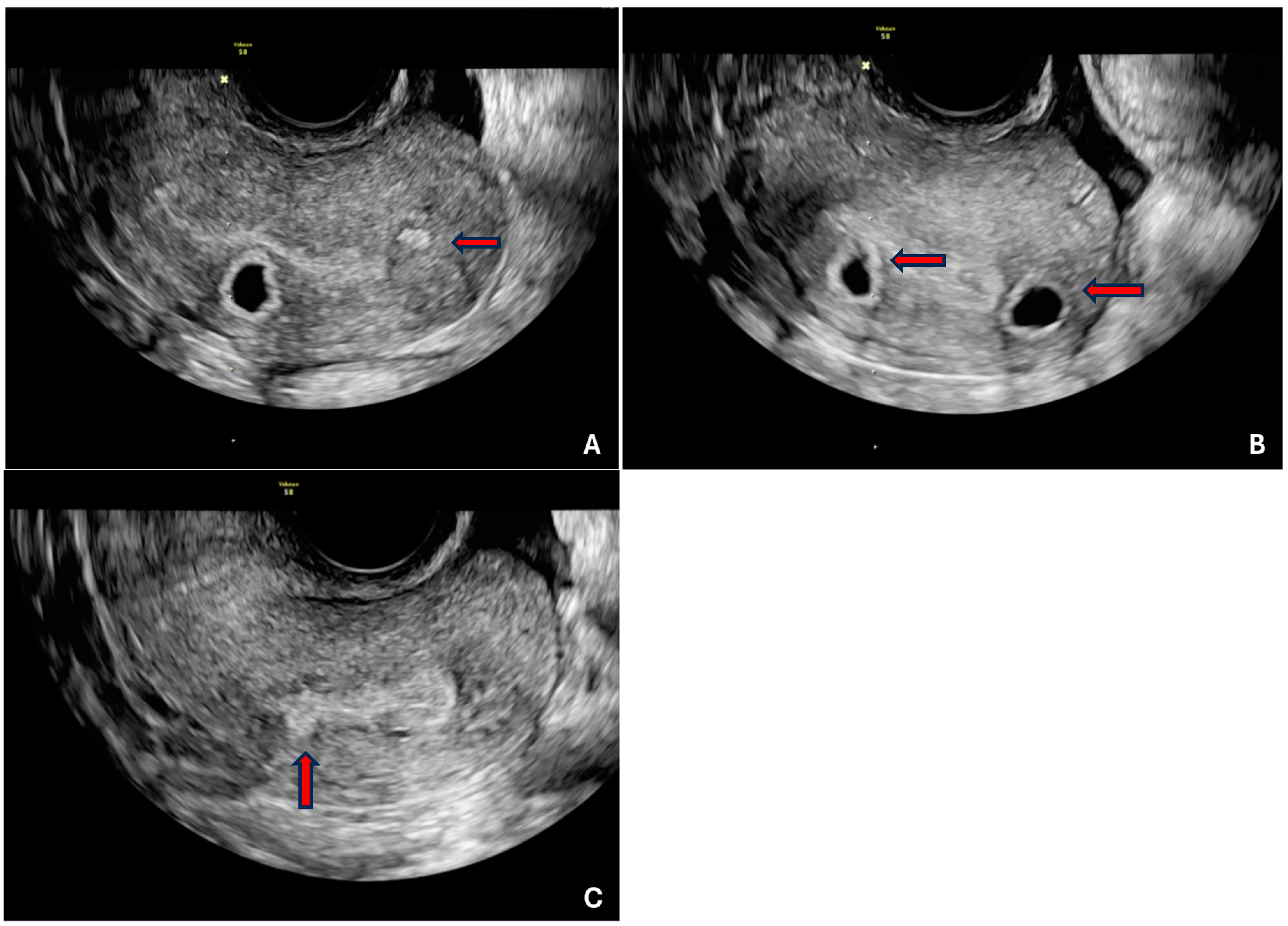
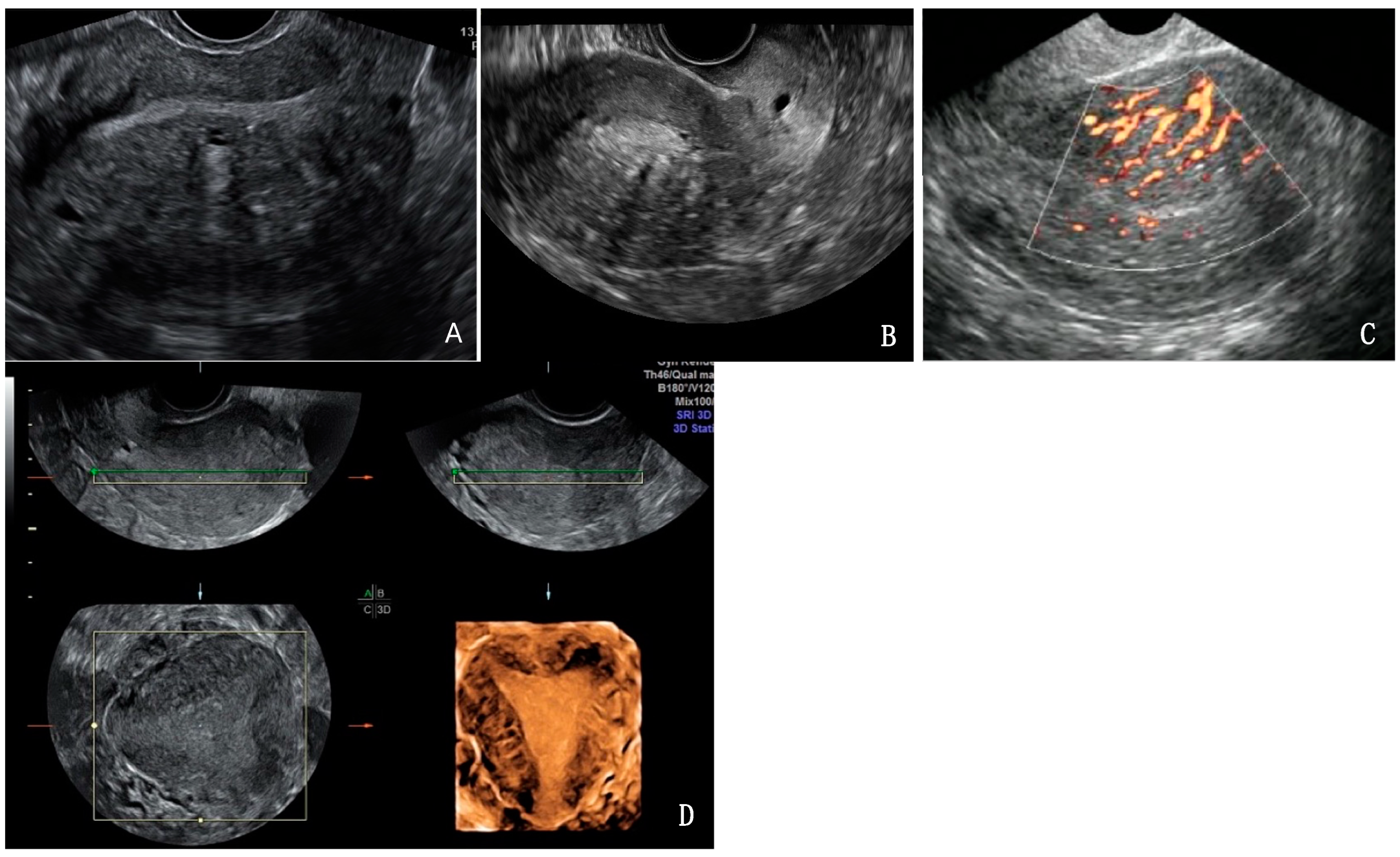
2.3. Ultrasound Data Analysis
2.4. Statistical Analysis
3. Results
3.1. Interobserver Agreement
3.2. Intra-Observer Agreement
4. Discussion
5. Conclusions
Author Contributions
Funding
Institutional Review Board Statement
Informed Consent Statement
Data Availability Statement
Conflicts of Interest
References
- Gordts, S.; Grimbizis, G.; Campo, R. Symptoms and classification of uterine adenomyosis, including the place of hysteroscopy in diagnosis. Fertil. Steril. 2018, 109, 380–388.e1. [Google Scholar] [CrossRef]
- Van den Bosch, T.; Van Schoubroeck, D. Ultrasound diagnosis of endometriosis and adenomyosis: State of the art. Best Pract. Res. Clin. Obstet. Gynaecol. 2018, 51, 16–24. [Google Scholar] [CrossRef] [PubMed]
- Maheshwari, A.; Gurunath, S.; Fatima, F.; Bhattacharya, S. Adenomyosis and subfertility: A systematic review of prevalence, diagnosis, treatment and fertility outcomes. Hum. Reprod. Update 2012, 18, 374–392. [Google Scholar] [CrossRef]
- Bazot, M.; Darai, E. Role of transvaginal sonography and magnetic resonance imaging in the diagnosis of uterine adenomyosis. Fertil. Steril. 2018, 109, 389–397. [Google Scholar] [CrossRef]
- Walsh, J.W.; Taylor, K.J.; Rosenfield, A.T. Gray scale ultrasonography in the diagnosis of endometriosis and adenomyosis. AJR Am. J. Roentgenol. 1979, 132, 87–90. [Google Scholar] [CrossRef] [PubMed]
- Andres, M.P.; Borrelli, G.M.; Ribeiro, J.; Baracat, E.C.; Abrao, M.S.; Kho, R.M. Transvaginal Ultrasound for the Diagnosis of Adenomyosis: Systematic Review and Meta-Analysis. J. Minim. Invasive Gynecol. 2018, 25, 257–264. [Google Scholar] [CrossRef] [PubMed]
- Lazzeri, L.; Morosetti, G.; Centini, G.; Monti, G.; Zupi, E.; Piccione, E.; Exacoustos, C. A sonographic classification of adenomyosis: Interobserver reproducibility in the evaluation of type and degree of the myometrial involvement. Fertil. Steril. 2018, 110, 1154–1161.e3. [Google Scholar] [CrossRef] [PubMed]
- Van den Bosch, T.; Dueholm, M.; Leone, F.P.G.; Valentin, L.; Rasmussen, C.K.; Votino, A.; Timmerman, D. Terms, definitions and measurements to describe sonographic features of myometrium and uterine masses: A consensus opinion from the Morphological Uterus Sonographic Assessment (MUSA) group. Ultrasound Obstet. Gynecol. 2015, 46, 284–298. [Google Scholar] [CrossRef]
- Rasmussen, C.K.; Hansen, E.S.; Dueholm, M. Inter-rater agreement in the diagnosis of adenomyosis by 2- and 3-dimensional transvaginal ultrasonography. J. Ultrasound Med. 2019, 38, 657–666. [Google Scholar] [CrossRef]
- Rasmussen, C.K.; Bosch, T.V.D.; Exacoustos, C.; Manegold-Brauer, G.; Benacerraf, B.R.; Froyman, W.; Landolfo, C.; Condorelli, M.; Egekvist, A.G.; Josefsson, H.; et al. Intra- and Inter-Rater Agreement Describing Myometrial Lesions Using Morphologic Uterus Sonographic Assessment: A Pilot Study. J. Ultrasound Med. 2019, 38, 2673–2683. [Google Scholar] [CrossRef] [PubMed]
- Harmsen, M.J.; Bosch, T.V.D.; de Leeuw, R.A.; Dueholm, M.; Exacoustos, C.; Valentin, L.; Hehenkamp, W.J.K.; Groenman, F.; De Bruyn, C.; Rasmussen, C.; et al. Consensus on revised definitions of Morphological Uterus Sonographic Assessment (MUSA) features of adenomyosis: Results of modified Delphi procedure. Ultrasound Obstet. Gynecol. 2022, 60, 118–131. [Google Scholar] [CrossRef] [PubMed]
- Neal, S.; Morin, S.; Werner, M.; Gueye, N.A.; Pirtea, P.; Patounakis, G.; Goodman, L. Three-dimensional ultrasound diagnosis of adenomyosis is not associated with adverse pregnancy outcome following single thawed euploid blastocyst transfer: Prospective cohort study. Ultrasound Obstet. Gynecol. 2020, 56, 611–617. [Google Scholar] [CrossRef] [PubMed]
- Andersson, J.K.; Mucelli, R.P.; Dueholm, M.; Fridsten, S.; Grigoriadis, A.; Guerriero, S.; Epstein, E. Inter-Rater Agreement for Diagnosing Adenomyosis Using Magnetic Resonance Imaging and Transvaginal Ultrasonography. Diagnostics 2023, 13, 2193. [Google Scholar] [CrossRef] [PubMed]
- D’Angelo, E.; Prat, J. Uterine sarcomas: A review. Gynecol. Oncol. 2010, 116, 131–139. [Google Scholar] [CrossRef] [PubMed]
- De Bruyn, C.; Ceusters, J.; Brande, K.V.; Timmerman, S.; Froyman, W.; Timmerman, D.; Van Rompuy, A.; Coosemans, A.; Bosch, T.V.D. Ultrasound features using MUSA terms and definitions in uterine sarcoma and leiomyoma: Cohort study. Ultrasound Obstet. Gynecol. 2024, 63, 683–690. [Google Scholar] [CrossRef] [PubMed]
- Alcazar, J.L.; Vara, J.; Usandizaga, C.; Ajossa, S.; Pascual, M.A.; Guerriero, S. Transvaginal ultrasound versus magnetic resonance imaging for diagnosing adenomyosis: A systematic review and head-to-head meta-analysis. Int. J. Gynaecol. Obstet. 2023, 161, 397–405. [Google Scholar] [CrossRef] [PubMed]
- Alson, S.; Jokubkiene, L.; Henic, E.; Sladkevicius, P. Prevalence of adenomyosis features in women scheduled for assisted reproductive treatment, using the Morphological Uterus Sonographic Assessment group definitions. Acta Obstet. Gynecol. Scand. 2024, 103, 1142–1152. [Google Scholar] [CrossRef]
- Taran, F.A.; Stewart, E.A.; Brucker, S. Adenomyosis: Epidemiology, Risk Factors, Clinical Phenotype and Surgical and Interventional Alternatives to Hysterectomy. Geburtshilfe Frauenheilkd. 2013, 73, 924–931. [Google Scholar] [CrossRef] [PubMed]
- Upson, K.; Missmer, S.A. Epidemiology of Adenomyosis. Semin. Reprod. Med. 2020, 38, 89–107. [Google Scholar] [CrossRef]
- Weiss, G.; Maseelall, P.; Schott, L.L.; Brockwell, S.E.; Schocken, M.; Johnston, J.M. Adenomyosis a variant, not a disease? Evidence from hysterectomized menopausal women in the Study of Women’s Health Across the Nation (SWAN). Fertil. Steril. 2009, 91, 201–206. [Google Scholar] [CrossRef] [PubMed]
- Dueholm, M.; Lundorf, E.; Sorensen, J.S.; Ledertoug, S.; Olesen, F.; Laursen, H. Reproducibility of evaluation of the uterus by transvaginal sonography, hysterosonographic examination, hysteroscopy and magnetic resonance imaging. Hum. Reprod. 2002, 17, 195–200. [Google Scholar] [CrossRef] [PubMed]
- Tellum, T.; Nygaard, S.; Lieng, M. Noninvasive Diagnosis of Adenomyosis: A Structured Review and Meta-analysis of Diagnostic Accuracy in Imaging. J. Minim. Invasive Gynecol. 2020, 27, 408–418.e3. [Google Scholar] [CrossRef]
- Dueholm, M. Transvaginal ultrasound for diagnosis of adenomyosis: A review. Best. Pract. Res. Clin. Obstet. Gynaecol. 2006, 20, 569–582. [Google Scholar] [CrossRef] [PubMed]
- Tellum, T.; Matic, G.V.; Dormagen, J.B.; Nygaard, S.; Viktil, E.; Qvigstad, E.; Lieng, M. Diagnosing adenomyosis with MRI: A prospective study revisiting the junctional zone thickness cutoff of 12 mm as a diagnostic marker. Eur. Radiol. 2019, 29, 6971–6981. [Google Scholar] [CrossRef]
- Raimondo, D.; Lazzeri, L.; Raffone, A.; Giorgi, M.; Orsini, B.; Verrelli, L.; Lenzi, J.; Travaglino, A.; De Meis, L.; Mollo, A.; et al. Sonographic Assessment of Uterine Biometry for the Diagnosis of Diffuse Adenomyosis in a Tertiary Outpatient Clinic. J. Pers. Med. 2022, 12, 1572. [Google Scholar] [CrossRef] [PubMed]
- Shen, J.; Zhang, C.J.P.; Jiang, B.; Chen, J.; Song, J.; Liu, Z.; He, Z.; Wong, S.Y.; Fang, P.-H.; Ming, W.-K. Artificial Intelligence Versus Clinicians in Disease Diagnosis: Systematic Review. JMIR Med. Inform. 2019, 7, e10010. [Google Scholar] [CrossRef]
- Raimondo, D.; Raffone, A.; Aru, A.C.; Giorgi, M.; Giaquinto, I.; Spagnolo, E.; Travaglino, A.; Galatolo, F.A.; Cimino, M.G.C.A.; Lenzi, J.; et al. Application of Deep Learning Model in the Sonographic Diagnosis of Uterine Adenomyosis. Int. J. Environ. Res. Public. Health 2023, 20, 1724. [Google Scholar] [CrossRef] [PubMed]

| Direct Features | Reviewer 1 (n = 53) | Reviewer 2 (n = 38) |
|---|---|---|
| One feature | 26 (49.1) | 17 (44.7) |
| Two features | 22 (41.5) | 18 (47.4) |
| Three features | 5 (9.4) | 3 (7.9) |
| Characteristics | Prevalence (n = 68) | Interobserver Agreement (κ) | 95% CI | Observed (%) | Expected (%) | ||
|---|---|---|---|---|---|---|---|
| Reviewer 1 | Reviewer 2 | ||||||
| Diagnosis of Adenomyosis (atleast one direct feature) | 53 (77.9) | 38 (55.8) | 0.27 | 0.06–0.48 | 66.1 | 53.2 | |
| Direct features | Myometrial cysts | 37 (52.8) | 25 (35.7) | 0.21 | −0.00–0.42 | 60.0 | 49.1 |
| Hyperechogenic islands | 34 (49.2) | 32 (46.3) | 0.24 | 0.01–0.47 | 62.3 | 50.0 | |
| Sub-endometrial lines/buds | 14 (21) | 4 (5.8) | 0.00 | −0.20–0.19 | 75.0 | 75.0 | |
| Indirect features | Globular Uterus | 7 (10) | 16 (22.8) | 0.54 | 0.29–0.79 | 87.1 | 71.7 |
| Asymmetrical thickening | 14 (20.2) | 28 (40.5) | 0.21 | 0.00–0.43 | 65.2 | 55.5 | |
| Fan shaped shadowing | 56 (80) | 28 (40) | 0.19 | 0.04–0.35 | 55.7 | 44.8 | |
| Trans-lesional vascularity | 25 (35.7) | 17 (24.2) | 0.39 | 0.17–0.62 | 74.2 | 57.3 | |
| Irregular JZ | 51 (75) | 43 (63.2) | 0.25 | 0.02–0.48 | 67.6 | 56.6 | |
| Interrupted JZ | 27 (39.7) | 18 (26.4) | 0.31 | 0.09–0.54 | 69.1 | 54.8 | |
| Direct Features | First Review(n = 53) | Second Review (n = 47) |
|---|---|---|
| One | 26 (49.1) | 27 (57.4) |
| Two | 22 (41.5) | 15 (31.9) |
| Three | 5 (9.4) | 5 (9.4) |
| Characteristics | Prevalence | Intra-Observer Agreement (κ) | 95% CI | Observed (%) | Expected (%) | ||
|---|---|---|---|---|---|---|---|
| First Review | Second Review | ||||||
| Diagnosis of adenomyosis (atleast one direct feature) | 53 (77.9) | 47 (68) | 0.13 | −0.10–0.37 | 54 | 40.7 | |
| Direct features | Myometrial cysts | 37 (52.8) | 21 (29.5) | 0.45 | 0.26–0.63 | 72 | 49.1 |
| Hyperechogenic islands | 34 (49.2) | 30 (42.3) | 0.32 | 0.09–0.53 | 66 | 50.3 | |
| Sub-endometrial lines/buds | 14 (21) | 21 (30.4) | 0.22 | −0.02–0.47 | 70.5 | 62.1 | |
| Indirect features | Globular uterus | 7 (10) | 5 (7) | 0.64 | 0.30–0.96 | 94.3 | 84.4 |
| Asymmetrical thickening | 14 (20.2) | 10 (14.1) | 0.80 | 0.61–0.98 | 94.2 | 71.4 | |
| Fan-shaped shadowing | 56 (80) | 53 (73.6) | 0.18 | −0.07–0.42 | 70.4 | 64.1 | |
| Trans-lesional vascularity | 25 (35.7) | 26 (36.6) | 0.36 | 0.13–0.58 | 70.4 | 53.9 | |
| Irregular JZ | 51 (75) | 40 (57.1) | 0.47 | 0.27–0.67 | 75.3 | 53.3 | |
| Interrupted JZ | 27 (39.7) | 14 (20) | 0.38 | 0.17–0.58 | 72.4 | 55.8 | |
Disclaimer/Publisher’s Note: The statements, opinions and data contained in all publications are solely those of the individual author(s) and contributor(s) and not of MDPI and/or the editor(s). MDPI and/or the editor(s) disclaim responsibility for any injury to people or property resulting from any ideas, methods, instructions or products referred to in the content. |
© 2025 by the authors. Licensee MDPI, Basel, Switzerland. This article is an open access article distributed under the terms and conditions of the Creative Commons Attribution (CC BY) license (https://creativecommons.org/licenses/by/4.0/).
Share and Cite
Kadam, N.; Khalid, S.; Jayaprakasan, K. How Reproducible Are the Ultrasound Features of Adenomyosis Defined by the Revised MUSA Consensus? J. Clin. Med. 2025, 14, 456. https://doi.org/10.3390/jcm14020456
Kadam N, Khalid S, Jayaprakasan K. How Reproducible Are the Ultrasound Features of Adenomyosis Defined by the Revised MUSA Consensus? Journal of Clinical Medicine. 2025; 14(2):456. https://doi.org/10.3390/jcm14020456
Chicago/Turabian StyleKadam, Nikit, Somia Khalid, and Kanna Jayaprakasan. 2025. "How Reproducible Are the Ultrasound Features of Adenomyosis Defined by the Revised MUSA Consensus?" Journal of Clinical Medicine 14, no. 2: 456. https://doi.org/10.3390/jcm14020456
APA StyleKadam, N., Khalid, S., & Jayaprakasan, K. (2025). How Reproducible Are the Ultrasound Features of Adenomyosis Defined by the Revised MUSA Consensus? Journal of Clinical Medicine, 14(2), 456. https://doi.org/10.3390/jcm14020456