Systematic Review

Effects of Bonded Rapid Palatal Expander on Vertical Dimension: A Systematic Review and Meta-Analysis

1
Department of Orthodontics, School of Dental Medicine, University of Pennsylvania, Philadelphia, PA 19104, USA
2
School of Dental Medicine, University of Pennsylvania, Philadelphia, PA 19104, USA
3
Department of Diagnostic Sciences, Tufts University School of Dental Medicine, Boston, MA 02111, USA
4
Department of Graduate Orthodontics, University of Detroit Mercy, Detroit, MI 48208, USA
*
Author to whom correspondence should be addressed.
The first two authors contributed equally to this work.
J. Clin. Med. 2025, 14(19), 7035; https://doi.org/10.3390/jcm14197035
Submission received: 2 September 2025 / Revised: 26 September 2025 / Accepted: 2 October 2025 / Published: 4 October 2025

Abstract

Objectives: The current study aimed to summarize the current evidence on vertical control provided by the bonded rapid palatal expander (BRPE) in pediatric patient populations within 6 months after expansion. Methods: Relevant studies were screened independently by two researchers from the eight databases MEDLINE (PubMed), Web of Science, SCOPUS, Embase, Cochrane, LILACS (Latin American and Caribbean Health Sciences Literature), LIVIVO and Google Scholar, and supplemented by a manual search of the reference lists from studies selected for full-manuscript reading. Relevant data from lateral cephalograms taken pre- and post-expansion was extracted. A meta-analysis was performed with RStudio and a risk of bias assessment of the included articles was completed. Results: Ten relevant studies were included for data extraction, although most had a high risk of bias. The meta-analysis revealed that within 6-month retention after BRPE treatment, there were (1) slight increases in total (0.83 mm), upper (0.57 mm), and lower (0.70 mm) facial height; (2) minimum change in the palatal plane angulation (−0.01°); (3) minimum change in the occlusal angulation (−0.04°); and (4) minimal mandibular plane angulation changes with 0.01° increase in SN-GoGn angle, 0.71° increase in SN-MP angle, 0.17° increase in FMA, and 0.82° increase in PP-GoGn angle. Conclusions: Current evidence indicates that BRPEs may not control or reduce the vertical dimension significantly within 6 months after expansion. Further high-quality studies, particularly on hyperdivergent patients, are needed to clarify whether bonded expanders offer advantages over traditional banded expanders in management of the vertical dimension.

1. Introduction

Orthodontic treatment frequently requires the correction or management of the three dimensions of occlusion: sagittal, vertical, and transverse. The vertical component is particularly important to manage in patients presenting with high angle growth patterns or tendencies toward open bite malocclusions [1]. Uncontrolled vertical growth can compromise both functional outcomes and esthetics, making vertical control a key consideration when planning treatment for a case [2].
Rapid palatal expansion (RPE) has long been used to correct maxillary transverse deficiencies. Traditional banded expanders, while effective in achieving skeletal expansion, have been associated with undesirable skeletal side effects such as downward displacement of the maxilla, downward and backward movement of the mandible, and an overall increase in lower facial height [3]. Lagravere et al. found in a systematic review that in patients treated with rapid maxillary expansion, the maxillary molar cusp extruded 0.53 mm in relation to the palatal plane, although this change is not clinically significant [4]. Nonetheless, these skeletal and dental changes can be problematic in hyperdivergent patients or those predisposed to vertical excess.
To address the issue mentioned above, bonded rapid palatal expanders (BRPEs) have gained popularity among clinicians as a modified approach to traditional RPEs. BRPEs are typically designed with acrylic coverage over the occlusal surfaces of posterior teeth, functioning similarly to bite blocks. Theoretically, this design feature infringes on the patient’s Freeway Space, resulting in a stretch of the elevator muscles beyond their normal resting length. As a result, an intrusive force is thus directed towards the maxillary and mandibular teeth from the tension in the elevator facial muscles [5]. This theory is echoed by some research, which demonstrated that bonded expanders decreased the amount of inferior movement of the maxilla during the expansion when comparing to banded RPE [5,6]. However, the evidence remains varied, with some studies reporting minimal vertical controlling effects from BRPE [7,8].
Given the importance of vertical control in achieving optimal orthodontic outcomes—particularly in patients with vertical growth tendencies, the evaluation of bonded expanders as a tool for managing the vertical dimension merits detailed investigation. This systematic review and meta-analysis aim to summarize and analyze the currently available information regarding the effect of the BRPE on the skeletal vertical dimension, as well as molar vertical position, within 6 months after expansion without the interference of other orthodontic appliances.

2. Materials and Methods

The current systematic review protocol is registered on Open Science Framework Registries (osf.io/u4r2j). All original articles were accessed through a thorough database search from the following electronic databases: MEDLINE (PubMed), Web of Science, SCOPUS, Embase, Cochrane, LILACS (Latin American and Caribbean Health Sciences Literature), ZBMed (LIVIVO) and Google Scholar, with an initial search finish date of 25 November 2024.

2.1. Study Selection Criteria

Using the population, interventions, comparison, and outcomes (PICOs) framework (Table 1), we conducted a systematic literature review on the effect of BRPEs on the vertical dimension. The inclusion criteria comprised (1) longitudinal studies (both prospective and retrospective) comparing pre- and post-expansion records, (2) use of lateral cephalometric radiographs to measure vertical parameters, and (3) use of an expander with posterior occlusal acrylic coverage to achieve expansion. The exclusion criteria were (1) BRPEs were not used or an alternative expander was used, such as a quad helix expander; (2) the study protocol included use of adjunctive orthodontic appliances other than BRPEs that could influence vertical parameters, such as a chin cup, facemask, or temporary anchorage devices (TADs), or orthodontic intervention was performed with the mandibular arch in conjunction with the maxillary expansion; (3) the article was a systematic review, case report, opinion, conference abstract, editorial, or master’s thesis; (4) the expansion protocol did not adhere to the rapid protocol; and (5) patients with craniofacial syndromes or other abnormalities were included in the study. No language or date restrictions were imposed. The PRISMA flow diagram illustrates the process of obtaining the final included articles (Figure 1).

2.2. Search Strategy

Our search strategy in all included databases was as follows: (“maxillary expander” AND “vertical”), (“maxillary expander” AND “intrusion”), (“Haas expander” AND “vertical”), (“Haas expander” AND “intrusion”), (“palatal expander” AND “vertical”), (“palatal expander” AND “intrusion”), (“Hyrax expander” AND “vertical”), and (“Hyrax expander” AND “intrusion”). We also performed a manual review of the references cited in the articles that we identified for full-text reading. The full texts of these articles were thoroughly evaluated and compared to the predetermined inclusion and exclusion criteria. Two authors (S. Horne and D.S.) independently carried out the literature search and screening to ensure the reliability and comprehensiveness of the results. In cases of discrepancies between the two authors, a third author (C.L.) was consulted for further discussion. All non-English articles were evaluated with the assistance of authors who were proficient in the written language (S. Habeb, H.C.C., and L.S.).

2.3. Risk of Bias/Quality Assessment

To assess the risk of bias, we modeled our protocol after that of a similar systematic review by Shen et al. [9]. Risk of bias was evaluated across four categories: study design, study measurements, statistical analysis, and other factors (Table 2). Two authors (S.H. and D.S.) independently assessed each article, and any discrepancies were resolved by a third author (C.L.). The articles [10,11] written in non-English language, Hasan et al. and Mara Galon et al., were assessed by authors proficient in the written language (S. Habeb and H.C.C., respectively). A final score for each article was calculated by dividing the number of criteria met by the total number of criteria (17). Based on this score, articles were classified as having a high, medium, or low risk of bias. An article was rated as high-risk if it did not meet or did not address ten or more of the relevant criteria. A low risk of bias was assigned when all criteria were clearly met. Articles that partially met the criteria or where the information was unclear were classified as medium-risk.

2.4. Data Extraction and Analysis

For all the articles included for further data analysis, relevant information was extracted from each article, including study type, sample size, gender, age range of patients, type and design of expander used, type of records, and timing of treatment records (Table 3), as well as the parameters used to evaluate skeletal changes in the vertical dimension and the vertical position of permanent first molars (Table 4).
After completing the systematic review and data collection process, we observed there was inconsistency in the timing of post-treatment imaging, with some studies using lateral cephalograms taken immediately after expansion (T2), while others used those taken after a retention period of up to six months (T3). After reviewing all the available data, meta-analysis was performed on any parameter that was measured by two or more studies and with the quantitative change between time points for these parameters in the format of mean ± standard deviation that either was directly stated in the study or could be calculated based on the report of the T1 and T3 values.

2.5. Statistical Analysis

The outcomes of this study that were analyzed included changes in (1) total, upper, and lower facial height, (2) palatal plane angulation and position, (3) vertical position of the upper and lower first molars as well as the occlusal plan angulation, and (4) overall skeletal measurements related to the mandibular lower border to assess the vertical dimension. The meta-analysis was performed on the extracted data using RStudio (version 2023.09.1+494, Posit Software, PBC, Boston, Massachusetts, USA) [16,17]. The meta-analysis was carried out employing a random effects model, and heterogeneity was evaluated for variance among studies using the Tau2 method (τ2). Results were presented as a Mean and 95% confidence interval [CI]. Sensitivity analysis and selective reporting within studies were not evaluated due to the limited number of studies included per analyzed variable.

3. Results

3.1. Literature Searching and Study Selections

Through the initial search of the seven databases, 5642 potentially relevant articles were identified (Figure 1). Following removal of duplicate articles, a total of 3352 articles remained for title screening and abstract review to remove irrelevant articles. Following title and abstract screening, 50 articles remained for full-text retrieval. Two articles could not be retrieved, and three additional articles were retrieved following reference list reviews. Finally, ten total articles met the inclusive and exclusive criteria and remained for the systematic review and analysis [5,6,7,8,10,11,12,13,14,15].

3.2. Risk of Bias

The quality of evidence from the ten studies included in our systematic review was evaluated using a risk of bias assessment (Table 2). Only Asanza et al. reported random allocation of treatment [6], while four studies did not perform randomization [12,13,14,15]. For the remaining studies, it was unclear whether randomization was performed [5,7,8,10,11]. None of the studies reported blinding of the examiner or statistician. Intra-rater reliability was assessed in five studies [7,10,12,13,14], and inter-rater reliability was assessed in one study [13]. Based on our analysis, none of the studies were classified as having a low risk of bias. Five studies were determined to have a medium risk of bias [7,10,12,13,14], and five were assessed as having a high risk of bias [5,6,8,11,15].

3.3. Demographic Data

The characteristics of the included study are shown in Table 3. Of the ten included studies, three were prospective, one was retrospective, and six did not clearly state whether they were prospective or retrospective studies. The expansion protocol for the studies was similar, with most using a typical rapid expansion protocol of two turns per day. All studies included lateral cephalometric radiographs taken before and after treatment, although the point at which the post-treatment records were taken ranged from immediately following expansion to up to 6 months retention.
Each study measured different vertical parameters, which is demonstrated in Table 4. Since a limited number of studies reported the measurements immediately after expansion (T2, i.e., within one month since treatment started), the meta-analysis was performed on the changes between T1 and T3 time points to show the effects of BRPEs after short-term retention.

3.4. Facial Height

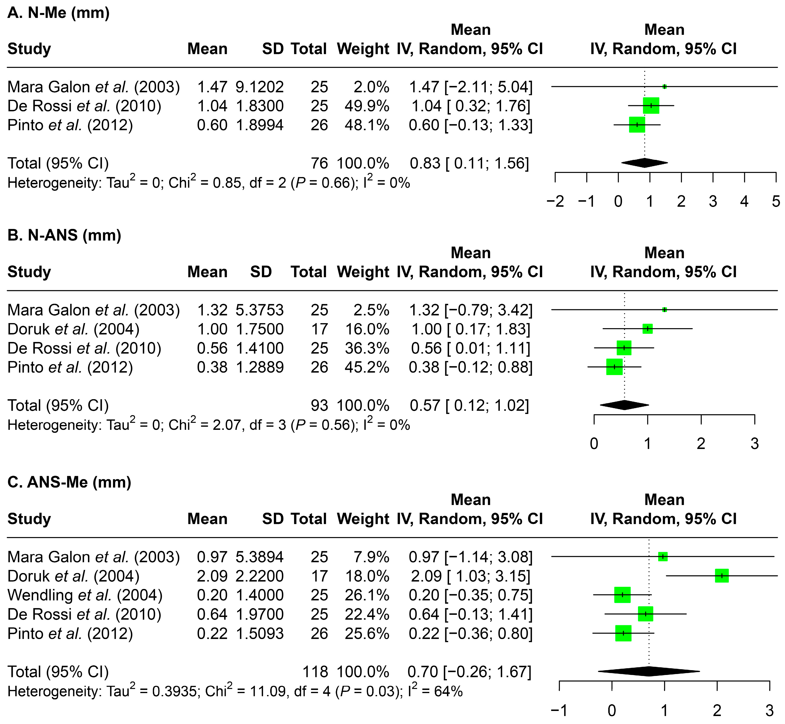
Changes in upper, lower, and total facial height were presented in Figure 2. Overall, a slight increase in facial height was evident for all three measurements. The greatest increase was seen in total facial height (N-Me, 0.83 mm), followed by upper facial height (N-ANS, 0.07 mm) and lower facial height (ANS-Me, 0.57 mm).

3.5. Palatal Plane Angulation and Position

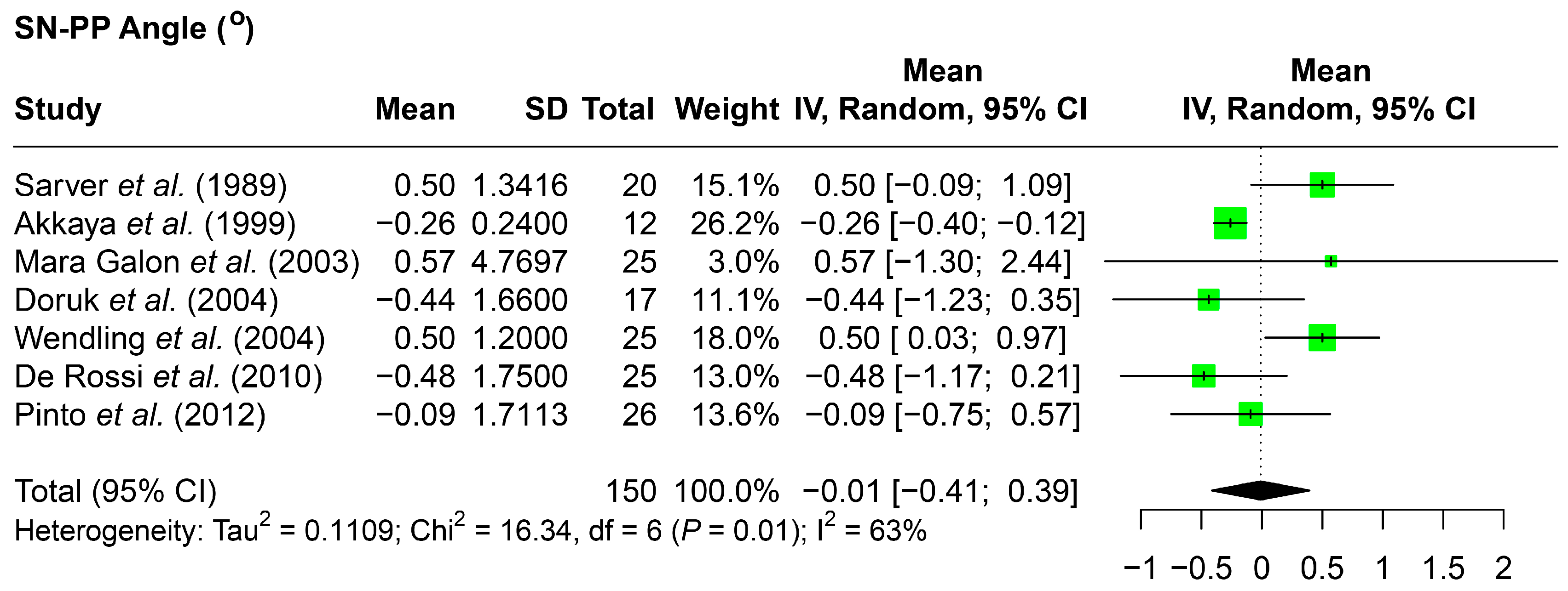
Limited data was available to assess the palatal plane vertical position, with two studies reporting a greater increase in the SN-ANS distance observed in comparison to the SN-PNS distance (Table 4) [5,6]. However, the meta-analysis revealed a minimal change in palatal plane angulation when compared to the SN plane (SN-PP angle, −0.01 degrees) (Figure 3).

3.6. Molar Position and Occlusal Plane Angulation

To evaluate the effect of bonded expanders on the vertical position of the upper and lower first molars, the distance of the upper first molar to the palatal plane and the lower first molar to the mandibular plane was reported (Table 4), with two studies reporting a reduction in the distance between the upper first molar to the palatal plane [6,13], and one study reporting an increase in the distance between the lower first molar to the mandibular plane [13]. However, no sufficient data could be utilized for a meta-analysis.
With the vertical position changes of the molars, the angle between the sella-nasion line and the occlusal plane (SN–occlusal plane) was also assessed, which demonstrated a little decrease (−0.04 degrees) (Figure 4).

3.7. Skeletal Assessments of the Vertical Dimension

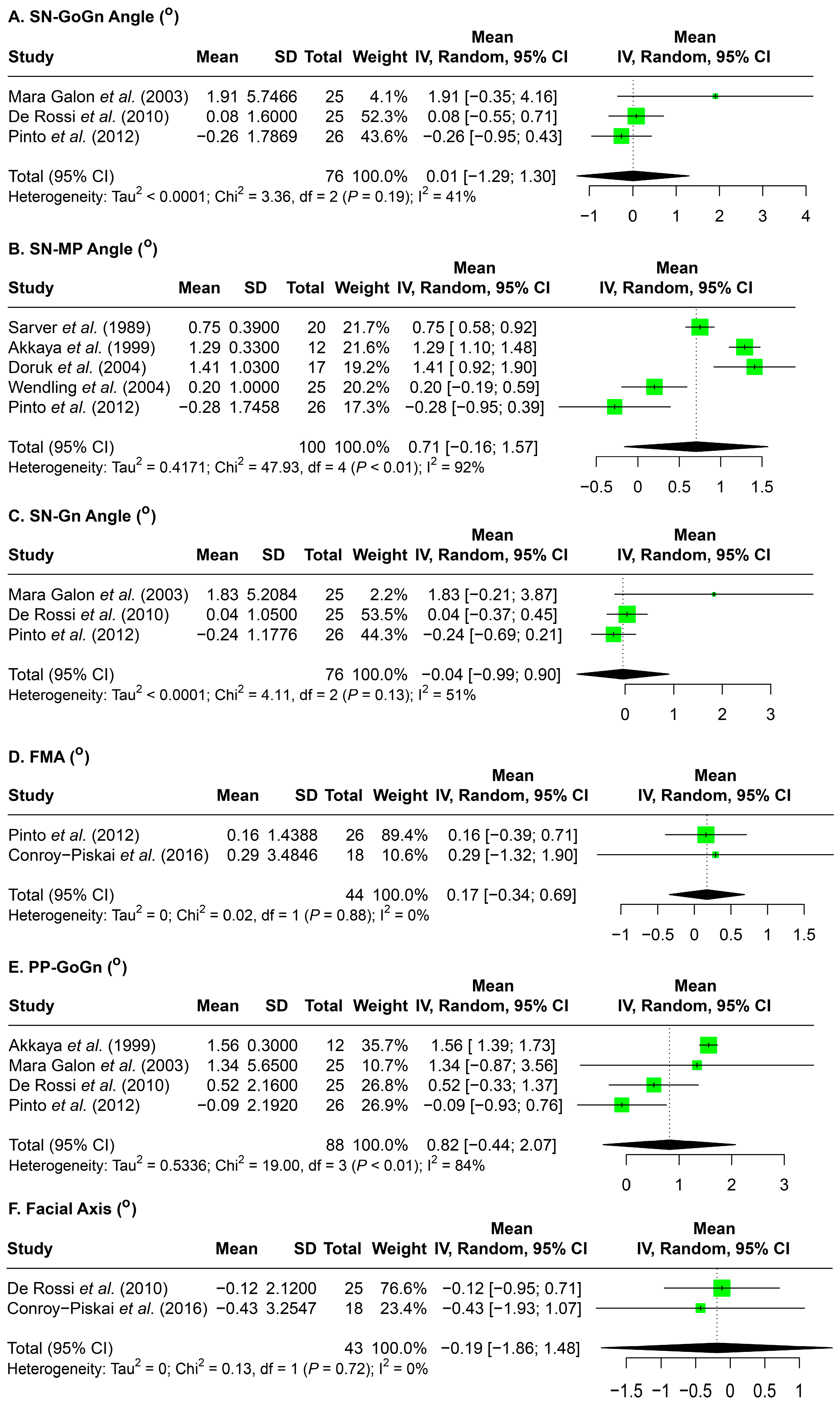
Lastly, the changes in the skeletal aspect of the vertical dimension in relation to the mandibular lower border were assessed (Figure 5). Overall, minimal changes were observed across these measurements. SN-Gn angle decreased by 0.04°, and facial axis by 0.19°. Increases were seen in SN-GoGn angle (0.01°), SN–mandibular plane angle (0.71°), FMA (0.17°) and PP-GoGn angle (0.82°).

4. Discussion

4.1. Summary of Evidence

The use of BRPEs with occlusal acrylic coverage, as opposed to banded RPEs, is a common orthodontic treatment approach aimed at minimizing increases in the vertical dimension during maxillary expansion. Despite its widespread use, no systematic review or meta-analysis to date has consolidated the evidence supporting this approach or clarified the mechanisms by which bonded expanders may control the vertical dimension. This systematic review and meta-analysis analyzed data from ten studies utilizing pre- and post-expansion lateral cephalometric radiographs to evaluate changes in the vertical dimension. Four outcomes were assessed: facial height, palatal plane position and angulation, vertical position of the upper and lower permanent first molars, and the vertical position of the mandibular lower border.
The literature search resulted in many studies with high variability in study characteristics; thus, only ten articles were included in our systematic review and nine articles were utilized in our meta-analysis. Overall, the mean changes in the skeletal parameters of the vertical dimension were small or insignificant within 6 months post-expansion. An important consideration when interpreting the results of the meta-analysis is that, as the involved subjects are children or teenagers, the observed mean changes in vertical parameters cannot be solely attributed to BRPE treatment. These changes may also reflect normal dentofacial growth. Buschang et al. reported that facial height increases by approximately 2.4 mm per year in males and 1.9 mm per year in females as part of typical growth patterns [18], while the current study found 0.83 mm of facial height increase after BRPEs intervention within 6 months post expansion. In terms of angular changes, an annual increase of 0.3 to 0.4 degrees in SN-MP and FMA is reported by Buschang et al. [18], and our current study demonstrated 0.71 degree and 0.17 degree changes, respectively. Therefore, bonded expanders likely do not result in an increase or decrease in the vertical dimension but may maintain the vertical dimension throughout treatment.
It is also important to consider if BRPEs provide a superior effect in vertical control when comparing to banded RPEs. A systematic review by Lione et al. that mixed both bonded and banded RPEs as well as mixed studies reporting short-term and long-term effects concluded that RPE resulted in a 1.7-degree increase in SN-MP angle and a 1.1-degree increase in SN-GoGn angle [19]. Another systematic review by Lagravere et al., which also combined banded and bonded Hyrax expanders, stated that immediately after expansion, there was a 1.97 increase in the SN-MP angle and a 1.65 degrees increase in the PP-MP angle [4]. The changes on the above parameters are about one degree larger than the result of our current study where a 0.71 degree increase in SN-MP, a 0.01 degree increase in SN-GoGn, and a 0.82 degree increase in PP-GoGn angle were revealed after bonded RPE treatment (Figure 5). Thus, the difference in the vertical effects of the banded and bonded RPE might be very minimal, with bonded expanders providing slightly more vertical control. In fact, Reed et al. compared patients treated with a banded versus a bonded RPE followed by edgewise orthodontics, and concluded that the banded RPE group had more vertical changes than the bonded RPE group, but most of the changes were less than 1 degree [20], which may not have clinical significance. However, it is worth noting that the initial vertical skeletal pattern of the involved subjects was not reported in these studies. Since vertical control is more critical for patients with a hyperdivergent pattern, further clinical studies that directly compare the short- and long-term effects of bonded and banded RPEs on patients with hyperdivergent pattern are needed. Such data will allow clinicians to individualize treatment planning based on both transverse and vertical treatment goals. A thorough understanding of these concepts will ultimately support orthodontists in delivering more predictable, efficient, and patient-specific care for individuals presenting with transverse maxillary deficiencies and varying vertical growth tendencies.

4.2. Limitations

There are several limitations to the current systematic review and meta-analysis that must be acknowledged. First, despite a comprehensive literature search, the number of studies meeting inclusion criteria was limited. The current body of published research specifically addressing the effects of BRPEs on the vertical dimension remains sparse. Consequently, our meta-analysis was based on a relatively small pool of studies and patients, which may reduce the statistical power and generalizability of our findings. Second, there was considerable heterogeneity among the included studies. This variability was reflected in the wide 95% confidence intervals observed for many of the mean changes in vertical measurements, making it challenging to draw definitive conclusions regarding the true effects of BRPEs on the vertical dimension. Furthermore, the outcomes evaluated in the meta-analysis represent mean changes between pre- and post-treatment lateral cephalometric tracings. These changes may reflect both treatment-related effects and natural growth and development. As such, it is not possible to isolate and attribute the observed skeletal or dentoalveolar changes solely to the use of bonded expanders. Future studies that include control groups or growth-matched comparisons will be critical in addressing this limitation.

5. Conclusions

The current research on bonded expanders and the treatment effects on the vertical dimension is limited and highly variable. Our systematic review and meta-analysis showed that bonded expanders may not control or reduce the vertical dimension significantly.

Author Contributions

Conceptualization, C.L.; methodology, S.H. (Sarah Horne) and C.L.; software, S.H. (Sarah Horne); validation, D.S.; formal analysis, S.H. (Sarah Horne), D.S., S.H. (Shahd Habeb), H.C.C., L.S., and C.L.; data curation, S.H. (Sarah Horne); writing—original draft preparation, S.H. (Sarah Horne) and D.S.; writing—review and editing, S.H. (Shahd Habeb), H.C.C., L.S., C.-H.C., and C.L.; visualization, C.L.; supervision, C.L.; project administration, C.L.; funding acquisition, C.L. All authors have read and agreed to the published version of the manuscript.

Funding

This work was supported by the American Association of Orthodontists Foundation (AAOF) Biomedical Research Award, International Orthodontic Foundation (IOF) Young Research Grant, the Department of Orthodontics, University of Pennsylvania School of Dental Medicine, the J. Henry O’Hern Jr. Pilot Grant, and University of Pennsylvania School of Dental Medicine, Joseph and Josephine Rabinowitz Award for Excellence in Research. The content is solely the responsibility of the authors and does not necessarily represent the official views of the funding agencies.

Institutional Review Board Statement

Not applicable.

Informed Consent Statement

Not applicable.

Data Availability Statement

All data generated or analyzed during this study are included in this published article.

Conflicts of Interest

The authors declare no conflicts of interest.

References

  1. Hsu, J.Y.; Cheng, J.H.; Feng, S.W.; Lai, P.C.; Yoshida, N.; Chiang, P.C. Strategic treatment planning for anterior open bite: A comprehensive approach. J. Dent. Sci. 2024, 19, 1328–1337. [Google Scholar] [CrossRef] [PubMed]
  2. Nielsen, I.L. Vertical malocclusions: Etiology, development, diagnosis and some aspects of treatment. Angle Orthod. 1991, 61, 247–260. [Google Scholar] [PubMed]
  3. Chung, C.H.; Font, B. Skeletal and dental changes in the sagittal, vertical, and transverse dimensions after rapid palatal expansion. Am. J. Orthod. Dentofac. Orthop. 2004, 126, 569–575. [Google Scholar] [CrossRef] [PubMed]
  4. Lagravere, M.O.; Heo, G.; Major, P.W.; Flores-Mir, C. Meta-analysis of immediate changes with rapid maxillary expansion treatment. J. Am. Dent. Assoc. 2006, 137, 44–53. [Google Scholar] [CrossRef] [PubMed]
  5. Sarver, D.M.; Johnston, M.W. Skeletal changes in vertical and anterior displacement of the maxilla with bonded rapid palatal expansion appliances. Am. J. Orthod. Dentofac. Orthop. 1989, 95, 462–466. [Google Scholar] [CrossRef] [PubMed]
  6. Asanza, S.; Cisneros, G.J.; Nieberg, L.G. Comparison of Hyrax and bonded expansion appliances. Angle Orthod. 1997, 67, 15–22. [Google Scholar] [PubMed]
  7. Pinto, F.M.P.; Abi-Ramia, L.B.P.; Stuani, A.S.; Stuani, M.B.S.; Artese, F. Vertical growth control during maxillary expansion using a bonded Hyrax appliance. Dent. Press. J. Orthod. 2012, 17, 101–107. [Google Scholar] [CrossRef]
  8. Rossi, M.S.; Stuani, M.B.S.; Silva, L.A.B. Cephalometric evaluation of vertical and anteroposterior changes associated with the use of bonded rapid maxillary expansion appliance. Dent. Press. J. Orthod. 2010, 15, 62–70. [Google Scholar]
  9. Shen, C.; Park, T.H.; Chung, C.H.; Li, C. Molar Distalization by Clear Aligners with Sequential Distalization Protocol: A Systematic Review and Meta-Analysis. J. Funct. Biomater. 2024, 15, 137. [Google Scholar] [CrossRef] [PubMed]
  10. Hasan, A.K.; Tizini, M.; Haffaf, R.A. Changes of the vertical dimension after expansion using bonded acrylic splint expander a clinical study. Tishreen Univ. J. Res. Sci. Stud. 2019, 41, 293–301. [Google Scholar]
  11. Galon, G.M.; Calçada, F.; Ursi, W.; Vilanova Queiroz, G.; Atta, J.; Almeida, G.A. Comparacao Cefalometrica entre os Aparelhos de ERM Bandado e Colado com Recobrimento Oclusal/Cephalometric Comparative Study between the Banded and the Bonded Rapid Maxillary Expansion Appliances. Rev. Dent. Press Ortodon. Ortop. Maxilar 2003, 8, 49–59. [Google Scholar]
  12. Akkaya, S.; Lorenzon, S.; Ucem, T.T. A comparison of sagittal and vertical effects between bonded rapid and slow maxillary expansion procedures. Eur. J. Orthod. 1999, 21, 175–180. [Google Scholar] [CrossRef] [PubMed]
  13. Conroy-Piskai, C.; Galang-Boquiren, M.T.; Obrez, A.; Viana, M.G.; Oppermann, N.; Sanchez, F.; Edgren, B.; Kusnoto, B. Assessment of vertical changes during maxillary expansion using quad helix or bonded rapid maxillary expander. Angle Orthod. 2016, 86, 925–933. [Google Scholar] [CrossRef] [PubMed]
  14. Doruk, C.; Bicakci, A.A.; Basciftci, F.A.; Agar, U.; Babacan, H. A comparison of the effects of rapid maxillary expansion and fan-type rapid maxillary expansion on dentofacial structures. Angle Orthod. 2004, 74, 184–194. [Google Scholar] [PubMed]
  15. Wendling, L.K.M.J.; James, A.; Franchi, L.; Baccetti, T. A Prospective Study of the Short-term Treatment Effects of the Acrylic-splint Rapid Maxillary Expander Combined with the Lower Schwarz Appliance. Angle Orthod. 2005, 75, 7–14. [Google Scholar] [PubMed]
  16. Harrer, M.; Cuijpers, P.; Furukawa, T.A.; Ebert, D.D. Doing Meta-Analysis with R: A Hands-On Guide; Chapman and Hall/CRC: Boca Raton, FL, USA, 2021. [Google Scholar]
  17. Harrer, M.; Cuijpers, P.; Furukawa, T.; Ebert, D.D. dmetar: Companion R Package For The Guide ‘Doing Meta-Analysis in R’, R package. version 0.1.0; GitHub: Francisco, CA, USA, 2019.
  18. Buschang, P.H.R.; Roldan, S.I.; Tadlock, L.P. Guidelines for assessing the growth and development of orthodontic patients. Semin. Orthod. 2017, 23, 321–335. [Google Scholar] [CrossRef]
  19. Lione, R.; Franchi, L.; Cozza, P. Does rapid maxillary expansion induce adverse effects in growing subjects? Angle Orthod. 2013, 83, 172–182. [Google Scholar] [CrossRef] [PubMed]
  20. Reed, N.; Ghosh, J.; Nanda, R.S. Comparison of treatment outcomes with banded and bonded RPE appliances. Am. J. Orthod. Dentofac. Orthop. 1999, 116, 31–40. [Google Scholar] [CrossRef] [PubMed]
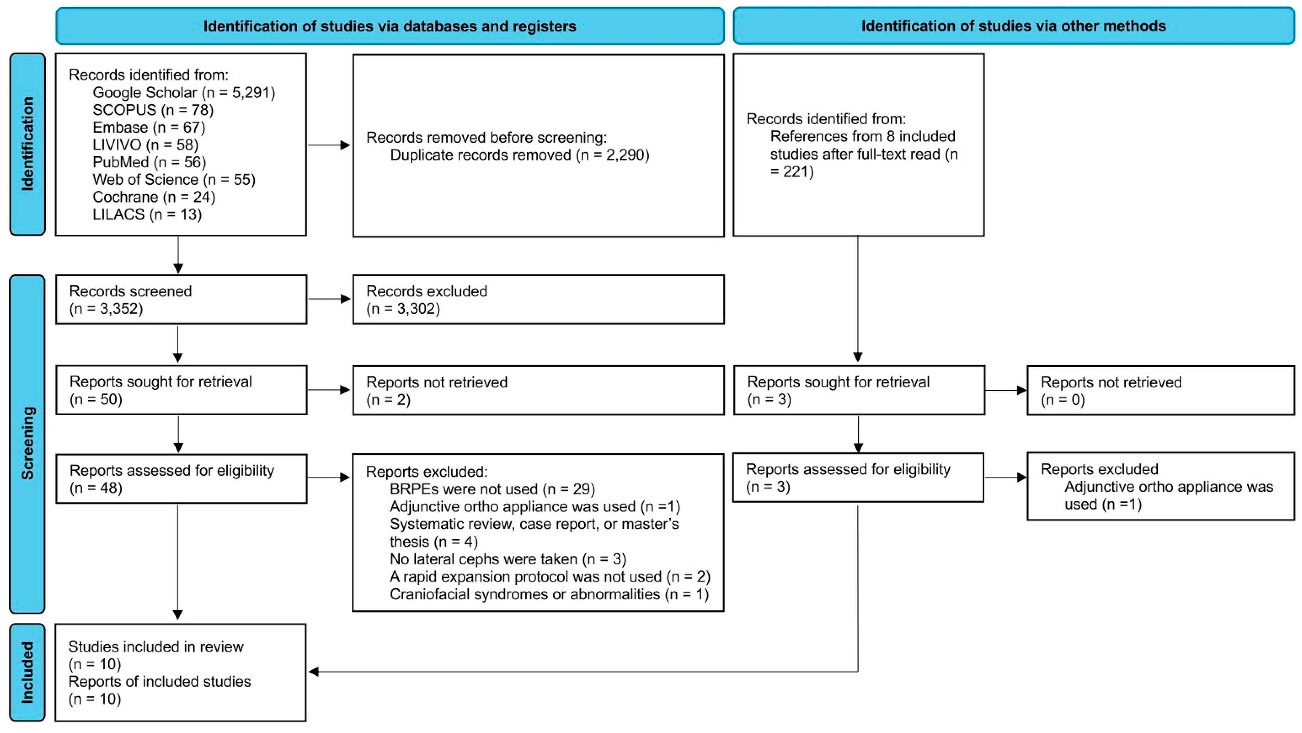
Figure 1. The PRISMA flow diagram demonstration the study identification and screening.
Jcm 14 07035 g001
Figure 2. Forest plots detailing the meta-analysis results for facial height: (A) N-Me, (B) N-ANS, and (C) ANS-Me. SD: standard deviation; CI: confidence interval; N: nasion; Me: menton; ANS: anterior nasal spine; mm: millimeters. Citation of involved references: [7,8,11,14,15].
Jcm 14 07035 g002
Figure 3. Forest plots detailing the meta-analysis results for the skeletal vertical parameters evaluating palatal plane angulation as SN-PP angle. SD: standard deviation; CI: confidence interval; SN: sella nasion line; PP: palatal plane. Citation of involved references: [5,7,8,11,12,14,15].
Jcm 14 07035 g003
Figure 4. Forest plot detailing the meta-analysis results for the SN–occlusal plane angle. SD: standard deviation; CI: confidence interval; SN: sella nasion line. Citation of involved references: [7,8,12,15].
Jcm 14 07035 g004
Figure 5. Forest plots detailing the results of the meta-analysis for overall skeletal measurements related to the mandibular lower border to assess the vertical dimension: (A) SN-GoGn angle, (B) SN-MP angle, (C) SN-Gn angle, (D) FMA, (E) PP-GoGn, and (F) facial axis. SD: standard deviation; CI: confidence interval; SN: sella nasion line; GoGn: gonion–gnathion line; MP: mandibular plane; Gn: gnathion; FMA: Frankfort–mandibular plane angle; PP: palatal plane. Citation of involved references: [5,7,8,11,12,13,14,15].
Jcm 14 07035 g005
Table 1. The PICO questions of this study.
CriteriaDescription
PopulationGrowing patients with a maxillary transverse discrepancy
InterventionBonded rapid palatal expander
ComparisonsPre-treatment records
OutcomeChange in skeletal and dentoalveolar vertical parameters
Table 2. Risk of bias assessment of the ten included studies. “+”: low risk of bias; “?”: medium risk of bias; “-”: high risk of bias.
Akkaya et al. (1999)
[12]
Asanza et al. (1997)
[6]
Conroy-Piskai et al. (2016)
[13]
De Rossi et al. (2010)
[8]
Doruk et al. (2004)
[14]
Hasan et al. (2019)
[10]
Mara Galon et al. (2003)
[11]
Pinto et al. (2012)
[7]
Sarver et al. (1989)
[5]
Wendling et al. (2005)
[15]
Study DesignObjective: objective clearly formulated++++++-+++
Sample size: considered adequate and estimated before collection of data?????+-?++
Baseline characteristics: similar baseline characteristics++++-+-+-+
Co-interventions (retainer, etc.)++++---+++
Randomization
      Random sampling---?-????-
      Random allocation of treatment-+-?-????-
Study MeasurementsMeasurement method: appropriate to the objective++++++-+++
Blind measurement: blinding
      Blinding (examiner)-----?----
      Blinding (statistician)-----?----
Reliability
      Reliability described? (intra-rater reliability)+-+-++?+--
      Adequate level of agreement? (inter-rater reliability)--+?-??---
Statistical AnalysisStatistical analysis
      Appropriate for data?++??+-???
      Combined subgroup analysis?????+-???
Cofounders (co-interventions): confounders included in analysis-----?----
Statistical significance level
      p-value stated?++++++-+++
      Confidence intervals stated?--+--+?---
OtherClinical significance++++++-+++
Total score781065100767
Percentage of the total41.1847.0658.8235.2929.4158.82041.1835.2941.18
Risk of biasMEDHIGHMEDHIGHMEDMEDHIGHMEDHIGHHIGH
Table 3. Characteristics of the ten included studies. F: female; M: male.
StudyStudy TypeMean Patient AgePatient Age RangeSample Size
(F/M)
Expansion ProtocolTreatment TimeTime of Post-Expansion Records
Akkaya et al. (1999) [12]Unclear11.96 years10.40–13.50 years12
(5/7)
2 turns/day0.70–1.60 monthsImmediately post expansion and at 3 months retention
Asanza et al. (1997) [6]UnclearNot specifiedNot specifiedNot specified2 turns/dayNot specifiedImmediately post expansion and at 3 months retention
Conroy-Piskai et al. (2016) [13]Retrospective102.89 ± 8.90 monthsNot specified18
(14/4)
2 turns/day12.28 ± 8.29 monthsAt 6 months retention
De Rossi et al. (2010) [8]Unclear8 years 5 months6 years 11 months–11 years 5 months25
(13/12)
1/2 turn/day20 days (14–26 days)At mean of 107 days
(90–124 days)
Doruk et al. (2004) [14]Unclear12.7 ±1.1 yearsNot specified17
(10/7)
1/2 turn/day26.47 ± 2.85 daysAt 90 days retention
(90.4 ± 6.7 days)
Hasan et al. (2019) [10]Prospective-6–12 years18
(F/M not specified)
1 turn/dayNot specifiedAt 6 months retention
Mara Galon et al. (2003) [11]Prospective-7.4–14.1 years25
(13/12)
3 turns/dayNot specifiedAt 4 months retention
Pinto et al. (2012) [7]Unclear8 years 5 months6 years 11 months–10 years 11 months26
(15/11)
2 turns/dayNot specifiedAt mean of 107 days
(90–124 days)
Sarver et al. (1989) [5]Unclear10.8 years7.5–16 years20
(14/6)
2 turns/dayNot specifiedImmediately post expansion
Wendling et al. (2005) [15]Prospective9 years 8 monthsNot specified25
(15/10)
1 turn/dayMean of 9.7 monthsAt 5 months retention
Table 4. The change in skeletal and dentoalveolar parameters between pre- and post-treatment records as measured on lateral cephalograms, extracted from the studies identified through our systematic review. T1: pre-treatment records; T2: immediately post-expansion records; T3: post-retention records. The data are presented as mean ± standard deviation, except the ones with additional notes. SE: standard error; N: nasion; ANS: anterior nasal spine; mm: millimeters; Me: menton; Ba: basion; Pt: Pt point; Gn: gnathion; S: sella; PP: palatal plane; PNS: posterior nasal spine; U6: upper first molar; MP: mandibular plane; FH: Frankfort Horizontal; Go: gonion; L6: lower first molar.
ParameterArticleT1T2T3T2-T1 DifferenceT3-T1 Difference
N-ANS (mm)De Rossi et al. (2010) [8]45.96 ± 2.92 46.52 ± 3.76 0.56 ± 1.41
Doruk et al. (2004) [14]55.32 ± 3.2556.62 ± 3.4456.32 ± 3.361.30 ± 2.391.00 ± 1.75
Mara Galon et al. (2003) [11]47.00 ± 3.718 48.316 ± 3.882
Pinto et al. (2012) [7]45.6777 ± 2.8383 46.0577 ± 3.0975 0.3800 ± 1.2889
N-Me (mm)De Rossi et al. (2010) [8]106.72 ± 5.07 107.76 ± 5.24 1.04 ± 1.83
Mara Galon et al. (2003) [11]107.860 ± 6.708 109.326 ± 6.179
Pinto et al. (2012) [7]108.5315 ± 4.8511 109.1273 ± 5.1055 0.5958 ± 1.8994
ANS-Me (mm)Asanza et al. (1997) [6]69.88 (range: 63.9 to 79.6) 0.05 (range: −1.70 to 2.00)
De Rossi et al. (2010) [8]63.08 ± 4.06 63.72 ± 3.92 0.64 ± 1.97
Doruk et al. (2004) [14]68.56 ± 4.6371.15 ± 5.5670.65 ± 5.372.59 ± 3.102.09 ± 2.22
Mara Galon et al. (2003) [11]62.975 ± 4.261 63.943 ± 3.300
Pinto et al. (2012) [7]62.8527 ± 3.7512 63.0704 ± 3.9912 0.2177 ± 1.5093
Wendling et al. (2004) [15]64.7 ± 5.2 0.2 ± 1.4
PP-Po-Or (°)Pinto et al. (2012) [7]4.1850 ± 2.7565 3.8612 ±2.7186 −0.3238 ± 1.4011
Condylion to gonion (mm)Wendling et al. (2004) [15]50.1 ± 3.8 0.7 ± 1.8
Gonion to pogonion (mm)Wendling et al. (2004) [15]71.6 ± 3.6 0.9 ± 1.6
Mandibular length (Co-Gn) (mm)Wendling et al. (2004) [15]108.5 ± 5.6 1.5 ± 1.2
Total facial height (NaBa-XiPm) (°)Conroy-Piskai et al. (2016) [13]65.92 ± 2.27 65.84 ± 2.49 −0.07
Lower facial height (ANS-Xi-Pm) (°)Conroy-Piskai et al. (2016) [13]48.28 ± 3.13 48.67 ± 3.16 0.38
Bjork sum (degrees)Hasan et al. (2019) [10]398.85 ± 5.09 399.51 ± 3.96
Y-axis (SN-SGn) (degrees)Hasan et al. (2019) [10]70.84 ± 4.6 71.17 ± 3.5
PFH/AFH (%)Hasan et al. (2019) [10]61.28 ± 3.4 60.93 ± 2.9
UFH/LFH (%)Hasan et al. (2019) [10]79.9 ± 5.1 79.3 ± 4.9
SN-PP angle (°)Asanza et al. (1997) [6]8.07 (range: 0 to 12.4) 1.25 (range: 0.20 to 2.80)
De Rossi et al. (2010) [8]7.88 ± 3.44 7.40 ± 3.31 −0.48 ± 1.75
Doruk et al. (2004) [14]9.65 ± 2.668.94 ± 2.349.21 ± 2.38−0.71 ± 1.87−0.44 ± 1.66
Mara Galon et al. (2003) [11]8.063 ± 3.462 8.636 ± 3.281
Pinto et al. (2012) [7]6.8831 ± 2.7199 6.7908 ± 2.8008 −0.0923 ± 1.7113
Sarver et al. (1989) [5] 0.50 ± 0.30 (SE)
Wendling et al. (2004) [15]6.8 ± 4.2 0.5 ± 1.2
SN-PNS (mm)Asanza et al. (1997) [6]44.0 (range: 41.6 to 49.4) 0.50 (range: −1.20 to 1.50)
Sarver et al. (1989) [5] 0.35 ± 0.18 (SE)
SN-ANS (mm)Asanza et al. (1997) [6]51.8 (range: 46.3 to 58.5) 1.50 (range: 0.40 to 4.20)
Sarver et al. (1989) [5] 1.25 ± 0.19 (SE)
SN–occlusal plane angle (°)Akkaya et al. (1999) [12]25.94 ± 1.1926.85 ± 1.1725.89 ± 0.97
De Rossi et al. (2010) [8]19.24 ± 3.97 19.00 ± 4.67 −0.24 ± 2.87
Pinto et al. (2012) [7]20.0415 ± 4.0931 20.1308 ± 4.3207 0.0893 ± 1.5746
Wendling et al. (2004) [15]20.8 ± 5.0 −0.3 ± 3.2
U6-PP (mm)Asanza et al. (1997) [6]22.23 (range: 17.0–26.0) −0.90 (range: −2.50 to 0.75)
Conroy-Piskai et al. (2016) [13]19.62 ± 1.79 19.29 ± 2.07 −0.33
L6-MP (mm)Conroy-Piskai et al. (2016) [13]26.79 ± 2.27 27.66 ± 2.62 0.87
SN-MP angle (°)Akkaya et al. (1999) [12]39.40 ± 0.8441.33 ± 0.9940.69 ± 0.85
Doruk et al. (2004) [14]39.09 ± 7.2640.94 ± 7.1740.50 ± 7.181.85 ± 1.371.41 ± 1.03
Pinto et al. (2012) [7]39.1738 ± 5.2802 38.8958 ± 5.2642 −0.2781 ± 1.7458
Sarver et al. (1989) [5] 0.75 ± 0.39
Wendling et al. (2004) [15]35.3 ± 6.1 0.2 ± 1.0
MP-FH angle (FMA) (°)Conroy-Piskai et al. (2016) [13]29.26 ± 2.32 29.55 ± 2.60 0.29
Pinto et al. (2012) [7]26.0965 ± 4.4902 26.2550 ± 4.5948 0.1585 ± 1.4388
PP-GoGn angle (°)De Rossi et al. (2010) [8]29.40 ± 4.17 29.92 ± 3.35 0.52 ± 2.16
Mara Galon et al. (2003) [11]27.642 ± 3.686 28.983 ± 4.282
Pinto et al. (2012) [7]30.1196 ± 4.0914 30.0346 ± 4.1125 −0.0850 ± 2.1920
SN-GoGn angle (°)De Rossi et al. (2010) [8]37.28 ± 5.31 37.36 ± 4.79 0.08 ± 1.60
Mara Galon et al. (2003) [11]35.706 ± 3.810 37.613 ± 4.302
Pinto et al. (2012) [7]37.0612 ± 5.2509 36.8019 ± 5.2126 −0.2593 ± 1.7869
SN-Gn angle (°)De Rossi et al. (2010) [8]68.88 ± 4.52 68.92 ± 4.61 0.04 ± 1.05
Mara Galon et al. (2003) [11]67.154 ± 3.581 68.986 ± 3.782
Pinto et al. (2012) [7]69.0262 ± 4.5551 68.7873 ± 4.5124 −0.2389 ± 1.1776
Facial axis (NaBa-PtGn) (°)Conroy-Piskai et al. (2016) [13]84.07 ± 2.22 83.64 ± 2.38 −0.43
De Rossi et al. (2010) [8]85.16 ± 3.28 85.04 ± 4.01 −0.12 ± 2.12
Disclaimer/Publisher’s Note: The statements, opinions and data contained in all publications are solely those of the individual author(s) and contributor(s) and not of MDPI and/or the editor(s). MDPI and/or the editor(s) disclaim responsibility for any injury to people or property resulting from any ideas, methods, instructions or products referred to in the content.