Systematic Review

Intracranial Metastases from Uterine Leiomyosarcoma: A Systematic Review and Case Illustration

1
Department of Radiology, Feinberg School of Medicine, Northwestern University, Chicago, IL 60611, USA
2
Department of Neurological Surgery, New York-Presbyterian Hospital Weill Cornell Medicine, New York, NY 10065, USA
3
Robert H. Lurie Comprehensive Cancer Center, Northwestern University, Chicago, IL 60611, USA
4
Department of Radiation Oncology, Feinberg School of Medicine, Northwestern University, Chicago, IL 60611, USA
5
Department of Neurology, Feinberg School of Medicine, Northwestern University, Chicago, IL 60611, USA
6
Department of Neurological Surgery, Feinberg School of Medicine, Northwestern University, Chicago, IL 60611, USA
7
Northwestern Medicine Malnati Brain Tumor Institute of the Lurie Comprehensive Cancer Center, Feinberg School of Medicine, Northwestern University, Chicago, IL 60611, USA
*
Author to whom correspondence should be addressed.
J. Clin. Med. 2025, 14(18), 6631; https://doi.org/10.3390/jcm14186631
Submission received: 29 July 2025 / Revised: 25 August 2025 / Accepted: 18 September 2025 / Published: 20 September 2025
(This article belongs to the Section Oncology)

Abstract

Background/Objectives: Brain metastasis from uterine leiomyosarcoma (ULMS) is an exceptionally rare complication of an aggressive malignancy. With fewer than 40 cases previously documented, a significant knowledge gap exists regarding its clinical course, management, and outcomes. This study provides the largest analysis of ULMS brain metastases to date, integrating a systematic literature review with a novel case report illustrating the disease’s uniquely rapid progression. Methods: Following PRISMA guidelines, we systematically reviewed four major databases to identify all reported cases of intracranial metastasis from ULMS. Data on patient demographics, clinico-radiological features, treatments, and survival were extracted and analyzed. Methodological quality was assessed using a modified Joanna Briggs Institute (JBI) tool. Results: We analyzed 34 studies with 39 individual cases. Additionally, this review was supplemented by one new illustrative case from our institution. The median patient age was 51.5 years, and most presented with focal neurological symptoms. Common imaging findings included hyperdense lesions on CT and homogeneously enhancing, dural-based masses on MRI, which mimic other intracranial pathologies. Though surgery was the most frequent intervention (76.9%), median survival after a brain metastasis diagnosis was a grim 5 months, with no significant difference observed between treatment modalities. Our illustrative case was remarkable for an extremely rapid volumetric doubling time averaging just 7.3 days. Conclusions: Brain metastasis from ULMS is a lethal event with an extremely poor prognosis. Nonspecific imaging features create diagnostic challenges, necessitating histopathological confirmation. Current therapies, including surgery and radiotherapy, offer palliative benefit but do not significantly alter survival. The aggressive biological behavior demonstrated here underscores the urgent need for increased clinical awareness and collaborative research to develop more effective management strategies and improve outcomes for this devastating diagnosis.

1. Introduction

Leiomyosarcoma (LMS) is an aggressive malignant tumor that originates from smooth muscle cells and belongs to the heterogeneous group of soft tissue sarcomas [1]. LMS includes several distinct subtypes, primarily differentiated by their anatomical site of origin, which dictates their biological behavior and clinical outcomes [2]. The primary subtypes include uterine LMS, retroperitoneal LMS, and LMS of the soft tissues of the extremities [1]. These variants display significant heterogeneity in their rates of growth, metastatic potential, and response to therapy. Key prognostic factors across all subtypes include tumor size, histological grade, anatomic location, tumor stage, and the feasibility of complete surgical resection [3].
A defining characteristic of LMS is its propensity for hematogenous metastasis [4]. The lungs are the most common site of distant spread, followed by the liver, particularly for LMS arising in the abdomen or uterus. However, the risk of metastasis varies substantially among subtypes [5]. For instance, cutaneous LMS has a much lower risk of spreading (5% to 10%) whereas more aggressive forms like uterine, vascular, and deep soft tissue LMS (especially retroperitoneal ones) have a much higher risk, around 30% to 40% or even more [3].
Uterine leiomyosarcoma (ULMS) is the most prevalent type of uterine sarcoma, although it accounts for only 1–2% of all uterine cancers. Like other LMS subtypes, ULMS typically metastasizes via the bloodstream, with the lungs, peritoneal and retroperitoneal cavities, and bones being the most frequent secondary sites [6]. The occurrence of brain metastases from ULMS is exceptionally rare, with only approximately 40 cases documented in the literature to date. The rarity of this complication has resulted in a scarcity of literature to guide clinical management [7].
This knowledge gap underscores the importance of a comprehensive analysis of reported instances to better understand the disease course in these patients. Such an evaluation is essential for clarifying the timeline to the development of brain metastases, the patterns of intracranial spread (e.g., solitary versus multiple lesions), and associated clinical presentations. Furthermore, a detailed review of these uncommon cases is essential for evaluating the effectiveness of various treatments, including surgery, radiation therapy (whole-brain or stereotactic), and systemic agents.
In this study, we present a comprehensive analysis of documented cases of ULMS with brain metastases, with a focus on imaging characteristics, treatment strategies, and survival outcomes. We also present a novel case that illustrates previously unreported tumor features, further highlighting the aggressive nature of ULMS brain metastases.

2. Materials and Methods

2.1. Study Design and Patient Selection

This retrospective study was approved by the Institutional Review Board and received waivers for both Health Insurance Portability and Accountability Act (HIPAA) authorization and written informed consent. The institutional electronic data warehouse (EDW) was searched from January 2004 to December 2022 to identify patients meeting the following inclusion criteria: diagnosis of biopsy-proven Soft Tissue Sarcoma and pre-treatment MRI of the primary tumor. During the dataset review, a single ULMS patient with brain metastases was identified. Patient confidentiality was strictly maintained, and all identifying information was anonymized.

2.2. Literature Search and Review Protocol

This systematic review was conducted and reported in accordance with the Preferred Reporting Items for Systematic Reviews and Meta-Analysis (PRISMA-2020) guidelines. The review was designed to address a research question developed using the PICO (Population, Intervention, Comparison, Outcome) framework [8]. Specifically, the review aimed to answer the following PICO-based question: In patients with ULMS, what are the clinical features, imaging findings, and outcomes associated with intracranial metastases, and how do different treatment modalities compare in terms of survival?
Systematic search was performed in PubMed, Google Scholar, Embase, and Web of Science using a predefined search strategy, with the final search conducted on 1 May 2025. The search strategy combined relevant keywords with medical subject headings (MeSH), where applicable. Search terms included combinations of: ((((((((((leiomyosarcoma) AND (brain)) OR (cerebral)) OR (intracranial)) OR (intracerebral))) OR (CNS)) OR (central nervous system)) AND (metastasis))) OR (metastatic). All the terms were searched in the title, abstracts, or keywords of all records (Supplemental Materials, Table S2).

2.3. Inclusion Criteria

Only retrospective case reports, case series, and other studies were identified for this review. Moreover, all eligible studies were enrolled in this study with no time and language restrictions.

2.4. Exclusion Criteria

Relevant exclusion criteria were: (1) diagnosis of any other sarcoma, (2) data were only published in abstract form, and (3) studies with incomplete data.

2.5. Data Search and Extraction

Two independent reviewers read the drafted articles in full text and extracted the following data: first author’s name, year of study, location of study, age, presenting symptoms, location of brain metastasis, tumor consistency, imaging features, contemporary lung metastasis, treatments, extent of resection in case of surgery, time from primary tumor to metastasis, survival after diagnosis of metastasis, cause of death, and immunohistochemistry characteristics. In case of any disagreement between the two reviewers, a third one made the final decision.

2.6. Quality Assessments and Analysis

Data extraction was independently carried out by two reviewers and encompassed a detailed assessment of study characteristics, patient demographics, common clinical features, radiological and histopathological findings, as well as follow-up data. To assess methodological quality and potential risk of bias, the included studies were evaluated using a modified version of the Joanna Briggs Institute (JBI) critical appraisal checklist for case reports, as previously described in the literature [9]. Discrepancies in data extraction or quality assessment were resolved through consensus or consultation with a third reviewer when necessary.
All statistical analyses were conducted using IBM SPSS Statistics version 29.0.2.0. Given the heterogeneity among the included studies, quantitative analysis was limited to descriptive statistics, including the calculation of means and ranges for relevant variables. As a result, data synthesis was predominantly qualitative in nature.

3. Results

3.1. Search Results and Study Characteristic

The systematic literature search, conducted using the predefined strategy, initially identified 2170 records. After removing 538 duplicates, 1632 unique articles remained for screening. Title and abstract screening excluded 1593 articles based on irrelevance to the research question. The full texts of the remaining 39 studies were assessed for eligibility. The most common reasons for exclusion at this stage were insufficient data (4 studies) and inability to access the full text (1 study). Following the application of all inclusion and exclusion criteria, 34 studies with a total of 39 individual cases were deemed eligible and included in the final analysis (Figure 1).

3.2. Patient Characteristics

From 1989 to 2025, 39 individual cases of brain metastases from uterine leiomyosarcoma (ULMS) were identified in 34 previously published studies. We also included one new case from our institution that met all inclusion criteria, bringing the total number of cases analyzed to 40. As expected, all patients were female, with a median age of 51.5 years (range: 26–70) (Table 1). For patients who were still alive at the time of data collection, survival was calculated to the last known follow-up date. This right-censoring of data is a key limitation, as it inherently underestimates the true survival time for this subgroup and contributes to a poorer overall prognostic outcome. Four patients were excluded from the survival analysis because their survival time was not reported or could not be estimated. Therefore, for the remaining 36 patients, the average and median survival times after diagnosis of brain metastasis were 10.37 months and 5 months, respectively.

3.3. Presentation Symptoms

All patients exhibited neurological symptoms corresponding to the anatomical location of the metastatic lesion, typically at the gray-white matter junction. Of the cases reviewed, 30 patients (75.0%) had a single brain lesion, 5 patients (12.5%) presented with two lesions, and another 5 patients (12.5%) had more than two intracranial lesions. Concurrent pulmonary metastases were documented in 29 out of 35 cases (82.86%) where lung involvement was reported. The median interval from initial diagnosis of the primary uterine tumor to the development of brain metastases was 31 (range 0–116) months (Table 1).

3.4. Radiological Features and Diagnosis

Nearly all patients underwent brain MRI (T1- and T2-weighted sequences), with or without accompanying CT imaging. Non-contrast CT scans, when performed, commonly revealed hyperattenuating lesions or evidence of osteolytic changes. Radiological findings were reported in 23 studies. Among MRI signal characteristics, T1 and T2 isointensity was observed in 4 (17.4%) cases, T2 hyperintensity in 5 (21.7%) cases, and a combination of T1 isointensity with T2 hyperintensity in 3 (13%) cases. Homogeneous contrast enhancement was the most frequently reported pattern, seen in 19 (82.6%) cases, while ring enhancement was described in 4 (17.4%) cases. Additional MRI features included necrosis in 14 (60.9%) cases and a dural-based lesion in 10 (43.5%) cases. (Supplementary Materials, Table S1).

3.5. Histopathology

Sixteen studies described histopathological features, consistently noting the presence of firm, moderately vascularized tissue with intermittent hemorrhagic areas. Immunohistochemical analysis was reported in 15 cases, with positive staining observed for actin in 12 (80%) cases, vimentin in 8 (53.3%) cases, and desmin in 8 (53.3%) cases (Table 1). While a comprehensive marker panel was often unavailable, the Ki-67 proliferation index was specifically reported in two instances [24,30], with values of approximately 20% and greater than 25%.

3.6. Treatment Approaches and Outcomes

Of the reported cases, 30 (76.9%) patients underwent surgical intervention, either alone or in combination with adjuvant therapies. GKRS, with or without chemotherapy, was administered to 5 (12.8%) patients, while 3 (7.7%) patients received whole-brain radiotherapy with or without chemotherapy. One (2.6%) patient was treated solely with chemotherapy (Table 1). Follow-up information was available for 35 subjects. Our new case was treated with Gamma Knife radiosurgery and chemotherapy. The median survival following the diagnosis of brain metastases was 5 months. At the time of publication, 10 patients (28.6%) were reported to be alive (Table 1).
To evaluate the impact of different treatment strategies on survival, patients were stratified into six groups (Table 2). A Kaplan–Meier plot visually demonstrates the survival probabilities for each treatment group over time (Figure 2). While an overall log-rank comparison of the groups did not show a statistically significant difference (p = 0.29), a specific pairwise test revealed a notable treatment effect: patients treated with surgery and chemotherapy had a significantly longer median survival (18.0 months) compared to those who received surgery alone (4.0 months, p = 0.03). No other pairwise comparisons between these treatment groups reached statistical significance.
Further analysis isolating the effect of chemotherapy confirmed its benefit. Patients who received chemotherapy as part of any treatment regimen had a median survival more than two times longer than those who did not (12 vs. 5 months). This finding was statistically significant (p = 0.04), strongly suggesting that including chemotherapy in the treatment plan is associated with improved survival for this patient cohort. A Cox proportional-hazards model further quantified the benefit of chemotherapy. It demonstrated that adding chemotherapy to a regimen reduced the hazard of death by approximately 70% compared to those without it. This finding reinforces that systemic therapy is a critical component of treatment for this aggressive disease.

3.7. Methodological Quality and Risk of Bias Assessments

A modified form of the JBI case series appraisal tool was employed to assess the methodological quality of the included studies (Table 3). The mean (SD) score was 7.1 (SD 1.03), reflecting good methodological quality. Lower-scoring studies lacked detailed pathology reports or follow-up assessments for patients and gave limited details on patient demographic characteristics and individual outcomes. The highest-scoring study offered a more complete account of the disease trajectory and patient follow-up. Nevertheless, the small sample sizes across the case series limit the broader applicability of the findings (Table 3. Additional details in Supplementary Materials, Table S3).

3.8. Illustrative Case

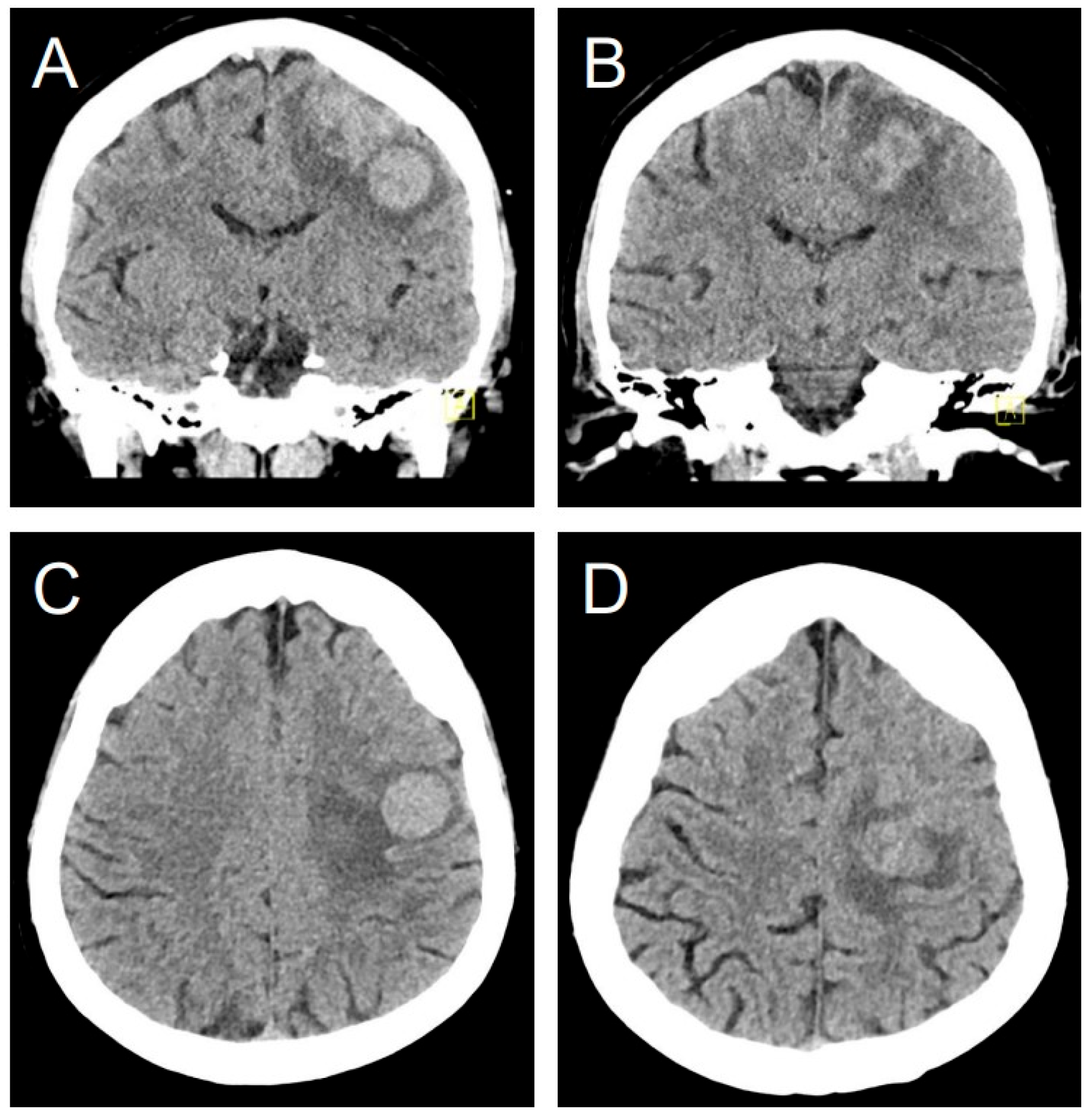
A 49-year-old female with a history of myomectomy 14 years prior, presented with right arm paresis, two months after being diagnosed with ULMS with concurrent lung metastases. Initial brain computed tomography (CT) revealed multiple hyperdense lesions with surrounding edema (Figure 3). A subsequent diagnostic magnetic resonance imaging (MRI) identified a total of four intracranial lesions, which appeared T1-weighted hypointense and T2-weighted hyperintense with homogeneous enhancement and central necrosis (Figure 4). The three largest lesions measured 11.8 × 11.2 × 11.3 mm, 22.6 × 17.6 × 21.3 mm, and 21.1 × 18.8 × 19.4 mm, with a mean Apparent Diffusion Coefficient (ADC) of 973 ± 37 × 10−6 mm2/s.
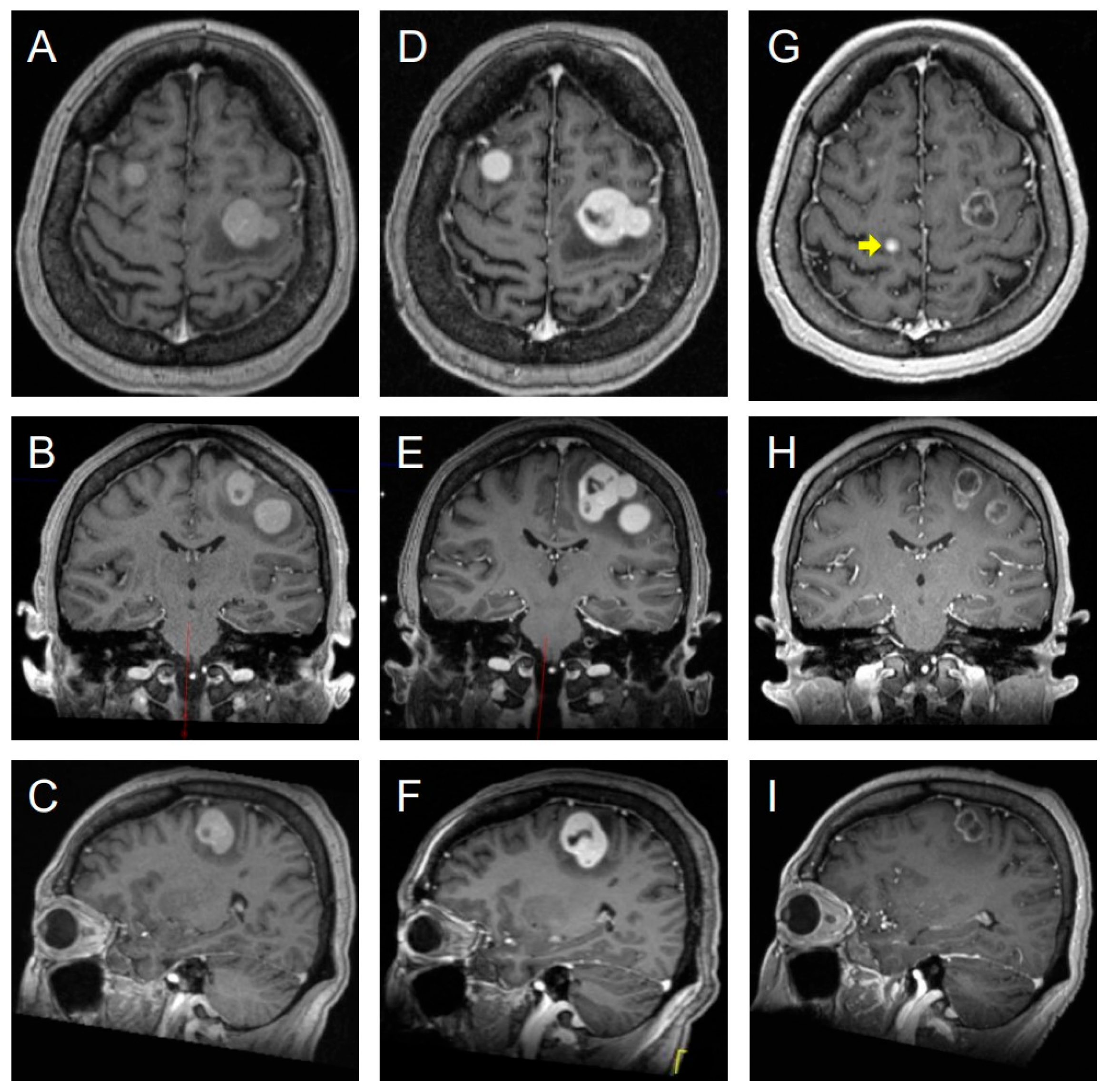
Given the findings, the patient was scheduled for GKRS. However, a pre-treatment planning MRI obtained just seven days later revealed aggressive disease progression. The total lesion count had increased to six, and the three largest lesions demonstrated a mean volumetric growth of 94% (range: 51–141%), corresponding to a mean volumetric doubling time of only 7.3 days (range: 5.5–11.7 days) (Figure 5) [42].
The patient proceeded with GKRS using frame-based immobilization. All lesions were treated to a median dose of 20 Gy, prescribed to the 50% isodose line, with 100% coverage of all visible and defined tumors. A two-month follow-up MRI confirmed a successful response in the targeted lesions, which showed a mean volumetric decrease of 73% (range: 62–86%) and a corresponding increase in the mean ADC to 2344 ± 270 × 10−6 mm2/s. Despite this effective local therapy, the development of new metastases was evident, with the total intracranial lesion count rising to twelve, signifying ongoing overall disease progression (Figure 5). Unfortunately, the patient ultimately succumbed to her systemic disease burden 20 months after the diagnosis of brain metastases.

4. Discussion

Brain metastasis from ULMS is an exceptionally rare and ominous complication of a disease already known for aggressive behavior and hematogenous spread. This analysis represents the largest collection of such cases to date, providing valuable insights into the clinical presentation, radiological features, treatment patterns, and outcomes associated with this disease entity. These key findings emphasize the aggressiveness of ULMS when it involves the brain, a median survival of just five months after diagnosis, underlining the serious diagnostic and therapeutic challenges.
A critical finding from our review is the radiological profile of these metastases. The frequent presentation as homogeneously enhancing, dural-based masses on MRI and hyperdense lesions on non-contrast CT creates a significant diagnostic challenge [4,6,43]. These features can, respectively, mimic more common intracranial pathologies such as meningiomas, particularly in patients without a known primary cancer, and hemorrhagic metastases from other primary sites like melanoma, renal cell carcinoma, or choriocarcinoma [4,6,43]. This radiological ambiguity underscores the need for a high index of suspicion. Histopathological confirmation is critical for an accurate diagnosis. The high prevalence of concurrent pulmonary metastases (82.86%) in our cohort suggests that in a ULMS patient with known lung involvement, the development of new neurological symptoms should immediately prompt investigation for intracranial spread.
The management of ULMS brain metastases remains a significant challenge with no established standard of care, a fact reflected in the variety of treatments employed in the reported cases [13,41]. Surgery was the most common intervention (76.9%), often utilized for diagnostic confirmation, cytoreduction, and palliation of mass effect. While a broad comparison of treatment modalities did not show an overall survival benefit, a more specific pairwise analysis revealed a critical difference: patients treated with surgery combined with chemotherapy had a significantly longer median survival than those treated with surgery alone (18.0 vs. 4.0 months, p = 0.03). This was the only treatment comparison to reach statistical significance. Furthermore, a focused analysis confirmed that the inclusion of chemotherapy in any regimen was the key factor associated with improved survival (p = 0.04). In contrast, a focused analysis revealed that the inclusion of chemotherapy was the only factor associated with significantly longer survival (18.0 vs. 5.0 months, p = 0.04), suggesting its critical role in improving outcomes [44,45]. A Cox proportional-hazards model reinforced this conclusion, showing that the addition of chemotherapy was associated with a roughly 70% reduction in the hazard of death compared to regimens without it. This finding underscores that achieving systemic control is likely the most critical component of the treatment strategy for this aggressive disease. This lack of a clear survival advantage may be attributable to several factors, including the small sample size, inherent selection bias in retrospective case reports (e.g., patients with better performance status are more likely to be offered surgery), and the overwhelming impact of systemic disease burden. The grim median survival of 5 months is unambiguously poor, even when compared to brain metastases from other aggressive cancers, suggesting an inherent biological aggressiveness once the central nervous system is involved.
Our case strikingly illustrates the clinical course and challenges associated with these tumors. The patient’s rapid radiological progression in just one week exemplifies their aggressive biological nature. Notably, her 20-month survival following Gamma Knife radiosurgery significantly exceeds the 4-month median observed in our review, suggesting that aggressive local therapy can achieve durable local control in select patients. As mentioned, the lesion doubled in size and, more critically, the number of lesions markedly proliferated within a single week, underscoring a concerning lack of response to the targeted therapy. This highlights the potential importance of initiating treatment as early as possible. Given this scant data, performing precise, regular neurologic examinations and obtaining brain imaging even with minimal neurologic changes may be of utmost importance to improve patient care. Conversely, our review of published cases indicates no significant difference in outcomes among various approaches to these metastases. This suggests that while local control of intracranial disease is crucial for neurological palliation, it may not alter the overall fatal course driven by widespread metastases. Ultimately, the prognosis appears to be dictated by the systemic tumor burden and its response to therapy.
This study is not without limitations. The foremost is its reliance on published case reports and small case series, which are inherently susceptible to publication bias (e.g., favoring the reporting of unusual or successful outcomes) and heterogeneity in reporting standards, treatment protocols, and follow-up duration across several decades. The small sample size (n = 40) limits the statistical power to detect significant differences between treatment groups and draw definitive conclusions about the optimal therapeutic strategy. The methodological quality assessment, while generally good, did reveal variability in the completeness of reporting, further constraining the depth of analysis.
Despite these limitations, our review offers important clinical insights. We recommend that clinicians maintain a high index of suspicion for brain metastases in ULMS patients, particularly those with confirmed pulmonary metastases, as this is a common pathway for disease spread. We propose a strategy of close, regular neurological examinations for this high-risk subset of patients. Furthermore, we advise that brain imaging, such as an MRI, be performed promptly at the first sign of even minimal neurological changes. This proactive approach, while not a universal screening program, could significantly reduce diagnostic delays and may allow for earlier intervention, potentially improving patient outcomes. A multidisciplinary approach is crucial to tailor therapy to the individual patient. This involves neurosurgeons, radiation oncologists, and medical oncologists who consider factors like the number and location of metastases, systemic disease status, and overall performance. Future research should focus on establishing a prospective, multicenter registry for rare metastases like these to enable more robust analysis. Furthermore, molecular and genomic profiling of these metastatic tumors is warranted to identify potential therapeutic targets that could lead to more effective systemic agents.

5. Conclusions

In conclusion, brain metastasis from ULMS is a rare but lethal event characterized by nonspecific imaging features and an extremely poor prognosis. While current treatments, including surgery and radiotherapy, may offer symptomatic benefits, they do not appear to significantly alter the grim overall survival. Increased awareness and collaborative, prospective research are urgently needed to develop more effective management strategies and improve outcomes for this patient population.

Supplementary Materials

The following supporting information can be downloaded at: https://www.mdpi.com/article/10.3390/jcm14186631/s1, Table S1: Radiological features and diagnosis. Table S2: Search strings. Table S3: JBI Case Reports Appraisal.

Author Contributions

Conceptualization, Y.S.V., A.P.-R., L.P.; methodology, A.P.-R., S.Z., Y.S.V.; software, Y.S.V.; validation, Y.S.V.; formal analysis, S.Z., A.P.-R., L.P., V.B.H., Y.S.V.; investigation, A.P.-R., S.Z., L.P., V.B.H., S.S., Y.S.V.; resources, C.K., K.S., L.K., A.A.B., H.S., R.A., T.A.T., Y.S.V.; data curation, A.P.-R., L.P., Y.S.V.; writing—original draft preparation, A.P.-R., S.Z., L.P.; writing—review and editing, A.P.-R., V.B.H., C.K., K.S., L.K., A.A.B., H.S., R.A., T.A.T., U.B., S.S., K.D., R.V.L., P.K., Y.S.V.; visualization, Y.S.V., A.P.-R., S.Z.; supervision, Y.S.V., A.P.-R., V.B.H., S.S., K.D., R.V.L., P.K., Y.S.V.; project administration, Y.S.V. All authors have read and agreed to the published version of the manuscript.

Funding

No support was received from any organization for this work.

Institutional Review Board Statement

The study was conducted in accordance with the Declaration of Helsinki and approved by the Institutional Review Board of Northwestern University. (#STU00217019 of 3 May 2025).

Informed Consent Statement

Patient consent was waived due to retrospective nature of the study.

Data Availability Statement

Data are contained within the article and supplementary materials.

Conflicts of Interest

The authors declare no conflicts of interest.

Abbreviations

The following abbreviations are used in this manuscript:
LMSLeiomyosarcoma
ULMSUterine Leiomyosarcoma
BMBrain Metastases
GKRSGamma-Knife Radiosurgery
WBRTWhole Brain Radiotherapy
CTComputer Tomography
MRIMagnetic Resonance Imaging
ADCApparent Diffusion Coefficient

References

  1. Hickman, A.; Siontis, B.L. Not All Leiomyosarcomas Are the Same: How to Best Classify LMS. Curr. Treat. Options Oncol. 2023, 24, 327–337. [Google Scholar] [CrossRef] [PubMed]
  2. Lacuna, K.; Bose, S.; Ingham, M.; Schwartz, G. Therapeutic advances in leiomyosarcoma. Front. Oncol. 2023, 13, 1149106. [Google Scholar] [CrossRef]
  3. Mocellin, S. Leiomyosarcoma. In Soft Tissue Tumors: A Practical and Comprehensive Guide to Sarcomas and Benign Neoplasms; Springer: Cham, Switzerland, 2021; pp. 477–481. [Google Scholar]
  4. Salvati, M.; Cervoni, L.; Caruso, R.; Gagliardi, F.M.; Delfini, R. Sarcoma metastatic to the brain: A Series of 15 Cases. Surg. Neurol. 1998, 49, 441–444. [Google Scholar] [CrossRef]
  5. Sabhavath, M.; Annamaraju, S.S.; Amanchi, N.R.; Bhavanam, K.R.; Kancha, R.K. Soft tissue sarcoma. In Biomedical Aspects of Solid Cancers; Springer: Cham, Switzerland, 2024; pp. 279–288. [Google Scholar]
  6. Jędrys, W.; Leśniak, A.; Borkowska, A.; Rutkowski, P.; Sobczuk, P. Brain metastases of sarcoma: A rare phenomenon in rare tumours. J. Cancer Res. Clin. Oncol. 2023, 149, 18271–18281. [Google Scholar] [CrossRef]
  7. Carrillo-Uzeta, A.A.; Varela-Avalos, E.; Agustin-Godinez, E.; Uehara-Gonzalez, J.A.; Medina-Romero, J.R.; Velazquez-Zamarripa, A.I.; Quiñones-González, A.M.; Romero-García, P.A. A neurosurgical approach to skull metastasis from uterine leiomyosarcoma: Illustrative case. J. Neurosurg. Case Lessons 2025, 9, CASE24769. [Google Scholar] [CrossRef]
  8. Page, M.J.; McKenzie, J.E.; Bossuyt, P.M.; Boutron, I.; Hoffmann, T.C.; Mulrow, C.D.; Shamseer, L.; Tetzlaff, J.M.; Akl, E.A.; Brennan, S.E.; et al. The PRISMA 2020 statement: An updated guideline for reporting systematic reviews. BMJ 2021, 372, 71. [Google Scholar] [CrossRef]
  9. Jordan, Z.; Pilla, B. From agenda to action: JBI Evidence Synthesis and the United Nations Sustainable Development Goals. JBI Evid. Synth. 2024, 22, 364–377. [Google Scholar] [CrossRef]
  10. Vaquero, J.; Martinez, R.; el Barkani, A.; Gomez-Angulo, J.C.; Escandon, J. Leiomyosarcoma metastatic to the brain with prolonged survival. J. Neurosurg. Sci. 1989, 33, 291–292. [Google Scholar] [PubMed]
  11. Prussia, P.R.; Clarke, H.A.; Mansoor, G.; Garriques, S.; Maheswaran, B. Uterine leiomyosarcoma with intracerebral metastasis: A case report. J. Natl. Med. Assoc. 1992, 84, 368–370. [Google Scholar] [PubMed]
  12. Wronski, M.; de Palma, P.; Arbit, E. Leiomyosarcoma of the uterus metastatic to brain: A case report and a review of the literature. Gynecol. Oncol. 1994, 54, 237–241. [Google Scholar] [CrossRef]
  13. Bindal, R.K.; Sawaya, R.E.; Leavens, M.E.; Taylor, S.H.; Guinee, V.F. Sarcoma metastatic to the brain: Results of surgical treatment. Neurosurgery 1994, 35, 185–190. [Google Scholar] [CrossRef]
  14. Uchino, M.; Endo, G.; Shibata, I.; Terao, H.; Kuramitsu, T.; Kushida, Y.; Nakamura, N. Uterine leiomyosarcoma metastasis to the skull—Case report. Neurol. Med. Chir. 1996, 36, 469–471. [Google Scholar] [CrossRef]
  15. Ziyal, I.M.; Musluman, M.; Bejjani, G.K.; Tanik, C.; Turkmen, C.S.; Aydin, Y. Cerebral metastasis of a uterine leiomyosarcoma—Case report. Neurol. Med. Chir. 1999, 39, 238–241. [Google Scholar] [CrossRef] [PubMed]
  16. Mawrin, C.; Kirches, E.; Dietzmann, K.; Weis, S. Uterine leiomyosarcoma metastatic to the brain stem. Arch. Gynecol. Obstet. 2002, 266, 119–121. [Google Scholar] [CrossRef]
  17. Yip, C.M.; Yang, K.C.; Lo, Y.S.; Liao, W.C.; Chen, J.Y.; Hsu, S.S. Skull metastasis from uterine leiomyosarcoma: A case report. Acta Neurol. Taiwan. 2006, 15, 109–113. [Google Scholar] [PubMed]
  18. Munakata, A.; Asano, K.; Hatayama, T.; Itoh, K.; Suzuki, S.; Ohkuma, H. Leiomyosarcoma of the uterus metastatic to the brain. No Shinkei Geka 2006, 34, 409–413. [Google Scholar] [PubMed]
  19. Melone, G.A.; D’Elia, A.; Brogna, C.; Salvati, M. Uterine leiomyosarcoma metastatic to the brain: Case report. Tumori 2008, 94, 856–860. [Google Scholar] [CrossRef]
  20. Kaya, A.O.; Büyükberber, S.; Yildiz, R.; Öztürk, B.; Karagülle, K.; Yaman, E. Uterine Leiomyosarcoma Presenting with Neurological Symptoms: Differential Diagnosis. Turk. Klin. J. Med. Sci. 2009, 29, 281–284. [Google Scholar]
  21. Benizelos, J.; Anagnostou, E.; Papathomas, T.; Spandos, V.; Kampas, L.; Rasala, V.; Sioutopoulou, D.; Destouni, C.; Tatsiou, Z.; Tsantila, I. Abstracts of the 22nd European Congress of Pathology, September 4–9, 2009, Florence, Italy. Virchows Arch. 2009, 455 (Suppl. 1), S1–S448. [Google Scholar] [CrossRef]
  22. Pereira, F.O.C.; Pereira, D.C.; de Castro Aguiar, R.; Lombardi, I.A.S.; Zanini, M.A. Leiomyosarcoma Metastatic to the skull and spine: A case report. Rev. Bras. Cancerol. 2011, 57, 63–66. [Google Scholar] [CrossRef]
  23. Honeybul, S.; Ha, T. Leiomyosarcoma of the uterus metastatic to the brain: A case report. Arch. Gynecol. Obs. 2009, 279, 391–393. [Google Scholar] [CrossRef]
  24. Venizelos, I.; Anagnostou, E.; Papathomas, T.; Spandos, V.; Marinopoulos, D.; Tsitsopoulos, P.; Tsonidis, C. A 57-year-old female with a cerebellar mass. Brain Pathol. 2011, 21, 351–354. [Google Scholar] [CrossRef]
  25. Yamada, S.; Yamada, S.M.; Nakaguchi, H.; Murakami, M.; Hoya, K.; Matsuno, A. A case of multiple brain metastases of uterine leiomyosarcoma with a literature review. Surg. Oncol. 2011, 20, e127–e131. [Google Scholar] [CrossRef]
  26. Mariniello, G.; Vergara, P.; Del Basso De Caro, M.L.; Maiuri, F. Intracranial dural metastasis from uterine leiomyosarcoma with orbital extension. Neurol. Sci. 2012, 33, 1173–1177. [Google Scholar] [CrossRef]
  27. Chen, H.; Xu, C.; Zhou, Y. Complete remission of leiomyosarcoma with lung and brain metastasis by chemoradiotherapy after surgery. Chin.-Ger. J. Clin. Oncol. 2013, 12, 502–504. [Google Scholar] [CrossRef]
  28. Shepard, M.J.; Fezeu, F.; Lee, C.C.; Sheehan, J.P. Gamma knife radiosurgery for the treatment of gynecologic malignancies metastasizing to the brain: Clinical article. J. Neurooncol. 2014, 120, 515–522. [Google Scholar] [CrossRef] [PubMed]
  29. Gurram, M.K.; Pulivarthi, S.; McGary, C.T.; Defillo, A. Brain and multiorgan metastases from uterine leiomyosarcoma. Tumori 2014, 100, e8–e13. [Google Scholar] [CrossRef]
  30. Abrahao, C.M.; Maluf, F.C. Uterine leiomyosarcoma with central nervous system metastases. Ecancermedicalscience 2015, 9, 515. [Google Scholar] [CrossRef]
  31. Kim, Y.H.; Park, I.K.; Min, G.E.; Jin, K.H.; Shin, J.H. A Case of Orbital Metastasis of Uterine Leiomyosarcoma With Intracranial Extension Presenting With Proptosis. Ophthalmic Plast. Reconstr. Surg. 2016, 32, e51–e52. [Google Scholar] [CrossRef] [PubMed]
  32. Inoue, K.; Tsubamoto, H.; Tomogane, Y.; Kamihigashi, M.; Shibahara, H. Pazopanib-mediated long-term disease stabilization after resection of a uterine leiomyosarcoma metastasis to the brain: A case report. Gynecol. Oncol. Rep. 2016, 17, 60–64. [Google Scholar] [CrossRef] [PubMed]
  33. Ahuja, A.; Agarwal, P.; Sardana, R.; Bhaskar, S. Extensively Metastasizing Leiomyosarcoma: A Diagnostic Challenge. J. Midlife Health 2017, 8, 148–150. [Google Scholar] [CrossRef] [PubMed]
  34. Sosa, P.; Cuadra, G.; Hidalgo, R. Metástasis meníngea de leiomiosarcoma uterino. Rep. Caso Revis. Lit. Neurocir. 2018, 29, 103–106. [Google Scholar]
  35. Chahdi, H.; Oukabli, M. Brain metastases from uterine leiomyosarcoma. Pan Afr. Med. J. 2018, 30, 90. [Google Scholar] [CrossRef]
  36. Imoumby, F.N.; Dokponou, Y.C.H.; Kouakou, F.; El Agouri, H.; El Asri, A.C.; Gazzaz, M. Brain metastasis of uterine leiomyosarcoma: A case report and review of the literature. Open J. Mod. Neurosurg. 2021, 11, 107. [Google Scholar] [CrossRef]
  37. Miki, K.; Samura, K.; Takahashi, K.; Kawashima, M. Treatment of skull metastasis from uterine leiomyosarcoma: A single-center experience with literature review. Interdiscip. Neurosurg. 2021, 23, 101004. [Google Scholar] [CrossRef]
  38. Soo, W.T.; Teo, E.G.; Mohamad, N.; Wong, A.S.H. Chronic Subdural Hematoma Caused by Calvarial and Dural Metastasis from Uterine Leiomyosarcoma. J. Neurosci. Rural Pr. 2022, 13, 351–353. [Google Scholar] [CrossRef]
  39. Rodríguez Delgado, J.E.; Morales Cruz, M.; López Hernández, C.J.; Rodríguez Morales, M.; Viscarra León, J.; Torres Álvarez, M.; Díaz Quiroz, G.; Aceves Chimal, J.L. Uterine Leiomyosarcoma: Infrequent Cardiac and Brain Metastasis A Case Report. J. Cardio-Thorac. Med. 2022, 10, 1099. [Google Scholar]
  40. Eatz, T.; Levy, A.; Merenzon, M.; Bystrom, L.; Berry, K.; Morell, A.; Bhatia, S.; Daggubati, L.; Higgins, D.; Schlumbrecht, M.; et al. Surgically Treated Brain Metastases from Uterine Origin: A Case Series and Systematic Review. World Neurosurg. 2023, 173, e91–e108. [Google Scholar] [CrossRef]
  41. Richards, H.; Alsalek, S.; Laiwalla, A.; Attiah, M.; Harary, M.; Kim, W.J.; Hirt, D.; Rahman, S.U. Brain and lung metastasis of uterine leiomyosarcoma: Illustrative case. J. Neurosurg. Case Lessons 2023, 5, CASE22557. [Google Scholar] [CrossRef]
  42. Seyal, A.R.; Parekh, K.; Velichko, Y.S.; Salem, R.; Yaghmai, V. Tumor growth kinetics versus RECIST to assess response to locoregional therapy in breast cancer liver metastases. Acad. Radiol. 2014, 21, 950–957. [Google Scholar] [CrossRef]
  43. Eminovic, S.; Orth, T.; Dell’Orco, A.; Baumgartner, L.; Morotti, A.; Wasilewski, D.; Guelen, M.S.; Scheel, M.; Penzkofer, T.; Nawabi, J. Clinical and imaging manifestations of intracerebral hemorrhage in brain tumors and metastatic lesions: A comprehensive overview. J. Neuro-Oncol. 2024, 170, 567–578. [Google Scholar] [CrossRef] [PubMed]
  44. Vogelbaum, M.A.; Brown, P.D.; Messersmith, H.; Brastianos, P.K.; Burri, S.; Cahill, D.; Dunn, I.F.; Gaspar, L.E.; Gatson, N.T.N.; Gondi, V.; et al. Treatment for Brain Metastases: ASCO-SNO-ASTRO Guideline. J. Clin. Oncol. 2022, 40, 492–516. [Google Scholar] [CrossRef] [PubMed]
  45. Mehta, M.P.; Paleologos, N.A.; Mikkelsen, T.; Robinson, P.D.; Ammirati, M.; Andrews, D.W.; Asher, A.L.; Burri, S.H.; Cobbs, C.S.; Gaspar, L.E.; et al. The role of chemotherapy in the management of newly diagnosed brain metastases: A systematic review and evidence-based clinical practice guideline. J. Neuro-Oncol. 2010, 96, 71–83. [Google Scholar] [CrossRef] [PubMed]
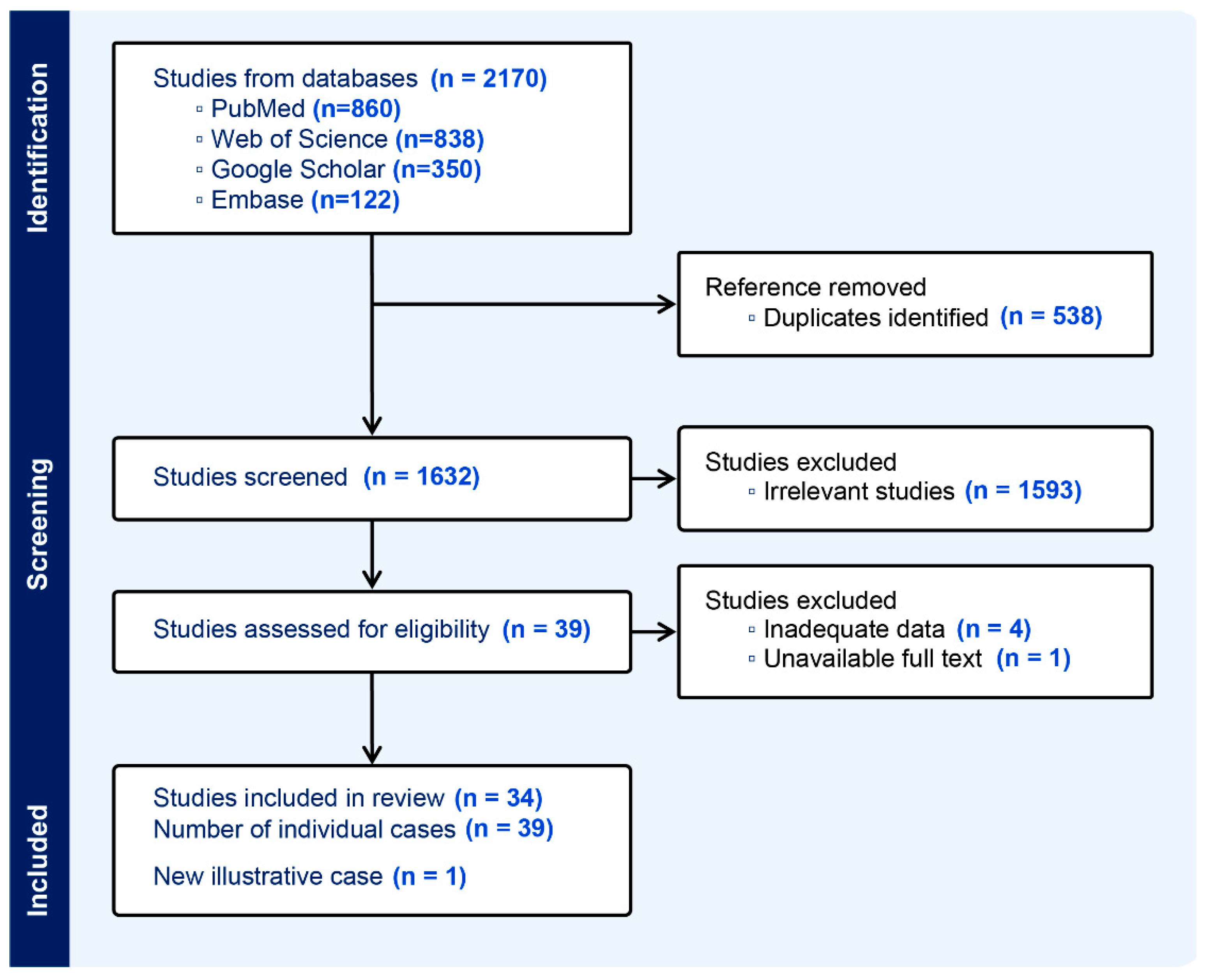
Figure 1. PRISMA diagram.
Jcm 14 06631 g001
Figure 2. Kaplan–Meier survival curves illustrating the probability of survival over time for patients in different treatment groups. A Cox proportional-hazards model was used to demonstrate the relative hazard of death for each group.
Jcm 14 06631 g002
Figure 3. Pretreatment non-contrast brain CT scan in coronal (A,B) and axial (C,D) views shows a hyperdense left posterior frontal lobe lesion with a marked vasogenic edema.
Jcm 14 06631 g003
Figure 4. Pretreatment axial non-contrast T1-weighted (A) and T2-weighted (B) brain MRI demonstrate two lesions, hypointense on T1W and hyperintense on T2W, located in the right and left frontal lobes, which show homogeneous enhancement on post-contrast T1W imaging (C) and are surrounded by extensive vasogenic edema on FLAIR (D).
Jcm 14 06631 g004
Figure 5. Post-contrast T1-weighted brain MRIs: Axial, coronal, and sagittal views across three time points. Columns (AC) represent the initial scan at admission. Columns (DF), taken one week later, demonstrate rapid lesion progression. Columns (GI) show the tumor’s response two months post- GKRS, highlighting an acceptable reduction in existing lesions despite the concurrent emergence of a new lesion (yellow arrow).
Jcm 14 06631 g005
Table 1. Patients’ characteristics.
AuthorAgeLung
Mets
Time
to BM
EORAdjuvant
for BM
Positive IHC
Markers
Cause of DeathSurvival
After BM,
Month
Vaquero
et al. [10]
26YesNSTChemoNSCNS50
Prussia
et al. [11]
36NS72TNoNSNSNS
Wronski
et al. [12]
60Yes78TWBRTNSSystemic30
Bindal
et al. [13]
55Yes116TWBRTNSSystemic14
Uchino
et al. [14]
54No48TChemoNSLung24
Salvati
et al. [4]
58Yes3TWBRTNSNS3
43Yes48TWBRTNSNS5
28Yes26TWBRTNSNS5
Ziyal
et al. [15]
38Yes72TWBRTNSCNS4
Mawrin
et al. [16]
59Yes36BxWBRTLaminin,
Vimentin
CNS2
Yip
et al. [17]
63NS15TNoActin,
Desmin
Systemic4
Munakata
et al. [18]
52No36BxGKRSNSNSNS
Melone
at al. [19]
57Yes12TWBRT + ChemoNS___At least 5
Kaya
et al. [20]
60Yes0___WBRT + ChemoActin,
Vimentin,
Desmin
NSNS
Benizelos
et al. [21]
51Yes6___WBRT + ChemoActin,
Desmin
Systemic3
Pereira
et al. [22]
55No60TNoActin,
Desmin
Systemic5
Honeybul
et al. [23]
42Yes10TNoNSSystemic2
Venizelos
et al. [24]
57Yes8STRWBRTActin,
Vimentin,
Desmin
Systemic1.5
Yamada
et al. [25]
50Yes28STRGKRSNSSystemic12
Mariniello
et al. [26]
57No8STRChemoActin,
Vimentin
CNS4
Chen
et al. [27]
54Yes48___GKRS + ChemoNS___At least 29
Shepard
et al. [28]
54NS24___GKRSNSSystemic1
Gurram
et al. [29]
59Yes84STRSRSNSSystemic2
Abrahao
et al. [30]
45Yes31TWBRT + ChemoNSNS27
Abrahao
et al. [30]
51Yes22TWBRT + ChemoNSSystemic16
Kim
et al. [31]
57Yes36__________Systemic1
Inoue
et al. [32]
48Yes77TChemoActin, EMA,
Vimentin,
Desmin
___At least 18
Ahuja
et al. [33]
60Yes6___ChemoActin,
Vimentin,
pan-CK
___At least 12
Sosa
et al. [34]
43Yes0TNoActinSystemic1
Chahdi
et al. [35]
46NS84TWBRTActin,
h-Caldesmon
NSNS
Imoumby
et al. [36]
46Yes60TWBRTActin,
Vimentin,
h-Caldesmon
Systemic5
Miki
et al. [37]
35NS36TWBRTNS___At least 17
70Yes96TNoNS___At least 15
51Yes48TWBRTNS___At least 7
Soo
et al. [38]
60No0TWBRT + ChemoNS___At least 6
Delgado
et al. [39]
33Yes10TWBRT + ChemoNSNSNS
Eatz
et al. [40]
55Yes5BxSRSNSSystemic2
Richards
et al. [41]
51Yes44TChemo +
SRS
Myosin___At least 2
Carrilo-Uzeta
et al. [7]
46No0TWBRTSMA,
h-Caldesmon
___At least 12
Our Case49Yes2___GKRS +
Chemo
Actin,
Vimentin
Systemic20
Mets: metastasis; EOR: extent of resection; NS: not specified; SMA: smooth muscle antigen; EMA: epithelial membrane antigen; Chemo: chemotherapy; WBRT: whole brain radiotherapy; GKRS: Gamma Knife radiosurgery; Bx: biopsy; T: total tumor removal; STR: subtotal tumor removal.
Table 2. Survival times for different therapeutic options.
TreatmentNumber of
Patients
Mean
Survival Time
Median
Survival Time
Surgery + Chemotherapy424 ± 19.218 (24–18)
Surgery55.4 ± 5.64 (5–2)
Surgery + RT139.3 ± 85 (12–4)
Surgery + RT + Chemotherapy511.2 ± 10.36 (16–5)
RT46.2 ± 9.22 (2–2)
With Chemotherapy1215.1 ± 13.912 (24–5)
Without Chemotherapy237.7 ± 7.45 (12–2)
Table 3. The JBI Case Reports Appraisal Scores.
CaseTotal Score
(out of 8)
Case
(Continuation)
Total Score
(out of 8)
Vaquero et al., 1989 [10]6Honeybul et al., 2009 [23]8
Prussia et al., 1992 [11]4.5Venizelos et al., 2011 [24]8
Wronski et al., 1994 [12]7Yamada et al., 2011 [25]8
Bindal et al., 1994 [13]7Mariniello et al., 2012 [26]8
Uchino et al., 1996 [14]7.5Sosa et al., 2018 [34]8
Salvati et al., 1998 [4]7Chahdi et al., 2018 [35]5
Ziyal et al., 1999 [15]7.5Kim et al., 2016 [31]5.5
Mawrin et al., 2002 [16]8Eatz et al., 2023 [40]7
Yip et al., 2006 [17]7.5Soo et al., 2022 [38]7.5
Chen et al., 2013 [27]7Carrillo-Uzeta et al., 2025 [7]8
Shepard et al., 2014 [28]6Abrahao et al., 2015 [30]7
Gurram et al., 2014 [29]8Delgado et al., 2022 [39]6
Munakata et al., 2006 [18]5Inoue et al., 2016 [32]8
Melone et al., 2008 [19]8Ahuja et al., 2017 [33]8
Kaya et al., 2009 [20]6Miki et al., 2021 [37]7.5
Benizelos et al., 2009 [21]7Richards et al., 2023 [41]8
Pereira et al., 2011 [22]8Imoumby et al., 2021 [36]8
Disclaimer/Publisher’s Note: The statements, opinions and data contained in all publications are solely those of the individual author(s) and contributor(s) and not of MDPI and/or the editor(s). MDPI and/or the editor(s) disclaim responsibility for any injury to people or property resulting from any ideas, methods, instructions or products referred to in the content.