1. Introduction
Uterine leiomyomas (fibroids or myomas) are the most common pelvic tumors in women of reproductive age. They usually cause menstrual irregularities, chronic pelvic pain, and infertility [
1]. They occur in at least half of American women of reproductive age, and their frequency and size increase with age [
2]. Fibroids represent an enormous public health burden for women and an economic cost to society. Various medical and surgical modalities are used to manage these patients. Although studies continue to be conducted on the best surgical modalities, the literature is not sufficiently clear on the pathophysiology of uterine fibroids. Strategies are needed to prevent, limit, and non-surgically treat the growth of these tumors.
Signal transducer and activator of transcription (STAT) proteins were identified in the early 1990s in conjunction with interferon (IFN)-mediated regulation of gene transcription [
3]. Today, it is known that various cytokines induce different STAT proteins. Seven STAT proteins have been identified in mammalian cells. These are known as STAT-1, STAT-2, STAT-3, STAT-4, STAT-5a, STAT-5b, and STAT-6. The stages of cytokine-mediated STAT protein activation are as follows: cytokines bind to their cognate receptors on the cell surface. Subsequently, oligomerization occurs. This oligomerization stimulates Janus kinase (JAK) proteins associated with the receptor via cross-phosphorylation. Activated JAKs bind to type I and II cytokine receptors and phosphorylate tyrosine residues on these receptors and on other JAKs. This phosphorylation allows STAT proteins to dimerize, forming homodimers or heterodimers, and translocate to the cell nucleus. Once in the nucleus, STAT proteins interact with specific response element sequences on DNA, stimulating the transcription of target genes. Uncontrolled STAT-3 and STAT-5 activities play a role in malignant transformation. STAT proteins are involved in carcinogenesis through two mechanisms, one of which is the continuous activation of STAT proteins. STAT-3 activity leads to an increase in vascular endothelial growth factor (VEGF) levels and plays a role in tumor angiogenesis [
4,
5].
The upregulation or downregulation of cytokines, which are vital mediators of the immune system, influences the development of diseases such as inflammation, infection, and cancer. IL-26, a member of the IL-10 family and IL-20 subfamily, exhibits diverse effects in various diseases, owing to its unique cationic structure. IL-26 is secreted by Th17 cells and is considered a semi-qunatiproinflammatory cytokine. By binding to the IL-26 receptor complex (IL-10R1/IL-20R2), it induces multiple signaling mediators, particularly STAT-1/STAT-3 [
6]. Due to its role in many pathological conditions, including inflammation, assessing IL-26 expression in tissues and biological fluids can be valuable, particularly for disease monitoring and prognosis [
7,
8,
9,
10]. Therefore, analyzing IL-26 expression in the myometrium and fibroids may be important for determining the role of cytokines in the development of leiomyomas. This study aimed to immunohistochemically detect STAT-3 and IL-26 expression in leiomyoma tissues and determine whether these signaling pathways play a role in leiomyoma pathophysiology.
2. Materials and Methods
2.1. Location of the Study
RTEU Training and Research Hospital.
2.2. Type of Research, Population, Sample, and Research Group
This case–control retrospective study included 38 patients aged 35–55 years who underwent hysterectomy due to uterine myoma at the RTEU Education and Research Hospital over a period of 5 years as the study group and patients who underwent hysterectomy for benign reasons other than fibroids as the control group. After extracting the records of patients who had undergone hysterectomy within the last five years from our archives, the final pathology reports of the surgical specimens were obtained from the pathology department. The indications for hysterectomy and the results of the pathology reports were recorded for each patient. Patients with pathological reports indicating uterine leiomyomas were included in the study group. The indication for hysterectomy in the study group was the presence of uterine fibroids. The control group comprised patients who underwent hysterectomy for benign gynecological reasons other than fibroids.
Tissue samples taken from some of the removed material (fibroid material of the study group and normal myometrium tissue of the control group) in the two groups of patients who underwent hysterectomy for their indications were evaluated immunohistochemically for STAT-3 and IL-26 activity. Since the primary outcome variable in the study was the staining of histopathological samples (such as hematoxylin and eosin) and these samples were used more than once, a sample size calculation was not required. Indications for hysterectomy in the myoma group were single or multiple uterine fibroids of various locations. In the control group, 14 patients underwent hysterectomy for dysfunctional uterine bleeding, 6 for multiple endometrial polyps, and 10 for grade 3 uterine prolapse. Endometrial polyp patients were those who had undergone hysteroscopic polypectomy but whose polyps had recurred.
Considering the roles of IL-26 and STAT-3 in tumorigenesis, angiogenesis, and inflammation [
4,
5,
6,
7], patients who were likely to affect the expression of these two cytokines were excluded from the study: those taking antiandrogen and lipid-lowering drugs with a history of intrauterine device insertion, Asherman syndrome, pelvic inflammatory disease, preoperative infectious disease, endometriosis, hydrosalpinx, previous pelvic surgery, recurrent abortion, and systemic and/or rheumatologic disease causing inflammation.
2.3. Ethical Approval
This study was conducted in compliance with the principles of the Declaration of Helsinki and was approved by the Ethics Committee of Recep Tayyip Erdoğan University (approval number: 2022/221). Written informed consent for the hysterectomy was obtained from all participants in the leiomyoma and control groups. Owing to the retrospective nature of the study, ethics committee approval was deemed sufficient for resection of paraffin blocks and immunostaining.
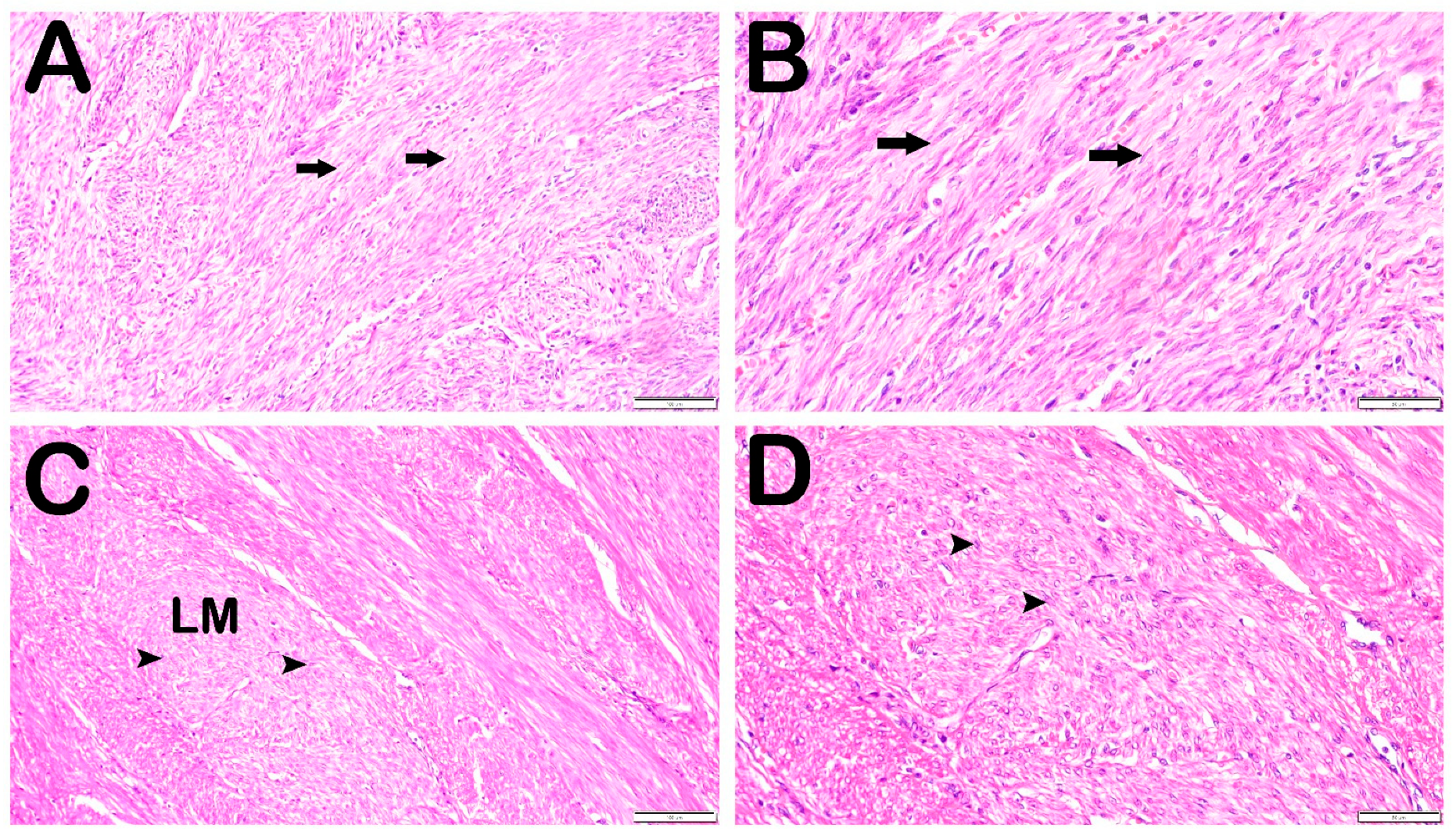
2.4. Histopathological Analysis
Biopsy specimens of uterine tissues were placed in tissue-tracking cassettes (Isolab GmbH, Eschau, Germany) and fixed in 10% phosphate-buffered formalin (Sigma-Aldrich, Darmstadt, Germany) for 24 h. Following fixation, dehydration (with increasing ethanol series, Merck KGAa, Darmstadt, Germany), mordanting (xylol, Merck KGAa, Darmstadt, Germany), and embedding in paraffin (Merck KGAa, Darmstadt, Germany) were performed according to routine histological follow-up procedures using a tissue-tracking device (Thermo Scientific Shendon Citadel 2000, Cheshire, England). Next, uterine tissue samples removed from the tissue-tracking cassettes were embedded in hard paraffin using a tissue embedding device (Leica 1150EGÜ, Leica Biosystems, Wetzlar, Germany) and blocked in tissue embedding cassettes (Merck KGAa, Darmstadt, Germany). The tissue samples were sectioned at 4–5 µm thickness with a rotary microtome (Leica RM2525, Lecia Biosystyems, Wetzlar, Germany) and stained with Harris hematoxylin and Eosin G (H&E; Merck, GmbH, Darmstadt, Germany).
2.5. Immunohistochemical Analysis
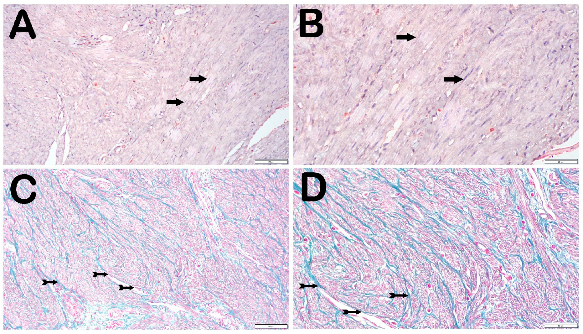
A total of 2–3 micrometer thick sections of myoma or myometrium tissue were placed on glass slides. The sections were stained with STAT-3 (ab109085, Abcam, Cambridge, UK) and IL-26 (ab224198, Abcam, Cambridge, UK) antibodies using a Bond-Max model (Leica Biosystems, Wetzlar, Germany) for automated immunohistochemical staining and in situ hybridization. For this purpose, uterine biopsy tissue sections were deparaffinized using Bond Dewax solution. Dehydrated peroxidase blockade was performed on biopsy tissues. Antigen retrieval was performed by heating the samples in ER2 solution (Leica Biosystems, Wetzlar, Germany) for 20 min. The cells were incubated with STAT-3 and IL-26 antibodies for 60 min. Tissues treated with secondary antibodies (ab205718, Abcam, Cambridge, UK) were stained with diaminobenzidine (DAB) using the Bond Polymer Refine Detection kit (Leica), and sections of biopsy specimens were stained with Harris hematoxylin (Bond Polymer Refine Detection, Leica, Wetzlar, Germany) for 10 min. After staining, the uterine tissue biopsy sections were covered with Entellan (Merck Gmbh, Darmstadt, Germany), examined, and photographed using a microscope. To assess nonspecific staining, negative controls were obtained by applying an equivalent amount of mouse or rabbit-specific IgG, instead of the primary antibody, to a portion of the uterine tissue samples.
Detailed information on the trichrome staining method can be found in the literature [
11]. Briefly, paraffin blocks containing myometrium and fibroid tissue were sectioned into 5 μm sections, heated, and mounted on a slide. Following deparaffinization with xylene, the sections were passed through a graded series of alcohol and rehydrated. The sections were treated with Weigert iron hematoxylin, distilled water, Biebrich red fuchsin, phosphotungstic-phosphomolybdic acid, aniline blue, and glacial acetic acid [
11]. The intensity of immunoreactivity in uterine biopsy sections stained with STAT-3 and IL-26 was determined using semi-quantitative analysis (
Table 1).
To avoid errors in the assessment of immunopositivity in our study, we included a negative isotype control. As the isotype control, we used the TRKB primary antibody (ab18987, Abcam, UK), which has no affinity for endometrial tissue.
2.6. Semi-Quantitative Analysis
Tissue samples incubated with primary antibodies against STAT-3 and IL-26 were scored as shown in
Table 1. Each preparation was scored by two histopathologists under a light microscope using 20 randomly selected fields at different magnifications (×20 and ×40). The histopathologists were blinded to the experimental group assignments. The immunostaining intensities of leiomyoma and myometrium samples were determined using the histo-score formula (histo-score = width × intensity). The extension h-score = (1 × % weak staining) + (2 × % moderate staining) + (3 × % strong staining) values were taken as the basis, while for density, (0: none, +0.5: little, +1: low, +2: moderate, +3: severe) values were taken [
12].
2.7. Statistical Analysis
The data obtained as a result of semi-quantitative analyses were analyzed using the Shapiro–Wilk test, Q-Q plots, Kurtosis-Skewness values, and Levene’s tests using SPSS 20.0 (IBM Corporation, Armonk, NJ, USA) statistical software, and the conformity to normal distribution was evaluated. Graphs were generated using GraphPad Prism 10.4.2 software (GraphPad Software, San Diego, CA, USA). Data are presented as mean ± standard error of the mean (SEM) for continuous variables. Statistical differences between groups were analyzed using an independent sample t-test. Statistical significance was set at p < 0.05.
4. Discussion
In this study, we investigated the expression levels of STAT-3 in uterine leiomyomas and evaluated the potential role of this transcription factor in fibroid pathogenesis. These findings suggest that STAT-3 is significantly increased in uterine leiomyomas and that this increase may be associated with various cellular mechanisms that support tumor growth.
Uterine leiomyomas are the most common benign tumors in women of reproductive age, and multiple mechanisms are involved in their development, including hormonal, genetic, and signaling pathway involvement. Recent studies have revealed that the STAT-3 pathway plays an important role in fibroid development.
STAT-3 is a transcription factor that is activated by cytokines and growth factors. After phosphorylation, it translocates to the nucleus and regulates several processes, including cell proliferation, the suppression of apoptosis, angiogenesis, and inflammatory response. These roles of STAT-3 make it an important regulator of tumorigenesis.
In a study conducted by Reschke et al. [
13], it was demonstrated that the leptin hormone increases cell proliferation and extracellular matrix (ECM) accumulation in uterine leiomyoma cells through the JAK2/STAT-3 and MAPK/ERK pathways. Leptin exerts these effects by stimulating the phosphorylation of STAT-3 in leiomyoma cells. The same study also emphasized that leptin inhibitors suppress these processes [
13].
Similarly, proinflammatory cytokines, such as interleukin-6 (IL-6), have been found to induce STAT-3 and increase the expression of ECM proteins (collagen I and fibronectin). Chegini reported that IL-6 increases proliferation and fibrotic response in leiomyoma cells via STAT-3 [
14].
In addition, the microRNA-29 family (specifically, miR-29b) inhibits leiomyoma cell proliferation and migration by suppressing the STAT-3 signaling pathway. Huang et al. demonstrated that miR-29b inhibits the expression of proliferative genes, such as STAT-3, Cyclin D1, and c-Myc, thereby suppressing tumor growth [
5].
These findings suggest that STAT-3 plays a multifaceted role in the growth and progression of uterine fibroids. STAT-3 regulates processes associated with tumor development, such as cell cycle progression, the inhibition of apoptosis, and ECM remodeling. Therefore, pharmacological targeting of STAT-3 early components in these signaling pathways may be a potential therapeutic strategy for controlling uterine fibroid growth. In particular, STAT-3 inhibitors are thought to be effective for hormone-independent treatment of fibroids.
Huang et al. [
5]. reported that the miR-29 and the positivity of STAT-3 early components in these signaling pathways opened avenues for further investigation into their roles in other tumor types. Understanding these mechanisms may lead to broader applications in cancer treatment beyond uterine leiomyoma and benefit a wider patient population. These studies, with findings similar to ours, provide valuable insights that may lead to innovative treatment options, improve patient care, and guide future research in the field of gynecological tumors.
However, most existing studies in this field are based on cell cultures and animal models. Further translational and clinical research on human tissues is needed to elucidate the role of STAT-3 in the pathogenesis of uterine fibroids. Simultaneously, the clinical efficacy and safety of STAT-3 inhibitors should be evaluated in future studies.
Uterine leiomyomas (fibroids) are among the most common benign tumors in women, and their development involves various molecular pathways. One of these mechanisms involves interleukins (ILs) and other cytokines. However, there is limited information on the expression and effects of IL-26 in uterine fibroids in the existing literature.
Proinflammatory cytokines play an important role in the pathogenesis of uterine myoma. For example, changes in cytokine levels, such as IL-1β, IL-6, TNF-α, IL-8, IL-12p70, and IFN-γ have been observed. Konenkov et al. observed a significant decrease in IFN-γ levels and a tendency to decrease IL-1β and TNF-α levels in the sera of patients with uterine fibroids. These changes may adversely affect the proliferation and differentiation of the uterine tissues [
15].
Furthermore, in another study, Isanbaeva et al. observed an increase in IL-1β, TGF-β2, and MCP-1 levels and a decrease in IL-2 levels in the serum of women with uterine fibroids. Changes in cytokine levels may be related to the growth of fibroids and may play a role in their progression [
16].
Konenkov et al. [
17] emphasized that the concentrations of important growth factors (IL-5, IL-7, G-CSF, VEGF, and PDGF) were decreased in the blood serum of women with uterine fibroids compared with healthy controls. Similarly, increased levels of peritoneal fluid interleukin-1 and the tumor necrosis factor in benign gynecological pathologies are evidence that uterine pathologies also affect the peritoneal microenvironment [
18]. In line with this, in uterine fibroids, the phagocytic ability of neutrophils is affected by the serum levels of IL-2, IL-17A, IL-6, and IL-4. In endometrial cancer, the degranulation ability of neutrophils is affected by serum IL-18 levels. This finding may provide evidence that neutrophil behavior in the presence of uterine fibroids differs from that in endometrial cancer [
19]. When these findings and our results are evaluated together, we suggest that fibroids affect the levels of different IL types in tissues, peritoneal fluid, and serum. The results may help healthcare providers understand the effects of uterine fibroids on fertility and reproductive outcomes. By understanding this role of growth, clinicians can better understand the characteristics and potential treatment options for this disease.
Since interleukins, along with other cytokines, trigger fibroid growth, IL-26 may also contribute to fibroid growth [
17,
20]. This idea is supported by the improvement in endometrial levels of many cytokines after the surgical resection of fibroids [
21]. However, the lack of specific studies on the expression and effects of IL-26 in uterine fibroids makes it difficult to clarify the role of this cytokine in fibroid pathogenesis. Considering the role of IL-26 in other inflammatory diseases, it may act on uterine fibroids via similar mechanisms. Therefore, prospective studies are needed to better understand the expression and effects of IL-26 in uterine fibroids.
This study evaluated the expression of STAT-3 and IL-26, two early components of the relevant signaling pathways in leiomyoma tissue in uterine fibroid development; some limitations should be emphasized. Assessing IL-26 and STAT-3 solely by immunohistochemistry prevented us from reaching a definitive conclusion. If mRNA and protein analyses had been performed in addition to histological analysis, we could have provided clearer data on the etiopathology of fibroids. Furthermore, because the cycle phases of the patients were unknown at the time of surgery, STAT-3 and IL-26 expression may have been affected by different phases. Since we did not subdivide the fibroid group into submucosal, intramural, and subserosal groups, we cannot comment on the effects of myoma number and location on the expression of these markers. To confirm the significance of our findings in fibroid development, prospective studies with larger groups of participants and investigations of multiple markers at the histological, protein, and mRNA levels are needed, especially considering the heterogeneous nature of uterine leiomyomas. In addition, Upon re-evaluation of our experimental design and antibody selection, we acknowledge that the anti-STAT-3 antibody used in our immunohistochemical analysis recognizes total STAT-3 and is not phospho-specific. Therefore, our study does not fully address the functional activation of the STAT-3 signaling pathway, which depends on the phosphorylated (active) form of STAT-3. Future studies incorporating phospho-specific STAT-3 antibodies will be necessary to better assess the relationship between total and activated STAT-3 and to validate our current findings. In addition, the STAT-3 and IL-26 positivity in our study needs to be supported by studies evaluating the biomarkers of mononuclear cells (such as CD45, CD68, CD4, etc.) in order to distinguish them from myocytes and mononuclear cells.


