Article

Transforaminal Lumbar Interbody Fusion (TLIF) with Expandable Banana-Shaped Interbody Spacers—Institutional 5-Year Experience

1
Spine Center, Cantonal Hospital St. Gallen, HOCH Health Ostschweiz & University of St. Gallen, 9000 St. Gallen, Switzerland
2
Department of Neurosurgery, Cantonal Hospital St. Gallen, HOCH Health Ostschweiz & University of St. Gallen, 9000 St. Gallen, Switzerland
3
Department of Orthopaedic Surgery, Cantonal Hospital St. Gallen, HOCH Health Ostschweiz & University of St. Gallen, 9000 St. Gallen, Switzerland
4
Department of Neurosurgery, University of Stanford, Stanford, CA 94305, USA
5
Neurosurgery Artificial Intelligence Lab, Stanford University School of Medicine, Stanford, CA 94305, USA
*
Author to whom correspondence should be addressed.
These authors contributed equally to the manuscript and should be considered as shared first authors.
These authors share last authorship.
J. Clin. Med. 2025, 14(15), 5402; https://doi.org/10.3390/jcm14155402 (registering DOI)
Submission received: 11 June 2025 / Revised: 20 July 2025 / Accepted: 28 July 2025 / Published: 31 July 2025
(This article belongs to the Section Orthopedics)

Abstract

Background: Transforaminal lumbar interbody fusion (TLIF) with static cages is a frequently performed procedure. Larger series focusing on the use of expandable TLIF spacers are less common. Methods: This retrospective, single-center observational cohort study reviewed consecutive patients treated by TLIF using expandable titanium interbody implants (ALTERA™, Globus Medical Inc., Audubon, PA, USA) for degenerative pathologies from L2-S1 between 11/2018 and 09/2023. Surgical parameters, adverse events, radiological outcomes (fusion rate, segmental lordosis, spinopelvic parameters), and clinical outcomes were analyzed through a mean postoperative follow-up of 12 months. Results: This study identified 270 patients (mean age 65 years, 50.4% female) who underwent TLIF with expandable interbody spacers at 324 levels. Clinical outcomes were good or excellent in 74.1% of patients at 3 months and 71.8% at 12 months. Radiographic fusion was achieved in 73.1% of assessable segments at 12 months. Segmental lordosis increased significantly from 17.8° preoperatively to 20.0° at 12 months (p < 0.001). Adverse event (AE) rates were acceptable across all timepoints, with no device failures or device-associated complications observed. Conclusions: This study demonstrates that TLIF with expandable titanium interbody implants was safe, associated with high fusion rates, and enabled significant restoration of segmental lordosis that was maintained during follow-up.

1. Introduction

Discogenic low-back pain arises from intervertebral disc degeneration involving multifactorial processes including mitochondrial dysfunction, advanced glycation end product accumulation, and inflammatory cascades [1]. When conservative management fails, surgical intervention with interbody fusion becomes necessary to restore spinal stability and alleviate symptoms.
Transforaminal lumbar interbody fusion (TLIF) is a frequently conducted surgical procedure, which allows for open or minimally invasive decompression and 360° fusion of a spinal motion segment via a single, transforaminal approach to the disc [2,3,4]. The use of an interbody cage helps to maintain or expand the intervertebral distance, allowing to increase foraminal height while helping with sagittal alignment restoration and increasing the chance of solid fusion [5]. Optimal spacer selection and placement may be challenging, however, as a certain implant height is required to obtain a fusion-inducing press fit according to Wolf’s law [6], while a too largely dimensioned implant can increase the risk of endplate injury and subsidence. The latter predisposes for instability, loss of lordosis, and suboptimal patient outcomes [7,8].
Expandable interbody spacers were developed to overcome these difficulties. They can be introduced into the disc space and positioned while at a contracted height, followed by controlled expansion until the desired device height and fit is obtained. In theory, the use of expandable cage technology may help to reduce the impaction force, endplate injury, and device subsidence [5]. Comparative analyses suggest that expandable cages may help to achieve a greater restoration of intervertebral distance (disc height) and segmental lordosis (SL), compared to static cages [5,9,10,11], however; this was not reproduced by other studies [12,13,14,15,16]. There have also been reports about cage failure with collapse, migration, and extrusion [17,18,19,20,21], highlighting the need for evaluating the safety of their use across a large sample.
The literature on expandable cage technology is limited, as mostly smaller case series and retrospective, single-center studies have been published so far [5,9,11,12,14,15,16,17,18,20,22,23,24,25,26,27,28,29,30]. The aim of this study was to complement the existing body of literature with our institutional experience, focusing on adverse events (AEs), clinical-radiological outcomes, and fusion rates in a large cohort.

2. Methods

2.1. Hospital Setting

The Cantonal Hospital of St. Gallen, Switzerland is an academic, tertiary teaching-hospital associated with the Medical School of St. Gallen. It serves a population of approximately 1,000,000 inhabitants. The Spine Center of Eastern Switzerland is formed mutually by twelve board-certified neurosurgeons or orthopedic spine surgeons and seven residents/physician assistants. Use and types of implants are unified. About 1100–1300 spine surgical procedures under general anesthesia are performed annually, including about 100–120 TLIF procedures annually. The ALTERA Expandable Integrated TLIF Spacer (Globus Medical Inc.) was introduced at our center in October 2018. This modern spacer is a banana-shaped, anteriorly placed, continuously expandable device made predominantly of titanium alloy with a central graft aperture. TLIF procedures were performed by all board-certified spine surgeons, or by fellows or senior residents under supervision.

2.2. Patient Identification

Unique patient numbers (UPNs) of patients, where an ALTERA spacer was employed until September 2023 were identified by electronic review of our hospital’s purchasing department. In addition, the surgery schedules were cross-checked for any TLIF procedures to make sure those with an expandable implant were included. Consecutive adult patients with degenerative pathology who underwent single- (Figure 1) or multi-level (Figure 2) lumbosacral (L2-S1) TLIF were included. Cases were excluded where static TLIF cages were employed or where expandable TLIF spacers had been inserted outside the approved range of CE marked indications (e.g., use in the thoracic spine, for trauma, tumors, or infection).

2.3. Data Collection and Variables

Eleven surgeons retrospectively reviewed the electronic patient charts to extract relevant information, based on a pre-specified codebook to classify and define variables. Baseline information, including age, sex, body mass index (BMI; in kg/m2) [31], smoking status [32,33], American Society of Anesthesiology (ASA) grading scale of surgical risk, Charlson Comorbidity Index [34] (CCI; stratified into very low (CCI 0), mild (CCI 1&2), moderate (CCI 3&4) and severe (CCI > 4) according to Keller et al. [35], and the Canadian clinical frailty scale [36] (ranging from 1 (very fit) to 9 (terminally ill)), was collected prospectively before each surgical procedure. Moreover, the indication for surgery was determined.
Surgical parameters included the spinal level where the TLIF was performed (between L2-S1), the total number of operated segments (e.g., 1 segment for L4/5; 2 segments for L3/L5), the type of expandable interbody spacer (8° or 15° lordotic), additional use of other types of interbody fusion procedures during the same anesthesia (e.g., anterior lumbar interbody fusion (ALIF), (extreme) lateral lumbar interbody fusion (XLIF/LLIF), posterior lumbar interbody fusion (PLIF), other), type of laminectomy (partial/complete), length of surgery (in minutes), estimated blood loss (EBL; in ml; defined by anesthesiologists from suction drainage and swabs), and intraoperative AEs.
Postoperative AEs at time of discharge from hospital were considered as documented within the discharge summary and the electronic patient record. Severity of AEs was classified according to the Therapy-Disability-Neurology (TDN) scoring system [37]. The same was recorded at 3 months (3M; AEs occurring between discharge and 3M follow-up) and at 12 months (12M; AEs occurring between 3M and 12M follow-up). Clinical outcome was graded according to Macnab into four categories, as best estimated from the follow-up letter and the electronic patient chart (excellent, good, fair, poor) [38,39,40]. To assess the degree and rate of fusion, intersomatic fusion was evaluated according to the Brantigan, Steffee & Fraser (BSF) classification [41], which distinguishes between obvious pseudarthrosis, intermediate type, and solid fusion (Figure 1). To evaluate the posterolateral fusion mass, the classification by Lenke et al. [42] was applied, which differentiated between definitely solid, possibly solid, probably not solid, and definitely not solid (Figure 1). A segment was considered radiographically fused if graded as either a solid fusion of the interbody space or a Lenke-type A or B grade of the posterolateral masses, or both. The radiographic fusion rate was then calculated based on the number of assessable segments, which were those with readable images for either intersomatic or posterolateral fusion sites, or both.
As computed tomography (CT) imaging was not available for all patients and ratings performed on (scoliosis) X-ray imaging were less certain, a “clinical pseudarthrosis” category was added, where the diagnosis of pseudarthrosis in patients without any pain was considered unlikely, but in patients with the typical delayed onset of recurring axial or radicular pain weeks to months after the index operation, pseudarthrosis as a failed attempt of spinal fusion was considered likely [43]. Diagnosis of “clinical pseudarthrosis” was based on both the clinical presentation in conjunction with available imaging studies (X-ray, CT, and Single Photon Emission Computed Tomography (SPECT)), after ruling out other causes of persistent pain.
The baseline sagittal Cobb angle (SL; e.g., upper endplate of L4 vertebra to lower endplate of L5 vertebra for the L4-L5 segment), as well as spinopelvic parameters including pelvic incidence (PI), lumbar lordosis (LL; from L1-S1), pelvic tilt (PT), sacral slope (SS), and C7-sagittal vertebral axis (C7 SVA), were measured using standing (scoliosis) or EOS X-ray images, whenever available. The patient-specific “ideal LL” was calculated using a web-based app (http://www.spinebit.io), which is based on the formulas by Le Huec and the European Spine Study Group [44,45,46]. Based on these values, the PI-LL mismatch (PI minus LL) and the ideal–actual LL mismatch (“ideal LL” minus actual LL) were determined. Postoperative radiological outcomes at 3M and 12M again included SL, LL, PT, SS, and C7 SVA. The intervertebral distance in mm was measured to determine the height of the disc space at its anterior, middle, and posterior aspect before and after surgery.

2.4. Surgical Technique

Pedicle screws were placed first to perform reduction maneuvers and to allow for some initial posterior distraction after uni- or bilateral partial facetectomies. Then, depending on the pathology and symptoms, either a uni- or bilateral complete facetectomy was performed to decompress the exiting nerve root and prepare the Kambin triangle(s) as entry zone(s) for the uni- or biportal TLIF procedure. The choice between the uniportal and biportal TLIF approach was determined based on several factors, including the extent of decompression required, need for bilateral cage placement, and surgeon preference. The uniportal approach was typically selected for single-level procedures with adequate unilateral access, while the biportal approach was employed when bilateral decompression or extensive coronal correction was required. After superficial discectomy, an intersomatic “Chiari” spreader was introduced for final segmental distraction, followed by a deep discectomy until the ALL was visualized. The ALL was preserved, in all patients. The TLIF cage was chosen in terms of width and height after trialing, and the desired degree of lordosis (8° or 15°) was chosen based on anatomical and physiological considerations. Following application of autogenous and/or allogeneic bone graft into the anterior aspect of the disc space, the TLIF cage was inserted, positioned, and expanded in its final position under serial fluoroscopic control. After releasing the introducer, the remaining disc space was filled with bone graft and posterior compression was applied as needed to increase SL.

2.5. Statistical Analysis

Statistical Analysis Software (SAS) (version 9.4; SAS Institute Inc., Cary, NC, USA) was used and employed mostly for descriptive statistics, reporting results as mean (standard deviation; SD) or count (percent). For the analysis of sagittal spinal parameters over time, outcomes were compared to the preoperative value using paired t-tests with p < 0.05 considered statistically significant.

2.6. Ethical Considerations

The Institutional Review Board (IRB) of Eastern Switzerland approved the study (BASEC ID 2023-01343). Retrospective collection, analysis, and publication of anonymized patient data were allowed with an institutional waiver for informed consent.

3. Results

3.1. Patient Cohort

This study identified a total cohort of 433 patients who underwent a TLIF procedure using an expandable ALTERA cage at 538 levels. After considering all predefined inclusion and exclusion criteria, 270 patients with TLIF performed at 324 levels remained in the analysis (Figure 3).
The mean age of the cohort was 64.6 years (SD 12.4); 50.4% of patients were female. A total of 66.3% of patients were overweight (BMI ≥ 25–30 kg/m2) or obese (BMI > 30 kg/m2), 41.9% were smokers or had a previous history of smoking, and approximately one out of four patients had moderate or severe comorbidities. All procedures were performed for a degenerative pathology, including spondylolisthesis, spinal stenosis with suspected instability, or (recurrent) disc herniation. More detailed baseline demographic information can be found in Table 1.
Table 2 summarizes procedure-related parameters. A total of 83% of TLIFs were performed between L4-S1, frequently as mono- or bisegmental fusion procedures or as part of longer constructs, including 3–4 segments of fusion; 13% of procedures entailed additional modalities of interbody fusion such as an ALIF, XLIF/LLIF, PLIF, or other approach. The average number of operated segments was 1.5 per patient (SD 0.7). The duration of surgery was about 4.5 h on average and the mean EBL was 556 mL (SD 446). Intraoperative AEs were encountered in 31 procedures (11.5%), of which the majority were incidental durotomies (74.2%).

3.2. Follow-Up and Reasons for Missing Data

Patients were discharged after a mean length of stay of 9.4 (SD 6.6) days. The 3-month follow-up was completed by n = 257 (95.2%) patients at a mean of 87.5 (SD 44.1) days postoperative. The 12-month follow-up was completed by n = 210 (77.8%) patients at a mean of 361.6 (SD 111.5) days postoperative. The main reasons for dropping out during the last follow-up included (i) surgeries, which were performed within less than 12 months, (ii) patients seeking further care at another facility, or iii) patients moving away from the area.

3.3. Clinical Outcomes

Between surgery and hospital discharge, AEs were noted in n = 53 patients (19.6%), of which most were anemia (24.5%), wound healing disorders (15.1%), or new neurological deficits/neuropathic pain (11.3%). Five (9.4%) complications were severe, and none led to death. Medical complications (67.9%) were more common than surgery-related complications (33.1%; Table 3).
At 3 months, AEs were noted in n = 27 patients (10.0%), of which most were new fractures/hardware-related issues (48.1%), wound healing disorders (33.3%), or adjacent segment disease (ASD; 14.8%). Most AEs were considered surgery-related (96.3%). Two AEs were graded as severe (7.4%), and there was no mortality at this time point. The clinical outcome was excellent or good in 74.1% (189/255) of patients with documented outcomes. Non-union was suspected in 7.5% of assessable TLIF segments.
At 12 months, AEs were noted in n = 25 patients (9.3%), of which most were ASD (52.0%), pseudarthrosis (24.0%), or new fractures/hardware-related issues (12.0%). Of AEs reported, one AE was graded severe (4%) and one related to discitis and sepsis was graded deadly (4.0%). The clinical outcome was excellent or good in 71.8% (148/206) of patients with documented outcomes. Radiographic fusion was observed in 73.1% of assessable TLIF segments, and clinical non-union was suspected in 12.0% of segments. No instance of cage collapse was observed through final follow-up.

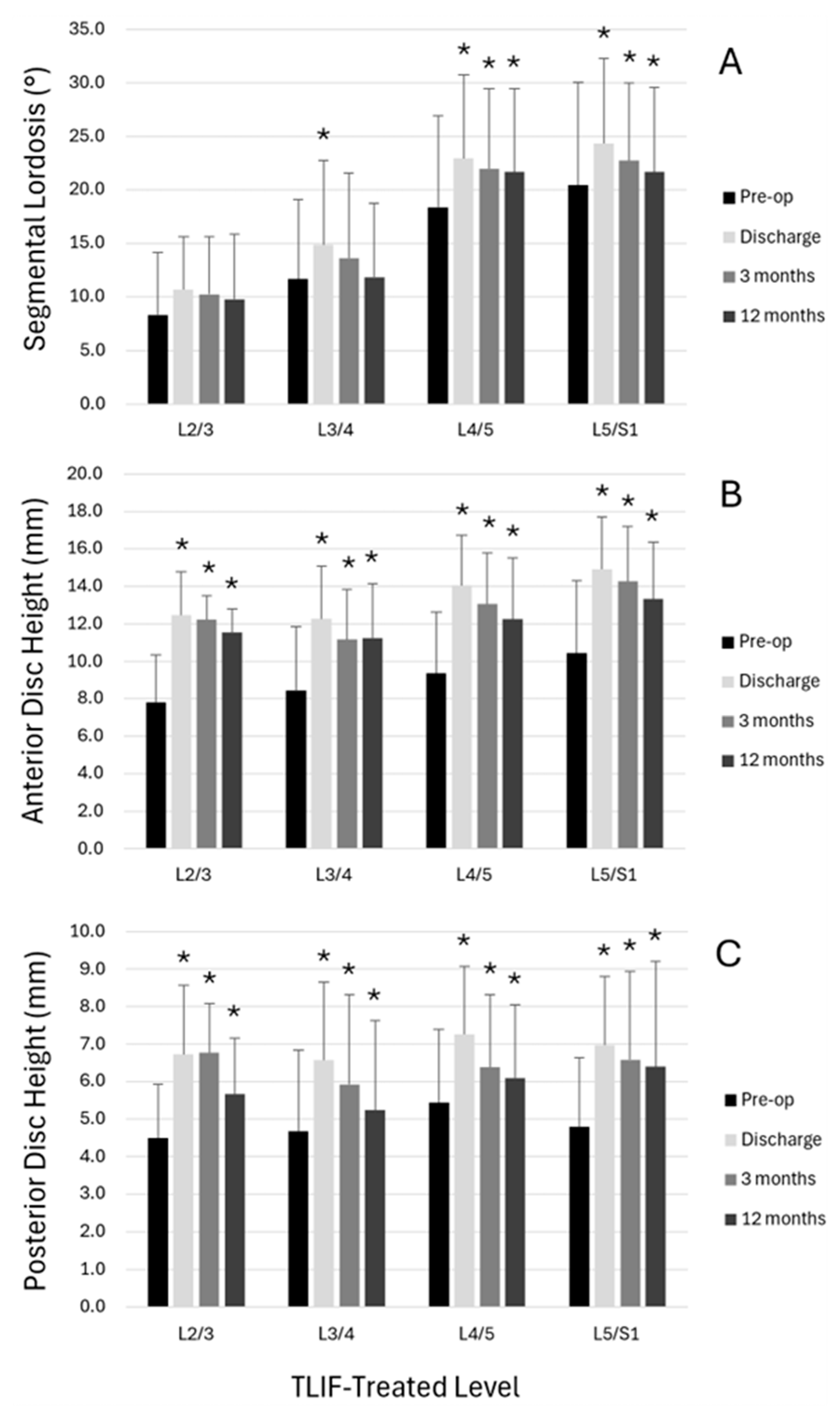
3.4. Radiological Outcomes

Table 4 illustrates the baseline spinopelvic measurements, as well as the evolution of the sagittal radiological parameters over time. There was a significant postoperative increase in both the global LL and SL, which were preserved over time at 12 months postoperatively, leading to significant decreases in both the PI-LL mismatch and ideal–actual LL mismatch. The intervertebral distance significantly increased by 4.5 mm in the anterior, 3.6 mm in the middle, and 1.9 mm in the posterior aspect of the disc space (all p < 0.001). This distance slightly decreased at the time of 3- and 12-month follow-up but remained significantly greater than prior to surgery.
Radiographic parameters specific to TLIF-treated segments were also evaluated by spinal level (Figure 4). Preoperatively, SL and disc space heights were observed to vary by level of treatment. SL was significantly increased at discharge from preoperative values at L3/4, L4/5, and L5/S1, and significant increases were sustained at 3 months and 12 months at L4/5 and L5/S1 (p < 0.05). Significant increases were observed in anterior and posterior disc space heights at all spinal levels and all postoperative times compared to preoperative measurements (p < 0.05).

4. Discussion

Expandable cage technology is relatively novel, and while numerous studies have explored the clinical impact and radiographic performance, the choice of interbody technology continues to incite development of new devices and considerable interest by clinicians. These factors have prompted the current analysis of an institutional 5-year experience consisting of 270 patients and 324 segments implanted with a specific anterior TLIF expandable spacer (ALTERA). The present study results may contribute to the decision-making pathway of patient selection and provide insight into contextualizing improvement when using expandable, banana-shaped spacers in TLIF procedures.
Notably, our spine center serves as a reference center for surrounding smaller and private hospitals and is responsible for taking care of patients that are rejected for surgery elsewhere, or that have suffered from complications at outside hospitals before. Despite our cohort including patients with considerable comorbidities associated with adverse outcomes, such as considerable degrees of frailty, poor bone quality, smoking, and obesity [31,32,33], our study reports detailed adverse events rates, including intraoperative (11.5%), at time of discharge (19.6%), and during 3- and 12-months of follow-up (~9–10%). Importantly, collapsed or loosened TLIF implants that migrated into the spinal canal or foramen, or that resulted in any kind of (neural) injury, as was previously reported for alternative expandable TLIF devices, were not identified [17,19,20,21,47]. Thus, the investigated ALTERA expandable TLIF device functioned reliably in our study series with respect to maintenance of the expanded device height.
Achieving appropriate spinopelvic alignment through restoration or preservation was considered a central goal of surgery for the present patient cohort. Even in short-construct lumbar fusion procedures for degenerative conditions, Leveque et al. have recommended that spinopelvic alignment be assessed and confirmed to be normal (PI-LL < 10°) before, during, and after surgery to enhance the likelihood of successful outcomes and reduce the risk of adjacent segment disease [48]. In the present study of degenerative (non-deformity) pathologies, the preoperative spinopelvic alignment measure of PI-LL averaged 5.5° and was within the normal range [47] for the majority of patients, indicating that in addition to symptomatic improvement, alignment preservation was a surgical goal for the majority of patients. Following treatment with fusion constructs including one or more expandable TLIF interbody cages, average PI-LL mismatch significantly improved by 2.2° by the final 12-month timepoint.
In addition to regional spinopelvic alignment changes, segment-specific changes were also observed. At TLIF-treated segments specifically, the use of expandable interbody cages allowed for a restoration of 4.0° of SL at the time of discharge, and 2.1° at 12-month follow-up. Our results compare well to Ledesma et al., who achieved 2.8° of additional SL at 6 weeks and 2.5° at 12 months in their cohort of 1- or 2-level MIS TLIF patients treated with an expandable interbody spacer [5]. Similarly, Chen et al. reported a gain of 1.4° and 2.0° immediately and at last follow-up in n = 30 patients treated by single-level TLIF with use of an expandable cage [24]. Our results demonstrate the inherent design of the anterior convexity angle of the banana-shaped expandable spacer and the anterior placement within the disc space, as anterior intervertebral distance significantly increased by an average of 2.9 mm at final follow up, thus supporting the recovery and preservation of SL correction [49]. While height changes were most pronounced in the anterior aspect of the disc space where cages were placed, posterior disc space height was also observed to increase significantly from the preoperative period to each postoperative period, potentially supplementing any direct decompression of posterior neural structures achieved via the TLIF and laminectomy procedures.
Our cohort captures the initial experience and technique evolution of using the banana-shaped expandable spacer over a 5-year period. Based on our experience on the use of expandable cages, surgeons may be tempted to spend less time on the discectomy and endplate preparation. It seems possible to insert the cage early and expand it before a thorough segmental release has been achieved, which may lead to endplate injury and cage subsidence. Observations of cage subsidence were most pronounced early in this case series, and these along with settling and bony remodeling may partially account for some of the postoperative trends seen in the spinopelvic alignment measures, notably the decreasing SL and disc space heights compared to discharge. Future research efforts focused on evaluating subsidence using a standardized grading scheme with correlation to patient characteristics and clinical outcomes may be desirable for various expandable cage technologies.
Overall, a successful outcome (rated as excellent or good) according to the Macnab criteria [39] was achieved by 74.1% of the patients with recorded outcomes at 3 months and 71.8% at 12 months, with 90.6% and 93.2% of patients demonstrating symptomatic improvement (rated from excellent to fair). Radiographic fusion was achieved in 73.1% of assessable segments at 12 months, while the interbody fusion rate was lower at 44.0%, with another 41.7% being BSF intermediate type. The interbody fusion outcome is similar to a rate (45.5%) recently reported by Liu et al. following a rigorous radiographic review of single-level TLIFs without rhBMP-2 using a modified BSF classification with minimum 2-year follow-up [50]. Lastly, the clinical non-union rates among assessable segments were 7.5% at 3-months and 12.0% at 12-months follow-up (Table 3), which is similar to those of prior studies reporting 6.7–6.9% after 1- or 2-level MIS TLIF [9,29], 7% at 12 months after 1- or 2-level MIS-TLIF [18,20], or 9.8% at 12 months after MIS-TLIF [28].

4.1. Implications for Practice

Based on our experience with the use of the ALTERA expandable TLIF spacer technology, it has been observed to be a well-working implant that functions reliably to gain disc height and SL, and achieve surgical goals. No collapsed implants were detected by 12 months, and no other AEs that were directly related to implant malfunction were recorded. While static TLIF cages may represent viable alternatives for “standard cases”, expandable cages, in our opinion, have certain advantages, especially in the settings of MIS approaches (possibility to insert a smaller implant and expand it in situ) and to assist with controlled correction of coronal and/or sagittal spinal malalignment by placing and expanding the cages in the desired location of the intervertebral space. While our study included both open and minimally invasive approaches, expandable cage technology may offer particular advantages in MIS-TLIF procedures by allowing insertion of a smaller-profile device that can subsequently be expanded in situ, potentially reducing tissue trauma and facilitating recovery. However, our study design did not specifically compare MIS versus open approaches, and future research should evaluate whether the theoretical advantages of expandable cages are more pronounced in minimally invasive settings.

4.2. Strengths and Weaknesses

Our study reports data from a large cohort of consecutive patients, treated with expandable TLIF spacers. This is an innovative technology, with increasing popularity, but on which there are limited data [5,9,10,11,12,14,15,16,17,18,20,22,23,24,25,26,27,28,29,30,51,52,53,54]. Despite its retrospective character, the missing data burden was reasonably low, especially for intraoperative and perioperative safety outcomes. Our series includes a very detailed record of perioperative AEs and grading of their severity using the TDN scale [37], which distinguishes it from previous studies.
Selection bias is likely to be present, as there was no control for the reason why patients were receiving an expandable TLIF spacer versus a static TLIF cage or any other type of anterior or posterior interbody fusion. The standardized use of patient-reported outcome measures (PROMs) in our center was only introduced in 2022, which is why this study classified the functional outcome using the Macnab criteria with a simple 4-tier scale [39,40,55]. A further limitation of this study is the absence of CT scan images for the assessment of the fusion status in all patients, as those are not routinely performed as part of the follow-up regimen in patients faring well. However, if patients developed clinically relevant symptoms on follow-up, CT or SPECT scans were ordered to rule out pseudarthrosis. Finally, there was no control group, as it was not the aim of the study to conduct a comparative analysis. In our series, 13% of patients (n ≈ 35) underwent additional interbody fusion procedures during the same anesthesia. While this represents a relatively small subset, it may have influenced overall outcomes, though the small numbers preclude meaningful subgroup analysis.

5. Conclusions

Data collected in this study suggest that the use of TLIF with an anteriorly placed expandable titanium interbody implant in this series promoted radiographic fusion, enabled correction and preservation of spinopelvic alignment, and was associated with an acceptable adverse event profile through 12 months of follow-up.

Author Contributions

M.N.S.: Conceptualization, Data curation, Formal analysis, Funding acquisition, Investigation, Methodology, Project administration, Visualization, Writing—original draft. L.B. (Lorenzo Bertulli): Conceptualization, Data curation, Formal analysis, Investigation, Visualization, Writing—original draft. G.F.: Investigation, Validation, Writing—review and editing. L.B. (Linda Bättig): Investigation, Validation, Writing—review and editing. Y.Y.: Investigation, Validation, Writing—review and editing. L.F.: Investigation, Validation, Writing—review and editing. F.K.: Investigation, Validation, Writing—review and editing. T.S.: Investigation, Validation, Writing—review and editing. F.C.S.: Investigation, Validation, Writing—review and editing. D.G.: Investigation, Validation, Writing—review and editing. S.M.: Investigation, Validation, Writing—review and editing. E.S.: Investigation, Validation, Writing—review and editing. A.V.: Investigation, Validation, Writing—review and editing. B.M.: Conceptualization, Formal analysis, Investigation, Supervision, Writing—review and editing. N.H.: Conceptualization, Formal analysis, Investigation, Supervision, Writing—review and editing. All authors have read and agreed to the published version of the manuscript.

Funding

A research grant was provided to the research account of Kantonsspital St. Gallen, Switzerland, by Globus Medical.

Institutional Review Board Statement

The Institutional Review Board (IRB) of Eastern Switzerland approved the study. Retrospective collection, analysis, and publication of anonymized patient data was allowed with an institutional waiver for informed consent. (BASEC ID 2023-01343); approval date: 20 July 2023.

Informed Consent Statement

Retrospective collection, analysis, and publication of anonymized patient data was allowed with an institutional waiver for informed consent.

Data Availability Statement

Dataset available on request from the authors.

Conflicts of Interest

The authors declare no conflict of interest.

References

  1. De Simone, M.; Choucha, A.; Ciaglia, E.; Conti, V.; Pecoraro, G.; Santurro, A.; Puca, A.A.; Cascella, M.; Iaconetta, G. Discogenic Low Back Pain: Anatomic and Pathophysiologic Characterization, Clinical Evaluation, Biomarkers, AI, and Treatment Options. J. Clin. Med. 2024, 13, 5915. [Google Scholar] [CrossRef] [PubMed]
  2. Gautschi, O.P.; Stienen, M.N.; Corniola, M.V.; Schaller, K. Minimal invasive surgery: Historical review, current status and perspective. Praxis 2014, 103, 1323–1329. [Google Scholar] [CrossRef] [PubMed]
  3. Harms, J.G.; Jeszenszky, D. Die posteriore, lumbale, interkorporelle Fusion in unilateraler transforaminaler Technik. Oper. Orthop. Traumatol. 1998, 10, 90–102. [Google Scholar] [CrossRef] [PubMed]
  4. Resnick, D.K.; Choudhri, T.F.; Dailey, A.T.; Groff, M.W.; Khoo, L.; Matz, P.G.; Mummaneni, P.; Watters, W.C., 3rd; Wang, J.; Walters, B.C.; et al. Guidelines for the performance of fusion procedures for degenerative disease of the lumbar spine. Part 7: Intractable low-back pain without stenosis or spondylolisthesis. J. Neurosurg. Spine 2005, 2, 670–672. [Google Scholar] [CrossRef]
  5. Ledesma, J.A.; Lambrechts, M.J.; Dees, A.; Thomas, T.; Hiranaka, C.G.; Kurd, M.F.; Radcliff, K.E.; Anderson, D.G. Static versus Expandable Interbody Fusion Devices: A Comparison of 1-Year Clinical and Radiographic Outcomes in Minimally Invasive Transforaminal Lumbar Interbody Fusion. Asian Spine J. 2023, 17, 61–74. [Google Scholar] [CrossRef]
  6. Cowin, S.C. Wolff’s law of trabecular architecture at remodeling equilibrium. J. Biomech. Eng. 1986, 108, 83–88. [Google Scholar] [CrossRef]
  7. Glassman, S.D.; Bridwell, K.; Dimar, J.R.; Horton, W.; Berven, S.; Schwab, F. The impact of positive sagittal balance in adult spinal deformity. Spine 2005, 30, 2024–2029. [Google Scholar] [CrossRef]
  8. Parisien, A.; Wai, E.K.; ElSayed, M.S.A.; Frei, H. Subsidence of Spinal Fusion Cages: A Systematic Review. Int. J. Spine Surg. 2022, 16, 1103–1118. [Google Scholar] [CrossRef]
  9. Hawasli, A.H.; Khalifeh, J.M.; Chatrath, A.; Yarbrough, C.K.; Ray, W.Z. Minimally invasive transforaminal lumbar interbody fusion with expandable versus static interbody devices: Radiographic assessment of sagittal segmental and pelvic parameters. Neurosurg. Focus 2017, 43, E10. [Google Scholar] [CrossRef]
  10. Lin, G.X.; Kim, J.S.; Kotheeranurak, V.; Chen, C.M.; Hu, B.S.; Rui, G. Does the application of expandable cages in TLIF provide improved clinical and radiological results compared to static cages? A meta-analysis. Front. Surg. 2022, 9, 949938. [Google Scholar] [CrossRef]
  11. Vaishnav, A.S.; Saville, P.; McAnany, S.; Kirnaz, S.; Wipplinger, C.; Navarro-Ramirez, R.; Hartl, R.; Yang, J.; Gang, C.H.; Qureshi, S.A. Retrospective Review of Immediate Restoration of Lordosis in Single-Level Minimally Invasive Transforaminal Lumbar Interbody Fusion: A Comparison of Static and Expandable Interbody Cages. Oper. Neurosurg. 2020, 18, 518–523. [Google Scholar] [CrossRef]
  12. Armocida, D.; Pesce, A.; Cimatti, M.; Proietti, L.; Santoro, A.; Frati, A. Minimally Invasive Transforaminal Lumbar Interbody Fusion Using Expandable Cages: Increased Risk of Late Postoperative Subsidence Without a Real Improvement of Perioperative Outcomes: A Clinical Monocentric Study. World Neurosurg. 2021, 156, e57–e63. [Google Scholar] [CrossRef]
  13. Dibble, C.F.; Zhang, J.K.; Greenberg, J.K.; Javeed, S.; Khalifeh, J.M.; Jain, D.; Dorward, I.; Santiago, P.; Molina, C.; Pennicooke, B.; et al. Comparison of local and regional radiographic outcomes in minimally invasive and open TLIF: A propensity score-matched cohort. J. Neurosurg. Spine 2022, 37, 384–394. [Google Scholar] [CrossRef]
  14. Khechen, B.; Haws, B.E.; Patel, D.V.; Yoo, J.S.; Guntin, J.A.; Cardinal, K.L.; Iyer, S.; Singh, K. Static Versus Expandable Devices Provide Similar Clinical Outcomes Following Minimally Invasive Transforaminal Lumbar Interbody Fusion. HSS J. 2020, 16, 46–53. [Google Scholar] [CrossRef]
  15. Stickley, C.; Philipp, T.; Wang, E.; Zhong, J.; Balouch, E.; O’Malley, N.; Leon, C.; Maglaras, C.; Manning, J.; Varlotta, C.; et al. Expandable cages increase the risk of intraoperative subsidence but do not improve perioperative outcomes in single level transforaminal lumbar interbody fusion. Spine J. 2021, 21, 37–44. [Google Scholar] [CrossRef]
  16. Yee, T.J.; Joseph, J.R.; Terman, S.W.; Park, P. Expandable vs. Static Cages in Transforaminal Lumbar Interbody Fusion: Radiographic Comparison of Segmental and Lumbar Sagittal Angles. Neurosurgery 2017, 81, 69–74. [Google Scholar] [CrossRef]
  17. Chang, C.C.; Chou, D.; Pennicooke, B.; Rivera, J.; Tan, L.A.; Berven, S.; Mummaneni, P.V. Long-term radiographic outcomes of expandable versus static cages in transforaminal lumbar interbody fusion. J. Neurosurg. Spine 2020, 34, 471–480. [Google Scholar] [CrossRef] [PubMed]
  18. Kim, C.W.; Doerr, T.M.; Luna, I.Y.; Joshua, G.; Shen, S.R.; Fu, X.; Wu, A.M. Minimally Invasive Transforaminal Lumbar Interbody Fusion Using Expandable Technology: A Clinical and Radiographic Analysis of 50 Patients. World Neurosurg. 2016, 90, 228–235. [Google Scholar] [CrossRef] [PubMed]
  19. Stein, I.C.; Than, K.D.; Chen, K.S.; Wang, A.C.; Park, P. Failure of a polyether-ether-ketone expandable interbody cage following transforaminal lumbar interbody fusion. Eur. Spine J. 2015, 24 (Suppl. 4), S555–S559. [Google Scholar] [CrossRef] [PubMed]
  20. Woodward, J.; Koro, L.; Richards, D.; Keegan, C.; Fessler, R.D.; Fessler, R.G. Expandable versus Static Transforaminal Lumbar Interbody Fusion Cages: 1-year Radiographic Parameters and Patient-Reported Outcomes. World Neurosurg. 2022, 159, e1–e7. [Google Scholar] [CrossRef]
  21. Hejrati, N.; Martens, B.; Jost, B.; Bozinov, O.; Stienen, M.N. Failure of an expandable lumbar interbody spacer–a critical analysis of secondary collapse, pseudoarthrosis and revision rates after thoracolumbar fusion. Eur. Spine J. 2025, 34, 182–190. [Google Scholar] [CrossRef] [PubMed]
  22. Boktor, J.G.; Pockett, R.D.; Verghese, N. The expandable transforaminal lumbar interbody fusion—Two years follow-up. J. Craniovertebral Junction Spine 2018, 9, 50–55. [Google Scholar] [CrossRef]
  23. Canseco, J.A.; Karamian, B.A.; DiMaria, S.L.; Patel, P.D.; Divi, S.N.; Chang, M.; Timmons, T.; Grewal, L.; Hallman, H.; Lee, J.K.; et al. Static Versus Expandable Polyether Ether Ketone (PEEK) Interbody Cages: A Comparison of One-Year Clinical and Radiographic Outcomes for One-Level Transforaminal Lumbar Interbody Fusion. World Neurosurg. 2021, 152, e492–e501. [Google Scholar] [CrossRef]
  24. Chen, C.; Li, Q.; Wang, W.; Ji, C.; Kang, Y.; Wang, C.; Zhang, H.; Zhang, M.; Zhou, H.; Feng, H.; et al. Comparison of the efficacy of expandable interbody fusion cage (EXP-IFC) and non-expandable interbody fusion cage (NE-IFC) in MIS-TLIF for lumbar degenerative diseases: A systematic retrospective study on 62 patients. Front. Surg. 2022, 9, 1008171. [Google Scholar] [CrossRef] [PubMed]
  25. Gelfand, Y.; Benton, J.; De la Garza-Ramos, R.; Yanamadala, V.; Yassari, R.; Kinon, M.D. Effect of Cage Type on Short-Term Radiographic Outcomes in Transforaminal Lumbar Interbody Fusion. World Neurosurg. 2020, 141, e953–e958. [Google Scholar] [CrossRef] [PubMed]
  26. Jitpakdee, K.; Sommer, F.; Gouveia, E.; Mykolajtchuk, C.; Boadi, B.; Berger, J.; Hussain, I.; Hartl, R. Expandable cages that expand both height and lordosis provide improved immediate effect on sagittal alignment and short-term clinical outcomes following minimally invasive transforaminal lumbar interbody fusion (MIS TLIF). J. Spine Surg. 2024, 10, 55–67. [Google Scholar] [CrossRef]
  27. Tassemeier, T.; Haversath, M.; Jager, M. Transforaminal lumbar interbody fusion with expandable cages: Radiological and clinical results of banana-shaped and straight implants. J. Craniovertebral Junction Spine 2018, 9, 196–201. [Google Scholar] [CrossRef]
  28. Virk, S.; Vaishnav, A.S.; Sheha, E.; Urakawa, H.; Sato, K.; Othman, Y.; Chaudhary, C.; Lee, R.; Cong, G.T.; Chaudhary, S.; et al. Combining Expandable Interbody Cage Technology with a Minimally Invasive Technique to Harvest Iliac Crest Autograft Bone to Optimize Fusion Outcomes in Minimally Invasive Transforaminal Lumbar Interbody Fusion Surgery. Clin. Spine Surg. 2021, 34, E522–E530. [Google Scholar] [CrossRef]
  29. Woodward, J.; Malone, H.; Witiw, C.D.; Kolcun, J.P.G.; Koro, L.; Keegan, K.C.; Ahmad, S.; Kerolus, M.G.; David, B.T.; Fessler, R.D.; et al. Transforaminal lumbar interbody fusion using a novel minimally invasive expandable interbody cage: Patient-reported outcomes and radiographic parameters. J. Neurosurg. Spine 2021, 35, 170–176. [Google Scholar] [CrossRef]
  30. Yearley, A.G.; Chalif, J.I.; Zaidi, H.A. Utility of Expandable Interbody Cages in Open Transforaminal Interbody Fusions: A Comparison With Static Cages. Cureus 2023, 15, e40262. [Google Scholar] [CrossRef]
  31. Stienen, M.N.; Joswig, H.; Smoll, N.R.; Corniola, M.V.; Schaller, K.; Hildebrandt, G.; Gautschi, O.P. Influence of Body Mass Index on Subjective and Objective Measures of Pain, Functional Impairment, and Health-Related Quality of Life in Lumbar Degenerative Disc Disease. World Neurosurg. 2016, 96, 570–577.e1. [Google Scholar] [CrossRef]
  32. Stienen, M.N.; Smoll, N.R.; Hildebrandt, G.; Schaller, K.; Gautschi, O.P. Influence of smoking status at time of surgery for herniated lumbar disk on postoperative pain and health-related quality of life. Clin. Neurol. Neurosurg. 2014, 122, 12–19. [Google Scholar] [CrossRef]
  33. Joswig, H.; Stienen, M.N.; Smoll, N.R.; Corniola, M.V.; Chau, I.; Schaller, K.; Hildebrandt, G.; Gautschi, O.P. Effects of Smoking on Subjective and Objective Measures of Pain Intensity, Functional Impairment, and Health-Related Quality of Life in Lumbar Degenerative Disk Disease. World Neurosurg. 2017, 99, 6–13. [Google Scholar] [CrossRef]
  34. Charlson, M.E.; Pompei, P.; Ales, K.L.; MacKenzie, C.R. A new method of classifying prognostic comorbidity in longitudinal studies: Development and validation. J. Chronic Dis. 1987, 40, 373–383. [Google Scholar] [CrossRef] [PubMed]
  35. Keller, K.; Schmitt, V.; Espinola-Klein, C.; Munzel, T.; Konstantinides, S.; Hobohm, L. Comorbidity burden assessed by the charlson comorbidity index and its influence on prognosis of patients with pulmonary embolism. Eur. Heart J. 2023, 44 (Suppl. 2), ehad655.2060. [Google Scholar] [CrossRef]
  36. Rockwood, K.; Song, X.; MacKnight, C.; Bergman, H.; Hogan, D.B.; McDowell, I.; Mitnitski, A. A global clinical measure of fitness and frailty in elderly people. CMAJ 2005, 173, 489–495. [Google Scholar] [CrossRef] [PubMed]
  37. Terrapon, A.P.R.; Zattra, C.M.; Voglis, S.; Velz, J.; Vasella, F.; Akeret, K.; Held, U.; Schiavolin, S.; Bozinov, O.; Ferroli, P.; et al. Adverse Events in Neurosurgery: The Novel Therapy-Disability-Neurology Grade. Neurosurgery 2021, 89, 236–245. [Google Scholar] [CrossRef]
  38. Joswig, H.; Hock, C.; Hildebrandt, G.; Schaller, K.; Stienen, M.N. Microscopic lumbar spinal stenosis decompression: Is surgical education safe? Acta Neurochir. 2016, 158, 357–366. [Google Scholar] [CrossRef]
  39. Macnab, I. Negative disc exploration. An analysis of the causes of nerve-root involvement in sixty-eight patients. J. Bone Jt. Surg. Am. 1971, 53, 891–903. [Google Scholar] [CrossRef]
  40. Tafazal, S.I.; Sell, P.J. Outcome scores in spinal surgery quantified: Excellent, good, fair and poor in terms of patient-completed tools. Eur. Spine J. 2006, 15, 1653–1660. [Google Scholar] [CrossRef]
  41. Brantigan, J.W.; Steffee, A.D. A carbon fiber implant to aid interbody lumbar fusion. Two-year clinical results in the first 26 patients. Spine 1993, 18, 2106–2107. [Google Scholar] [CrossRef]
  42. Lenke, L.G.; Bridwell, K.H.; Bullis, D.; Betz, R.R.; Baldus, C.; Schoenecker, P.L. Results of in situ fusion for isthmic spondylolisthesis. J. Spinal Disord. 1992, 5, 433–442. [Google Scholar] [CrossRef]
  43. Raizman, N.M.; O’Brien, J.R.; Poehling-Monaghan, K.L.; Yu, W.D. Pseudarthrosis of the spine. J. Am. Acad. Orthop. Surg. 2009, 17, 494–503. [Google Scholar] [CrossRef] [PubMed]
  44. Laouissat, F.; Sebaaly, A.; Gehrchen, M.; Roussouly, P. Classification of normal sagittal spine alignment: Refounding the Roussouly classification. Eur. Spine J. 2018, 27, 2002–2011. [Google Scholar] [CrossRef]
  45. Pizones, J.; Moreno-Manzanaro, L.; Sanchez Perez-Grueso, F.J.; Vila-Casademunt, A.; Yilgor, C.; Obeid, I.; Alanay, A.; Kleinstuck, F.; Acaroglu, E.R.; Pellise, F.; et al. Restoring the ideal Roussouly sagittal profile in adult scoliosis surgery decreases the risk of mechanical complications. Eur. Spine J. 2020, 29, 54–62. [Google Scholar] [CrossRef] [PubMed]
  46. Sebaaly, A.; Grobost, P.; Mallam, L.; Roussouly, P. Description of the sagittal alignment of the degenerative human spine. Eur. Spine J. 2018, 27, 489–496. [Google Scholar] [CrossRef]
  47. Kim, P.D.; Baron, E.M.; Levesque, M. Extrusion of expandable stacked interbody device for lumbar fusion: Case report of a complication. Spine 2012, 37, E1155–E1158. [Google Scholar] [CrossRef]
  48. Leveque, J.A.; Segebarth, B.; Schroerlucke, S.R.; Khanna, N.; Pollina, J., Jr.; Youssef, J.A.; Tohmeh, A.G.; Uribe, J.S. A Multicenter Radiographic Evaluation of the Rates of Preoperative and Postoperative Malalignment in Degenerative Spinal Fusions. Spine 2018, 43, E782–E789. [Google Scholar] [CrossRef] [PubMed]
  49. Lin, G.X.; He, L.R.; Nan, J.N.; Xu, W.B.; Xiao, K.; Que, Z.; Jhang, S.W.; Chen, C.M.; Zhu, M.T.; Rui, G. Comparing Outcomes of Banana-Shaped and Straight Cages in Transforaminal Lumbar Interbody Fusion for Lumbar Degenerative Diseases: A Systematic Review and Meta-Analysis. Neurospine 2024, 21, 261–272. [Google Scholar] [CrossRef]
  50. Liu, J.; Xie, R.; Chin, C.T.; Rajagopalan, P.; Duan, P.G.; Li, B.; Burch, S.; Berven, S.H.; Mummaneni, P.V.; Chou, D. Comparison of Lumbosacral Fusion Grade in Patients after Transforaminal and Anterior Lumbar Interbody Fusion with Minimum 2-Year Follow-Up. Orthop. Surg. 2023, 15, 2334–2341. [Google Scholar] [CrossRef]
  51. Alvi, M.A.; Kurian, S.J.; Wahood, W.; Goyal, A.; Elder, B.D.; Bydon, M. Assessing the Difference in Clinical and Radiologic Outcomes Between Expandable Cage and Nonexpandable Cage Among Patients Undergoing Minimally Invasive Transforaminal Interbody Fusion: A Systematic Review and Meta-Analysis. World Neurosurg. 2019, 127, 596–606.e1. [Google Scholar] [CrossRef] [PubMed]
  52. Macki, M.; Hamilton, T.; Haddad, Y.W.; Chang, V. Expandable Cage Technology-Transforaminal, Anterior, and Lateral Lumbar Interbody Fusion. Oper. Neurosurg. 2021, 21 (Suppl. 1), S69–S80. [Google Scholar] [CrossRef] [PubMed]
  53. Subramanian, T.; Merrill, R.K.; Shahi, P.; Pathania, S.; Araghi, K.; Maayan, O.; Zhao, E.; Shinn, D.; Kim, Y.E.; Kamil, R.; et al. Predictors of Subsidence and its Clinical Impact After Expandable Cage Insertion in Minimally Invasive Transforaminal Interbody Fusion. Spine 2023, 48, 1670–1678. [Google Scholar] [CrossRef]
  54. Weinstein, M.A.; Ayala, G.A.; Roura, R.; Christmas, K.N.; Warren, D.H.; Simon, P. Transforaminal lumbar interbody fusion with an expandable interbody device: Two-year clinical and radiographic outcomes. N. Am. Spine Soc. J. 2023, 16, 100286. [Google Scholar] [CrossRef]
  55. Maldaner, N.; Stienen, M.N. Subjective and Objective Measures of Symptoms, Function, and Outcome in Patients with Degenerative Spine Disease. Arthritis Care Res. 2020, 72 (Suppl. 10), 183–199. [Google Scholar] [CrossRef] [PubMed]
Figure 1. Case vignette of a 74-year-old male with right-sided radicular pain corresponding to both L4 and L5 dermatomes, as well as facet joint pain with short-term response to repeated injections. (A) Standing sagittal X-ray studies demonstrate degenerative grade I spondylolisthesis at L4/5 and collapsed disc height. PI 58° (Roussouly type 3), PT 23°, SS 35°, LL L1-S1 52°, SL at L4/5 of 18°. (B) Preoperative sagittal paramedian MRI on the right side, illustrating the foraminal stenosis due to disc protrusion and facet joint disease. A single-level TLIF was performed with unilateral facetectomy and implantation of a Globus Medical ALTERA spacer (9–13 mm expandable, 10 × 31 mm, 8° lordotic). (C) Standing X-ray imaging before discharge, demonstrating an increase in SL by 11° (to 29°). The patient made an unremarkable, excellent recovery with complete improvement of symptoms at 3 and 12 months. X-ray studies at 12 months in sagittal (D) and coronal (E) view. PT 12°, SS 46°, LL L1-S1 68°, SL at L4/5 29°. An abdominal CT scan, performed for an oncological workup about 2 years postoperatively showed evidence of solid intersomatic fusion ((F); classified as BSF-type 3) and unilateral solid fusion mass ((G); classified as Lenke-type B).
Jcm 14 05402 g001
Figure 2. Case vignette of a 64-year-old male with history of three prior lumbar decompression procedures (hemilaminectomy L5, foraminotomy L5, bilateral decompression L3/4), presenting in our outpatient clinic with neurogenic claudication, recurring spinal stenosis, severe foraminal stenosis at multiple levels, and motor deficit for elevation of the left foot (BMRC grade 2/5). He received multiple epidural steroid injections and facet blocks with only temporary improvement of symptoms. (A,B) Standing preoperative whole-spine X-ray in anterior–posterior and lateral projection (PI 29°, Roussouly type 1, PT 5°, SS 24°, LL L1-S1 46° (ideal: 43° according to www.spinebit.io, accessed on: 27 July 2025), SL L4/S1 28° (ideal: 39° according to www.spinebit.io)). (C) Sagittal MRI demonstrating spinal stenosis at L2/3, L3/4, and L4/5. (D) Sagittal CT scan with evidence of degenerative disc disease and vacuum sign at L3/4, L4/5, and L5/S1. Considering the severity of his symptoms (VAS back pain 7/10, VAS leg pain 10/10, Oswestry Disability Index (ODI) 53.3/100%, Core Outcome Measures Index (COMI) Back 8.14/10), a fusion procedure was proposed entailing indirect decompression at L2/3 via extreme lateral lumbar interbody fusion (XLIF), as well as direct decompression with TLIF at L3-S1. (E,F) Postoperative standing whole-spine X-ray in anterior–posterior and lateral projection demonstrates good restoration of SL between L4-S1 (37°) and between L1-S1 (42°). The patient recovered well, without suffering from intra- or postoperative complications. At 12 months, he presented with significant improvement of preoperative symptoms (VAS back pain 3/10, VAS leg pain 1/10, ODI 22/100%, COMI Back 2.28/10) and without signs of pseudarthrosis.
Jcm 14 05402 g002
Figure 3. Flowchart of inclusion and exclusion of patients undergoing TLIF with use of an expandable ALTERA spacer between November 2018 and September 2023 in our institution.
Jcm 14 05402 g003
Figure 4. Radiographic outcomes by spinal level showing (A) segmental lordosis with significant sustained improvements at L4/5 and L5/S1 levels, (B) anterior disc space height with significant increases maintained at all levels, and (C) posterior disc space height demonstrating sustained improvements across all treated levels. (* p < 0.05 vs. preoperative values).
Jcm 14 05402 g004
Table 1. Demographic information of n = 270 patients undergoing thoracolumbar fusion surgery including the use of an expandable interbody spacer on 324 levels. Data are presented as count (percent) or mean (standard deviation; SD). ASA = American Society of Anesthesiology; BMI = body mass index; CCI = Charlson comorbidity index; CFI = Canadian frailty index.
Variable
Age (years)64.6 (SD 12.4)
Sex
Female136 (50.4%)
Male134 (49.6%)
BMI categories, according to the WHO
Underweight (<18.5 kg/m2)2 (0.7%)
Healthy (18.5–24.9 kg/m2)89 (33.0%)
Overweight (25–30 kg/m2)100 (37.0%)
Obese (>30 kg/m2)79 (29.3%)
Smoking status
Nonsmoker157 (58.1%)
Smoker85 (31.5%)
Former smoker28 (10.4%)
ASA grade
I7 (2.6%)
II169 (62.6%)
III90 (33.3%)
IV4 (1.5%)
CCI severity
Very low113 (41.9%)
Mild103 (38.1%)
Moderate37 (13.7%)
Severe17 (6.3%)
CFI severity
Very fit22 (8.1%)
Well82 (30.4%)
Managing well97 (35.9%)
Vulnerable57 (21.1%)
Mildly frail8 (3.0%)
Moderately frail3 (1.1%)
Severely frail1 (0.4%)
Degenerative disease type
(Recurrent) disc herniation42 (15.6%)
Spinal stenosis with instability62 (23.0%)
Degenerative spondylolisthesis75 (27.8%)
Isthmic spondylolisthesis38 (14.1%)
Other53 (19.6%)
Totaln = 270 (100%)
Table 2. Surgery-specific information of n = 270 patients undergoing lumbar fusion surgery including the use of an expandable interbody spacer in n = 324 segments. Data are presented as count (percent) or mean (standard deviation, range) of patients (*), segments (**), or adverse events (***). AE = adverse event; ALIF = anterior lumbar interbody fusion; L = lumbar; LLIF = lateral lumbar interbody fusion; PLIF = posterior lumbar interbody fusion; S = sacral; TLIF = transforaminal lumbar interbody fusion; XLIF = extreme lateral lumbar interbody fusion.
Variable
TLIF segment **
L2/311 (3.4%)
L3/4 44 (13.6%)
L4/5164 (50.6%)
L5/S1105 (32.4%)
Number of operated segments *1.5 (SD 0.7, range: 1–4)
Type of interbody spacer **
8° lordotic161 (49.7%)
15° lordotic 163 (50.3%)
Other types of interbody fusion employed *
None234 (86.7%)
XLIF/LLIF7 (2.6%)
ALIF14 (5.2%)
PLIF 12 (4.4%)
Other3 (1.1%)
Type of laminectomy **
Partial 231 (71.3%)
Complete93 (28.7%)
Length of surgery, in minutes *263 (SD 89, range: 124–725)
Estimated blood loss, in milliliters *556 (SD 446, range: 50–3600)
Intraoperative AE *
No239 (88.5%)
Yes31 (11.5%)
Type of intraoperative AE ***
Dural lesion23 (74.2%)
Hardware-related5 (16.1%)
Osseous injury2 (6.5%)
Excessive bleeding1 (3.2%)
Table 3. Information on surgical adverse events (AEs) and outcome at discharge, 90 days and 12 months in n = 270 patients undergoing lumbar fusion surgery including the use of an expandable interbody spacer in n = 324 segments. Data are presented as count (percent) or mean (standard deviation). n/a = not applicable; TDN = Therapy-Disability-Neurology.
VariableDischarge90 Days12 Months
AE
No217 (80.4%)227 (84.1%)185 (68.5%)
Yes53 (19.6%)27 (10.0%)25 (9.3%)
Missing data0 (0%)16 (5.9%)60 (22.2%)
Type of AE #
Medical36 (67.9%)1 (3.7%)0 (0%)
Surgical17 (33.1%)26 (96.3%)25 (100%)
TDN grade of AE
1 (mild AE)7 (13.2%)1 (3.7%)0 (0%)
2 (mild to moderate AE)22 (41.5%)2 (7.4%)5 (20.0%)
3 (moderate AE)19 (35.8%)22 (81.5%)18 (72.0%)
4 (severe AE)5 (9.4%)2 (7.4%)1 (4.0%)
5 (death)0 (0%)0 (0%)1 (4.0%)
Clinical outcomen/a
Excellent92 (34.1%)93 (34.5%)
Good97 (35.9%)55 (20.4%)
Fair42 (15.6%)44 (16.3%)
Poor24 (8.9)14 (5.2%)
Missing data15 (5.6%)64 (23.7%)
Posterolateral fusionn/aAssessable levels n = 220Assessable levels n = 209
Definitively not solid11 (5.0%)17 (8.1%)
Probably not solid138 (62.7%)51 (24.4%)
Possibly solid55 (25.0%)83 (39.7%)
Definitively solid16 (7.3%)58 (27.8%)
Intersomatic fusionn/aAssessable levels n = 221Assessable levels n = 216
Fusion10 (4.5%)95 (44.0%)
Intermediate type183 (82.8%)90 (41.7%)
Pseudarthrosis28 (12.7%)31 (14.4%)
Any radiographic fusionn/aAssessable levels n = 222Assessable levels n = 216
Fused71 (32.0%)158 (73.1%)
Not fused151 (68.0%)58 (26.9%)
Clinical non-unionn/aAssessable levels n = 279Assessable levels n = 249
No258 (92.5%)219 (88.0%)
Yes21 (7.5%)30 (12.0%)
Cage collapsen/an/aAssessable levels n = 250
No250 (100%)
Yes0 (0.0%)
# Type of AEs at discharge: n = 13 anemia (24.5%), n = 8 wound healing disorder/infection/hematoma (15.1%), n = 6 new neurological deficit/neuropathic pain (11.3%), n = 5 other AEs (9.4%), n = 4 pulmonary embolism (7.5%), n = 3 congestive heart failure (5.7%), n = 3 urinary tract infection (1.4%), n = 3 fracture/hardware-related issues (5.7%), n = 3 delirium (5.7%), n = 3 renal failure (0.9%), n = 2 pneumonia (3.8%). Type of AEs at 90 days: n = 13 fracture/hardware-related issues (48.1%), n = 9 wound healing disorder/infection/hematoma (33.3%), n = 4 adjacent segment disease/proximal junctional kyphosis or failure (14.8%), n = 1 pneumonia (3.7%). Type of AEs at 12 months: n = 13 adjacent segment disease/proximal junctional kyphosis or failure (52%), n = 6 pseudarthrosis (24%), n = 3 fracture/hardware-related issues (12%), n = 3 wound healing disorder/infection/hematoma (12%).
Table 4. Information on spinopelvic parameters and intervertebral distance in n = 270 patients undergoing thoracolumbar fusion surgery including the use of an expandable interbody spacer in 324 segments. Data are presented as count (percent) or mean (standard deviation), comparing measurements at follow-up with preoperative measurements. ° = degrees, LL = lumbar lordosis, C7 SVA = C7 sagittal vertical axis, PI = pelvic incidence, PT = pelvic tilt, SS = sacral slope.
Spinopelvic ParametersPreoperativeDischarge90 Days Postoperative12 Months Postoperative
PI, in °57.1 (12.1)
Total LL, in °51.6 (14.2)49.8 (12.7)0.00353.6 (12.5)0.02553.8 (12.3)0.004
SS, in °38.0 (9.7)37.0 (8.8)0.03038.5 (9.0)0.81639.1 (8.6)0.030
PT, in °19.0 (8.4)19.9 (8.9)0.03718.8 (8.6)0.85018.1 (9.1)0.072
Segmental lordosis, in °17.8 (9.3)21.9 (8.6)<0.00120.8 (8.2)<0.00120.0 (8.6)<0.001
C7 SVA, in cm4.2 (3.9)4.1 (3.3)0.1735.0 (3.5)0.4685.8 (4.6)0.514
Ideal LL, in °58.0 (7.4)
Ideal-actual LL mismatch, in °6.4 (12.2)8.2 (11.1)0.0034.5 (11.2)0.0254.0 (11.4)0.004
PI-LL mismatch, in °5.5 (11.9)7.2 (11.8)0.0033.7 (11.7)0.0323.3 (12.4)0.004
Intervertebral distance, in mm
Anterior disc space 9.6 (3.5)14.0 (2.9)<0.00113.2 (2.9)<0.00112.5 (3.2)<0.001
Middle disc space8.2 (2.8)11.8 (2.3)<0.00111.0 (2.2)<0.00110.5 (2.4)<0.001
Posterior disc space5.1 (1.9)7.1 (1.9)<0.0016.4 (2.1)<0.0016.1 (2.3)<0.001
Disclaimer/Publisher’s Note: The statements, opinions and data contained in all publications are solely those of the individual author(s) and contributor(s) and not of MDPI and/or the editor(s). MDPI and/or the editor(s) disclaim responsibility for any injury to people or property resulting from any ideas, methods, instructions or products referred to in the content.

