
The Perivascular Fat Attenuation Index: Bridging Inflammation and Cardiovascular Disease Risk


Abstract

1. Introduction
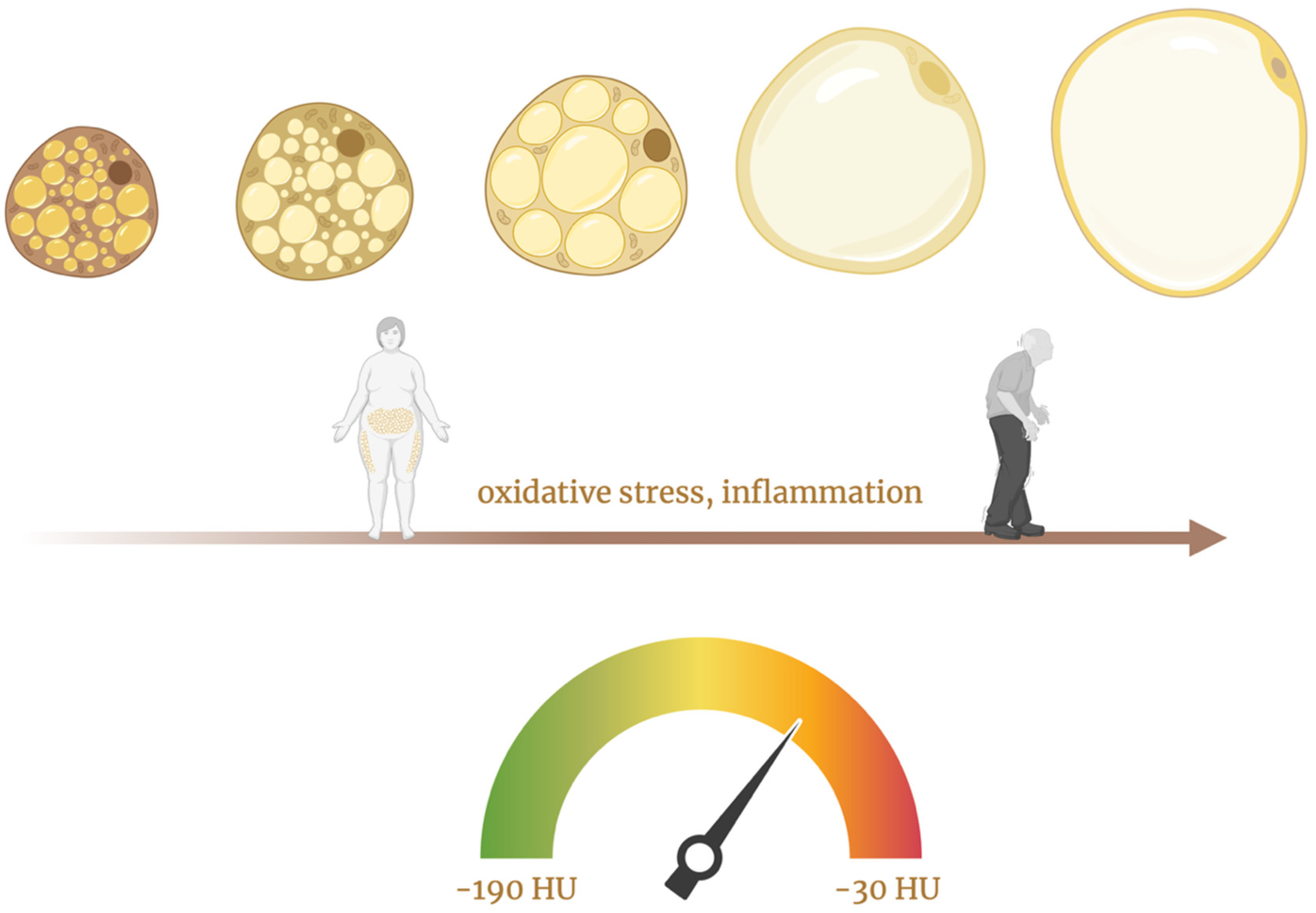
2. The Role of PVAT

3. The Perivascular Fat Attenuation Index (pFAI)
4. A New Frontier in Cardiovascular Care
5. Unravelling the Link to Systemic Inflammation

| Domain | Widely-Used Technique | Main Strengths | Key Limitations |
|---|---|---|---|
| Conventional risk scores | Framingham, SCORE2, QRISK3, etc. | Low cost, point-of-care use | Underestimate SID CV-risk by ≥30% [51,52,57] |
| Disease-activity indices | PASI (psoriasis), DAS-28 (RA), SLEDAI, CDAI (IBD) | Track primary-disease severity | Poor surrogate for CV-outcome |
| Systemic inflammatory biomarkers | hs-CRP, IL-6, TNF-α, etc. | Inexpensive, serial follow-up | Reflect global inflammation, poor correlation with coronary events or FAI values, high biological variability |
| Anatomical CT/US imaging | CAC-score, CCTA, carotid IMT | Quantifies plaque burden and morphology | Detects late, structural disease, insensitive to active inflammation, operator dependent |
| Molecular/functional imaging | 18F-FDG-PET/CT, 18F-NaF-PET/CT | Direct signal of cellular activity | Expensive, limited availability, higher radiation, FDG spill-over in coronaries |
| Invasive plaque imaging | IVUS, OCT | Plaque anatomy | Invasive, focal, unsuitable for longitudinal screening |
| CT-derived FAI | FAI on routine CCTA | Segment-specific coronary-inflammation metric, therapy-responsive | Requires CCTA and AI-based standardization (FAI-Score) |
6. Addressing the Limitations of FAI
7. Conclusions
Author Contributions
Funding
Data Availability Statement
Acknowledgments
Conflicts of Interest
Abbreviations
| AI | Artificial Intelligence |
| ARD | Autoimmune Rheumatic Disease |
| ASCVD | Atherosclerotic Cardiovascular Disease |
| CABG | Coronary Artery Bypass Graft |
| CAD | Coronary Artery Disease |
| CAC | Coronary artery calcium |
| CCS | Coronary artery Calcium Score |
| CCTA | Coronary Computed Tomography Angiography |
| CDAI | Crohn’s Disease Activity Index |
| CRISP-CT | Cardiovascular RISk Prediction using Computed Tomography |
| CT | Computed Tomography |
| CVD | Cardiovascular Disease |
| DAS-28 | Disease Activity Score in 28 joints |
| FAI | Fat Attenuation Index |
| FFR | Fractional Flow Reserve |
| FIB-4 | Fibrosis-4 Index for Liver Fibrosis |
| 18F-FDG-PET/CT | Positron-emission tomography/CT with 18F-fluorodeoxyglucose |
| HES | Hypereosinophilic Syndrome |
| HFS | Hepamet Fibrosis Score |
| HLA | Human Leukocyte Antigen |
| HRP | High-Risk Plaque |
| HU | Hounsfield Units |
| IBD | Inflammatory Bowel Disease |
| IFN-γ | Interferon-gamma |
| IL-1RA | Interleukin-1 Receptor Antagonist |
| IL-6 | Interleukin 6 |
| IL-10 | Interleukin 10 |
| IL-12/23 | Interleukin 12 and 23 |
| IL-17 | Interleukin 17 |
| IL-1β | Interleukin-1 Beta |
| IL-23 | Interleukin 23 |
| IMT | Intima–media thickness |
| IVUS | Intravascular ultrasound |
| INOCA | Ischemia and Non-Obstructive Coronary Arteries |
| LAD | Left Anterior Descending Artery |
| LCx | Left Circumflex Artery |
| LDL | Low-Density Lipoprotein |
| LSM | Liver Stiffness Measurement |
| MASLD | Metabolic dysfunction-associated steatotic liver disease |
| MCP-1 | Monocyte Chemoattractant Protein-1 |
| MINOCA | Myocardial Infarction with Non-Obstructive Coronary Arteries |
| 8F-NaF-PET/CT | PET/CT with 18F-sodium fluoride |
| NF-κB | Nuclear Factor Kappa B |
| OCT | Optical coherence tomography |
| PASI | Psoriasis Area and Severity Index |
| PET/CT | Positron-emission tomography/computed tomography |
| pFAI | Perivascular Fat Attenuation Index |
| PCAT | Pericoronary Adipose Tissue |
| PTX3 | Pentraxin 3 |
| PVAT | Perivascular Adipose Tissue |
| tPVAT | Thoracic Periaortic Adipose Tissue |
| aPVAT | Abdominal Periaortic Adipose Tissue |
| mPVAT | Mesenteric Perivascular Adipose Tissue |
| QRISK3 | QRESEARCH risk estimator version 3 |
| RA | Rheumatoid Arthritis |
| RCA | Right Coronary Artery |
| SCORE2 | European Systematic Coronary Risk Evaluation, version 2 |
| SIDs | Systemic inflammatory diseases |
| SLE | Systemic Lupus Erythematosus |
| SLEDAI | Systemic Lupus Erythematosus Disease Activity Index |
| TNF-α | Tumor Necrosis Factor Alpha |
| VMI | Virtual Monoenergetic Image |
References
- Antonopoulos, A.S.; Sanna, F.; Sabharwal, N.; Thomas, S.; Oikonomou, E.K.; Herdman, L.; Margaritis, M.; Shirodaria, C.; Kampoli, A.-M.; Akoumianakis, I.; et al. Detecting human coronary inflammation by imaging perivascular fat. Sci. Transl. Med. 2017, 9, eaal2658. [Google Scholar] [CrossRef]
- Chong, B.; Jayabaskaran, J.; Jauhari, S.M.; Chan, S.P.; Goh, R.; Kueh, M.T.W.; Li, H.; Chin, Y.H.; Kong, G.; Anand, V.V.; et al. Global burden of cardiovascular diseases: Projections from 2025 to 2050. Eur. J. Prev. Cardiol. 2024, zwae281. [Google Scholar] [CrossRef]
- Fernández-Gutiérrez, B.; Perrotti, P.P.; Gisbert, J.P.; Domènech, E.; Fernández-Nebro, A.; Cañete, J.D.; Ferrándiz, C.; Tornero, J.; García-Sánchez, V.; Panés, J.; et al. Cardiovascular disease in immune-mediated inflammatory diseases. Medicine 2017, 96, e7308. [Google Scholar] [CrossRef]
- Nava, E.; Llorens, S. The Local Regulation of Vascular Function: From an Inside-Outside to an Outside-Inside Model. Front. Physiol. 2019, 10, 729. [Google Scholar] [CrossRef]
- Li, X.; Ma, Z.; Zhu, Y.Z. Regional Heterogeneity of Perivascular Adipose Tissue: Morphology, Origin, and Secretome. Front. Pharmacol. 2021, 12, 697720. [Google Scholar] [CrossRef]
- Simantiris, S.; Pappa, A.; Papastamos, C.; Korkonikitas, P.; Antoniades, C.; Tsioufis, C.; Tousoulis, D. Perivascular Fat: A Novel Risk Factor for Coronary Artery Disease. Diagnostics 2024, 14, 1830. [Google Scholar] [CrossRef]
- Ajoolabady, A.; Pratico, D.; Lin, L.; Mantzoros, C.S.; Bahijri, S.; Tuomilehto, J.; Ren, J. Inflammation in atherosclerosis: Pathophysiology and mechanisms. Cell Death Dis. 2024, 15, 817. [Google Scholar] [CrossRef]
- Antoniades, C.; Tousoulis, D.; Vavlukis, M.; Fleming, I.; Duncker, D.J.; Eringa, E.; Manfrini, O.; Antonopoulos, A.S.; Oikonomou, E.; Padró, T.; et al. Perivascular adipose tissue as a source of therapeutic targets and clinical biomarkers. Eur. Heart J. 2023, 44, 3827–3844. [Google Scholar] [CrossRef]
- Sigdel, S.; Udoh, G.; Albalawy, R.; Wang, J. Perivascular Adipose Tissue and Perivascular Adipose Tissue-Derived Extracellular Vesicles: New Insights in Vascular Disease. Cells 2024, 13, 1309. [Google Scholar] [CrossRef]
- Grigoras, A.; Amalinei, C.; Balan, R.A.; Giusca, S.E.; Caruntu, I.D. Perivascular adipose tissue in cardiovascular diseases—An update. Anatol. J. Cardiol. 2019, 22, 219–231. [Google Scholar] [CrossRef]
- Li, Y.; Chen, Z.; Xiao, Y.; Li, X. Cross-talks between perivascular adipose tissue and neighbors: Multifaceted nature of nereids. Front. Pharmacol. 2024, 15, 1442086. [Google Scholar] [CrossRef]
- Yuvaraj, J.; Cheng, K.; Lin, A.; Psaltis, P.J.; Nicholls, S.J.; Wong, D.T.L. The Emerging Role of CT-Based Imaging in Adipose Tissue and Coronary Inflammation. Cells 2021, 10, 1196. [Google Scholar] [CrossRef]
- Shi, H.; Wu, H.; Winkler, M.A.; de Chantemèle, E.J.B.; Lee, R.; Kim, H.W.; Weintraub, N.L. Perivascular adipose tissue in autoimmune rheumatic diseases. Pharmacol. Res. 2022, 182, 106354. [Google Scholar] [CrossRef]
- Muffová, B.; Králová Lesná, I.; Poledne, R. Physiology and Pathobiology of Perivascular Adipose Tissue: Inflammation-Based. Physiol. Res. 2024, 73, 929–941. [Google Scholar] [CrossRef]
- Oikonomou, E.K.; Marwan, M.; Desai, M.Y.; Mancio, J.; Alashi, A.; Hutt Centeno, E.; Thomas, S.; Herdman, L.; Kotanidis, C.P.; Thomas, K.E.; et al. Non-invasive detection of coronary inflammation using computed tomography and prediction of residual cardiovascular risk (the CRISP CT study): A post-hoc analysis of prospective outcome data. Lancet Lond. Engl. 2018, 392, 929–939. [Google Scholar] [CrossRef]
- Tan, N.; Dey, D.; Marwick, T.H.; Nerlekar, N. Pericoronary Adipose Tissue as a Marker of Cardiovascular Risk: JACC Review Topic of the Week. J. Am. Coll. Cardiol. 2023, 81, 913–923. [Google Scholar] [CrossRef]
- Němečková, E. Figure 1. [Internet]. 2025. Available online: https://BioRender.com/ms2hh4k (accessed on 30 June 2025).
- Rami, A.Z.A.; Hamid, A.A.; Anuar, N.N.M.; Aminuddin, A.; Ugusman, A. Exploring the Relationship of Perivascular Adipose Tissue Inflammation and the Development of Vascular Pathologies. Mediat. Inflamm. 2022, 2022, 2734321. [Google Scholar] [CrossRef]
- Antoniades, C.; Kotanidis, C.P.; Berman, D.S. State-of-the-art review article. Atherosclerosis affecting fat: What can we learn by imaging perivascular adipose tissue? J. Cardiovasc. Comput. Tomogr. 2019, 13, 288–296. [Google Scholar] [CrossRef]
- Sowka, A.; Dobrzyn, P. Role of Perivascular Adipose Tissue-Derived Adiponectin in Vascular Homeostasis. Cells 2021, 10, 1485. [Google Scholar] [CrossRef]
- Lei, X.; Qiu, S.; Yang, G.; Wu, Q. Adiponectin and metabolic cardiovascular diseases: Therapeutic opportunities and challenges. Genes Dis. 2023, 10, 1525–1536. [Google Scholar] [CrossRef]
- Ajuwon, K.M.; Spurlock, M.E. Adiponectin inhibits LPS-induced NF-kappaB activation and IL-6 production and increases PPARgamma2 expression in adipocytes. Am. J. Physiol. Regul. Integr. Comp. Physiol. 2005, 288, R1220–R1225. [Google Scholar] [CrossRef]
- Wolf, A.M.; Wolf, D.; Rumpold, H.; Enrich, B.; Tilg, H. Adiponectin induces the anti-inflammatory cytokines IL-10 and IL-1RA in human leukocytes. Biochem. Biophys. Res. Commun. 2004, 323, 630–635. [Google Scholar] [CrossRef]
- Gruzdeva, O.; Dyleva, Y.; Belik, E.; Sinitsky, M.; Stasev, A.; Kokov, A.; Brel, N.; Krivkina, E.; Bychkova, E.; Tarasov, R.; et al. Relationship between Epicardial and Coronary Adipose Tissue and the Expression of Adiponectin, Leptin, and Interleukin 6 in Patients with Coronary Artery Disease. J. Pers. Med. 2022, 12, 129. [Google Scholar] [CrossRef]
- Nosalski, R.; Guzik, T.J. Perivascular adipose tissue inflammation in vascular disease. Br. J. Pharmacol. 2017, 174, 3496–3513. [Google Scholar] [CrossRef]
- Queiroz, M.; Sena, C.M. Perivascular adipose tissue: A central player in the triad of diabetes, obesity, and cardiovascular health. Cardiovasc. Diabetol. 2024, 23, 455. [Google Scholar] [CrossRef]
- Němečková, E. Figure 2. [Internet]. 2025. Available online: https://BioRender.com/m0l9f3r (accessed on 30 June 2025).
- Sagris, M.; Antonopoulos, A.S.; Simantiris, S.; Oikonomou, E.; Siasos, G.; Tsioufis, K.; Tousoulis, D. Pericoronary fat attenuation index—A new imaging biomarker and its diagnostic and prognostic utility: A systematic review and meta-analysis. Eur. Heart J.-Cardiovasc. Imaging 2022, 23, e526–e536. [Google Scholar] [CrossRef]
- Oikonomou, E.K.; Antonopoulos, A.S.; Schottlander, D.; Marwan, M.; Mathers, C.; Tomlins, P.; Siddique, M.; Klüner, L.V.; Shirodaria, C.; Mavrogiannis, M.C.; et al. Standardized measurement of coronary inflammation using cardiovascular computed tomography: Integration in clinical care as a prognostic medical device. Cardiovasc. Res. 2021, 117, 2677–2690. [Google Scholar] [CrossRef]
- Van Der Bijl, P.; Kuneman, J.H.; Bax, J.J. Pericoronary adipose tissue attenuation: Diagnostic and prognostic implications. Eur. Heart J.-Cardiovasc. Imaging 2022, 23, e537–e538. [Google Scholar] [CrossRef]
- Mergen, V.; Ried, E.; Allmendinger, T.; Sartoretti, T.; Higashigaito, K.; Manka, R.; Euler, A.; Alkadhi, H.; Eberhard, M. Epicardial Adipose Tissue Attenuation and Fat Attenuation Index: Phantom Study and In Vivo Measurements With Photon-Counting Detector CT. Am. J. Roentgenol. 2021, 218, 822–829. [Google Scholar] [CrossRef]
- Biradar, B.; Valakkada, J.; Ayappan, A.; Kannath, S.; Sasidharan, B.; Alex, A. Right coronary artery pericoronary fat attenuation index as a future predictor for acute coronary events in nonobstructive coronary artery disease—A prospective single centre study. Clin. Radiol. 2025, 82, 106774. [Google Scholar] [CrossRef]
- Ding, Y.; Shan, D.; Han, T.; Liu, Z.; Wang, X.; Dou, G.; Xin, R.; Guo, Z.; Chen, G.; Jing, J.; et al. Incremental Prognostic Value of Perivascular Fat Attenuation Index in Patients with Diabetes with Coronary Artery Disease. Radiol. Cardiothorac. Imaging 2025, 7, e240242. [Google Scholar] [CrossRef]
- Mátyás, B.B.; Benedek, I.; Raț, N.; Blîndu, E.; Parajkó, Z.; Mihăilă, T.; Benedek, T. Assessing the Impact of Long-Term High-Dose Statin Treatment on Pericoronary Inflammation and Plaque Distribution—A Comprehensive Coronary CTA Follow-Up Study. Int. J. Mol. Sci. 2024, 25, 1700. [Google Scholar] [CrossRef]
- Zhang, X.; Cao, Z.; Xu, J.; Guan, X.; He, H.; Duan, L.; Ji, L.; Liu, G.; Guo, Q.; You, Y.; et al. Peri-coronary fat attenuation index combined with high-risk plaque characteristics quantified from coronary computed tomography angiography for risk stratification in new-onset chest pain individuals without acute myocardial infarction. PLoS ONE 2024, 19, e0304137. [Google Scholar] [CrossRef]
- Lee, S.-E.; Chang, H.-J.; Sung, J.M.; Park, H.-B.; Heo, R.; Rizvi, A.; Lin, F.Y.; Kumar, A.; Hadamitzky, M.; Kim, Y.J.; et al. Effects of Statins on Coronary Atherosclerotic Plaques: The PARADIGM Study. JACC Cardiovasc. Imaging 2018, 11, 1475–1484. [Google Scholar] [CrossRef]
- Suzuki, K.; Kinoshita, D.; Yuki, H.; Niida, T.; Sugiyama, T.; Yonetsu, T.; Araki, M.; Nakajima, A.; Seegers, L.M.; Dey, D.; et al. Higher Noncalcified Plaque Volume Is Associated with Increased Plaque Vulnerability and Vascular Inflammation. Circ. Cardiovasc. Imaging 2024, 17, e015769. [Google Scholar] [CrossRef]
- Kuneman, J.H.; van Rosendael, S.E.; van der Bijl, P.; van Rosendael, A.R.; Kitslaar, P.H.; Reiber, J.H.C.; Jukema, J.W.; Leon, M.B.; Ajmone Marsan, N.; Knuuti, J.; et al. Pericoronary Adipose Tissue Attenuation in Patients with Acute Coronary Syndrome Versus Stable Coronary Artery Disease. Circ. Cardiovasc. Imaging 2023, 16, e014672. [Google Scholar] [CrossRef]
- Yu, Y.; Ding, X.; Yu, L.; Dai, X.; Wang, Y.; Zhang, J. Increased coronary pericoronary adipose tissue attenuation in diabetic patients compared to non-diabetic controls: A propensity score matching analysis. J. Cardiovasc. Comput. Tomogr. 2022, 16, 327–335. [Google Scholar] [CrossRef]
- Gaibazzi, N.; Martini, C.; Botti, A.; Pinazzi, A.; Bottazzi, B.; Palumbo, A.A. Coronary Inflammation by Computed Tomography Pericoronary Fat Attenuation in MINOCA and Tako-Tsubo Syndrome. J. Am. Heart Assoc. 2019, 8, e013235. [Google Scholar] [CrossRef]
- Port, J.J.; Weber, B.N.; Kadiyala, M. Inflammation and INOCA: Can Fat Attenuation Indexing by Coronary CT Angiography Help Identify Coronary Inflammation? JACC Case Rep. 2025, 30, 103215. [Google Scholar] [CrossRef]
- Zuo, L.; Tian, Z.; Zhou, B.; Hou, M.; Chen, Y.; Han, P.; Ma, C.; Wu, X.; Yu, D. Perivascular fat attenuation index value and plaque volume increased in non-target lesions of coronary arteries after stenting. Eur. Radiol. 2024, 34, 4233–4242. [Google Scholar] [CrossRef]
- Adolf, R.; Krinke, I.; Datz, J.; Cassese, S.; Kastrati, A.; Joner, M.; Schunkert, H.; Wall, W.; Hadamitzky, M.; Engel, L.-C. Specific calcium deposition on pre-procedural CCTA at the time of percutaneous coronary intervention predicts in-stent restenosis in symptomatic patients. J. Cardiovasc. Comput. Tomogr. 2025, 19, 9–16. [Google Scholar] [CrossRef]
- Huang, S.; Yu, X.; Yang, B.; Xu, T.; Gu, H.; Wang, X. Predictive value of pericoronary fat attenuation index for graft occlusion after coronary artery bypass grafting. Jpn. J. Radiol. 2024, 43, 612–621. [Google Scholar] [CrossRef]
- Dai, X.; Deng, J.; Yu, M.; Lu, Z.; Shen, C.; Zhang, J. Perivascular fat attenuation index and high-risk plaque features evaluated by coronary CT angiography: Relationship with serum inflammatory marker level. Int. J. Cardiovasc. Imaging 2020, 36, 723–730. [Google Scholar] [CrossRef]
- Ridker, P.M.; Everett, B.M.; Thuren, T.; MacFadyen, J.G.; Chang, W.H.; Ballantyne, C.; Fonseca, F.; Nicolau, J.; Koenig, W.; Anker, S.D.; et al. Antiinflammatory Therapy with Canakinumab for Atherosclerotic Disease. N. Engl. J. Med. 2017, 377, 1119–1131. [Google Scholar] [CrossRef]
- Asenjo-Lobos, C.; González, L.; Bulnes, J.F.; Roque, M.; Muñoz Venturelli, P.; Rodríguez, G.M. Cardiovascular events risk in patients with systemic autoimmune diseases: A prognostic systematic review and meta-analysis. Clin. Res. Cardiol. 2024, 113, 246–259. [Google Scholar] [CrossRef]
- Němečková, E. Figure 3. [Internet]. 2025. Available online: https://BioRender.com/qj5lw27 (accessed on 30 June 2025).
- Crowson, C.S.; Matteson, E.L.; Roger, V.L.; Therneau, T.M.; Gabriel, S.E. Usefulness of Risk Scores to Estimate the Risk of Cardiovascular Disease in Patients with Rheumatoid Arthritis. Am. J. Cardiol. 2012, 110, 420–424. [Google Scholar] [CrossRef]
- Crowson, C.S.; Gabriel, S.E.; Semb, A.G.; Van Riel, P.L.C.M.; Karpouzas, G.; Dessein, P.H.; Hitchon, C.; Pascual-Ramos, V.; Kitas, G.D.; Trans-Atlantic Cardiovascular Consortium for Rheumatoid Arthritis. Rheumatoid arthritis-specific cardiovascular risk scores are not superior to general risk scores: A validation analysis of patients from seven countries. Rheumatology 2017, 56, 1102–1110. [Google Scholar] [CrossRef]
- Zhu, L.; Singh, M.; Lele, S.; Sahakian, L.; Grossman, J.; Hahn, B.; McMahon, M. Assessing the validity of QRISK3 in predicting cardiovascular events in systemic lupus erythematosus. Lupus Sci. Med. 2022, 9, e000564. [Google Scholar] [CrossRef]
- Colaco, K.; Ocampo, V.; Ayala, A.P.; Harvey, P.; Gladman, D.D.; Piguet, V.; Eder, L. Predictive Utility of Cardiovascular Risk Prediction Algorithms in Inflammatory Rheumatic Diseases: A Systematic Review. J. Rheumatol. 2020, 47, 928–938. [Google Scholar] [CrossRef]
- Innala, L.; Möller, B.; Ljung, L.; Magnusson, S.; Smedby, T.; Södergren, A.; Öhman, M.-L.; Rantapää-Dahlqvist, S.; Wållberg-Jonsson, S. Cardiovascular events in early RA are a result of inflammatory burden and traditional risk factors: A five year prospective study. Arthritis Res. Ther. 2011, 13, R131. [Google Scholar] [CrossRef]
- Tarkin, J.M.; Joshi, F.R.; Evans, N.R.; Chowdhury, M.M.; Figg, N.L.; Shah, A.V.; Starks, L.T.; Martin-Garrido, A.; Manavaki, R.; Yu, E.; et al. Detection of Atherosclerotic Inflammation by 68Ga-DOTATATE PET Compared to [18F]FDG PET Imaging. J. Am. Coll. Cardiol. 2017, 69, 1774–1791. [Google Scholar] [CrossRef]
- Antoniades, C.; Antonopoulos, A.S.; Deanfield, J. Imaging residual inflammatory cardiovascular risk. Eur. Heart J. 2020, 41, 748–758. [Google Scholar] [CrossRef]
- Elnabawi, Y.A.; Oikonomou, E.K.; Dey, A.K.; Mancio, J.; Rodante, J.A.; Aksentijevich, M.; Choi, H.; Keel, A.; Erb-Alvarez, J.; Teague, H.L.; et al. Association of Biologic Therapy With Coronary Inflammation in Patients With Psoriasis as Assessed by Perivascular Fat Attenuation Index. JAMA Cardiol. 2019, 4, 885. [Google Scholar] [CrossRef]
- Weber, B.; Liao, K.P.; DiCarli, M.; Blankstein, R. Cardiovascular Disease Prevention in Individuals with Underlying Chronic Inflammatory Disease. Curr. Opin. Cardiol. 2021, 36, 549–555. [Google Scholar] [CrossRef]
- Dairov, A.; Issabekova, A.; Sekenova, A.; Shakhatbayev, M.; Ogay, V. Prevalence, incidence, gender and age distribution, and economic burden of psoriasis worldwide and in Kazakhstan. J. Clin. Med. Kazakhstan 2024, 21, 18–30. [Google Scholar] [CrossRef]
- Nussbaum, L.; Chen, Y.L.; Ogg, G.S. Role of regulatory T cells in psoriasis pathogenesis and treatment. Br. J. Dermatol. 2021, 184, 14–24. [Google Scholar] [CrossRef]
- Fitch, E.; Harper, E.; Skorcheva, I.; Kurtz, S.E.; Blauvelt, A. Pathophysiology of Psoriasis: Recent Advances on IL-23 and Th17 Cytokines. Curr. Rheumatol. Rep. 2007, 9, 461–467. [Google Scholar] [CrossRef]
- Sieminska, I.; Pieniawska, M.; Grzywa, T.M. The Immunology of Psoriasis—Current Concepts in Pathogenesis. Clin. Rev. Allergy Immunol. 2024, 66, 164–191. [Google Scholar] [CrossRef]
- Liu, L.; Cui, S.; Liu, M.; Huo, X.; Zhang, G.; Wang, N. Psoriasis Increased the Risk of Adverse Cardiovascular Outcomes: A New Systematic Review and Meta-Analysis of Cohort Study. Front. Cardiovasc. Med. 2022, 9, 829709. [Google Scholar] [CrossRef]
- Ahlehoff, O.; Gislason, G.H.; Charlot, M.; Jørgensen, C.H.; Lindhardsen, J.; Olesen, J.B.; Abildstrøm, S.Z.; Skov, L.; Torp-Pedersen, C.; Hansen, P.R. Psoriasis is associated with clinically significant cardiovascular risk: A Danish nationwide cohort study. J. Intern. Med. 2011, 270, 147–157. [Google Scholar] [CrossRef]
- Gelfand, J.M.; Neimann, A.L.; Shin, D.B.; Wang, X.; Margolis, D.J.; Troxel, A.B. Risk of myocardial infarction in patients with psoriasis. JAMA 2006, 296, 1735–1741. [Google Scholar] [CrossRef]
- Prodanovich, S.; Kirsner, R.S.; Kravetz, J.D.; Ma, F.; Martinez, L.; Federman, D.G. Association of psoriasis with coronary artery, cerebrovascular, and peripheral vascular diseases and mortality. Arch. Dermatol. 2009, 145, 700–703. [Google Scholar] [CrossRef]
- Smith, A.; Karahasan, A.; Yi, D.; Yapabandara, S.; Elhindi, J.; Fernandez-Penas, P.; Chow, C.; Zaman, S. Biologic Therapy and Cardiometabolic Risk in Psoriasis: A Retrospective Review. Dermatol. Ther. 2025, 15, 201–212. [Google Scholar] [CrossRef]
- Farina, C.J.; Davidson, M.H.; Shah, P.K.; Stark, C.; Lu, W.; Shirodaria, C.; Wright, T.; Antoniades, C.A.; Nilsson, J.; Mehta, N.N. Inhibition of oxidized low-density lipoprotein with orticumab inhibits coronary inflammation and reduces residual inflammatory risk in psoriasis: A pilot randomized, double-blind placebo-controlled trial. Cardiovasc. Res. 2024, 120, 678–680. [Google Scholar] [CrossRef]
- Hollan, I.; Scott, H.; Saatvedt, K.; Prayson, R.; Mikkelsen, K.; Nossent, H.C.; Kvelstad, I.L.; Liang, M.H.; Førre, O.T. Inflammatory rheumatic disease and smoking are predictors of aortic inflammation: A controlled study of biopsy specimens obtained at coronary artery surgery. Arthritis Rheum. 2007, 56, 2072–2079. [Google Scholar] [CrossRef]
- Hollan, I.; Nebuloni, M.; Bottazzi, B.; Mikkelsen, K.; Førre, O.T.; Almdahl, S.M.; Mantovani, A.; Fagerland, M.W.; Aukrust, P.; Meroni, P.L.; et al. Pentraxin 3, a novel cardiovascular biomarker, is expressed in aortic specimens of patients with coronary artery disease with and without rheumatoid arthritis. Cardiovasc. Pathol. Off. J. Soc. Cardiovasc. Pathol. 2013, 22, 324–331. [Google Scholar] [CrossRef]
- Cainzos-Achirica, M.; Glassner, K.; Zawahir, H.S.; Dey, A.K.; Agrawal, T.; Quigley, E.M.M.; Abraham, B.P.; Acquah, I.; Yahya, T.; Mehta, N.N.; et al. Inflammatory Bowel Disease and Atherosclerotic Cardiovascular Disease: JACC Review Topic of the Week. J. Am. Coll. Cardiol. 2020, 76, 2895–2905. [Google Scholar] [CrossRef]
- Weber, B.N.; Paik, J.J.; Aghayev, A.; Klein, A.L.; Mavrogeni, S.I.; Yu, P.B.; Mukherjee, M. Novel Imaging Approaches to Cardiac Manifestations of Systemic Inflammatory Diseases: JACC Scientific Statement. J. Am. Coll. Cardiol. 2023, 82, 2128–2151. [Google Scholar] [CrossRef]
- Karpouzas, G.A.; Rezaeian, P.; Ormseth, S.R.; Hollan, I.; Budoff, M.J. Epicardial Adipose Tissue Volume As a Marker of Subclinical Coronary Atherosclerosis in Rheumatoid Arthritis. Arthritis Rheumatol. 2021, 73, 1412–1420. [Google Scholar] [CrossRef]
- Zheng, H.; Sechi, L.A.; Navarese, E.P.; Casu, G.; Vidili, G. Metabolic dysfunction-associated steatotic liver disease and cardiovascular risk: A comprehensive review. Cardiovasc. Diabetol. 2024, 23, 346. [Google Scholar] [CrossRef]
- Kidoh, M.; Oda, S.; Sueta, D.; Egashira, K.; Hayashi, H.; Nakaura, T.; Nagayama, Y.; Yamamoto, Y.; Tsujita, K.; Hirai, T. Serial assessment of coronary artery inflammation using cardiac CT in anthracycline chemotherapy for breast cancer. Eur. Radiol. 2025, 35, 3897–3906. [Google Scholar] [CrossRef]

| Study | Patient Population/Scanner | Basic PCAT Window (HU) | “High-Risk” FAI Cut-Off |
|---|---|---|---|
| Antonopoulos et al. 2017 [1] | Mixed elective CCTA referrals, N = 273; 64-slice CT | –190 to –30 | –70.1 |
| CRISP-CT (Oikonomou 2018) [15] | Stable chest pain or screening, N = 3909; ≥64-slice CT | –190 to –30 | –70.1 |
| Sagris et al. 2022 (meta-analysis) [28] | Ten CCTA outcome studies, N ≈ 16,000 (pooled) | –190 to –30 | –70.0 |
| Biradar et al. 2025 [32] | Non-obstructive CAD with atypical chest pain, N = 302; 256-slice CT | –190 to –30 | –77.3 |
| Ding et al. 2025 [33] | Type-2 diabetes undergoing CCTA, N = 510; dual-source CT | –190 to –30 | –75.0 |
| Mátyás et al. 2024 [34] | Follow-up after ≥12 months high-dose statin, N = 118; 320-row CT | –190 to –30 | –70.0 |
| Zhang et al. 2024 [35] | New-onset chest pain, troponin-negative, N = 524; 128-slice CT | –190 to –30 | –71.7 |
| Author (Year) | Study Type | Sample Size | Population | Main Finding | CT Modality |
|---|---|---|---|---|---|
| Elnabawi et al. 2019 [56] | Prospective cohort | 82 | Psoriasis patients | FAI significantly reduced after 1 year of biologic treatment | Conventional energy-integrating detector CT (EID-CT) |
| Farina et al. 2024 [67] | Randomized placebo-controlled trial | 40 | Psoriasis patients treated with orticumab | Orticumab reduced FAI despite unchanged lipid levels | Dual-source CT (Siemens Somatom Force) |
| Weber et al. 2023 [71] | Cross-sectional imaging study | 54 | Patients with systemic lupus erythematosus (SLE) | Higher perivascular HU values indicating inflammation | Third-generation dual-source CT (Somatom Force) |
| Karpouzas et al. 2021 [72] | Imaging sub-study (CIRT–Rheumatoid Arthritis) | 130 | Patients with rheumatoid arthritis | Higher FAI values correlated with systemic inflammation and disease activity | Sixty-four-slice CT scanner (GE Discovery) |
Disclaimer/Publisher’s Note: The statements, opinions and data contained in all publications are solely those of the individual author(s) and contributor(s) and not of MDPI and/or the editor(s). MDPI and/or the editor(s) disclaim responsibility for any injury to people or property resulting from any ideas, methods, instructions or products referred to in the content. |
© 2025 by the authors. Licensee MDPI, Basel, Switzerland. This article is an open access article distributed under the terms and conditions of the Creative Commons Attribution (CC BY) license (https://creativecommons.org/licenses/by/4.0/).
Share and Cite
Němečková, E.; Krása, K.; Malý, M. The Perivascular Fat Attenuation Index: Bridging Inflammation and Cardiovascular Disease Risk. J. Clin. Med. 2025, 14, 4753. https://doi.org/10.3390/jcm14134753
Němečková E, Krása K, Malý M. The Perivascular Fat Attenuation Index: Bridging Inflammation and Cardiovascular Disease Risk. Journal of Clinical Medicine. 2025; 14(13):4753. https://doi.org/10.3390/jcm14134753
Chicago/Turabian StyleNěmečková, Eliška, Kryštof Krása, and Martin Malý. 2025. "The Perivascular Fat Attenuation Index: Bridging Inflammation and Cardiovascular Disease Risk" Journal of Clinical Medicine 14, no. 13: 4753. https://doi.org/10.3390/jcm14134753
APA StyleNěmečková, E., Krása, K., & Malý, M. (2025). The Perivascular Fat Attenuation Index: Bridging Inflammation and Cardiovascular Disease Risk. Journal of Clinical Medicine, 14(13), 4753. https://doi.org/10.3390/jcm14134753